Neck-2
`
Dr. Heba Kalbouneh
Associate Professor of Anatomy and Histology

Triangles of the neck

Lower border of mandible
Line between angle of
mandible and mastoid
Superior nuchal line
Anterior border of trapezius
Clavicle
Midline
Side of the neck

Sternocleidomastoid divides
the neck into anterior and
posterior triangles

Anterior triangle

Posterior triangle

Posterior triangle
Floor:
Muscles covered by
prevertebral fascia
Boundaries:
Posterior border of sternocleidomastoid
Anterior border of trapezius
Middle third of clavicle
Apex: occipital bone posterior to mastoid
Is on the lateral aspect of the neck in
direct continuity with the upper limb

Posterior Triangle
Floor:
Splenius capitis
Levator scapulae
Posterior scalene
Middle scalene
Anterior scalene
Roof:
Investing layer of
cervical fascia that
surrounds
sternocleidomastoid
and trapezius muscles
Splenius capitis
Levator scapulae
Posterior scalene
Middle scalene
Anterior scalene
Sternomastoid
Trapezius

Thyroid
gland
Trachea
Esophagus
Cervical
vertebra
Investing layer
Prevertebral layer
Carotid sheath
Cross section through the neck
(below hyoid)
Infrahyoid muscles
Pretracheal layer

Supraclavicular
(subclavian) triangle
Occipital triangle
Posterior triangle
The posterior triangle of the
neck is further subdivided
by the inferior belly of the
omohyoid muscle into a
large occipital triangle
above and a small
supraclavicular (subclavian)
triangle below

Contents 5 Veins:
1. External jugular vein
2. Anterior jugular vein
3. Transverse cervical vein
4. Suprascapular vein
5. Subclavian vein
4 Arteries:
1. Occipital artery
2. Subclavian artery (3rd part)
3. Transverse cervical artery
4. Suprascapular artery
3 Nerves
1. Brachial plexus (trunks)
2. Accessory nerve
3. Branches of cervical plexus
1 Muscle
Inferior belly of omohyoid

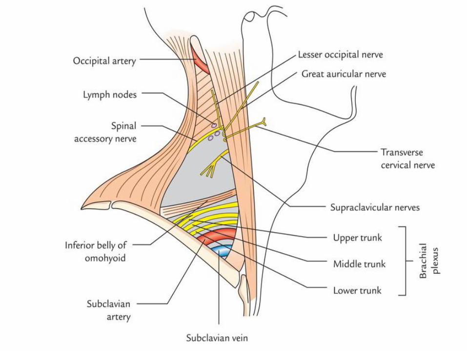
Spinal accessory nerve
is embedded in the
investing layer of deep
fascia (Which forms the
roof of the posterior
triangle), stretched
between two muscles
The superficial
location of the spinal
accessory nerve as it
crosses the posterior
cervical triangle makes
it susceptible to injury
Transverse
cervical nerve
Great auricular nerve
Lesser occipital nerve
Supraclavicular nerves
Trunks of
brachial
plexus
Spinal accessory nerve

Inferior thyroid artery
Transverse cervical artery
Suprascapular artery
1st 2nd
3rd
Common
carotid artery
Thyrocervical
trunk
Vertebral artery

Occipital artery
Transverse cervical artery
Suprascapular artery
3rd part of subclavian artery

External jugular vein
Anterior jugular vein
Transverse cervical vein
Suprascapular vein

Inferior belly of omohyoid

Structures piercing
the roof of posterior
triangle (investing
layer of deep fascia)

Anterior triangle
Boundaries:
- Superiorly:
Body of the mandible
- Posteriorly:
Sternocleidomastoid
- Anteriorly:
Midline
It is further subdivided into
1. Carotid triangle
2. Submandibular (digastric) triangle
3. Submental triangle
4. Muscular triangle
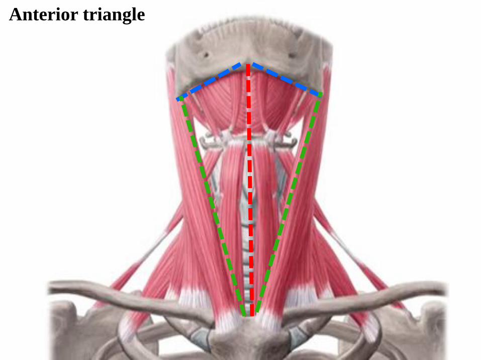
Anterior triangle

Omohyoid Trapezius
Digastric
Sternocleidomastoid
Remember
4 Muscles
3 Bones
2 Lines


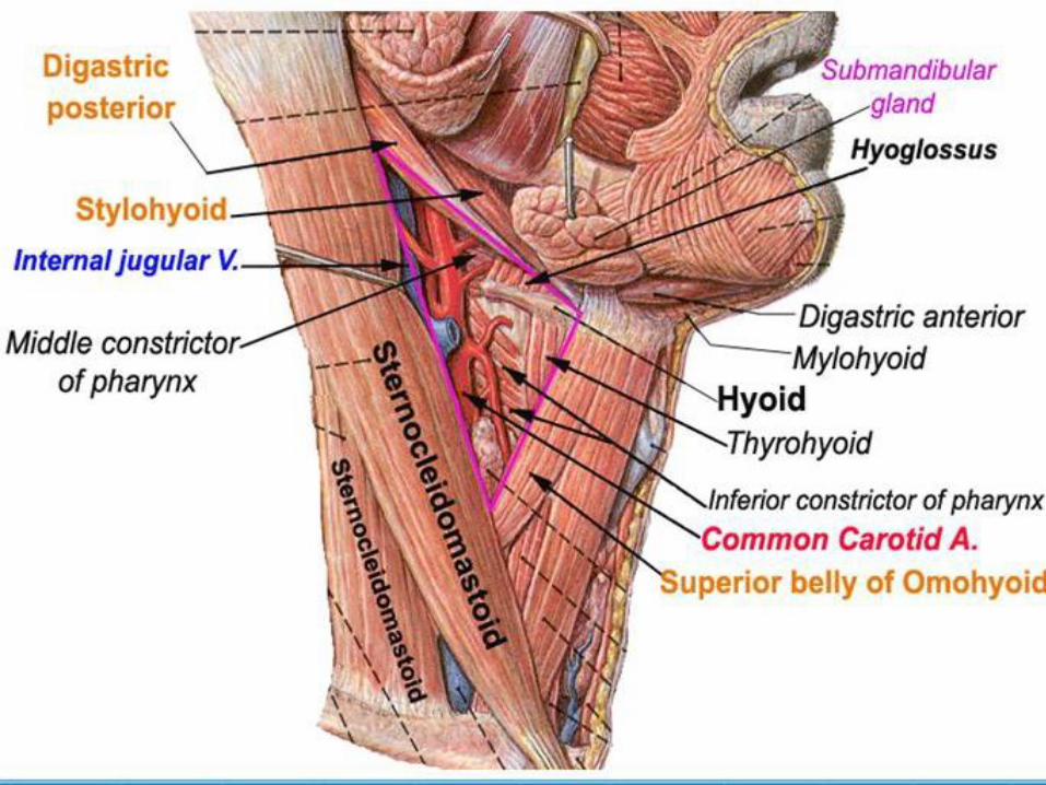
Carotid triangle
Boundaries:
Superior:
Posterior belly of digastric
Lateral:
Sternocleidomastoid
Inferior:
Superior belly of omohyoid
The main contents of carotid
triangle are:
1- Common carotid artery
2- External carotid artery
(and lower 5 branches)
3- Internal carotid artery
4- Internal jugular vein
6- Vagus nerve
7- Accessory nerve
8- Hypoglossal nerve
9- Ansa cervicalis

IJV
ICA ECA
CCA

11th
10th
12th
Ansa Cervicalis

To check carotid pulse
Place your index and middle
fingers on the neck to the side of
larynx (in carotid triangle), under
the angle of the mandible

Boundaries:
Superiorly:
Body of mandible
Anteriorly:
Anterior belly of digastric
Posteriorly:
Posterior belly of digastric
Submandibular triangle
(Digastric)
Is located underneath body of
mandible
Contents:
1. Submandibular gland
2. Submandibular lymph nodes
3. Facial artery
4. Facial vein

Submandibular gland
Submandibular LNs
Facial artery
Facial vein

The boundaries:
Superiorly:
Hyoid bone
Medially:
Imaginary midline of the neck
Supero-laterally:
Superior belly of omohyoid
Infero-laterally:
Sternocleidomastoid
Muscular triangle
It is a slightly ‘dubious’ triangle, in
reality having four boundaries
Contents:
1- Infrahyoid muscles
2- Larynx
3- Trachea
4- Thyroid and parathyroid glands
5- Pharynx
6- Esophagus

Muscular triangle
Hyoid bone
Imaginary midline of
the neck.
Superior belly of
omohyoid
Anterior border of
sternocleidomastoid
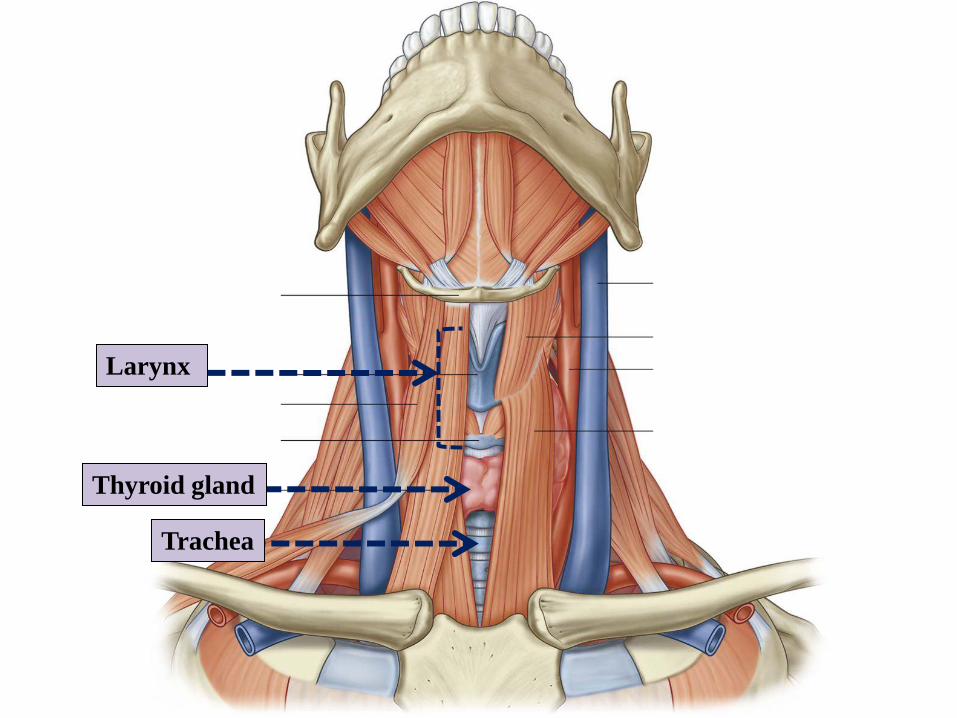
Larynx
Trachea
Thyroid gland

Imaginary midline of
the neck.
Hyoid bone
Anterior belly of
digastric
Boundaries:
Inferiorly:
Hyoid bone
Medially:
Midline of the neck
Laterally:
Anterior belly of digastric
Floor:
Mylohyoid muscle
Submental triangle
Is situated underneath the chin
Contents: Submental lymph nodes

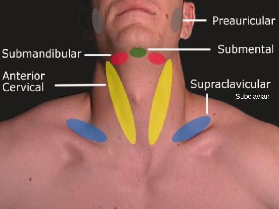
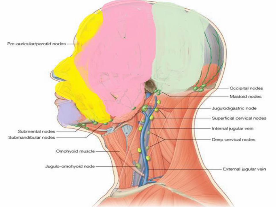
Lymphatic drainage of head and
neck

1- Submandibular nodes
2- Submental nodes
4- Mastoid nodes
5- Occipital nodes
3- Pre-auricular/ parotid nodes
Lymph nodes of face and scalp
Form a ring around
the lower part of head
Five groups of lymph nodes
Lymph nodes of the neck
Superficial Deep

The superficial cervical
nodes are a collection of
lymph nodes along the
external jugular vein on
the superficial surface of
sternocleidomastoid
Superficial cervical nodes
Vertical along
superficial veins

Deep cervical nodes
Deep Cervical Lymph nodes
1- Median group:
-Retropharyngeal, prelaryngeal,
pretracheal and paratracheal
2- Lateral group: At the side of the neck
along internal jugular vein:
- Upper & lower deep cervical nodes

Prelaryngeal lymph nodes
Pretracheal and
paratracheal lymph
nodes
Median group of deep cervical
lymph nodes

Pretracheal layer posterior to the pharynx is called
Buccopharyngeal fascia
Posterior
Anterior
Esophagus
Thyroid
gland
Trachea
Pretracheal layer


At side of neck along internal
jugular vein
Lateral group of deep cervical
lymph nodes
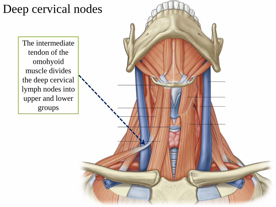
The intermediate
tendon of the
omohyoid
muscle divides
the deep cervical
lymph nodes into
upper and lower
groups
Deep cervical nodes

Jugulo-omohyoid node
is at or just inferior to
the intermediate tendon
of omohyoid
Jugulo-digastric Node is
where posterior belly of
digastric crosses internal
jugular vein
Deep cervical nodes

Two important nodes in the deep
cervical group
1 – Jugulo-digastric node
This large node is where
posterior belly of digastric
crosses the internal jugular vein
and receives lymphatic drainage
from the tonsils and tongue
Enlarged jugulodigastric
lymph nodes are commonly
found in tonsillitis
2 - Jugulo-omohyoid node
it is at or just inferior to the
intermediate tendon of omohyoid
This node receives lymphatic
drainage from the tongue
Deep cervical nodes along Internal
jugular vein

Fate of lymph drainage of head & neck

Pulse points
Where to take
arterial pulses in the
head and neck

How to outline the anterior and
posterior triangles of the neck
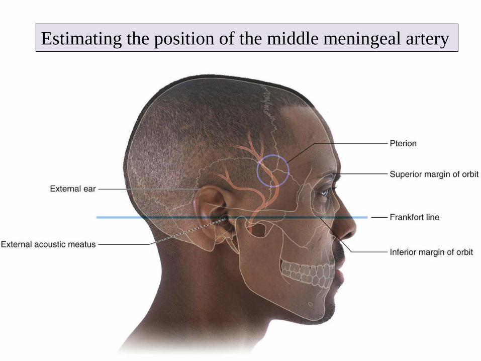
Estimating the position of the middle meningeal artery

Major features
of the face
Recommended