CHAPTER ONE
Neuroimaging Applicationsin DystoniaKristina Simonyan1Department of Otolaryngology, Massachusetts Eye and Ear Infirmary, Boston, MA, United StatesDepartment of Neurology, Massachusetts General Hospital, Boston, MA, United StatesHarvard Medical School, Boston, MA, United States1Corresponding author: e-mail address: [email protected]
Contents
1. Multi-modal Neuroimaging Application in Dystonia 22. Structural Neuroimaging of Dystonia 4
2.1 Gray Matter Structural Organization 42.2 White Matter Structural Organization 7
3. Functional Neuroimaging of Dystonia 113.1 Positron Emission Tomography 113.2 Functional MRI During Task Production and Resting State 123.3 Dystonia as a Functional Network Disorder 15
4. Neuroreceptor Mapping Studies in Dystonia 174.1 Dopaminergic Neurotransmission 174.2 GABAergic Neurotransmission 20
5. Contribution of Neuroimaging to Understanding the Pathophysiologyof Dystonia 21
References 22
Abstract
Dystonia is a neurological disorder characterized by involuntary, repetitive movements.Although the precise mechanisms of dystonia development remain unknown, thediversity of its clinical phenotypes is thought to be associated with multifactorial path-ophysiology, which is linked not only to alterations of brain organization, but also envi-ronmental stressors and gene mutations. This chapter will present an overview of thepathophysiology of isolated dystonia through the lens of applications of major neuro-imaging methodologies, with links to genetics and environmental factors that play aprominent role in symptom manifestation.
Dystonia is a neurological movement disorder, which is characterized by
sustained or intermittent muscle contractions, causing abnormal, often
repetitive movements, postures, or both (Albanese et al., 2013). Isolated
dystonia (formerly known as primary), where dystonic symptoms are the
International Review of Neurobiology, Volume 143 # 2018 Elsevier Inc.ISSN 0074-7742 All rights reserved.https://doi.org/10.1016/bs.irn.2018.09.007
1
only disease manifestation, is a rare disorder with a prevalence of about 3–30per 100,000 in the general population (Asgeirsson, Jakobsson, Hjaltason,
Jonsdottir, & Sveinbjornsdottir, 2006; de Carvalho Aguiar & Ozelius,
2002; Nutt, Muenter, Melton, Aronson, & Kurland, 1988). It typically
develops spontaneously and progresses into a chronic debilitating condi-
tion, oftentimes severely impacting not only the freedom of movements
but also various other aspects of patient’s life, leading to continuous stress,
social embarrassment, and even loss of employment. Dystonia comprises a
large number of clinical syndromes, with adult-onset focal dystonia being
the most common phenotype, followed by segmental dystonia, and much
rare early (childhood)-onset generalized dystonia. Some forms of dystonia
selectively affect higher-order motor control, resulting in impairments of
unique patterns of highly learned behaviors, such as writing, speaking, or
playing a musical instrument.
The exact pathophysiological mechanisms of isolated dystonia remain
unknown. Based on the current state of knowledge, the diversity of clinical
phenotypes of dystonia is thought to be associated with multifactorial path-
ophysiology, which is linked not only to alterations of brain organization,
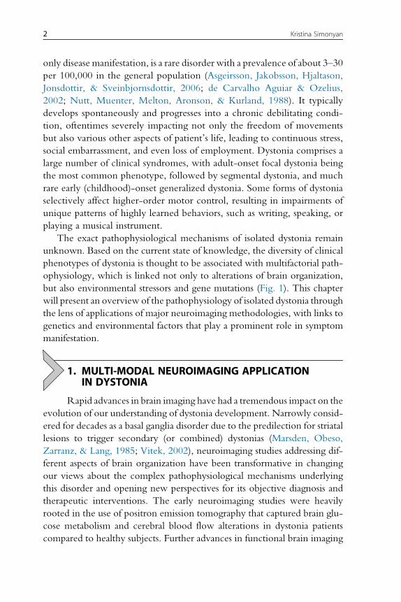
but also environmental stressors and gene mutations (Fig. 1). This chapter
will present an overview of the pathophysiology of isolated dystonia through
the lens of applications of major neuroimaging methodologies, with links to
genetics and environmental factors that play a prominent role in symptom
manifestation.
1. MULTI-MODAL NEUROIMAGING APPLICATIONIN DYSTONIA
Rapid advances in brain imaging have had a tremendous impact on the
evolution of our understanding of dystonia development. Narrowly consid-
ered for decades as a basal ganglia disorder due to the predilection for striatal
lesions to trigger secondary (or combined) dystonias (Marsden, Obeso,
Zarranz, & Lang, 1985; Vitek, 2002), neuroimaging studies addressing dif-
ferent aspects of brain organization have been transformative in changing
our views about the complex pathophysiological mechanisms underlying
this disorder and opening new perspectives for its objective diagnosis and
therapeutic interventions. The early neuroimaging studies were heavily
rooted in the use of positron emission tomography that captured brain glu-
cose metabolism and cerebral blood flow alterations in dystonia patients
compared to healthy subjects. Further advances in functional brain imaging
2 Kristina Simonyan
using functional magnetic resonance imaging (MRI) and magnetoenceph-
alography as well as structural imaging using diffusion weighted and high-
resolution MR sequences provided deeper insights into brain regional
alterations at both structural and functional levels. Finally, relatively recent
applications of mathematical modeling of larger-scale brain networks rev-
ealed global neural abnormalities that were not recognized in initial studies,
reinforcing the notion that isolated dystonia is a network disorder. In par-
allel, neuroreceptor mapping studies with positron emission tomography
andmagnetic resonance spectroscopy helped define the aberrations of dopa-
minergic and GABAergic neurotransmission across different forms of dys-
tonia as one of the potential underlying causes of altered network function.
Recent studies have also attempted to link and understand the impact of
gene mutations on the development of brain abnormalities in dystonia.
Fig. 1 Schematic representation of multifactorial pathophysiology of isolated dystoniawith major factors including abnormal brain structural, functional and neurochemicalorganization, underlying gene mutations, and environmental triggers, a combinationof which leads to the manifestation of dystonic symptoms.
3Neuroimaging Applications in Dystonia
Starting with the first discovery of DYT1 (TOR1A) gene for early-onset
generalized dystonia in 1997 (Ozelius et al., 1997), several studies have been
designed to elucidate distinct brain disorganization in manifesting and non-
manifestingmutation carriers. Further on, rapid advances in next-generation
whole-exome sequencing led to identification of additional dystonia genes,
including DYT6 (THAP 1), DYT24 (ANO3), DYT25 (GNAL), DYT4
(TUBB4), and DYT23 (CIZ1), with the last two still pending an indepen-
dent confirmation (Klein, 2014). These mutations are known to predomi-
nantly cause segmental dystonias that affect adjacent body regions. Again,
neuroimaging studies revealed important associations between some of these
genetic factors and neural aberrancies.
Arguably, the least explored aspect in the pathophysiology of dystonia is
the role of environmental variables, such as extended sun exposure for
blepharospasm, neck trauma and surgery in cervical dystonia, viral infections
in laryngeal dystonia, and overuse practice in focal hand dystonia and musi-
cian’s dystonia. The current effort is directed to elucidating the possible
environmental stressors that may influence the development of brain alter-
ations and by this trigger the manifestation of dystonic symptoms.
2. STRUCTURAL NEUROIMAGING OF DYSTONIA
2.1 Gray Matter Structural OrganizationThe absence of gross structural abnormalities on conventional MRI is one
of the clinical hallmarks of dystonia. In fact, it is often a criterion con-
firming the differential diagnosis of dystonia. On the other hand, detailed
evaluation of high-resolution T1-weighted and diffusion-weighted MR
sequences in numerous neuroimaging studies led to identification of
microstructural cortical and subcortical alterations across all forms of isolated
dystonia.
One of the first studies dates back to late 1990s, which applied MRI-
based stereological volumetry to reveal a 10% increase of putaminal volume
in patients with blepharospasm and focal hand dystonia (Black, Ongur, &
Perlmutter, 1998). With the focused investigation on the striatum, this
finding was contemporary and supportive of prevailing knowledge at
the time that dystonia is a basal ganglia disorder due to intrinsic structural
changes. However, further development of methodologies, such as voxel-
based morphometry and cortical thickness analyses, allowed for evaluation
of volumetric changes within and/or between patient and control groups
based on statistical parametric mapping. Analytic pipelines for both
4 Kristina Simonyan
methodologies use a refined and fully automated approach of segmentation
of high-resolution T1-weighted brain images into gray matter, white mat-
ter, and cerebrospinal fluid. Voxel-based morphometry measures the quan-
tity of tissue within a voxel and is dependent on local cortical thickness
and/or cortical surface area. In contrast, cortical thickness measurements
are based on determining the inner and outer cortical boundaries or sur-
faces and have a high sensitivity in detecting cortical changes, especially
those prone to gene mutations. Both methodologies are complimentary,
with voxel-based morphometry providing information about the volumet-
ric organization of the entire brain and cortical thickness measurements
being more limited to the cortical ribbon.
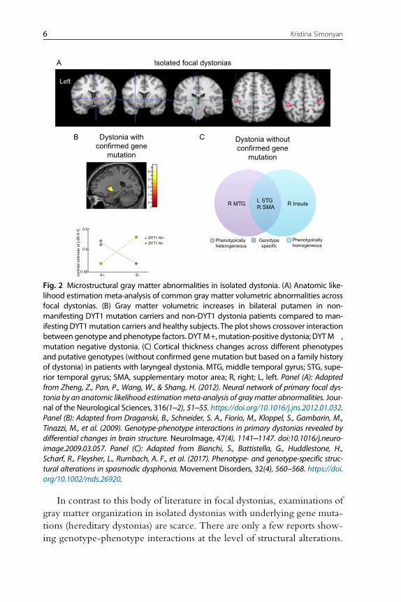
The use of these analytic methods made it apparent that putaminal vol-
umetric changes are indeed a common feature of gray matter alterations
across different forms of dystonia, including both focal and generalized phe-
notypes (Draganski et al., 2009; Draganski, Thun-Hohenstein, Bogdahn,
Winkler, & May 2003; Etgen, Muhlau, Gaser, & Sander, 2006; Granert,
Peller, Jabusch, Altenmuller, & Siebner, 2011; Obermann et al., 2007;
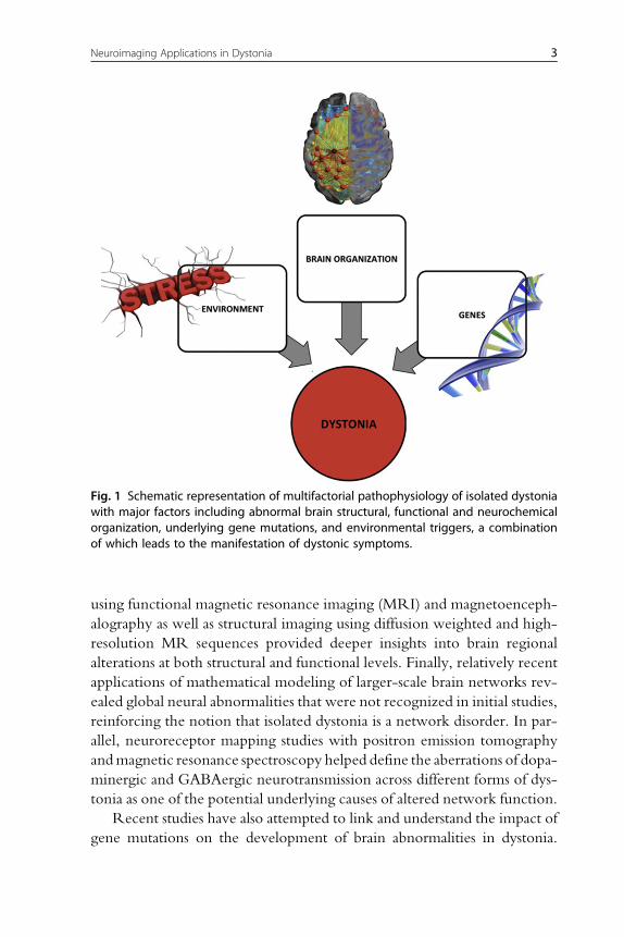
Pantano et al., 2011; Simonyan & Ludlow, 2012) (Fig. 2A). In addition, these
studies revealed that gray matter changes in focal dystonias further extend to
the other divisions of the basal ganglia, most commonly including caudate
nucleus and globus pallidus, as well as thalamus and cerebellum (Delmaire
et al., 2005; Draganski et al., 2003; Egger et al., 2007; Filip et al., 2017;
Obermann et al., 2007; Pantano et al., 2011; Simonyan & Ludlow, 2012;
Waugh et al., 2016; Zeuner et al., 2015). More importantly, structural alter-
ations were shown to involve some of the key cortical regions, responsible for
sensorimotor processing, integration and motor execution, such as fronto-
parietal, supplementary motor and primary sensorimotor areas (Delmaire
et al., 2005; Draganski et al., 2003; Egger et al., 2007; Garraux et al., 2004;
Granert, Peller, Jabusch, et al., 2011; Horovitz, Gallea, Najee-Ullah, &
Hallett, 2013; Martino et al., 2011; Pantano et al., 2011; Ramdhani et al.,
2014; Simonyan & Ludlow, 2012). Among these, primary sensorimotor
changes tended to localize to dystonia-affected body regions within the sen-
sorimotor homunculus. For example, gray matter volumetric increases in
patients with focal hand dystonia were observed in the hand area of sensori-
motor cortex (Delmaire et al., 2005; Egger et al., 2007; Garraux et al., 2004),
whereas in patients with laryngeal dystonia these were found within the lar-
ynx representation (Bianchi et al., 2017; Kostic et al., 2016; Simonyan &
Ludlow, 2012), further showing relevance of cortical aberrations to the path-
ophysiology of distinct forms of dystonia.
5Neuroimaging Applications in Dystonia
In contrast to this body of literature in focal dystonias, examinations of
gray matter organization in isolated dystonias with underlying gene muta-
tions (hereditary dystonias) are scarce. There are only a few reports show-
ing genotype-phenotype interactions at the level of structural alterations.
0.36
cont
rast
est
ima
te a
t [-2
6-5-
4]
S+ S–
DYT1 M+Phenotypicallyheterogeneous
Phenotypicallyhomogeneous
R MTG R InsulaL STGR SMA
Dystonia withconfirmed gene
mutation
A
B C
Left
Dystonia withoutconfirmed gene
mutation
Isolated focal dystonias
Genotypespecific
00.5
11.5
22.5
33.5
44.5
5
DYT1 M–
0.4
0.5
Fig. 2 Microstructural gray matter abnormalities in isolated dystonia. (A) Anatomic like-lihood estimation meta-analysis of common gray matter volumetric abnormalities acrossfocal dystonias. (B) Gray matter volumetric increases in bilateral putamen in non-manifesting DYT1 mutation carriers and non-DYT1 dystonia patients compared to man-ifesting DYT1mutation carriers and healthy subjects. The plot shows crossover interactionbetween genotype and phenotype factors. DYTM+,mutation-positive dystonia; DYTM�,mutation negative dystonia. (C) Cortical thickness changes across different phenotypesand putative genotypes (without confirmed gene mutation but based on a family historyof dystonia) in patients with laryngeal dystonia. MTG, middle temporal gyrus; STG, supe-rior temporal gyrus; SMA, supplementary motor area; R, right; L, left. Panel (A): Adaptedfrom Zheng, Z., Pan, P., Wang, W., & Shang, H. (2012). Neural network of primary focal dys-tonia by an anatomic likelihood estimationmeta-analysis of graymatter abnormalities. Jour-nal of the Neurological Sciences, 316(1–2), 51–55. https://doi.org/10.1016/j.jns.2012.01.032.Panel (B): Adapted from Draganski, B., Schneider, S. A., Fiorio, M., Kloppel, S., Gambarin, M.,Tinazzi, M., et al. (2009). Genotype-phenotype interactions in primary dystonias revealed bydifferential changes in brain structure. NeuroImage, 47(4), 1141–1147. doi:10.1016/j.neuro-image.2009.03.057. Panel (C): Adapted from Bianchi, S., Battistella, G., Huddlestone, H.,Scharf, R., Fleysher, L., Rumbach, A. F., et al. (2017). Phenotype- and genotype-specific struc-tural alterations in spasmodic dysphonia. Movement Disorders, 32(4), 560–568. https://doi.org/10.1002/mds.26920.
6 Kristina Simonyan
One study revealed that non-manifestingDYT1mutation carriers and non-
DYT1 dystonia patients have significantly increased bilateral putaminal
volume compared to manifesting DYT1mutation carriers and healthy sub-
jects (Draganski et al., 2009) (Fig. 2B). Another study reported a volumetric
increase of globus pallidus in patients with generalized dystonia, although
the DYT1 status was not known in this cohort (Egger et al., 2007). As gene
discovery for isolated focal dystonias remains stagnant and extremely chal-
lenging due, in part, to rare availability of large families, low penetrance,
and variable expressivity, the design of neuroimaging studies has also been
limited by somewhat arbitrary assignments of experimental cohorts based
on a confirmed family history of dystonia only. Based on such approach,
one recent study in patients with sporadic and familial forms of laryngeal
dystonia identified phenotype-specific graymatter abnormalities in primary
and associative areas of motor control that were distinct from genotype-
specific changes within the cortical regions controlling sensory processing
(Bianchi et al., 2017) (Fig. 2C).
Along with the progress in mapping and characterizing gray matter
organization in dystonia, questions pertaining to the exact cellular mech-
anisms and causes of gray matter alterations as revealed by voxel-based
morphometry and cortical thickness measurements remain poorly under-
stood. It is possible that gray matter volumetric increases in some forms of
dystonia, especially in musician’s dystonia and other task-specific dystonias,
may be due to abnormal motor training and overuse that generally lead to
volumetric increases in related brain regions in healthy populations (Ceccarelli
et al., 2009; Draganski et al., 2004; Granert, Peller, Gaser, et al., 2011; Quallo
et al., 2009). Another possible mechanisms may involve the formation of
abnormal new connections by dendritic spine growth and axonal remodeling,
and/or the strengthening of existing synaptic connections (Chklovskii, 2004;
Chklovskii, Mel, & Svoboda, 2004; Holtmaat, Wilbrecht, Knott, Welker, &
Svoboda, 2006; Sur&Rubenstein, 2005). Future ultra-high-resolution imag-
ing of graymatter, ideally coupled with postmortem neuropathology, may be
able to reveal which of these causative mechanisms are responsible for
observed neuroimaging changes across dystonias.
2.2 White Matter Structural OrganizationMost of the knowledge about white matter organization in isolated
dystonia comes from the use of diffusion tensor imaging, which is a
form of diffusion-weighted imaging that examines axonal organization
7Neuroimaging Applications in Dystonia
in brain tissue. It is based on the principal of random displacement of water
molecules and differential quantification of their diffusivity in parallel and
perpendicular directions along the axonal course. Diffusion imaging mea-
sures include anisotropy indices (e.g., fractional anisotropy) to quantify the
directionality of water diffusivity, reflecting axonal integrity and tissue
coherence. Other diffusivity indices (e.g., mean diffusivity, trace of diffu-
sion tensor) examine the magnitude of water movement independent of
the direction and provide information about the organization of the extra-
cellular space and the intracellular water content. Similar to voxel-based
morphometry and cortical thickness measurements, diffusivity measures
are sensitive to microstructural white matter abnormalities not evident
on conventional MRI.
In addition to the assessment of regional white matter organization, dif-
fusion imaging methods allow for in vivo reconstruction of the white matter
tracts using deterministic and probabilistic tractography. This is considered
as a valid method for examination of white matter connectivity due to its
capability to model fiber direction(s) per voxel (single with deterministic
tractography and multiple with probabilistic tractography), providing real-
istic estimates of the wiring strength of gross white matter projections.
However, tractography is still limited for mapping the connectivity between
brain regions with high fiber complexity and uncertainty about the fiber
directions, especially in the presence of dense local cortico-cortical connec-
tions. The likelihood of tractography is excessively higher in estimation of
the tracts from the regions of interest to immediately surrounding areas
than those in more distant brain regions, leading to the distance bias. Sim-
ilarly, the tractography algorithm is prone to higher propagation of tracts
to larger targets and even to generating false-positive tracts. Moreover,
tractography by-passes monosynaptic connections and does not allow infer-
ring directionality of the connection, which makes it difficult to assess the
structural influence of one brain region upon another.While a powerful ana-
lytic method for in vivo examination of structural connectivity in humans,
the results of diffusion tractography warrant a careful comparison to those
obtained with neuroanatomical tract tracing methods in non-human pri-
mates, most importantly to avoid identification of spurious “novel” connec-
tivity and its impairments in diseased populations.
Studies assessing regional white matter organization in isolated dystonia
have been mostly focused on the use of various diffusivity measures between
and within different groups of patients and healthy subjects. The general
unifying finding across these studies is identification of reduced axonal
8 Kristina Simonyan
integrity and increased water diffusivity, involving both cortical and sub-
cortical structures along the cortico-striato-pallido-thalamic and cerebello-
thalamo-cortical pathways (Blood et al., 2012; Bonilha et al., 2007;
Colosimo et al., 2005; Delmaire et al., 2009; Fabbrini et al., 2008; Horovitz,
Ford, Najee-Ullah, Ostuni, & Hallett, 2012; Prell et al., 2013; Simonyan
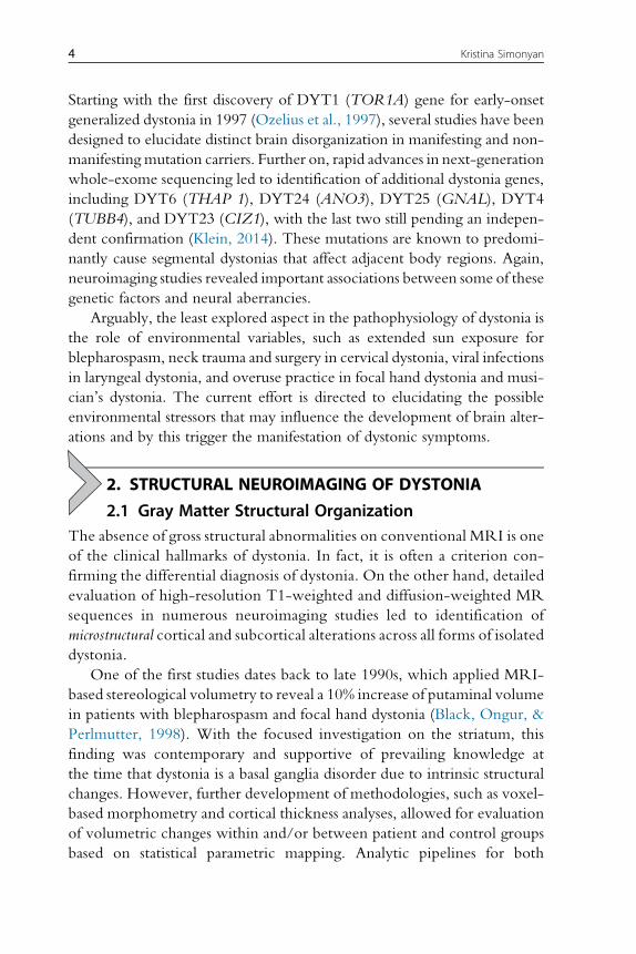
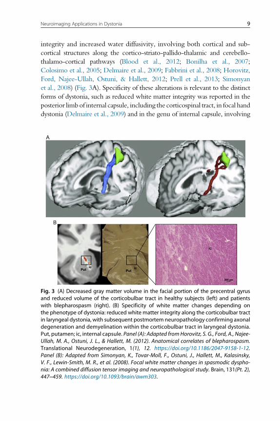
et al., 2008) (Fig. 3A). Specificity of these alterations is relevant to the distinct
forms of dystonia, such as reduced white matter integrity was reported in the
posterior limbof internal capsule, including the corticospinal tract, in focal hand
dystonia (Delmaire et al., 2009) and in the genu of internal capsule, involving
Fig. 3 (A) Decreased gray matter volume in the facial portion of the precentral gyrusand reduced volume of the corticobulbar tract in healthy subjects (left) and patientswith blepharospasm (right). (B) Specificity of white matter changes depending onthe phenotype of dystonia: reduced white matter integrity along the corticobulbar tractin laryngeal dystonia, with subsequent postmortem neuropathology confirming axonaldegeneration and demyelination within the corticobulbar tract in laryngeal dystonia.Put, putamen; ic, internal capsule. Panel (A): Adapted fromHorovitz, S. G., Ford, A., Najee-Ullah, M. A., Ostuni, J. L., & Hallett, M. (2012). Anatomical correlates of blepharospasm.Translational Neurodegeneration, 1(1), 12. https://doi.org/10.1186/2047-9158-1-12.Panel (B): Adapted from Simonyan, K., Tovar-Moll, F., Ostuni, J., Hallett, M., Kalasinsky,V. F., Lewin-Smith, M. R., et al. (2008). Focal white matter changes in spasmodic dyspho-nia: A combined diffusion tensor imaging and neuropathological study. Brain, 131(Pt. 2),447–459. https://doi.org/10.1093/brain/awm303.
9Neuroimaging Applications in Dystonia
corticobulbar tract, in laryngeal dystonia (Simonyan et al., 2008). The latter
finding has been substantiated by neuropathological evidence of decreased axo-
nal density and myelin content, potentially underlying the change in water
diffusivity (Simonyan et al., 2008) (Fig. 3B).
Among the hereditary forms of dystonia, both manifesting and non-
manifesting DYT1 carriers showed reduced white matter integrity in sub-
gyral white matter of sensorimotor cortex, albeit with greater abnormalities
in manifesting than non-manifesting carriers (Carbon, Kingsley, et al., 2004;
Carbon, Kingsley, Tang, Bressman, & Eidelberg, 2008). A similar finding
was observed in the study of manifesting DYT6 carriers (Cheng et al.,
2012). In addition, DYT1 carriers showed abnormal white matter organiza-
tion of dorsal pontine tegmentum adjacent to the left superior cerebellar
peduncle (Carbon et al., 2008). Diffusion tractography, examining the cere-
bellar involvement, identified reduced integrity in the cerebellar lobule VI
adjacent to dentate nucleus in DYT1 and DYT6 manifesting carriers as well
as reduced thalamo-cortical connectivity in non-manifesting dystonia gene
carriers (Argyelan et al., 2009).On the other hand, substantial phenotypic var-
iability of hereditary dystonia appears to be linked to reduced integrity of
somatotopic projections related to asymptomatic body regions (Vo, Sako,
Niethammer, et al., 2015). Similarly, non-manifesting carriers showed addi-
tionally reduced connectivity within the distal thalamo-cortical segment
of the cerebello-thalamo-cortical tract, pointing to the protective neural
mechanisms underlying themanifestation of hereditary dystonia. Corroborat-
ing these findings, an ex vivo study in the heterozygous DYT1 knock-in
mouse model showed reduced connectivity of the cerebello-thalamic,
thalamo-cortical and thalamo-striatal pathways in mutants compared to wild
type (Ulug et al., 2011). Another study reported deficits of free water diffu-
sivity and widespread increases in functional connectivity of the stratumwith
somatosensory cortex, thalamus, cerebellum and brainstem in the conditional
knock-outmice of theDYT1 protein torsinA (DeSimone et al., 2017). As the
cerebello-thalamo-cortical pathway facilitates intracortical inhibition via pro-
jections to interneurons in the sensorimotor cortex (Molinari, Filippini, &
Leggio, 2002), these findings may suggest the presence of the intermediary
pattern in non-manifesting carriers that may act as a buffer against the aberrant
outflow from the proximal part of this pathway (Niethammer, Carbon,
Argyelan, & Eidelberg, 2011).
Taken together, the use of volumetric and diffusion imaging helped
establish the presence of microstructural alterations of gray and white matter
in dystonia that are not restricted to the putamen but rather extend to
10 Kristina Simonyan
surrounding subcortical and further cortical and cerebellar regions, predom-
inantly within the basal ganglia-thalamo-cortical and cerebello-thalamo-
cortical circuitries. Identification of structural alterations specific to the
disorder genotype and phenotype has been important for providing a critical
step toward future delineation of imaging markers and the potential targets
for novel therapeutic interventions in this disorder.
3. FUNCTIONAL NEUROIMAGING OF DYSTONIA
3.1 Positron Emission TomographyEarly studies in dystonia relied heavily on the use of positron emission
tomography to capture the cerebral blood flow and glucose metabolism
in patient within-group and between-group comparisons with healthy sub-
jects. Positron emission tomography is based on radioactivity emitted after a
radiolabeled tracer is injected intravenously. The nucleus of the radioisotope
emits a positron, which collides with an electron in the tissue and in the pro-
cess converts mass to energy in the form of two photons. The tomograph
uses scintillation crystals placed around the subject’s head to detect these
photons. The crystals absorb the photons, producing light that is converted
into an electrical signal.
One of the most commonly used tracers to quantify brain activity via
glucose metabolism in dystonia is [18F]-fluorodeoxyglucose ([18F]FDG),
which is taken up by brain regions depending onmetabolic needs. As regional
metabolism is dependent on synaptic activity, changes in tracer uptake corre-
late with changes in regional neuronal activity. Using [18F]FDG, several
studies reported abnormal metabolism in basal ganglia, thalamus, cerebellum,
and sensorimotor cortex in patients with focal dystonia (Esmaeli-Gutstein,
Nahmias, Thompson, Kazdan, & Harvey, 1999; Hutchinson et al., 2000;
Kerrison et al., 2003;Magyar-Lehmann et al., 1997; Suzuki et al., 2007).Dur-
ing induced sleep in patients with blepharospasm, hypometabolic activity was
additionally found in the frontal eye field (Brodmann area 8), which is
involved in planning of complex movements and is associated with supra-
nuclear control of the eyelid opening (Hutchinson et al., 2000). Evaluation
of the effectiveness of botulinum toxin treatment in blepharospasm showed
an association with normalization of metabolic activity in the cerebellum
and pons (Suzuki et al., 2007).
Studies in hereditary dystonias also identified distinct patterns of regional
metabolic activity. Specifically, DYT1 carriers showed metabolic increases
in the striatum and cerebellum; DYT6 carriers had metabolic reductions in
11Neuroimaging Applications in Dystonia
the putamen, thalamus, cerebellar cortex, and upper brainstem; and DYT11
mutation carriers had both metabolic increases in the deep cerebellar nuclei
and vermis and metabolic decreases in the medial prefrontal cortex and pre-
supplementary motor area (Carbon & Eidelberg, 2009; Carbon, Su, et al.,
2004). The findings in DYT1 patients were substantiated in the Tor1a het-
erozygous knock-out mouse, which showed metabolic abnormalities in the
striatum and cerebellum as well as sensorimotor cortex and subthalamic
nucleus (Vo, Sako, Dewey, Eidelberg, & Ulug, 2015).
Another commonly used radiotracer in dystonia research is [15O] H2O,
which allowed the assessment of the regional cerebral blood flow during
production of both symptomatic and asymptomatic tasks while in the
scanner (Ali et al., 2006; Ceballos-Baumann et al., 1995; Ibanez, Sadato,
Karp, Deiber, & Hallett, 1999; Lerner et al., 2004; Playford, Passingham,
Marsden, & Brooks, 1998). Similar to glucose metabolism, the regional
cerebral blood flow appeared to be abnormal in the basal ganglia, thalamus,
cerebellum, and sensorimotor cortex in focal dystonia. In addition, wider
spread abnormalities were identified involving prefrontal, posterior parietal
and temporal regions necessary for motor planning and sensorimotor
processing. Examination of the regional covariance pattern during writing
and at rest in patients with writer’s cramp further revealed reduced correla-
tions between putamen and premotor cortex and between bilateral
premotor cortex (Ibanez et al., 1999), suggesting that not only regional brain
activity but also functional connectivity may be abnormal in dystonia. Defi-
cient activity and connectivity of the preparatory cortical regions and basal
ganglia pointed to a loss of inhibition during the generation of motor com-
mands, likely arising from the striatal region.
3.2 Functional MRI During Task Production and Resting StateThe development of functional MRImethodology in early 1990s (Kwong
et al., 1992; Ogawa, Lee, Nayak, & Glynn, 1990) has completely changed
the landscape of neuroimaging studies examining brain activity and func-
tional connectivity in dystonia. Functional MRI is based on a blood
oxygen-level dependent contrast, which captures the hemodynamic
response by relying on differential magnetic susceptibility of oxyhemo-
globin and deoxyhemoglobin due to changes during neuronal activity.
Functional MRI has been used during various tasks, both eliciting and
not eliciting dystonic movements. Similar to positron emission tomogra-
phy and structural imaging studies, this line of research again confirmed
12 Kristina Simonyan
the presence of alterations in basal ganglia, thalamus, cerebellum and sen-
sorimotor cortex across different forms of isolated dystonia and hinted
to abnormal integration of sensorimotor information flow within the
basal ganglia-thalamo-cortical and cerebello-thalamo-cortical circuitries
(e.g. Baker, Andersen, Morecraft, & Smith, 2003; Burciu et al., 2017;
Butterworth et al., 2003; Haslinger, Altenmuller, Castrop, Zimmer, &
Dresel, 2010; Hu, Wang, Liu, & Zhang, 2006; Islam et al., 2009;
Kadota et al., 2010; Pujol et al., 2000; Simonyan & Ludlow, 2010). Addi-
tionally, some studies pointed to abnormal sensory processing by primary
somatosensory cortex that may contribute to the pathophysiology of dys-
tonia (Dresel, Haslinger, Castrop, Wohlschlaeger, & Ceballos-Baumann,
2006; Haslinger et al., 2010; Simonyan & Ludlow, 2010), whereas others
mapped abnormal somatotopy of digit representation in primary somato-
sensory cortex and putamen in focal hand dystonia (Butterworth et al., 2003;
Delmaire et al., 2005; Nelson, Blake, & Chen, 2009). Studies examining the
effects of botulinum toxin injections on brain activity in patients with focal
dystonia provided largely controversial results, showing either modulated
or non-modulated brain activity following the treatment (Ali et al., 2006;
Dresel et al., 2011; Haslinger et al., 2005; Nevrly et al., 2018). On the other
hand, subclinical abnormalities in sensory discrimination were described
as a mediational endophenotype of dystonia (Hutchinson et al., 2013) and
linked to alterations in primary somatosensory and middle frontal cortices
(Termsarasab et al., 2016). Importantly, differential associations between
abnormal sensory discrimination and functional abnormalities were observed
depending on the phenotype and genotype of dystonia, including greater cer-
ebellar involvement in familial laryngeal dystonia cases and greater putaminal
and cortical sensorimotor inclusion in different phenotypes of laryngeal dys-
tonia (Termsarasab et al., 2016).
Another step forward in understanding the neuroimaging pathophysiol-
ogy of dystonia came with the development of resting-state functional MRI,
which relies on the measurement of low frequency physiological fluctua-
tions in the BOLD signal, reflecting the functional brain organization during
various activation states (Biswal, Yetkin, Haughton, & Hyde, 1995; Smith
et al., 2009). The resting-state functional MRI approach proved to be useful
in circumventing the challenges associated with the implementation of the
task-related designs across different forms of dystonia, which exhibit distinct
symptoms, thus making the choice of a single, commonly affected task pro-
duction unfeasible. Furthermore, because the explanation of functional
changes across distinct dystonia phenotypes and genotypes may sometimes
13Neuroimaging Applications in Dystonia
be ambiguous due to a combination of motor and sensory components,
examination of the resting-state activity and connectivity provides a more
uniform and coherent understanding of neural alterations.
Resting-state functional MRI studies in focal hand dystonia found
decreased connectivity in primary somatosensory region with concomitant
increases in the putamen as well as functional decoupling of dorsal premotor
cortex fromparietal cortex (Delnooz,Helmich, Toni, & van deWarrenburg,
2012; Mohammadi et al., 2012). In cervical dystonia, alterations were found
not only in sensorimotor but also in visual and executive control networks
(Delnooz, Pasman, Beckmann, & van de Warrenburg, 2013), whereas
changes in blepharospasm were related to the default-mode network
(Yang et al., 2013). It was further shown that vulnerable connectivity of pri-
mary sensorimotor and inferior parietal cortices in laryngeal dystonia is
tightly associated with the polygenic risk of dystonia, likely representing
an endophenotypic imaging marker of this disorder; genes contributing to
the polygenic score are involved in synaptic transmission and neuron devel-
opment (Battistella, Fuertinger, Fleysher, Ozelius, & Simonyan, 2016;
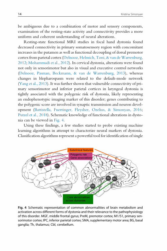
Putzel et al., 2018). Schematic knowledge of functional alterations in dysto-
nia can be viewed in Fig. 4.
Using these findings, a few studies started to probe existing machine
learning algorithms in attempt to characterize neural markers of dystonia.
Classification algorithms represent a powerful tool for identification of single
Fig. 4 Schematic representation of common abnormalities of brain metabolism andactivation across different forms of dystonia and their relevance to the pathophysiologyof this disorder. MGF, middle frontal gyrus; PreM, premotor cortex; M1/S1, primary sen-sorimotor cortex; IPC, inferior parietal cortex; SMA, supplementary motor area; BG, basalganglia; Th, thalamus; Cbl, cerebellum.
14 Kristina Simonyan
traits or a combination of features that characterize and separate two or
more classes of objects or subjects. Algorithmic classifiers using functional
MRI voxel-wise time series have been successfully applied in several neu-
rodegenerative disorders (Fornari, Maeder, Meuli, Ghika, & Knyazeva,
2012; Janousova, Schwarz, & Kasparek, 2015; Yourganov et al., 2014).
The first study in dystonia used multivariate classification algorithm of linear
discriminant analysis (LDA) based on the measures of abnormal functional
resting-state connectivity in primary sensorimotor, premotor and inferior
parietal regions, achieving 71% accuracy in classifying laryngeal dystonia
and healthy controls (Battistella et al., 2016). It further improved its accuracy
in classifying familial vs sporadic laryngeal patients at 81% and remained at
the same 71% accuracy level when considering different (adductor and
abductor) phenotypes. As such, this study used neuroimaging data to disam-
biguate focal dystonia from a normal state and differentiate the disorder
based not on clinical evaluations of its symptoms but of neuroimaging het-
erogeneity, opening new avenues to the development of disorder-specific
diagnostic biomarkers.
3.3 Dystonia as a Functional Network DisorderStarting with the positron emission tomography studies in generalized dys-
tonia that showed a presence of the abnormal metabolic network (Eidelberg
et al., 1998, 1995; Niethammer et al., 2011), the overall concept of functional
alterations not being limited to the basal ganglia, as historically proposed,
has evolved into propositions of focal dystonia, too, to represent a functional
network disorder (Lehericy, Tijssen, Vidailhet, Kaji, & Meunier, 2013;
Neychev, Gross, Lehericy, Hess, & Jinnah, 2011; Quartarone & Hallett,
2013; Ramdhani & Simonyan, 2013; Zoons, Booij, Nederveen, Dijk, &
Tijssen, 2011). This concept has been recently experimentally substantiated
in a study that used a graph theoretical approach to probe the organization
of large-scale functional networks across different forms of focal dystonia.
Compared to healthy subjects, patients showed altered network architecture,
which was characterized by an abnormal breakdown of the basal ganglia-
thalamo-cerebellar community, a loss of pivotal regions of information trans-
fer (hubs) in the premotor cortex, and a pronounced decline in sensorimotor
and inferior parietal cortical connectivity (Battistella, Termsarasab,Ramdhani,
Fuertinger, & Simonyan, 2017) (Fig. 5A). These findings pointed to the uni-
fied pathophysiological mechanism underlying different forms of dystonia due
to common network alterations, while suggesting the concurrent presence of
15Neuroimaging Applications in Dystonia
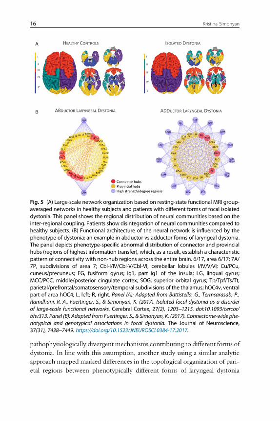
pathophysiologically divergent mechanisms contributing to different forms of
dystonia. In line with this assumption, another study using a similar analytic
approach mapped marked differences in the topological organization of pari-
etal regions between phenotypically different forms of laryngeal dystonia
Fig. 5 (A) Large-scale network organization based on resting-state functional MRI group-averaged networks in healthy subjects and patients with different forms of focal isolateddystonia. This panel shows the regional distribution of neural communities based on theinter-regional coupling. Patients show disintegration of neural communities compared tohealthy subjects. (B) Functional architecture of the neural network is influenced by thephenotype of dystonia; an example in abductor vs adductor forms of laryngeal dystonia.The panel depicts phenotype-specific abnormal distribution of connector and provincialhubs (regions of highest information transfer), which, as a result, establish a characteristicpattern of connectivity with non-hub regions across the entire brain. 6/17, area 6/17; 7A/7P, subdivisions of area 7; CbI-I/IV/Cbl-V/Cbl-VI, cerebellar lobules I/IV/V/VI; Cu/PCu,cuneus/precuneus; FG, fusiform gyrus; Ig1, part Ig1 of the insula; LG, lingual gyrus;MCC/PCC, middle/posterior cingulate cortex; SOG, superior orbital gyrus; Tp/Tpf/Ts/Tt,parietal/prefrontal/somatosensory/temporal subdivisions of the thalamus; hOC4v, ventralpart of area hOC4; L, left; R, right. Panel (A): Adapted from Battistella, G., Termsarasab, P.,Ramdhani, R. A., Fuertinger, S., & Simonyan, K. (2017). Isolated focal dystonia as a disorderof large-scale functional networks. Cerebral Cortex, 27(2), 1203–1215. doi:10.1093/cercor/bhv313. Panel (B): Adapted from Fuertinger, S., & Simonyan, K. (2017). Connectome-wide phe-notypical and genotypical associations in focal dystonia. The Journal of Neuroscience,37(31), 7438–7449. https://doi.org/10.1523/JNEUROSCI.0384-17.2017.
16 Kristina Simonyan
(Fuertinger & Simonyan, 2017) (Fig. 5B). Moreover, the interface between
sporadic genotype and most common adductor phenotype yielded distinct
functional communities of interacting regions that were primarily governed
by intramodular hub regions. On the other hand, the interface between less
common familial genotype and abductor phenotype was associated with
numerous long-ranging hub regions and an abnormal integration of left thal-
amus and basal ganglia.
Another aspect of network abnormality is how one region exerts its influ-
ences upon another. Although the current analytic methodology, dynamic
causal modeling, has severe limitations such as the total number of regions
to be realistically explored cannot exceed 4 or 5, rendering examination
of whole brain connectivity not feasible, it has nevertheless been useful in
showing malfunctioning intracortical connections between primary motor
cortex and supplementary motor area as well as abnormal reciprocal excit-
atory connectivity in the cortico-cerebellar circuitry during performance
of a motor task in patients with writer’s cramp (Rothkirch et al., 2018).
Future studies should focus on the development of new analytic tools to assess
the mechanistic properties of abnormal functional network in dystonia by
examining its effective connectivity.
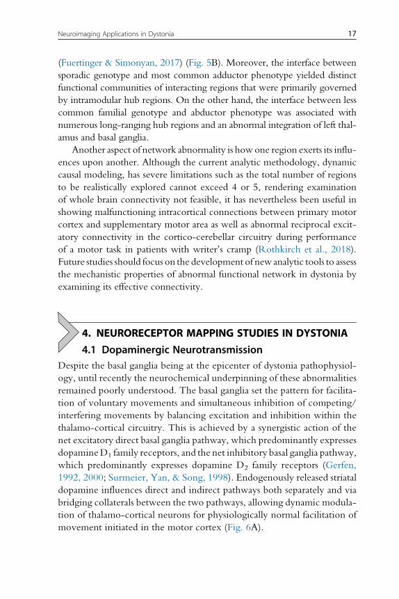
4. NEURORECEPTOR MAPPING STUDIES IN DYSTONIA
4.1 Dopaminergic NeurotransmissionDespite the basal ganglia being at the epicenter of dystonia pathophysiol-
ogy, until recently the neurochemical underpinning of these abnormalities
remained poorly understood. The basal ganglia set the pattern for facilita-
tion of voluntary movements and simultaneous inhibition of competing/
interfering movements by balancing excitation and inhibition within the
thalamo-cortical circuitry. This is achieved by a synergistic action of the
net excitatory direct basal ganglia pathway, which predominantly expresses
dopamineD1 family receptors, and the net inhibitory basal ganglia pathway,
which predominantly expresses dopamine D2 family receptors (Gerfen,
1992, 2000; Surmeier, Yan, & Song, 1998). Endogenously released striatal
dopamine influences direct and indirect pathways both separately and via
bridging collaterals between the two pathways, allowing dynamic modula-
tion of thalamo-cortical neurons for physiologically normal facilitation of
movement initiated in the motor cortex (Fig. 6A).
17Neuroimaging Applications in Dystonia
It has been suggested that abnormal dopamine levels may modulate
striatal synaptic plasticity in dystonia (Breakefield et al., 2008; Hallett,
2004; Todd & Perlmutter, 1998), while increased D1-mediated excitation
and decreased D2 receptor-mediated inhibition may alter the balance
between the basal ganglia direct and indirect pathways, cause overall disin-
hibition of the thalamocortical circuitry, and contribute to dystonia muscle
contractions during performance of fine motor tasks (Hallett, 1993).
glutamate
Substantia nigrapars compacta
Substantia nigrapars compacta
Substantia nigrapars reticulata
Substantia nigrapars reticulata
StriatumD1R(+)D2R(–)
Motor Cortex
A
C
BNormal State Isolated Dystonia
Healthy Subjects Healthy Subjects Laryngeal DystoniaWriter’s Cramp
StriatumD1R(+)D2R(–)
Motor Cortex
Globus pallidusexternal segment
Globus pallidusexternal segment
Globus pallidusinternal segment
Globus pallidusinternal segment
Brainstem BrainstemThalamus
D1
leftleftleftleft
D1×D2
D1×D2×DA
D1×DAD2×DA
D2 DA
ThalamusSubthalamic nucleus Subthalamic nucleus
dopamineGABA
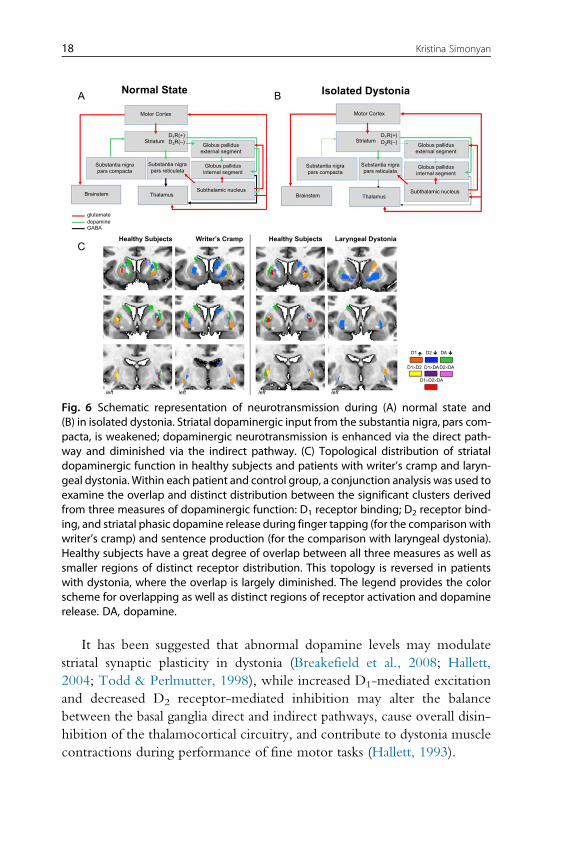
Fig. 6 Schematic representation of neurotransmission during (A) normal state and(B) in isolated dystonia. Striatal dopaminergic input from the substantia nigra, pars com-pacta, is weakened; dopaminergic neurotransmission is enhanced via the direct path-way and diminished via the indirect pathway. (C) Topological distribution of striataldopaminergic function in healthy subjects and patients with writer’s cramp and laryn-geal dystonia. Within each patient and control group, a conjunction analysis was used toexamine the overlap and distinct distribution between the significant clusters derivedfrom three measures of dopaminergic function: D1 receptor binding; D2 receptor bind-ing, and striatal phasic dopamine release during finger tapping (for the comparison withwriter’s cramp) and sentence production (for the comparison with laryngeal dystonia).Healthy subjects have a great degree of overlap between all three measures as well assmaller regions of distinct receptor distribution. This topology is reversed in patientswith dystonia, where the overlap is largely diminished. The legend provides the colorscheme for overlapping as well as distinct regions of receptor activation and dopaminerelease. DA, dopamine.
18 Kristina Simonyan
Using PET or single-photon emission computed tomography with
specialized radioligands to target striatal dopamine receptors, decreased
striatal dopamine D2 receptor binding at rest was found in patients with
both focal and generalized forms of dystonia as well as non-manifesting
DYT1 and DYT6 gene carriers (Asanuma et al., 2005; Berman, Hallett,
Herscovitch, & Simonyan, 2013; Berger et al., 2007; Carbon et al., 2009;
Horie et al., 2009; Horstink et al., 1997; Naumann et al., 1998; Perlmutter
et al., 1997; Simonyan, Berman,Herscovitch,&Hallett, 2013). Among these,
two studies leveraged the ability of endogenously released phasic dopamine to
displace the bound radiotracer in order to assess dopaminergic function during
symptomatic and asymptomatic tasks (Berman et al., 2013; Simonyan et al.,
2013). It was reported that patients with focal hand dystonia and laryngeal
dystonia exhibit abnormally decreased striatal phasic dopamine release during
symptomatic task production (finger tapping and speaking, respectively),
whereas the levels of striatal dopamine release during asymptomatic tasks
(speaking and finger tapping, respectively) are abnormally increased. While
there is no apparent neurodegeneration or cell loss within the basal ganglia,
experimentally reduced striatal D2 receptor binding was also observed in
the dtszmutant hamster and associatedwith increased striatal dopamine release
during the manifestation of dystonic episodes (Hamann & Richter, 2004;
Nobrega, Richter, Tozman, Jiwa, & Loscher, 1996). Decreased neurorecep-
tor binding at rest is thought to reflect decreased D2 receptor availability due
to decreased receptor density and/or increased tonic dopamine levels in the
synapses. This contributes to disinhibition within the indirect basal ganglia
pathway and leads to an inability to suppress unwanted “nearby” motor con-
tractions during the production of specific actions, a well-established abnor-
mality in isolated dystonia.On the other hand, decreased phasic dopaminergic
activity may represent a disorder-specific pathophysiological trait involved in
generation of dystonic symptoms, whereas increased dopamine release during
unaffected and unrelated motor tasks may be due to compensatory adaptation
of the nigrostriatal dopaminergic system.
In terms of involvement of the direct basal ganglia pathway in dystonia,
the recent study revealed increased availability of striatal dopamine D1
receptors, suggesting hyperactivity of the direct pathway in patients with
focal hand dystonia and laryngeal dystonia (Simonyan, Cho, Hamzehei
Sichani, Rubien-Thomas, & Hallett, 2017). This dopaminergic alteration
followed a well-known somatotopic organization of the striatum, with
changes localized to the striatal hand and larynx representations, respectively.
19Neuroimaging Applications in Dystonia
Furthermore, dopaminergic dysfunctions involved both associative (anterior)
and sensorimotor (posterior) striatal subdivisions, potentially having direct
impact not only on hyperexcitability of motor cortex but also on parietal
and prefrontal projection regions, which are responsible for the control
of sensorimotor integration and preparation to motor execution and which
are known to have abnormal activity and connectivity in dystonia. In
fact, these cortical alterations may be a result of propagation of abnormal
dopaminergic function via influencing beta oscillations within different
striato-cortical loops.
When examining topological distribution of D1 and D2 receptor abnor-
malities, abnormal segregation of hyperfunctional direct and hypofunctional
indirect pathways within the striatum became apparent, with negligible, if
any, overlap between the two pathways (Fig. 6C). Moreover, the loss of
overlap between the regions of dopamine D1 and D2 receptor availability
and phasic dopamine release suggested complex disorganization of a
nigro-striatal input.
Overall, these data showed that disorganization of striatal dopaminergic
neurotransmission is of a global scale involving both the direct and indirect
basal ganglia pathways and representing a common pathophysiological trait
in dystonia.
4.2 GABAergic NeurotransmissionA loss of surround inhibition is considered as one of the main pathophysi-
ological features of isolated dystonia (Quartarone & Hallett, 2013). Initial
studies employing magnetic resonance spectroscopy to quantify GABAergic
function in dystonia patients reported that GABA levels are significantly
decreased in sensorimotor cortex and lentiform nucleus in patients with
writer’s cramp compared to healthy subjects (Levy & Hallett, 2002). This
findings failed a replication in the follow up study that used a somewhat dif-
ferent analytic approach (Herath, Gallea, van der Veen, Horovitz, & Hallett,
2010). Subsequent positron emission studieswith [11C] flumazenil radiotracer
produced more stable and reproducible results, demonstrating reduced bind-
ing of the ligand to GABAA receptors in primary sensorimotor, premotor,
anterior cingulate, supplementarymotor area, inferior parietal and insular cor-
tex as well as caudate nucleus and cerebellum with some variations
of affected regions across different forms of dystonia, including carriers
of DYT1 mutation (Berman et al., 2018; Gallea et al., 2018; Garibotto
20 Kristina Simonyan
et al., 2011; Simonyan, 2017). This GABAergic deficiency may result from
the loss or decreased density of GABAA receptors, abnormal binding prop-
erties of these receptors, or decreasedGABA synthesis due to a loss of inhib-
itory interneurons in dystonia. One study showed that decreased GABAA
receptor availability in inferior parietal cortex is associated with increased
gray matter volume (Simonyan, 2017) and increased brain activity (Gallea
et al., 2018). Given its dysfunctional connectivity with sensorimotor
regions and an association with the polygenic risk of dystonia (Gallea,
Horovitz, Najee-Ullah, & Hallett, 2016; Putzel et al., 2018), inferior
parietal cortex may be critical for setting off the disinhibition within the
dystonic network.
Taken together, these studies re-evaluated the involvement of the basal
ganglia circuitry in the pathophysiology of isolated dystonia as follows
(Simonyan et al., 2017). Attenuated and topologically misplaced nigro-
striatal dopamine release acts upon upregulated direct basal ganglia pathway
and downregulated indirect pathway, which leads to overly excessive excit-
atory striatal output via the direct pathway in the presence of decreased
inhibitory striatal output via the indirect pathway (Fig. 6B). This, collec-
tively, disinhibits the thalamus and propagates to the motor cortex and other
sensorimotor cortical regions, potentially underlying dissociations between
activity in the striatum and sensorimotor cortex in the development of the
dystonia-characteristic cortico-striatal loop.
5. CONTRIBUTION OF NEUROIMAGINGTO UNDERSTANDING THE PATHOPHYSIOLOGYOF DYSTONIA
Based on advanced neuroimaging methodologies and analytic tech-
niques, some unifying conclusions about neural alterations in isolated dys-
tonia can be drawn. First, brain changes are not restricted to the basal
ganglia; rather, these extend to other subcortical, cerebellar and sensorimo-
tor cortical regions, forming a dysfunctional network with the major impair-
ments within the striato-thalamo-cortical and cerebellar-thalamo-cortical
pathways. Second, genotype and phenotype interactions appear to differen-
tially impact brain network disorganization in dystonia, leading to distinct
manifestations of the disorder. Third, altered dopaminergic and GABAergic
neurotransmission underlies circuit abnormalities by influencing the balance
21Neuroimaging Applications in Dystonia
between the basal ganglia direct and indirect pathways, propagating to dis-
inhibition and hyperexcitability of cortical regions that are involved not only
in the sensorimotor control but also important for the sensorimotor integra-
tion and preparation to the movement execution.
The progress in the field of neuroimaging methodologies continues hav-
ing a direct impact on unraveling dystonic brain disorganization, piece-by-
piece. With the further development of advanced imaging methodologies,
the studies will be adequately powered and designed to provide yet lacking
explanations of whether wide-ranging neural changes are causative, com-
pensatory, or both in isolated dystonia. This, in turn, will be crucial for iden-
tification of novel criteria for enhanced and objective diagnosis of dystonia as
well as for the development of new therapeutic and neurosurgical approaches
to target these aberrations.
REFERENCESAlbanese, A., Bhatia, K., Bressman, S. B., Delong, M. R., Fahn, S., Fung, V. S., et al. (2013).
Phenomenology and classification of dystonia: A consensus update. Movement Disorders,28(7), 863–873. https://doi.org/10.1002/mds.25475.
Ali, S. O., Thomassen, M., Schulz, G. M., Hosey, L. A., Varga, M., Ludlow, C. L., et al.(2006). Alterations in CNS activity induced by botulinum toxin treatment in spasmodicdysphonia: An H215O PET study. Journal of Speech, Language, and Hearing Research:JSLHR, 49(5), 1127–1146. https://doi.org/10.1044/1092-4388(2006/081).
Argyelan,M., Carbon,M.,Niethammer,M.,Ulug, A.M., Voss, H.U., Bressman, S. B., et al.(2009). Cerebellothalamocortical connectivity regulates penetrance in dystonia.The Jour-nal of Neuroscience, 29(31), 9740–9747. https://doi.org/10.1523/JNEUROSCI.2300-09.2009.
Asanuma, K., Ma, Y., Okulski, J., Dhawan, V., Chaly, T., Carbon, M., et al. (2005).Decreased striatal D2 receptor binding in non-manifesting carriers of the DYT1 dystoniamutation. Neurology, 64(2), 347–349. https://doi.org/10.1212/01.WNL.0000149764.34953.BF.
Asgeirsson, H., Jakobsson, F., Hjaltason, H., Jonsdottir, H., & Sveinbjornsdottir, S. (2006).Prevalence study of primary dystonia in Iceland. Movement Disorders, 21(3), 293–298.https://doi.org/10.1002/mds.20674.
Baker, R. S., Andersen, A. H., Morecraft, R. J., & Smith, C. D. (2003). A functional mag-netic resonance imaging study in patients with benign essential blepharospasm. Journal ofNeuro-Ophthalmology, 23(1), 11–15.
Battistella, G., Fuertinger, S., Fleysher, L., Ozelius, L. J., & Simonyan, K. (2016). Corticalsensorimotor alterations classify clinical phenotype and putative genotype of spasmodicdysphonia. European Journal of Neurology, 23(10), 1517–1527. https://doi.org/10.1111/ene.13067.
Battistella, G., Termsarasab, P., Ramdhani, R. A., Fuertinger, S., & Simonyan, K. (2017).Isolated focal dystonia as a disorder of large-scale functional networks. Cerebral Cortex,27(2), 1203–1215. https://doi.org/10.1093/cercor/bhv313.
Berger, H. J., van der Werf, S. P., Horstink, C. A., Cools, A. R., Oyen, W. J., &Horstink, M. W. (2007). Writer’s cramp: Restoration of striatal D2-binding after suc-cessful biofeedback-based sensorimotor training. Parkinsonism & Related Disorders,13(3), 170–173. https://doi.org/10.1016/j.parkreldis.2006.09.003.
22 Kristina Simonyan
Berman, B. D., Hallett, M., Herscovitch, P., & Simonyan, K. (2013). Striatal dopaminergicdysfunction at rest and during task performance in writer’s cramp. Brain, 136,3645–3658. Pt. 12. https://doi.org/10.1093/brain/awt282.
Berman, B. D., Pollard, R. T., Shelton, E., Karki, R., Smith-Jones, P. M., & Miao, Y.(2018). GABAA receptor availability changes underlie symptoms in isolated cervical dys-tonia. Frontiers in Neurology, 9, 188. https://doi.org/10.3389/fneur.2018.00188.
Bianchi, S., Battistella, G., Huddlestone, H., Scharf, R., Fleysher, L., Rumbach, A. F., et al.(2017). Phenotype- and genotype-specific structural alterations in spasmodic dysphonia.Movement Disorders, 32(4), 560–568. https://doi.org/10.1002/mds.26920.
Biswal, B., Yetkin, F. Z., Haughton, V. M., & Hyde, J. S. (1995). Functional connectivity inthe motor cortex of resting human brain using echo-planar MRI. Magnetic Resonance inMedicine, 34(4), 537–541.
Black, K. J., Ongur, D., & Perlmutter, J. S. (1998). Putamen volume in idiopathic focal dys-tonia. Neurology, 51(3), 819–824.
Blood, A. J., Kuster, J. K.,Woodman, S. C., Kirlic, N.,Makhlouf,M. L.,Multhaupt-Buell, T. J.,et al. (2012). Evidence for altered basal ganglia-brainstem connections in cervical dysto-nia. PLoS One, 7(2), e31654. https://doi.org/10.1371/journal.pone.0031654.
Bonilha, L., de Vries, P. M., Vincent, D. J., Rorden, C., Morgan, P. S., Hurd, M. W., et al.(2007). Structural white matter abnormalities in patients with idiopathic dystonia.Move-ment Disorders, 22(8), 1110–1116. https://doi.org/10.1002/mds.21295.
Breakefield, X. O., Blood, A. J., Li, Y., Hallett, M., Hanson, P. I., & Standaert, D. G. (2008).The pathophysiological basis of dystonias. Nature Reviews. Neuroscience, 9(3), 222–234.https://doi.org/10.1038/nrn2337.
Burciu, R. G., Hess, C. W., Coombes, S. A., Ofori, E., Shukla, P., Chung, J. W., et al.(2017). Functional activity of the sensorimotor cortex and cerebellum relates to cervicaldystonia symptoms. Human Brain Mapping, 38(9), 4563–4573. https://doi.org/10.1002/hbm.23684.
Butterworth, S., Francis, S., Kelly, E., McGlone, F., Bowtell, R., & Sawle, G. V. (2003).Abnormal cortical sensory activation in dystonia: An fMRI study. Movement Disorders,18(6), 673–682. https://doi.org/10.1002/mds.10416.
Carbon, M., & Eidelberg, D. (2009). Abnormal structure-function relationships in hereditarydystonia. Neuroscience, 164(1), 220–229. https://doi.org/10.1016/j.neuroscience.2008.12.041.
Carbon, M., Kingsley, P. B., Su, S., Smith, G. S., Spetsieris, P., Bressman, S., et al. (2004).Microstructural white matter changes in carriers of the DYT1 gene mutation. Annals ofNeurology, 56(2), 283–286. https://doi.org/10.1002/ana.20177.
Carbon,M., Kingsley, P. B., Tang, C., Bressman, S., & Eidelberg, D. (2008). Microstructuralwhite matter changes in primary torsion dystonia. Movement Disorders, 23(2), 234–239.https://doi.org/10.1002/mds.21806.
Carbon, M., Niethammer, M., Peng, S., Raymond, D., Dhawan, V., Chaly, T., et al.(2009). Abnormal striatal and thalamic dopamine neurotransmission: Genotype-related features of dystonia. Neurology, 72(24), 2097–2103. https://doi.org/10.1212/WNL.0b013e3181aa538f.
Carbon, M., Su, S., Dhawan, V., Raymond, D., Bressman, S., & Eidelberg, D. (2004).Regional metabolism in primary torsion dystonia: Effects of penetrance and genotype.Neurology, 62(8), 1384–1390.
Ceballos-Baumann, A. O., Passingham, R. E., Warner, T., Playford, E. D., Marsden, C. D.,& Brooks, D. J. (1995). Overactive prefrontal and underactive motor cortical areas inidiopathic dystonia. Annals of Neurology, 37(3), 363–372. https://doi.org/10.1002/ana.410370313.
Ceccarelli, A., Rocca, M. A., Pagani, E., Falini, A., Comi, G., & Filippi, M. (2009). Cog-nitive learning is associated with gray matter changes in healthy human individuals:A tensor-based morphometry study. NeuroImage, 48(3), 585–589. https://doi.org/10.1016/j.neuroimage.2009.07.009.
23Neuroimaging Applications in Dystonia
Cheng, F. B.,Wan, X. H., Feng, J. C., Ma, L. Y., Hou, B., Feng, F., et al. (2012). Subcellulardistribution of THAP1 and alterations in the microstructure of brain white matter inDYT6 dystonia. Parkinsonism & Related Disorders, 18(8), 978–982. https://doi.org/10.1016/j.parkreldis.2012.05.008.
Chklovskii, D. B. (2004). Synaptic connectivity and neuronal morphology: Two sides of thesame coin. Neuron, 43(5), 609–617. https://doi.org/10.1016/j.neuron.2004.08.012.
Chklovskii, D. B., Mel, B. W., & Svoboda, K. (2004). Cortical rewiring and informationstorage. Nature, 431(7010), 782–788. https://doi.org/10.1038/nature03012.
Colosimo, C., Pantano, P., Calistri, V., Totaro, P., Fabbrini, G., & Berardelli, A. (2005).Diffusion tensor imaging in primary cervical dystonia. Journal of Neurology, Neurosurgery,and Psychiatry, 76(11), 1591–1593. https://doi.org/10.1136/jnnp.2004.056614.
de Carvalho Aguiar, P. M., & Ozelius, L. J. (2002). Classification and genetics of dystonia.Lancet Neurology, 1(5), 316–325.
Delmaire, C., Krainik, A., Tezenas du Montcel, S., Gerardin, E., Meunier, S., Mangin, J. F.,et al. (2005). Disorganized somatotopy in the putamen of patients with focal handdystonia.Neurology, 64(8), 1391–1396. https://doi.org/10.1212/01.WNL.0000158424.01299.76.
Delmaire, C., Vidailhet, M.,Wassermann, D., Descoteaux, M., Valabregue, R., Bourdain, F.,et al. (2009). Diffusion abnormalities in the primary sensorimotor pathways in writer’scramp.Archives of Neurology, 66(4), 502–508. https://doi.org/10.1001/archneurol.2009.8.
Delnooz, C. C., Helmich, R. C., Toni, I., & van de Warrenburg, B. P. (2012). Reducedparietal connectivity with a premotor writing area in writer’s cramp.Movement Disorders,27(11), 1425–1431. https://doi.org/10.1002/mds.25029.
Delnooz, C. C., Pasman, J. W., Beckmann, C. F., & van deWarrenburg, B. P. (2013). Task-free functional MRI in cervical dystonia reveals multi-network changes that partiallynormalize with botulinum toxin. PLoS One, 8(5), e62877. https://doi.org/10.1371/journal.pone.0062877.
DeSimone, J. C., Pappas, S. S., Febo, M., Burciu, R. G., Shukla, P., Colon-Perez, L. M.,et al. (2017). Forebrain knock-out of torsinA reduces striatal free-water and impairswhole-brain functional connectivity in a symptomatic mouse model of DYT1 dystonia.Neurobiology of Disease, 106, 124–132. https://doi.org/10.1016/j.nbd.2017.06.015.
Draganski, B., Gaser, C., Busch, V., Schuierer, G., Bogdahn, U., & May, A. (2004). Neu-roplasticity: Changes in grey matter induced by training. Nature, 427(6972), 311–312.https://doi.org/10.1038/427311a.
Draganski, B., Schneider, S. A., Fiorio, M., Kloppel, S., Gambarin, M., Tinazzi, M., et al.(2009). Genotype-phenotype interactions in primary dystonias revealed by differentialchanges in brain structure. NeuroImage, 47(4), 1141–1147. https://doi.org/10.1016/j.neuroimage.2009.03.057.
Draganski, B., Thun-Hohenstein, C., Bogdahn, U., Winkler, J., & May, A. (2003). “Motorcircuit” graymatter changes in idiopathic cervical dystonia.Neurology, 61(9), 1228–1231.
Dresel, C., Bayer, F., Castrop, F., Rimpau, C., Zimmer, C., & Haslinger, B. (2011). Bot-ulinum toxin modulates basal ganglia but not deficient somatosensory activation inorofacial dystonia. Movement Disorders, 26(8), 1496–1502. https://doi.org/10.1002/mds.23497.
Dresel, C., Haslinger, B., Castrop, F., Wohlschlaeger, A. M., & Ceballos-Baumann, A. O.(2006). Silent event-related fMRI reveals deficient motor and enhanced somatosensoryactivation in orofacial dystonia. Brain, 129(Pt. 1), 36–46. https://doi.org/10.1093/brain/awh665.
Egger, K., Mueller, J., Schocke, M., Brenneis, C., Rinnerthaler, M., Seppi, K., et al. (2007).Voxel based morphometry reveals specific gray matter changes in primary dystonia.Movement Disorders, 22(11), 1538–1542. https://doi.org/10.1002/mds.21619.
Eidelberg, D., Moeller, J. R., Antonini, A., Kazumata, K., Nakamura, T., Dhawan, V., et al.(1998). Functional brain networks in DYT1 dystonia. Annals of Neurology, 44(3),303–312. https://doi.org/10.1002/ana.410440304.
24 Kristina Simonyan
Eidelberg, D., Moeller, J. R., Ishikawa, T., Dhawan, V., Spetsieris, P., Przedborski, S.,et al. (1995). The metabolic topography of idiopathic torsion dystonia. Brain, 118,1473–1484. Pt. 6.
Esmaeli-Gutstein, B., Nahmias, C., Thompson, M., Kazdan, M., & Harvey, J. (1999). Pos-itron emission tomography in patients with benign essential blepharospasm. OphthalmicPlastic & Reconstructive Surgery, 15(1), 23–27.
Etgen, T., Muhlau, M., Gaser, C., & Sander, D. (2006). Bilateral grey-matter increase in theputamen in primary blepharospasm. Journal of Neurology, Neurosurgery, and Psychiatry,77(9), 1017–1020. https://doi.org/10.1136/jnnp.2005.087148.
Fabbrini, G., Pantano, P., Totaro, P., Calistri, V., Colosimo, C., Carmellini, M., et al.(2008). Diffusion tensor imaging in patients with primary cervical dystonia and inpatients with blepharospasm. European Journal of Neurology, 15(2), 185–189. https://doi.org/10.1111/j.1468-1331.2007.02034.x.
Filip, P., Gallea, C., Lehericy, S., Bertasi, E., Popa, T., Marecek, R., et al. (2017). Disruptionin cerebellar and basal ganglia networks during a visuospatial task in cervical dystonia.Movement Disorders, 32(5), 757–768. https://doi.org/10.1002/mds.26930.
Fornari, E., Maeder, P., Meuli, R., Ghika, J., & Knyazeva, M. G. (2012). Demyelinationof superficial white matter in early Alzheimer’s disease: A magnetization transferimaging study. Neurobiology of Aging, 33(2), 428.e7–19. https://doi.org/10.1016/j.neurobiolaging.2010.11.014.
Fuertinger, S., & Simonyan, K. (2017). Connectome-wide phenotypical and genotypicalassociations in focal dystonia. The Journal of Neuroscience, 37(31), 7438–7449. https://doi.org/10.1523/JNEUROSCI.0384-17.2017.
Gallea, C., Herath, P., Voon, V., Lerner, A., Ostuni, J., Saad, Z., et al. (2018). Loss of inhi-bition in sensorimotor networks in focal hand dystonia. NeuroImage. Clinical, 17, 90–97.https://doi.org/10.1016/j.nicl.2017.10.011.
Gallea, C., Horovitz, S. G., Najee-Ullah, M., & Hallett, M. (2016). Impairment of a parieto-premotor network specialized for handwriting in writer’s cramp. Human Brain Mapping,37(12), 4363–4375. https://doi.org/10.1002/hbm.23315.
Garibotto, V., Romito, L. M., Elia, A. E., Soliveri, P., Panzacchi, A., Carpinelli, A., et al.(2011). In vivo evidence for GABA(A) receptor changes in the sensorimotor systemin primary dystonia. Movement Disorders, 26(5), 852–857. https://doi.org/10.1002/mds.23553.
Garraux, G., Bauer, A., Hanakawa, T., Wu, T., Kansaku, K., & Hallett, M. (2004). Changesin brain anatomy in focal hand dystonia.Annals of Neurology, 55(5), 736–739. https://doi.org/10.1002/ana.20113.
Gerfen, C. R. (1992). The neostriatal mosaic: Multiple levels of compartmental organization.Trends in Neurosciences, 15(4), 133–139.
Gerfen, C. R. (2000). Molecular effects of dopamine on striatal-projection pathways. Trendsin Neurosciences, 23(10 Suppl), S64–S70.
Granert, O., Peller, M., Gaser, C., Groppa, S., Hallett, M., Knutzen, A., et al. (2011).Manualactivity shapes structure and function in contralateral human motor hand area.NeuroImage, 54(1), 32–41. https://doi.org/10.1016/j.neuroimage.2010.08.013.
Granert, O., Peller, M., Jabusch, H. C., Altenmuller, E., & Siebner, H. R. (2011). Senso-rimotor skills and focal dystonia are linked to putaminal grey-matter volume in pianists.Journal of Neurology, Neurosurgery, and Psychiatry, 82(11), 1225–1231. https://doi.org/10.1136/jnnp.2011.245811.
Hallett, M. (1993). Physiology of basal ganglia disorders: An overview. The Canadian Journalof Neurological Sciences, 20(3), 177–183.
Hallett, M. (2004). Dystonia: Abnormal movements result from loss of inhibition.Advances inNeurology, 94, 1–9.
Hamann, M., & Richter, A. (2004). Striatal increase of extracellular dopamine levels duringdystonic episodes in a genetic model of paroxysmal dyskinesia. Neurobiology of Disease,16(1), 78–84. https://doi.org/10.1016/j.nbd.2004.01.005.
25Neuroimaging Applications in Dystonia
Haslinger, B., Altenmuller, E., Castrop, F., Zimmer, C., & Dresel, C. (2010). Sensorimotoroveractivity as a pathophysiologic trait of embouchure dystonia. Neurology, 74(22),1790–1797. https://doi.org/10.1212/WNL.0b013e3181e0f784.
Haslinger, B., Erhard, P., Dresel, C., Castrop, F., Roettinger, M., & Ceballos-Baumann, A. O. (2005). “Silent event-related” fMRI reveals reduced sensorimotoractivation in laryngeal dystonia. Neurology, 65(10), 1562–1569. https://doi.org/10.1212/01.wnl.0000184478.59063.db.
Herath, P., Gallea, C., van der Veen, J. W., Horovitz, S. G., & Hallett, M. (2010). In vivoneurochemistry of primary focal hand dystonia: A magnetic resonance spectroscopicneurometabolite profiling study at 3T. Movement Disorders, 25(16), 2800–2808.https://doi.org/10.1002/mds.23306.
Holtmaat, A., Wilbrecht, L., Knott, G. W., Welker, E., & Svoboda, K. (2006). Experience-dependent and cell-type-specific spine growth in the neocortex. Nature, 441(7096),979–983. https://doi.org/10.1038/nature04783.
Horie, C., Suzuki, Y., Kiyosawa, M., Mochizuki, M., Wakakura, M., Oda, K., et al. (2009).Decreased dopamine D receptor binding in essential blepharospasm. Acta NeurologicaScandinavica, 119(1), 49–54. https://doi.org/10.1111/j.1600-0404.2008.01053.x.
Horovitz, S. G., Ford, A., Najee-Ullah, M. A., Ostuni, J. L., & Hallett, M. (2012). Anatom-ical correlates of blepharospasm.Translational Neurodegeneration, 1(1), 12. https://doi.org/10.1186/2047-9158-1-12.
Horovitz, S. G., Gallea, C., Najee-Ullah, M., & Hallett, M. (2013). Functional anatomy ofwriting with the dominant hand. PLoS One, 8(7), e67931. https://doi.org/10.1371/journal.pone.0067931.
Horstink, C. A., Praamstra, P., Horstink, M.W., Berger, H. J., Booij, J., & VanRoyen, E. A.(1997). Low striatal D2 receptor binding as assessed by [123I]IBZM SPECT in patientswith writer’s cramp. Journal of Neurology, Neurosurgery, and Psychiatry, 62(6), 672–673.
Hu, X. Y., Wang, L., Liu, H., & Zhang, S. Z. (2006). Functional magnetic resonance imag-ing study of writer’s cramp. Chinese Medical Journal, 119(15), 1263–1271.
Hutchinson,M., Kimmich, O.,Molloy, A.,Whelan, R., Molloy, F., Lynch, T., et al. (2013).The endophenotype and the phenotype: Temporal discrimination and adult-onset dys-tonia. Movement Disorders, 28(13), 1766–1774. https://doi.org/10.1002/mds.25676.
Hutchinson, M., Nakamura, T., Moeller, J. R., Antonini, A., Belakhlef, A., Dhawan, V.,et al. (2000). Themetabolic topography of essential blepharospasm: A focal dystonia withgeneral implications. Neurology, 55(5), 673–677.
Ibanez, V., Sadato, N., Karp, B., Deiber, M. P., & Hallett, M. (1999). Deficient activation ofthe motor cortical network in patients with writer’s cramp. Neurology, 53(1), 96–105.
Islam, T., Kupsch, A., Bruhn, H., Scheurig, C., Schmidt, S., & Hoffmann, K. T. (2009).Decreased bilateral cortical representation patterns in writer’s cramp: A functional mag-netic resonance imaging study at 3.0 T.Neurological Sciences, 30(3), 219–226. https://doi.org/10.1007/s10072-009-0045-7.
Janousova, E., Schwarz, D., & Kasparek, T. (2015). Combining various types of classifiers andfeatures extracted from magnetic resonance imaging data in schizophrenia recognition.Psychiatry Research, 232(3), 237–249. https://doi.org/10.1016/j.pscychresns.2015.03.004.
Kadota, H., Nakajima, Y., Miyazaki, M., Sekiguchi, H., Kohno, Y., Amako, M., et al.(2010). An fMRI study of musicians with focal dystonia during tapping tasks. Journalof Neurology, 257(7), 1092–1098. https://doi.org/10.1007/s00415-010-5468-9.
Kerrison, J. B., Lancaster, J. L., Zamarripa, F. E., Richardson, L. A., Morrison, J. C.,Holck, D. E., et al. (2003). Positron emission tomography scanning in essential bleph-arospasm. American Journal of Ophthalmology, 136(5), 846–852.
Klein, C. (2014). Genetics in dystonia. Parkinsonism & Related Disorders, 20(Suppl. 1),S137–S142. https://doi.org/10.1016/S1353-8020(13)70033-6.
26 Kristina Simonyan
Kostic, V. S., Agosta, F., Sarro, L., Tomic, A., Kresojevic, N., Galantucci, S., et al. (2016).Brain structural changes in spasmodic dysphonia: A multimodal magnetic resonanceimaging study. Parkinsonism & Related Disorders, 25, 78–84. https://doi.org/10.1016/j.parkreldis.2016.02.003.
Kwong, K. K., Belliveau, J. W., Chesler, D. A., Goldberg, I. E., Weisskoff, R. M.,Poncelet, B. P., et al. (1992). Dynamic magnetic resonance imaging of human brainactivity during primary sensory stimulation. Proceedings of the National Academy of Sciencesof the United States of America, 89(12), 5675–5679.
Lehericy, S., Tijssen, M. A., Vidailhet, M., Kaji, R., & Meunier, S. (2013). The anatomicalbasis of dystonia: Current view using neuroimaging.Movement Disorders, 28(7), 944–957.https://doi.org/10.1002/mds.25527.
Lerner, A., Shill, H., Hanakawa, T., Bushara, K., Goldfine, A., & Hallett, M. (2004).Regional cerebral blood flow correlates of the severity of writer’s cramp symptoms.NeuroImage, 21(3), 904–913. https://doi.org/10.1016/j.neuroimage.2003.10.019.
Levy, L. M., & Hallett, M. (2002). Impaired brain GABA in focal dystonia. Annals of Neu-rology, 51(1), 93–101.
Magyar-Lehmann, S., Antonini, A., Roelcke, U., Maguire, R. P., Missimer, J., Meyer, M.,et al. (1997). Cerebral glucose metabolism in patients with spasmodic torticollis. Move-ment Disorders, 12(5), 704–708. https://doi.org/10.1002/mds.870120513.
Marsden, C. D., Obeso, J. A., Zarranz, J. J., & Lang, A. E. (1985). The anatomical basis ofsymptomatic hemidystonia. Brain, 108, 463–483. Pt. 2.
Martino, D., Di Giorgio, A., D’Ambrosio, E., Popolizio, T., Macerollo, A., Livrea, P., et al.(2011). Cortical gray matter changes in primary blepharospasm: A voxel-based morphom-etry study. Movement Disorders, 26(10), 1907–1912. https://doi.org/10.1002/mds.23724.
Mohammadi, B., Kollewe, K., Samii, A., Beckmann, C. F., Dengler, R., & Munte, T. F.(2012). Changes in resting-state brain networks in writer’s cramp.Human Brain Mapping,33(4), 840–848. https://doi.org/10.1002/hbm.21250.
Molinari, M., Filippini, V., & Leggio, M. G. (2002). Neuronal plasticity of interrelatedcerebellar and cortical networks. Neuroscience, 111(4), 863–870.
Naumann, M., Pirker, W., Reiners, K., Lange, K. W., Becker, G., & Brucke, T. (1998).Imaging the pre- and postsynaptic side of striatal dopaminergic synapses in idiopathic cer-vical dystonia: A SPECT study using [123I] epidepride and [123I] beta-CIT. MovementDisorders, 13(2), 319–323. https://doi.org/10.1002/mds.870130219.
Nelson, A. J., Blake, D. T., & Chen, R. (2009). Digit-specific aberrations in the primarysomatosensory cortex in writer’s cramp. Annals of Neurology, 66(2), 146–154. https://doi.org/10.1002/ana.21626.
Nevrly, M., Hlustik, P., Hok, P., Otruba, P., Tudos, Z., & Kanovsky, P. (2018). Changes insensorimotor network activation after botulinum toxin type A injections in patients withcervical dystonia: A functional MRI study. Experimental Brain Research, 236(10),2627–2637. https://doi.org/10.1007/s00221-018-5322-3.
Neychev, V. K., Gross, R. E., Lehericy, S., Hess, E. J., & Jinnah, H. A. (2011). The func-tional neuroanatomy of dystonia. Neurobiology of Disease, 42(2), 185–201. https://doi.org/10.1016/j.nbd.2011.01.026.
Niethammer, M., Carbon, M., Argyelan, M., & Eidelberg, D. (2011). Hereditary dystonia asa neurodevelopmental circuit disorder: Evidence from neuroimaging. Neurobiology ofDisease, 42(2), 202–209. https://doi.org/10.1016/j.nbd.2010.10.010.
Nobrega, J. N., Richter, A., Tozman, N., Jiwa, D., & Loscher, W. (1996). Quantitativeautoradiography reveals regionally selective changes in dopamine D1 and D2 receptorbinding in the genetically dystonic hamster. Neuroscience, 71(4), 927–937.
Nutt, J. G., Muenter, M. D., Melton, L. J., 3rd, Aronson, A., & Kurland, L. T. (1988). Epi-demiology of dystonia in Rochester, Minnesota. Advances in Neurology, 50, 361–365.
27Neuroimaging Applications in Dystonia
Obermann, M., Yaldizli, O., De Greiff, A., Lachenmayer, M. L., Buhl, A. R., Tumczak, F.,et al. (2007). Morphometric changes of sensorimotor structures in focal dystonia. Move-ment Disorders, 22(8), 1117–1123. https://doi.org/10.1002/mds.21495.
Ogawa, S., Lee, T. M., Nayak, A. S., & Glynn, P. (1990). Oxygenation-sensitive contrast inmagnetic resonance image of rodent brain at high magnetic fields. Magnetic Resonance inMedicine, 14(1), 68–78.
Ozelius, L. J., Hewett, J. W., Page, C. E., Bressman, S. B., Kramer, P. L., Shalish, C., et al.(1997). The early-onset torsion dystonia gene (DYT1) encodes an ATP-binding protein.Nature Genetics, 17(1), 40–48. https://doi.org/10.1038/ng0997-40.
Pantano, P., Totaro, P., Fabbrini, G., Raz, E., Contessa, G. M., Tona, F., et al. (2011).A transverse and longitudinal MR imaging voxel-based morphometry study in patientswith primary cervical dystonia. AJNR. American Journal of Neuroradiology, 32(1), 81–84.https://doi.org/10.3174/ajnr.A2242.
Perlmutter, J. S., Stambuk, M. K., Markham, J., Black, K. J., McGee-Minnich, L.,Jankovic, J., et al. (1997). Decreased [18F]spiperone binding in putamen in idiopathicfocal dystonia. The Journal of Neuroscience, 17(2), 843–850.
Playford, E. D., Passingham, R. E., Marsden, C. D., & Brooks, D. J. (1998). Increased acti-vation of frontal areas during arm movement in idiopathic torsion dystonia. MovementDisorders, 13(2), 309–318. https://doi.org/10.1002/mds.870130218.
Prell, T., Peschel, T., Kohler, B., Bokemeyer, M. H., Dengler, R., Gunther, A., et al. (2013).Structural brain abnormalities in cervical dystonia. BMC Neuroscience, 14, 123. https://doi.org/10.1186/1471-2202-14-123.
Pujol, J., Roset-Llobet, J., Rosines-Cubells, D., Deus, J., Narberhaus, B., Valls-Sole, J., et al.(2000). Brain cortical activation during guitar-induced hand dystonia studied by func-tional MRI. NeuroImage, 12(3), 257–267. https://doi.org/10.1006/nimg.2000.0615.
Putzel, G.G., Battistella, G.,Rumbach, A. F.,Ozelius, L. J., Sabuncu,M.R.,& Simonyan, K.(2018). Polygenic risk of spasmodic dysphonia is associated with vulnerable sensorimotorconnectivity.Cerebral Cortex, 28(1), 158–166. https://doi.org/10.1093/cercor/bhw363.
Quallo, M. M., Price, C. J., Ueno, K., Asamizuya, T., Cheng, K., Lemon, R. N., et al.(2009). Gray and white matter changes associated with tool-use learning in macaquemonkeys. Proceedings of the National Academy of Sciences of the United States of America,106(43), 18379–18384. https://doi.org/10.1073/pnas.0909751106.
Quartarone, A., & Hallett, M. (2013). Emerging concepts in the physiological basis of dys-tonia. Movement Disorders, 28(7), 958–967. https://doi.org/10.1002/mds.25532.
Ramdhani, R. A., Kumar, V., Velickovic, M., Frucht, S. J., Tagliati, M., & Simonyan, K.(2014). What’s special about task in dystonia? A voxel-based morphometry and diffu-sion weighted imaging study. Movement Disorders, 29(9), 1141–1150. https://doi.org/10.1002/mds.25934.
Ramdhani, R. A., & Simonyan, K. (2013). Primary dystonia: Conceptualizing the disorderthrough a structural brain imaging lens. Tremor and Other Hyperkinetic Movements(New York, N. Y.), 3.
Rothkirch, I., Granert, O., Knutzen, A., Wolff, S., Govert, F., Pedersen, A., et al. (2018).Dynamic causal modeling revealed dysfunctional effective connectivity in both, thecortico-basal-ganglia and the cerebello-cortical motor network in writers’ cramp.NeuroImage. Clinical, 18, 149–159. https://doi.org/10.1016/j.nicl.2018.01.015.
Simonyan, K. (2017). Inferior parietal cortex as a hub of loss of inhibutuib and maladaptiveplasticity. In Paper presented at the Annual Meeting of Americal Academy of Neurology, Boston.
Simonyan, K., Berman, B. D., Herscovitch, P., & Hallett, M. (2013). Abnormal striataldopaminergic neurotransmission during rest and task production in spasmodic dyspho-nia. The Journal of Neuroscience, 33(37), 14705–14714. https://doi.org/10.1523/JNEUROSCI.0407-13.2013.
28 Kristina Simonyan
Simonyan, K., Cho, H., Hamzehei Sichani, A., Rubien-Thomas, E., & Hallett, M. (2017).The direct basal ganglia pathway is hyperfunctional in focal dystonia. Brain, 140(12),3179–3190. https://doi.org/10.1093/brain/awx263.
Simonyan, K., & Ludlow, C. L. (2010). Abnormal activation of the primary somatosensorycortex in spasmodic dysphonia: An fMRI study. Cerebral Cortex, 20(11), 2749–2759.https://doi.org/10.1093/cercor/bhq023.
Simonyan, K., & Ludlow, C. L. (2012). Abnormal structure-function relationship in spas-modic dysphonia. Cerebral Cortex, 22(2), 417–425. https://doi.org/10.1093/cercor/bhr120.
Simonyan, K., Tovar-Moll, F., Ostuni, J., Hallett, M., Kalasinsky, V. F., Lewin-Smith, M. R.,et al. (2008). Focal white matter changes in spasmodic dysphonia: A combined diffusiontensor imaging and neuropathological study. Brain, 131(Pt. 2), 447–459. https://doi.org/10.1093/brain/awm303.
Smith, S.M., Fox, P. T., Miller, K. L., Glahn, D. C., Fox, P.M.,Mackay, C. E., et al. (2009).Correspondence of the brain’s functional architecture during activation and rest. Proceed-ings of the National Academy of Sciences of the United States of America, 106(31), 13040–13045.https://doi.org/10.1073/pnas.0905267106.
Sur, M., & Rubenstein, J. L. (2005). Patterning and plasticity of the cerebral cortex. Science,310(5749), 805–810. https://doi.org/10.1126/science.1112070.
Surmeier, D. J., Yan, Z., & Song, W. J. (1998). Coordinated expression of dopamine recep-tors in neostriatal medium spiny neurons. Advances in Pharmacology, 42, 1020–1023.
Suzuki, Y., Mizoguchi, S., Kiyosawa, M., Mochizuki, M., Ishiwata, K., Wakakura, M.,et al. (2007). Glucose hypermetabolism in the thalamus of patients with essential bleph-arospasm. Journal of Neurology, 254(7), 890–896. https://doi.org/10.1007/s00415-006-0468-5.
Termsarasab, P., Ramdhani, R. A., Battistella, G., Rubien-Thomas, E., Choy, M.,Farwell, I. M., et al. (2016). Neural correlates of abnormal sensory discrimination inlaryngeal dystonia. NeuroImage. Clinical, 10, 18–26. https://doi.org/10.1016/j.nicl.2015.10.016.
Todd, R. D., & Perlmutter, J. S. (1998). Mutational and biochemical analysis of dopamine indystonia: Evidence for decreased dopamine D2 receptor inhibition. Molecular Neurobiol-ogy, 16(2), 135–147. https://doi.org/10.1007/BF02740641.
Ulug, A. M., Vo, A., Argyelan, M., Tanabe, L., Schiffer, W. K., Dewey, S., et al. (2011).Cerebellothalamocortical pathway abnormalities in torsinA DYT1 knock-in mice. Pro-ceedings of the National Academy of Sciences of the United States of America, 108(16),6638–6643. https://doi.org/10.1073/pnas.1016445108.
Vitek, J. L. (2002). Pathophysiology of dystonia: A neuronal model. Movement Disorders,17(Suppl. 3), S49–S62.
Vo, A., Sako, W., Dewey, S. L., Eidelberg, D., & Ulug, A. M. (2015). 18FDG-microPETand MR DTI findings in Tor1a+/� heterozygous knock-out mice.Neurobiology of Dis-ease, 73, 399–406. https://doi.org/10.1016/j.nbd.2014.10.020.
Vo, A., Sako,W., Niethammer, M., Carbon, M., Bressman, S. B., Ulug, A.M., et al. (2015).Thalamocortical connectivity correlates with phenotypic variability in dystonia. CerebralCortex, 25(9), 3086–3094. https://doi.org/10.1093/cercor/bhu104.
Waugh, J. L., Kuster, J. K., Levenstein, J. M., Makris, N., Multhaupt-Buell, T. J.,Sudarsky, L. R., et al. (2016). Thalamic volume is reduced in cervical and laryngealdystonias. PLoS One, 11(5), e0155302. https://doi.org/10.1371/journal.pone.0155302.
Yang, J., Luo, C., Song, W., Chen, Q., Chen, K., Chen, X., et al. (2013). Altered regionalspontaneous neuronal activity in blepharospasm: A resting state fMRI study. Journal ofNeurology, 260(11), 2754–2760. https://doi.org/10.1007/s00415-013-7042-8.
29Neuroimaging Applications in Dystonia

Yourganov, G., Schmah, T., Churchill, N. W., Berman, M. G., Grady, C. L., &Strother, S. C. (2014). Pattern classification of fMRI data: Applications for analysisof spatially distributed cortical networks. NeuroImage, 96, 117–132. https://doi.org/10.1016/j.neuroimage.2014.03.074.
Zeuner, K. E., Knutzen, A., Granert, O., Gotz, J., Wolff, S., Jansen, O., et al. (2015).Increased volume and impaired function: The role of the basal ganglia in writer’s cramp.Brain and Behavior: A Cognitive Neuroscience Perspective, 5(2), e00301. https://doi.org/10.1002/brb3.301.
Zoons, E., Booij, J., Nederveen, A. J., Dijk, J. M., & Tijssen, M. A. (2011). Structural, func-tional and molecular imaging of the brain in primary focal dystonia—A review.NeuroImage, 56(3), 1011–1020. https://doi.org/10.1016/j.neuroimage.2011.02.045.
30 Kristina Simonyan
Recommended


![J. Irwin J. Schwartzmanl Study Design: Case Report Wall Dystonia and CRPS.pdfforms of dystonia can occur that involve all limbs [7,8]; however dystonia of axial muscles (intercostal,](https://img.pdfslide.net/doc/110x75/60277a5699a9ad280a71f846/j-irwin-j-schwartzmanl-study-design-case-report-wall-dystonia-and-crpspdf-forms.jpg)