Neurologic and Neurosurgical Neurologic and Neurosurgical Emergencies in the ICUEmergencies in the ICUEmergencies in the ICUEmergencies in the ICU
OverviewOverview
Review definitions of altered consciousness/comaReview cranial nerve exam for mid/hind brain injuryReview physiology of intracranial pressure and basis f it ifor monitoring
Review Dx and Tx of Status epilepticus
Acute stroke intervention
Subarachnoid hemorrhage• Subarachnoid hemorrhage

Altered Consciousness and ComaAltered Consciousness and Coma
Consciousness requires arousal (coming from the brainstemConsciousness requires arousal (coming from the brainstem reticular formation) and content (the cerebral hemispheres)
Alt ti i i t fAlterations in consciousness stem from:• Disorders affecting the reticular formation• Disorders affecting both cerebral hemispheres• Disorders affecting the connections between the brainstem and
the hemispheres

Altered Consciousness and ComaAltered Consciousness and Coma
DefinitionsDefinitions• Delirium: classically, altered awareness with motor and
sympathetic hyperactivity, often with sleeplessness, hallucinations, and delusionshallucinations, and delusions
- More recently used to describe any acute change in consciousness short of coma, as a synonym for encephalopathy
• Obtundation: the patient appears to sleep much of the day but has some spontaneous arousals
Altered Consciousness and ComaAltered Consciousness and Coma
Stupor: the patient lies motionless unless aroused but willStupor: the patient lies motionless unless aroused but will awaken with stimulation; localizes or withdraws from noxious stimuli
C th ti t k d t d bl tComa: the patient makes no understandable response to stimulation but may display abnormal flexor (decorticate) or extensor (decerebrate) posturing
Altered Consciousness and ComaAltered Consciousness and Coma
Examining the patient with altered consciousness:Examining the patient with altered consciousness:• ABCs - insure adequate oxygenation and blood pressure before
proceeding• Be certain that the blood glucose is at least normal• Be certain that the blood glucose is at least normal• If there is any reason to suspect thiamine deficiency, administer
100 mg thiamine IV
Altered Consciousness and ComaAltered Consciousness and Coma
The purpose of the coma examination is to determine whether the upper brainstem is functioning.• Brainstem dysfunction means immediate imaging.• Bilateral hemispheral dysfunction leads initially to metabolic or p y y
toxic diagnoses.Four domains to examine:• Pupillary responsesPupillary responses• Extraocular movements• Respiratory pattern
M t• Motor responses

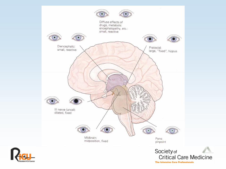
ParasympatheticParasympatheticcontrol of pupil size
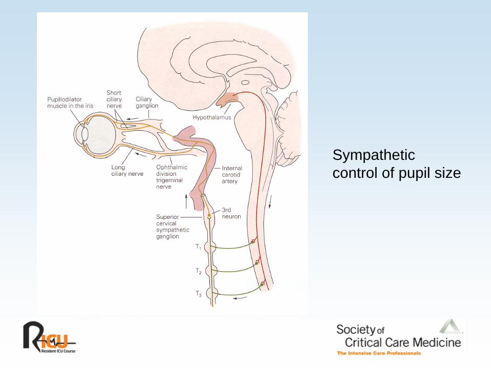
SympatheticSympatheticcontrol of pupil size
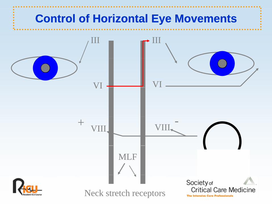
Control of Horizontal Eye MovementsControl of Horizontal Eye Movements
III III
VI VIVI
VIII VIII -+
MLF
Neck stretch receptors
Assessing Eye MovementsAssessing Eye Movements
Spontaneous horizontal conjugate eye movements prove that theSpontaneous horizontal conjugate eye movements prove that the brainstem centers for eye movement are intact.• These overlap the portion of the reticular formation necessary for
consciousness.consciousness.• Therefore, coma in a patient with roving horizontal conjugate eye
movements is not due to brainstem dysfunction.
Assessing Eye MovementsAssessing Eye Movements
If there are no spontaneous eye movements, attempt to trigger p y , p ggthem.• In the absence of cervical spine disease, test cervico-ocular
reflexes (“dolls’ eyes”):- Turning the head to the right should cause the eyes to go left,
and vice versa.- Same meaning as spontaneous movements regarding the
brain stembrain stem- Partial responses mean a problem involving the brainstem or
cranial nerves (use the diagram to determine where the problem lies)problem lies).
Assessing Eye MovementsAssessing Eye Movements
Vestibulo-ocular testing (“cold calorics”)Vestibulo ocular testing ( cold calorics )• Check for tympanic membrane perforation first• 50 - 60 mL ice water in one extra-ocular canal using soft tubing
(e g from a butterfly; do not use an IV catheter which can(e.g., from a butterfly; do not use an IV catheter, which can penetrate the tympanic membrane)
• Tonic deviation of both eyes toward cold ear indicates intact brainstem functionbrainstem function.
• Wait for one ear to warm up before testing the other ear.

Assessing Eye MovementsAssessing Eye Movements
Nystagmus away from the cold ear is due to cortical correction of the brainstem-induced eye movement and
th ti t i t tmeans the patient is not comatose.

Respiratory Patterns in ComaRespiratory Patterns in Coma
Cheyne – Stokes respiration: bilateral hemispheralCheyne Stokes respiration: bilateral hemispheral dysfunction Central reflex hyperpnea: midbrain dysfunction causing neurogenic pulmonary edemaneurogenic pulmonary edema• rarely see true central neurogenic hyperventilation with this lesion;
central hyperventilation is common with increased ICP

Respiratory Patterns in ComaRespiratory Patterns in Coma
Apneustic respiration (inspiratory cramp lasting up to 30 sec):Apneustic respiration (inspiratory cramp lasting up to 30 sec): pontine lesion
Cluster breathing (Biot breathing): pontine lesion
Ataxic respiration: pontomedullary junction lesion
Motor ResponsesMotor Responses
Defensive, avoidance, or withdrawal - indicative of corticalDefensive, avoidance, or withdrawal indicative of cortical function (the patient is not comatose)Flexor (decorticate) posturing - the cortex is not in control of the spinal cord but the midbrain (red nucleus) isthe spinal cord, but the midbrain (red nucleus) isExtensor (decerebrate) posturing - the midbrain is not in control but the pontomedullary region (vestibular nuclei) isGoing from flexion to extension indicates worsening; extension to flexion, improvement

Increased Intracranial PressureIncreased Intracranial Pressure
The volume of the skull is a constant (Monro-KellieThe volume of the skull is a constant (Monro Kellie hypothesis) which contains:• Brain• Blood• Blood• CSF
An increase in the volume of any of these or the introduction of alien tissue (e.g., tumor) will raise ICP.

Increased Intracranial PressureIncreased Intracranial Pressure
Initially, the ICP rises slowly as volume is added (CSF and then y, y (blood exits the skull)
But as the volume increases to rise, compliance worsens and the pressure rises rapidly:• This impairs arterial blood flow, producing ischemia• Focal increases in volume also cause herniation from high
pressure compartments to lower pressure ones.• Cerebral Perfusion Pressure = MAP – ICP (or CVP)
• Keep CPP > 60, ICP < 20• Keep Brain O2 (discussed later) > 20
Increased Intracranial PressureIncreased Intracranial Pressure
The standard theory of coma due to rostro-caudal brainstemThe standard theory of coma due to rostro caudal brainstem movement has been supplanted by Ropper’s lateral shift theory.
Shift is often heralded by a third cranial nerve palsy (usually causing a dilated pupil before failure of extra-ocular movements).
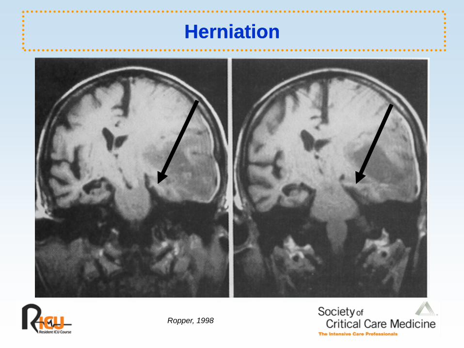
HerniationHerniation
Ropper, 1998
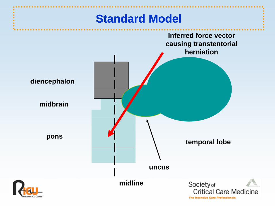
Standard ModelStandard ModelInferred force vector
causing transtentorialherniation
diencephalon
midbrain
ponstemporal lobe
uncus
midline
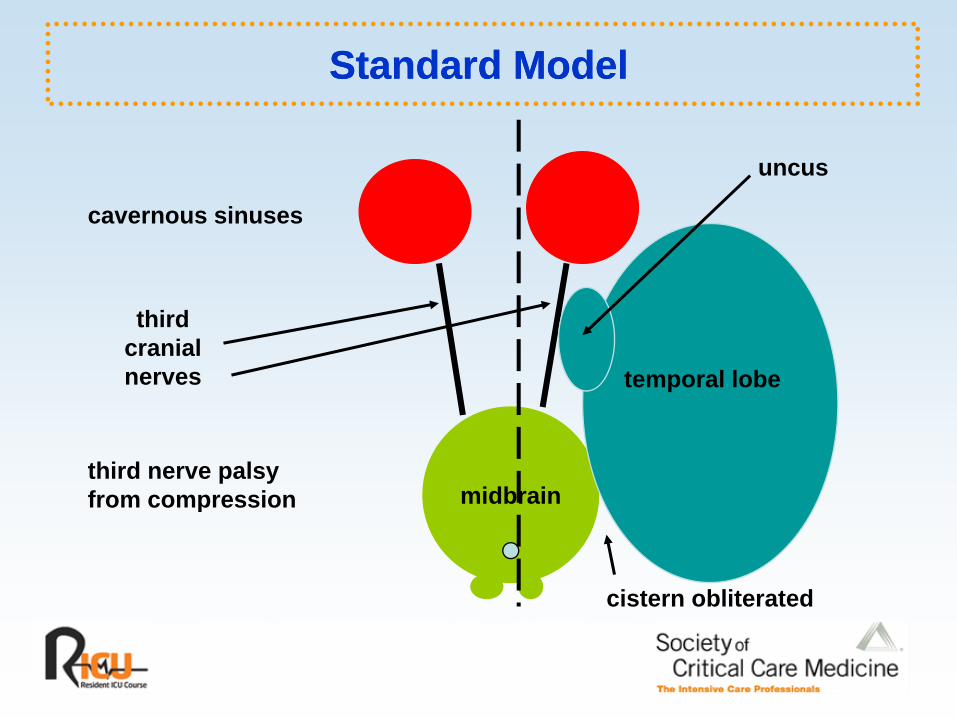
Standard ModelStandard Model
uncus
cavernous sinuses
temporal lobe
thirdcranialnerves
midbrainthird nerve palsyfrom compression
cistern obliterated
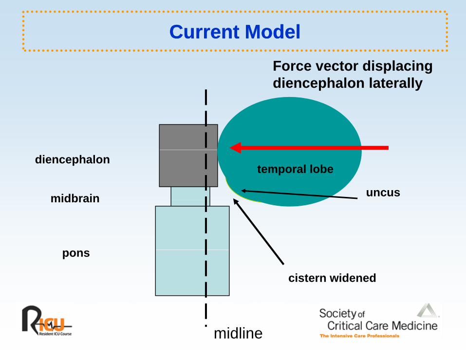
Current ModelCurrent ModelForce vector displacingdiencephalon laterally
diencephalon
midbrain
temporal lobe
uncusmidbrain
pons
cistern widened
midline
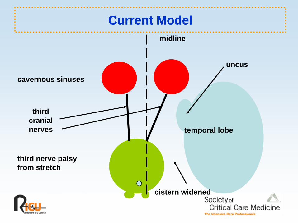
Current ModelCurrent Model
uncus
midline
uncus
cavernous sinuses
l l b
thirdcranial
temporal lobenerves
third nerve palsythird nerve palsyfrom stretch
cistern widenedcistern widened
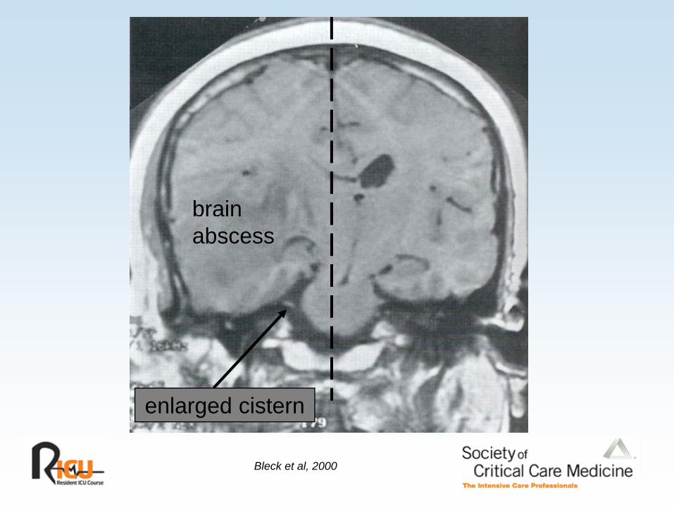
brainabscess
enlarged cistern
Bleck et al, 2000
Coma ScalesComa Scales
Standard: Glasgow coma scale (GCS)
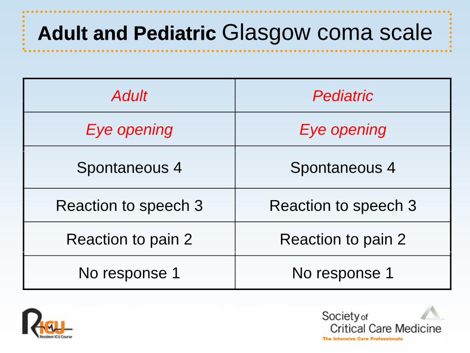
Adult and Pediatric Adult and Pediatric Glasgow coma scale
Adult PediatricAdult Pediatric
Eye opening Eye opening
Spontaneous 4 Spontaneous 4
R ti t h 3 R ti t h 3Reaction to speech 3 Reaction to speech 3
Reaction to pain 2 Reaction to pain 2
No response 1 No response 1
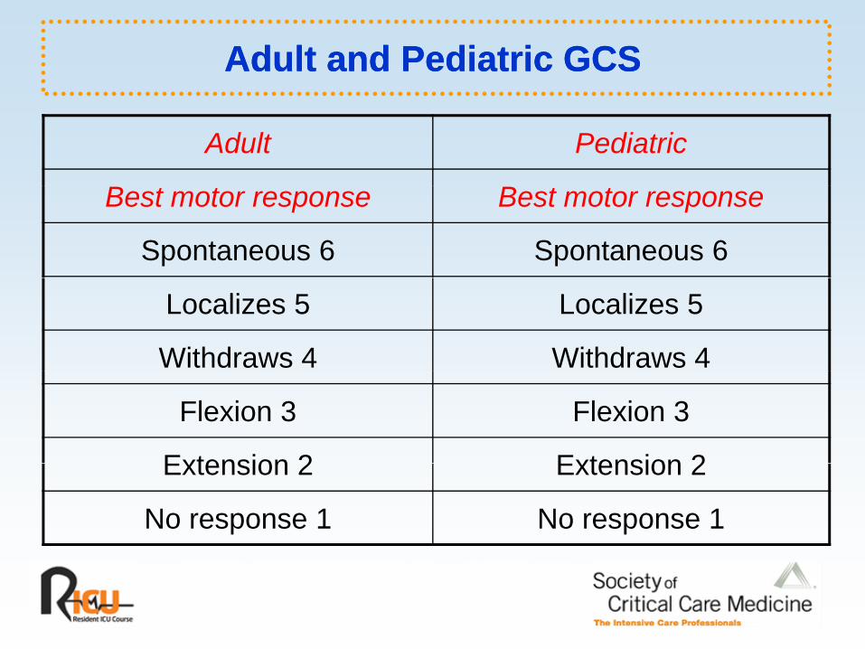
Adult and Pediatric GCSAdult and Pediatric GCS
Adult Pediatric
Best motor response Best motor response
Spontaneous 6 Spontaneous 6
Localizes 5 Localizes 5
Withdraws 4 Withdraws 4
Flexion 3 Flexion 3
Extension 2 Extension 2Extension 2 Extension 2
No response 1 No response 1

Adult and Pediatric GCSAdult and Pediatric GCS
Adult Verbal
Oriented 5
Confused 4
Inappropriate words 3
Incomprehen-sible sounds 2sounds 2
No response 1
M it i S d ti St tM it i S d ti St tMonitoring Sedation Status over Monitoring Sedation Status over Time in ICU Patients: Reliability Time in ICU Patients: Reliability yy
and Validity of the Richmond and Validity of the Richmond AgitationAgitation--Sedation ScaleSedation ScaleAgitationAgitation--Sedation ScaleSedation Scale
JAMA. 2003;289:2983-91.

Increased Intracranial PressureIncreased Intracranial Pressure
ManagementManagement• Make plans to correct the underlying pathophysiology if possible.• Airway control and prevention of hypercapnea are crucial:
Wh i t b ti ti t ith l t d ICP thi t l- When intubating patients with elevated ICP use thiopental, etomidate, or intravenous lidocaine to blunt the increase in ICP associated with laryngoscopy and tube passage.
ICP it i ll d d t id th• ICP monitoring usually needed to guide therapy• Cortical Oximetry (Lycox catheter) may be beneficial
• Measures O2 tension in parenchyma in area of catheter• Keep Brain O2 > 20
Increased Intracranial PressureIncreased Intracranial Pressure
Posture and head positionPosture and head position• Avoid jugular vein compression
- Head should be in neutral position- Cervical collars should not be too tight
• Elevation of the head and trunk may improve jugular venous return.

Increased Intracranial PressureIncreased Intracranial Pressure
Hyperventilation (PaCO 30-35 mmHg) works byHyperventilation (PaCO2 30-35 mmHg) works by decreasing blood flow and should be reserved for emergency treatment and only for brief periods.• The major determinant of arteriolar caliber is the extracellular
pH, not actually the PaCO2, but this is the parameter we can control.
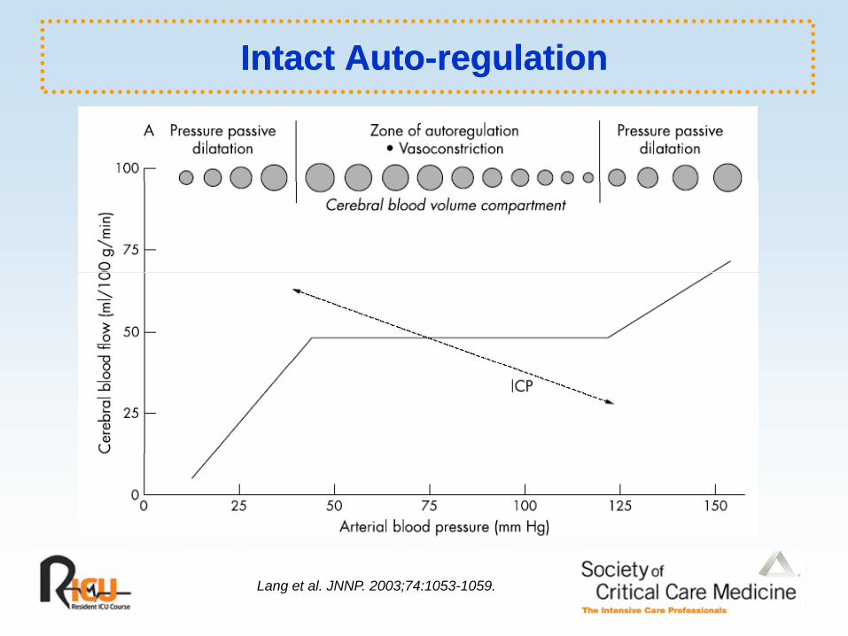
Intact AutoIntact Auto--regulationregulation
Lang et al. JNNP. 2003;74:1053-1059.

Defective AutoDefective Auto--regulationregulation

Increased Intracranial PressureIncreased Intracranial Pressure
Pharmacologic optionsPharmacologic options• Mannitol 0.25 gm/kg q4h (may need to increase dose over time)• Hypertonic saline (requires central line)
- 3%3%- 7.5%- 23.4% (30 mL over 10 min)
• Steroids only for edema around tumors or abscesses (not for use• Steroids only for edema around tumors or abscesses (not for use in trauma or cerebrovascular disease)
Increased Intracranial PressureIncreased Intracranial Pressure
SedationSedation• Benzodiazepines• Propofol• Barbiturates• Barbiturates
Works by decreasing cerebral metabolic rate, which is coupled to blood flowblood flow• Requires autoregulation, which often fails in patients with elevated ICP• Often causes a drop in MAP, impairing cerebral perfusion and thus
requiring vasopressors (e g norepinephrine)requiring vasopressors (e.g., norepinephrine)
Increased Intracranial PressureIncreased Intracranial Pressure
High dose barbituratesHigh-dose barbiturates• E.g., pentobarbital 5 – 12 mg/kg load followed by infusion to
control ICPV l d d• Very rarely needed
Increased Intracranial PressureIncreased Intracranial Pressure
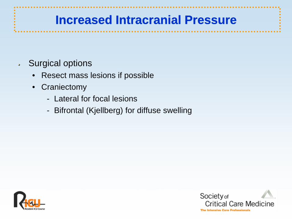
Surgical optionsSurgical options• Resect mass lesions if possible• Craniectomy
- Lateral for focal lesions- Bifrontal (Kjellberg) for diffuse swelling
Classification of Classification of Neurogenic Respiratory FailureNeurogenic Respiratory FailureNeurogenic Respiratory FailureNeurogenic Respiratory Failure
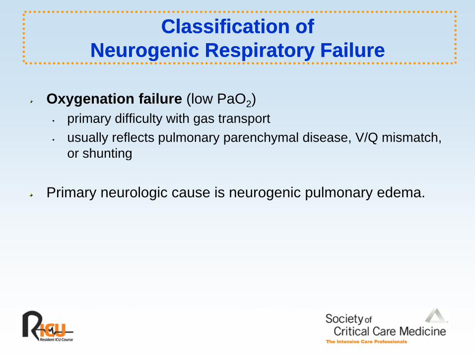
Oxygenation failure (low PaO2)Oxygenation failure (low PaO2)• primary difficulty with gas transport• usually reflects pulmonary parenchymal disease, V/Q mismatch,
or shuntingor shunting
Primary neurologic cause is neurogenic pulmonary edema.

Neurogenic Pulmonary EdemaNeurogenic Pulmonary Edema
A state of increased lung water (interstitial and sometimesA state of increased lung water (interstitial and sometimes alveolar):• as a consequence of acute nervous system disease
in the absence of• in the absence of - cardiac disorders (CHF),- pulmonary disorders (ARDS), or- hypervolemia
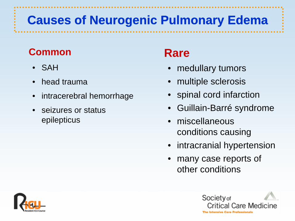
Causes of Neurogenic Pulmonary EdemaCauses of Neurogenic Pulmonary Edema
Common Rare• SAH
• head trauma
intracerebral hemorrhage
• medullary tumors• multiple sclerosis• spinal cord infarction• intracerebral hemorrhage
• seizures or status epilepticus
• spinal cord infarction• Guillain-Barré syndrome• miscellaneous
conditions causing • intracranial hypertension• many case reports of• many case reports of
other conditions

Classification of Classification of Neurogenic Respiratory FailureNeurogenic Respiratory FailureNeurogenic Respiratory Failure Neurogenic Respiratory Failure
Ventilatory failure (inadequate minute ventilation [VE] for theVentilatory failure (inadequate minute ventilation [VE] for the volume of CO2 produced):
• In central respiratory failure, the brainstem response to CO2 is inadequate, and the PaCO2 begins to rise earlythe PaCO2 begins to rise early.
• In neuromuscular ventilatory failure, the tidal volume begins to fall, and the PaCO2 is initially normal (or low).

Causes of Neurogenic Ventilatory FailureCauses of Neurogenic Ventilatory Failure
Most common causes are:Most common causes are:• Myasthenia gravis• Guillain-Barré syndrome• Critical illness polyneuropathy, myopathy• Cervical spine disease
Many rarer causes
Management of Neurogenic Ventilatory Management of Neurogenic Ventilatory FailureFailureFailureFailure
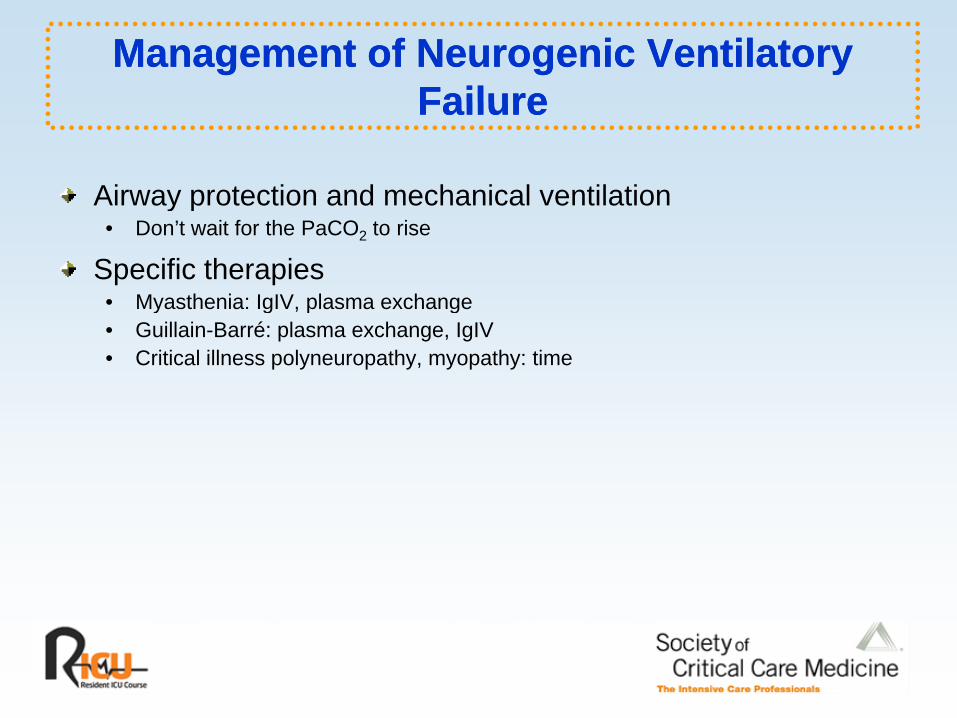
Airway protection and mechanical ventilationAirway protection and mechanical ventilation• Don’t wait for the PaCO2 to rise
Specific therapies• Myasthenia: IgIV, plasma exchangeMyasthenia: IgIV, plasma exchange• Guillain-Barré: plasma exchange, IgIV• Critical illness polyneuropathy, myopathy: time
Status EpilepticusStatus Epilepticus
Types of status epilepticus:Types of status epilepticus:• Convulsive• Nonconvulsive
Status EpilepticusStatus Epilepticus
DefinitionDefinition• Typically diagnosed after 30 min of either:
- Continuous seizure activity I t itt t i ith t b t- Intermittent seizures without recovery between
• Don’t wait for 30 min to treat:- Seizures become more difficult to treat the longer they last.- More systemic complications occur (e.g., aspiration).- Most seizures end spontaneously within 7 min in adults and
12 min in children:• These are reasonable points to start treating to terminate
seizures in order to prevent the establishment of status.

Status EpilepticusStatus Epilepticus
Initial treatmentInitial treatment• Lorazepam (Ativan) IV 0.1 mg/kg• Alternatives:
• PropofolPropofol- Phenobarbital IV 20 mg/kg- Valproate IV 20 - 30 mg/kg
• If IV access cannot be establishedIf IV access cannot be established,- Midazolam (Versed)(buccal, nasal, IM)
Failure of the first drug given in adequate dosage constitutesFailure of the first drug given in adequate dosage constitutes refractory status.
Status EpilepticusStatus Epilepticus
Treatment of refractory status (RSE)Treatment of refractory status (RSE)• Midazolam 0.2 mg/kg loading dose with immediate infusion 0.1 – 2.0 mg/kg/hr
- Must have EEG monitoring and demonstrate seizure suppression- After 12 hours free of seizures attempt to taper
M d th d ( h t i h b bit l ) t t- May need other drugs (e.g., phenytoin, phenobarbital ) to prevent recurrence• Other options for RSE
- Propofol
- Pentobarbital
Acute Stroke InterventionAcute Stroke Intervention
Intravenous thrombolysis is indicated for patients with:Intravenous thrombolysis is indicated for patients with:• A clinical diagnosis of ischemic stroke• A CT scan excluding intracerebral hemorrhage• Onset of symptoms less than 3 hours before starting treatmentOnset of symptoms less than 3 hours before starting treatment• No contraindications
rt-PA 0.9 mg/kg (up to 90 mg)• 10% bolus remainder over 60 min• 10% bolus, remainder over 60 min

Acute Stroke InterventionAcute Stroke Intervention
Between 3 and 6 hours intra-arterial therapy may be anBetween 3 and 6 hours, intra arterial therapy may be an option
No role for acute heparin in evolving or completed stroke• May be needed later for secondary prevention in patients with atrial fibrillation

Intracerebral HemorrhageIntracerebral Hemorrhage
Hypertensive hemorrhages occur in the:Hypertensive hemorrhages occur in the:• Putamen• Thalamus• Pons• Cerebellum
Patients with hemorrhages elsewhere, or without a history of hypertension need to be worked up for underlying vascularhypertension, need to be worked up for underlying vascular lesions or a bleeding diathesis.
Intracerebral HemorrhageIntracerebral Hemorrhage
For supratentorial hemorrhage the major determinant ofFor supratentorial hemorrhage, the major determinant of survival is hemorrhage volume:
• < 30 mL usually survive• > 60 mL frequently dieq y
Patients with cerebellar hemorrhages often benefit from surgical evacuation
P d b f i l fi di d l• Proceed before cranial nerve findings develop.
Intracerebral HemorrhageIntracerebral Hemorrhage
Management remains controversialManagement remains controversial• Airway control• Lowering mean arterial pressure may limit hemorrhage growth• Correct coagulopathy• Surgical intervention not routinely useful
- May be helpful with superficial lesions

Subarachnoid HemorrhageSubarachnoid Hemorrhage
Most commonly due to trauma and then ruptured aneurysmMost commonly due to trauma and then ruptured aneurysm
Present with sudden headache, often diminished consciousness
• Focal findings suggest intracerebral hemorrhage, which may occur due to dissection of blood from the bleeding aneurysm into the cortex.

Current Management Strategies for SAHCurrent Management Strategies for SAH
Early definitive aneurysm obliterationEarly definitive aneurysm obliteration
Induce hypertension and increase cardiac output to treat vasospasm if it develops (after aneurysm is clipped)
Biggest risk between days 3-7 following bleed
Nimodipine or nicardipine to relieve or ameliorate the effects of vasospasmof vasospasm

Current Management Strategies for SAHCurrent Management Strategies for SAH
Interventional neuroradiologic techniques (e.g., angioplastyInterventional neuroradiologic techniques (e.g., angioplasty and intra-arterial verapamil or nicardipine infusion) to treat vasospasm Ventricular drainage to treat hydrocephalusVentricular drainage to treat hydrocephalus
Complications of Aneurysmal SAHComplications of Aneurysmal SAH
Rebleeding Arrhythmias and otherRebleedingCerebral vasospasmVolume disturbances
Arrhythmias and other cardiovascular complicationsCNS infections
Osmolar disturbancesSeizures
CNS infectionsOther complications of critical illness

Aneurysmal RebleedingAneurysmal Rebleeding
Risk of rebleeding from unsecured aneurysms:Risk of rebleeding from unsecured aneurysms:• about 4% on the first post-bleed day • about 1.5% per day up to day 28
Mortality of rebleeding following the diagnosis of SAH y g g gexceeds 75%. Rebleeding is more frequent in:
• patients with higher grades of SAH• patients with higher grades of SAH • women• those with systolic blood pressures over 170 mmHg

Volume and Osmolar DisturbancesVolume and Osmolar Disturbances
Reported in about 30% of SAH patients Most common problem is cerebral salt wasting
• SIADH should not be diagnosed in the period of risk for vasospasm.• Acute SAH patients should never be allowed to become volume depleted.• The primary problem is excess of natriuretic factors, with secondary water
retention to attempt to maintain volume (converse of SIADH).
Volume and Osmolar DisturbancesVolume and Osmolar Disturbances
Prophylaxis: maintain adequate salt intakeProphylaxis: maintain adequate salt intake• (e.g., 3L+ saline/d)• some use mineralocorticoid supplementation
If hypo-osmolality occurs need to increase the osmolality ofIf hypo osmolality occurs, need to increase the osmolality of the fluids administered to exceed that of the urine excreted
• hypertonic saline (1.8% - 3%) as needed• some also give supplemental salt enterallysome also give supplemental salt enterally

ConclusionConclusion
• Brain losses auto-regulation for up to 7Brain losses auto regulation for up to 7 days post-injury– CPP can be affected directlyCPP can be affected directly– Tx guided by cortical oximetry and ICP monitoring
• Minimize secondary insultsMinimize secondary insults– Keep CPP > 60 (or SBP > 100)– Keep PCO2 30-35eep CO 30 35
• Time = Brain in injury and CVA– Surgical intervention ASAP when neededSurgical intervention ASAP when needed
Head TraumaHead Trauma

Secondary Injury in Head TraumaSecondary Injury in Head Trauma
Hypoxia and hypotension are the 2 major causes ofHypoxia and hypotension are the 2 major causes of secondary CNS injury following head trauma.
Even in the best intensive care units, these complications occur frequently.
Preventing hypoxia and hypotension could have the greatest effect of any currently available treatment for head traumaeffect of any currently available treatment for head trauma.

Fluid Thresholds and Outcome from Severe Fluid Thresholds and Outcome from Severe Brain InjuryBrain InjuryBrain InjuryBrain Injury
Retrospective study (from the NIH multicenter hypothermiaRetrospective study (from the NIH multicenter hypothermia trial data) of the effect on GOS of ICP, MAP, CPP, and fluid balance at 6 months after injury
Univariate predictors of poor outcome: • ICP > 25 mm Hg• MAP < 70 mm Hg or
CPP 60 H d fl id b l 594 L• CPP < 60 mm Hg and fluid balance < -594 mL
Clifton et al. Crit Care Med 2002;30:739–745.

Fluid Thresholds and Outcome from Severe Fluid Thresholds and Outcome from Severe Brain InjuryBrain InjuryBrain InjuryBrain Injury
Conclusions: Exceeding thresholds of ICP MAP CPP andConclusions: Exceeding thresholds of ICP, MAP, CPP, and fluid volume may be detrimental to severe brain injury outcome.
Fluid balance lower than -594 mL was associated with an adverse effect on outcome, independent of its relationship to intracranial pressure, mean arterial pressure, or cerebral perfusion pressure.

Diffuse Axonal InjuryDiffuse Axonal Injury
An active process triggered by the injury that takes about 24An active process triggered by the injury that takes about 24 hours to develop in humansMay occur without any radiographic abnormalityFrequently seen in areas of radiographically apparent “shear injury”
• this latter finding usually occurs at the grey-white junction
Is a major cause of long-term disability

Rosner View of Cerebral Blood FlowRosner View of Cerebral Blood Flow

Oxygenation MonitoringOxygenation Monitoring
Jugular bulb catheterJugular bulb catheter• jugular venous blood oxygen saturation
- A-V differences in saturation, content, lactate
Direct cortical oxygen sensors (Licox)Direct cortical oxygen sensors (Licox)

Management Management
Resuscitation and airway managementResuscitation and airway management• avoid hypoxia and hypotension• concomitant cervical spine lesions• methods of intubation
orotracheal with inline stabilization- orotracheal with inline stabilization• no nasal tubes (tracheal or gastric)
- fiberoptic• posture and head position
ff t ICP d CPP- effects on ICP and CPP
Management Management
Antiseizure drugsAntiseizure drugs• phenytoin 20 mg/kg • only for the first week for patients without seizures
Free radical scavengersg• potential future therapies
Nutrition and GI bleeding prophylaxisTh b b li h l iThromboembolism prophylaxis

Category Description % of pts Good/moderate Severe/vegetative Dead
No CT data 2.3 5.9 0.0 94.1
Diffuse injury I No visible pathology on CT 7.0 61.6 28.8 9.6
Diffuse injury IICisterns visible, shift 0 – 5 mm, no high or mixed density lesion 23.7 34.5 52.0 13.5Diffuse injury II no high or mixed density lesion > 25 cm3
23.7 34.5 52.0 13.5
Diffuse injury III (swelling)
Cisterns compressed or absent, shift 0 – 5 mm, no high or mixed density lesion > 25 20.5 16.4 49.7 34.0
cm3
Diffuse injury IV (shift)
Shift > 5 mm, no high or mixed density lesion > 25 cm3 4.3 6.2 37.6 56.2
Evacuated mass lesion
Any lesion surgically evacuated 37.0 22.8 38.4 38.8
Nonevacuated mass
High or mixed density lesion > 25 cm3 not surgically 4.8 11.1 36.1 52.8mass g yevacuated
Brainstem injury (no brainstem reflexes by physical exam) 0.4 0.0 33.3 66.7
Marshall et al. J Neurotrauma. 1992;9 Suppl 1:S287-92.
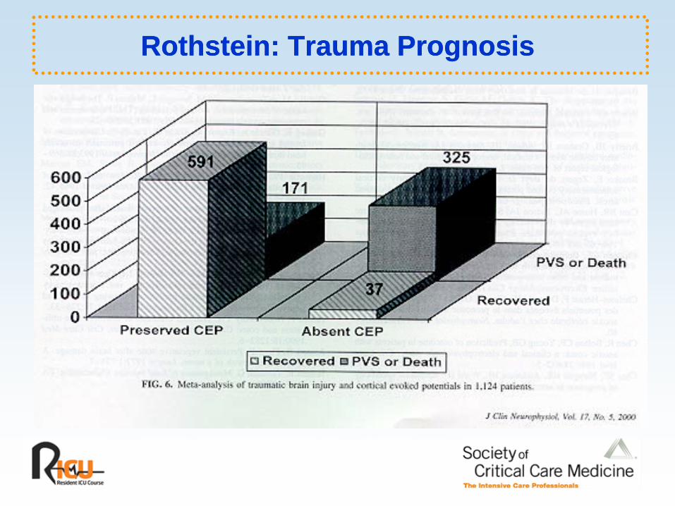
Rothstein: Trauma PrognosisRothstein: Trauma Prognosis


Return to WorkReturn to Work
Ab t 40% f ith l i d 30% fAbout 40% of persons with paraplegia and 30% of persons with tetraplegia (quadriplegia) eventually return to work.


Complete SCIComplete SCI
L f ll f ti b l th l l f th l iLoss of all function below the level of the lesion
Typically associated with spinal shock

Types of Incomplete SCITypes of Incomplete SCI
Central cord syndromeCentral cord syndrome
Anterior cord syndrome
Brown-Sequard syndromeBrown Sequard syndrome
Spinal cord injury without radiologic abnormality (SCIWORA)
Central Cord SyndromeCentral Cord Syndrome
Typically results from an extension injuryTypically results from an extension injury
Greater impairment of upper than lower extremity function
Urinary retentionUrinary retention
Sparing of sacral sensation

M d tModerate
MarkedMarked

Anterior Cord SyndromeAnterior Cord Syndrome
Due either to:Due either to: • Compression of the anterior portion of the cord by a vertebral body• Anterior spinal artery occlusion
Presents with preservation of dorsal column functionPresents with preservation of dorsal column function (vibration and position sense)
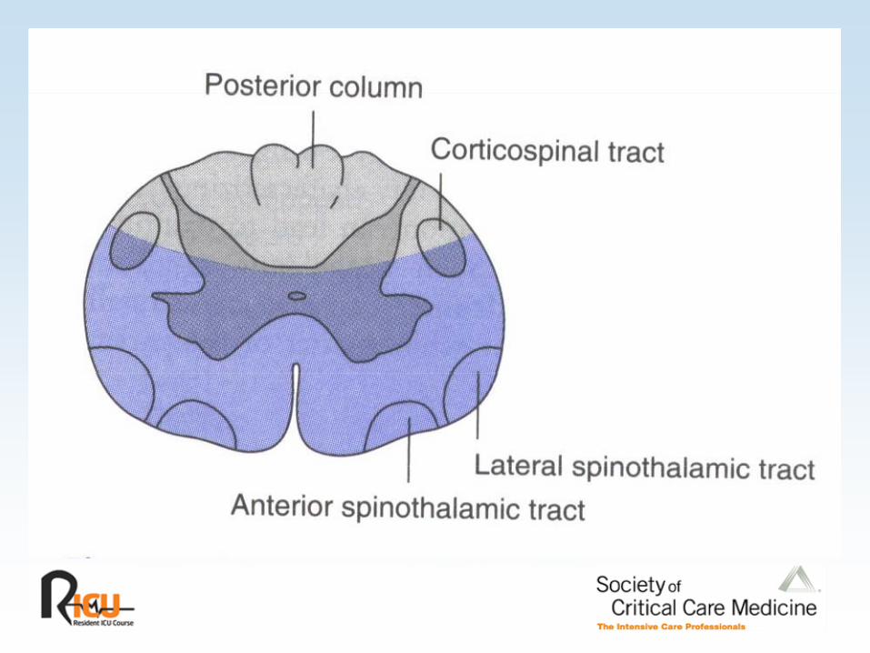

BrownBrown--Sequard SyndromeSequard Syndrome
H i ti f th dHemisection of the cord
Usually due to penetrating injury


Spinal Cord Injury Without Radiologic Spinal Cord Injury Without Radiologic Abnormality (SCIWORA)Abnormality (SCIWORA)Abnormality (SCIWORA)Abnormality (SCIWORA)
No bony abnormalities on plain film or CTNo bony abnormalities on plain film or CT• MRI may show abnormalities
Usually in children; symptoms may be transient at firstShould probably lead to immobilization to prevent subsequent development of cord damage
Secondary InjurySecondary Injury
After the initial macroscopic injury secondary injuries are anAfter the initial macroscopic injury, secondary injuries are an important cause of disability:
• Movement of unstable spine• Vascular insufficiencyy• Free radical induced damage
Neural Control of Blood Pressure and Neural Control of Blood Pressure and Blood FlowBlood FlowBlood FlowBlood Flow
Complete lesions above T1 will therefore eliminate allComplete lesions above T1 will therefore eliminate all sympathetic outflow.
Lesions between T1 and T6 will preserve sympathetic tone in the head and upper extremities but deny it to the adrenals and the lower extremities.
Lesions between T6 and the lumbar cord will preserveLesions between T6 and the lumbar cord will preserve adrenal innervation but denervate the lower extremities.

CNS Disturbances Affecting the CNS Disturbances Affecting the Cardiovascular SystemCardiovascular SystemCardiovascular SystemCardiovascular System
“Spinal” shock• Actually refers to the acute loss of tendon reflexes and muscle tone
below the level of a spinal cord lesion• However, neurogenic hypotension is very common and can be profound
with spinal cord lesions above T1:with spinal cord lesions above T1:- In the series of Vale et al, 40% of patients with complete cervical spinal cord
lesions were in neurogenic shock on presentation.• Hypotension in spinal shock is typically accompanied by bradycardia,
reflecting loss of cardiac sympathetic efferents and unopposed vagalreflecting loss of cardiac sympathetic efferents and unopposed vagal tone:
- These patients are unable to mount a tachycardic response to volume depletion.B f th i dil ti th b t till h l t d- Because of their vasodilation they are warm, but may still have elevated venous lactate concentrations.
CNS Disturbances Affecting the CNS Disturbances Affecting the Cardiovascular SystemCardiovascular SystemCardiovascular SystemCardiovascular System
• It is tempting to treat this hypotension with volume expansion, even if the patient is not volume depleted.
- Initially this is appropriate as venous return is frequently reduced. - However, this must be pursued cautiously.
• If the patient is conscious, making urine, and the venous lactate is decreasing, the MAP i b bl d tMAP is probably adequate.
• Neurogenic pulmonary edema is common in patients with cervical spinal cord lesions, complicating their management.
• These patients commonly develop pulmonary vascular redistribution, interstitial d i d A DO d i l l l d t PCWP i th 18edema, increased AaDO2, and on occasional alveolar edema at PCWPs in the 18
- 20 mmHg range:- May provide important clues to the mechanisms of NPE

Management of Cardiovascular Shock After Management of Cardiovascular Shock After Spinal Cord InjurySpinal Cord InjurySpinal Cord InjurySpinal Cord Injury
Always suspect associated injuries:Always suspect associated injuries:• Usual symptoms and physical findings may be absent due to the spinal cord
injury.
Volume resuscitation cannot be guided solely by physical g y y p yfindings:
• Hypotension and bradycardia will persist regardless of the volume of saline or colloid administered.
R l th i i d i t ith i tReplace the missing adrenergic tone with α-agonists (phenylephrine or norepinephrine depending on heart rate).
March 2002

Spinal Perfusion Pressure ManagementSpinal Perfusion Pressure Management
Developed by analogy to cerebral perfusion pressureDeveloped by analogy to cerebral perfusion pressure management
• Attempt to prevent cord ischemia by raising blood pressure.- Assumes that the same secondary injury mechanisms (hypotension and hypoxia)- Assumes that the same secondary injury mechanisms (hypotension and hypoxia)
worsen the outcome from spinal cord injury as in head injury• NASCIS II and III provide an inference that oxygen-derives free radicals
worsen outcome after spinal cord injury.
Spinal Perfusion Pressure ManagementSpinal Perfusion Pressure Management
Vale et al applied cerebral perfusion pressure managementVale et al applied cerebral perfusion pressure management principles to 77 patients with cervical and thoracic cord injuries.
• Place PA catheters and arterial linesPlace PA catheters and arterial lines• Maintained MAP > 85 mmHg
- Used “fluids, colloids, and vasopressors”• Did not specify how much of what
Vale FL et al J Neurosurg 1997;87:239-246

Spinal Perfusion Pressure ManagementSpinal Perfusion Pressure Management
30% of patients with complete cervical injuries were able to30% of patients with complete cervical injuries were able to walk at 1 year
• 20% had regained bladder function
“M h b tt th hi t i l t l t i th“Much better than historical controls or reports in the literature”

Penetrating Injuries of the Spinal CordPenetrating Injuries of the Spinal Cord
In a series 67 patients with penetrating injuries of the cordIn a series 67 patients with penetrating injuries of the cord, only 7% of patients presented with neurogenic shock:
• 74% of patients had significant blood loss, felt to explain their hypotension.
Zipnick RI et al J Trauma 1993;35:578-582

CNS Disturbances Affecting the CNS Disturbances Affecting the Cardiovascular SystemCardiovascular SystemCardiovascular SystemCardiovascular System
Autonomic dysreflexia:Autonomic dysreflexia:• Patients with lesions above T5 may develop hypertension and profuse sweating
in response to a distended viscus (usually the bladder).• Presumably represents adrenal release of catecholamines via spinal cord
pathways not being controlled by brainstem centers
Silver JR Spinal Cord 2000;38:229-233

Neurogenic Ventilatory Disturbance Neurogenic Ventilatory Disturbance Syndromes: Spinal Cord DisordersSyndromes: Spinal Cord DisordersSyndromes: Spinal Cord DisordersSyndromes: Spinal Cord Disorders
Lesions above or at C4Lesions above or at C4• Phrenic nerve failure
Lesions between C4 – T6L f t l i t t l t ti h t ll t i k d i• Loss of parasternal intercostal contraction causes chest wall to sink during inspiration, decreasing the tidal volume
• Loss of sympathetic innervation to the lungs can also prompt bronchospasm (imbalance of parasympathetic and sympathetic tone).

ManagementManagement
ABCsABCs• If intubation needed, use in-line stabilization
- Direct laryngoscopy vs. fiberoptic• Maintain blood pressure with volume, packed RBCs, vasopressors as needed
Prevent secondary injury• Log-rolling
C id it t h d i jConsider concomitant head injury
ManagementManagement
PharmacologicPharmacologic• Methylprednisolone 30 mg/kg bolus then 5.4 mg/kg/h for 23 – 47 hours
depending on latency from the injury- Starting 0 – 3 hours from injury: 23 hours duration- 3 – 8 hours: 47 hours3 8 hours: 47 hours- After 8 hours, do not start- Although there is still debate about its efficacy, this is often considered the “standard of
care.”- Not likely to be an anti-edema effect, since tirilazad (a non-glucocorticoid free radical
) i i l tscavenger) is equivalent.
Blood PressureBlood Pressure
No standards or guidelinesNo standards or guidelines
Options:• Avoid or correct hypotension (systolic BP < 90 mmHg)• Maintaining MAP between 85 and 90 mmHg for the first 7 days is recommended

DVT ProphylaxisDVT Prophylaxis
StandardsStandards• Either:
- LMW heparin, rotating bed, adjusted dose heparin (1.5 x control aPTT), or a combination of these, or
- Low-dose unfractionated heparin plus sequential compression devices or electrical p p q pstimulation
Guidelines• Low-dose unfractionated heparin alone is insufficient.• Oral anticoagulation alone probably not indicated
DVT ProphylaxisDVT Prophylaxis
OptionsOptions• 3-month duration of prophylaxis• Use IVC filters for patients failing anticoagulation or intolerant of it
Recommended