21
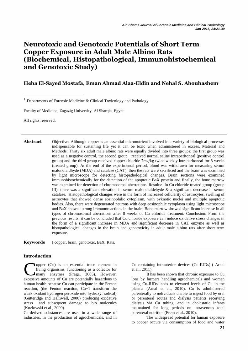
Ain Shams Journal of Forensic Medicine and Clinical Toxicology Jan 2015, 24:21-30
Neurotoxic and Genotoxic Potentials of Short Term Copper Exposure in Adult Male Albino Rats (Biochemical, Histopathological, Immunohistochemical and Genotoxic Study)
Heba El-Sayed Mostafa, Eman Ahmad Alaa-Eldin and Nehal S. Abouhashem1 1 Departments of Forensic Medicine & Clinical Toxicology and Pathology
Faculty of Medicine, Zagazig University, Al Sharqia, Egypt
All rights reserved.
Introduction
opper (Cu) is an essential trace element in
living organisms, functioning as a cofactor for
many enzymes (Fraga, 2005). However,
excessive amounts of Cu are potentially hazardous to
human health because Cu can participate in the Fenton
reaction, (the Fenton reaction, Cu+1 transform the
weak oxidant hydrogen peroxide into hydroxyl radical)
(Gutteridge and Halliwell, 2000) producing oxidative
stress and subsequent damage to bio molecules
(Kozlowski et al., 2009).
Cu-derived substances are used in a wide range of
industries, in the production of agrochemicals, and in
Cu-containing intrauterine devices (Cu-IUDs) ( Arnal
et al., 2011).
It has been shown that chronic exposure to Cu
ions by farmers handling agrochemicals and women
using Cu-IUDs leads to elevated levels of Cu in the
plasma (Arnal et al., 2010). Cu is administered
parenterally to individuals unable to ingest food by oral
or parenteral routes and dialysis patients receiving
dialysis via Cu tubing, and in cholestatic infants
maintained for long periods on intravenous total
parenteral nutrition (Frem et al., 2010).
The widespread potential for human exposure
to copper occurs via consumption of food and water
Abstract Objective: Although copper is an essential micronutrient involved in a variety of biological processes
indispensable for sustaining life yet it can be toxic when administered in excess. Material and
Methods: Thirty six adult male albino rats were equally divided into three groups; the first group was
used as a negative control, the second group received normal saline intraperitoneal (positive control
group) and the third group received copper chloride 7mg/kg twice weekly intraperitoneal for 8 weeks
(treated group). At the end of the experimental period, blood was withdrawn for measuring serum
malondialdhyde (MDA) and catalase (CAT), then the rats were sacrificed and the brain was examined
by light microscope for detecting histopathological changes. Brain sections were examined
immunohistochemically for the detection of the apoptotic BaX protein and finally, the bone marrow
was examined for detection of chromosomal aberrations. Results: In Cu chloride treated group (group
III), there was a significant elevation in serum malondialdehyde & a significant decrease in serum
catalase. Histopathological changes were in the form of increased cellularity of astrocytes, swelling of
astrocytes that showed dense eosinophilic cytoplasm, with pyknotic nuclei and multiple apoptotic
bodies. Also, there were degenerated neurons with deep eosinophilc cytoplasm using light microscope
and BaX showed strong immunoreactions in the brain. Bone marrow showed significant increase in all
types of chromosomal aberrations after 8 weeks of Cu chloride treatment. Conclusion: From the
previous results, it can be concluded that Cu chloride exposure can induce oxidative stress changes in
the form of a significant increase in MDA and significant decrease in CAT enzyme as well as
histopathological changes in the brain and genotoxicity in adult male albino rats after short term
exposure.
Keywords I copper, brain, genotoxic, BaX, Rats.
C
22 Mostafa et al., / Ain Shams J Forensic Med Clin Toxicol, Jan 2015 (24):21-30
contaminated with copper and also by inhalation of
industrial or cosmetics contained dust or fumes with it
(Mitra et al., 2012).
Various studies have demonstrated that
patients with neurodegenerative diseases had elevated
Cu concentrations in their plasma, suggesting a direct
or indirect involvement of Cu overload in the
progression and/or the etiology of neurologic diseases.
Ingestion of excessive Cu through drinking water and
vitamin supplements is at least partly responsible for
the development of Alzheimer’s disease in developed
countries, which to date is reaching epidemic
proportions (Arnal et al., 2014).
Copper is an essential metal for all living
organisms and is a component of many metalloproteins
such as antioxidant enzyme Cu–Zn superoxide
dismutase (Cu, Zn, -SOD) and cytochrome oxidase .
However, copper also plays a role in the pathogenesis
of neurodegenerative disease such as Alzheimer’s
disease (AD) (Lu et al., 2006).
Copper ions themselves, in the presence of
reducing agents such as ascorbate or glutathione,
causes various kinds of damage to DNA, and also it
helps other agents to damage DNA and chromatin, via
production of reactive oxygen species (ROS), Hence, it
is accused of direct relation to mutagenicity and
genotoxicity (Mitra et al., 2013).
The oxidative damage causes disruption of
lipid bi-layers, alterations in protein function, and
perhaps also altered gene expression. This potential
consequence is programmed as cell death, or apoptosis
(Linder, 2012).
A pro-apoptotic molecule (Bax) becomes an
integral membrane protein and cross-linkable as a
homodimer following a death stimulus and blocks the
anti-apoptotic effect of Bcl-2 ( Xu et al., 2012).
The Bcl-2 gene has been found to inhibit
apoptosis. Bax can also dimerise with itself, and
appears to promote apoptosis when overproduced. The
balance of Bax and Bcl-2 in a cell is one of the critical
factors determining whether the cell will undergo
apoptosis .Bax mRNA has been found to be up-
regulated and Bcl-2 mRNA down-regulated in mouse
brain neurons undergoing apoptosis. Various studies
showed increased levels of Bax protein in the rat and
gerbil hippocampus following cerebral ischemia
(MacGibbon et al., 1997).
Genotoxicity is a general term for any type of
DNA or chromosome damage or alteration. It includes
not only gene mutations, chromosome breaks and
rearrangements, but also interaction with or damage to
DNA, interference with the DNA replication or repair
processes (Zeiger and Hill, 2010).
The present work aimed to evaluate the
potential neurotoxic and genotoxic effects following
intraperitoneal injection of copper chloride (Cu cl2) for
8 weeks in adult male albino rats.
Materials and Methods Materials
a) Chemicals
1- Copper: copper (II) chloride dihydrate crystals (Cu
cl2) purchased from Biodiagnostic co. 29 El- Tahrer
Street- Dokki- Giza, Eygpt.
2- Normal saline: (0.9%Nacl solution) from a local
pharmacy.
b) Animals
Ethical consideration of the study:
-All ethically approved conditions used for animal
housing & handling were considered. -The animals
were acclimatized to experimental conditions prior to
the start of dosing for a period of 14 days to ascertain
their physical well-being and to exclude any diseased
animals (Semler, 1992). -Promotion of high slandered
of care & animal well-being at all times. Painless
procedures were performed with appropriate sedation
to avoid distress & pain.
A total of 36 adult male albino rats, weighing
180-200 gm, were kept on copper-free rat chow and
water ad libitum at the Breeding Animal House of the
Faculty of Medicine, Zagazig University.
Acclimatization period lasted for 2 weeks. The animals
were housed in filter-top plastic cages at a temperature
(23 ± 1°C), humidity (55 ± 5%) and artificially
illuminated (12:12 hours light: dark cycle) in a room
free from any source of chemical contamination. All
rats received human care in compliance with the
guidelines of the medical research ethics committee
(Institute of Laboratory Animal Resources et al., 1996).
c) Experimental Design:
The rats were divided into 3 numerically equal groups,
12 rats for each group. Six rats subjected to oxidative
stress markers measurements & neurotoxic studies and
the remaining 6 rats for chromosomal (genotoxic)
studies. They were divided as follows:
Group I (negative control group): Kept
without treatment for 8 weeks to measure the basic
parameters to be compared with other groups.
Group II (positive control group): Each rat in
this group received normal saline (vehicle for copper
chloride) twice weekly intraperitoneally for 8 weeks.
Group III (copper chloride group): Each rat of
this group received copper chloride (7mg/kg)
representing 1/10 of Ld 50 according to (Paget and
Branes,1964) which is equal to 1mg/kg in mice twice
weekly intraperitoneally for 8 weeks according to
Mitra et al. (2012) .
After the end of 8 weeks, 6 rats from each
group were anesthetized by ether inhalation for
drawing blood samples from the retro–orbital plexuses
of veins for performing the biochemical studies
malondialdehyde (MDA) and catalase (CAT). Then the
anesthetized rats were sacrificed & specimens from the
brain were taken. The remaining 6 rats in each group
were subjected to chromosomal studies.
Methods
1- Biochemical Study
Venous blood samples were collected from the retro-
orbital plexus of veins of the animals by capillary glass
23 Mostafa et al., / Ain Shams J Forensic Med Clin Toxicol, Jan 2015 (24):21-30
tubes after under light ether anaesthesia. The collected
blood was used as follows: Blood samples (about 3
mL) were collected in clean test tubes without
anticoagulant then the sera were separated by
centrifugation of blood 3000 r.p.m for 10 minutes. The
supernatant sera were pipetted off using fine tipped
automatic pipettes and stored in deep freeze at –20 C
until used for estimating serum MDA (Ohkawa et al.,
1979) and CAT (Aebi et al., 1984).
2- Histopathological examination
a) Hematoxylin & Eosin stain
After 8 weeks, 6 rats from each group were
anaesthetized with ether inhalation then sacrificed. The
brain was immediately dissected out and grossly
inspected to assess any abnormalities. Tissues from
brain were obtained as 5-mm thick sections, fixed with
10 % neutral l formalin for more than 12 hours
,dehydrated, embedded in paraffin, sectioned at5 μm,
mounted
b) on glass slides, and stained with
hematoxylin and eosin (H&E) stain (Wilson
and Gamble, 2002)
Immunohistochemical procedure according to
(Bancroft and Gamble, 2002):
Paraffin sections 3-5 um were deparaffinized in the
oven at 56 ˚C for 30 minutes, and inserted in xylene for
30 minutes. Tissues were rehydrated in descending
grades of alcohol 95%, 85%, and then 75% for 5
minutes each. Slides were rinsed with distilled water
for 5 minutes. Antigen retrieval was performed by
boiling in sodium citrate buffer (0.001M, pH 6) for 15
minutes in microwave. Endogenous peroxidase activity
was blocked by incubation with hydrogen peroxide for
10 minutes, then rinsed with distilled water.
Primary antibodies (2-3 drops of a Bax Ab-4
mouse monoclonal antibody) were applied on each
slide to cover the specimen overnight at 4˚C. After
washing 3 times with phosphate buffer saline (PBS),
the sections were incubated with biotinylated
secondary antibodies for 30 min. This was followed by
incubation with streptavidin-biotin-peroxidase
complex. After 3 rinses with PBS, the slides were
incubated with diaminobenzidine for 15 min. The
slides were then rinsed with H2O and counerstained
with hematoxylin for 3 minutes. This was followed by
washing in cold running water, then washing in
distilled water. Sections were dehydrated in ascending
grades of alcohol and cleared with xylene, then
coverslipped and examined.
3- Chromosomal study
After 8 weeks, 6 rats from each group were
anaesthetized with ether before sacrifice. The bone
marrow was processed using some modifications in
the method described by (Rabello-Gay and Ahmed,
1980). 1-Colchicine (BDH Chemicals Ltd Poole,
England) was prepared as 0.5% solution and injected
intramuscularly in the rats at a dose of 0.25 ml/100 gm
body weight. 2-Two hours after colchicine injection,
the rats were sacrificed by decapitation and both
femora were dissected and cut at both ends. 3-Four
milliliters of freshly prepared 0.075 M potassium
chloride pre-warmed at 37ºC were injected from one
end of the femur while collecting the cell suspension at
the other end in a centrifuge tube. 4-The cell
suspensions were incubated at 37ºC for one hour then,
centrifuged for five minutes at 1000 rpm and the
supernatant was discarded. 5-The cells were re-
suspended in 2 ml of freshly prepared fixative (3:1,
methanol:glacial acetic acid) and the tubes were
stoppered and left for at least 30 minutes up to 24
hours at 4ºC. 6-The cell suspension was centrifuged
for 3 minutes at 1000 rpm and the supernatant was
discarded. The cells were washed by 4 mL of the
fixative, centrifuged and the supernatant was discarded
for 2-3 times. 7-Finally, 1 ml of the fixative was added
to each cell precipitate to obtain a condensed cell
suspension. 8-The cell suspensions were dropped and
spread on clean glass slides previously put in cold 70%
ethanol. 9-The spreads were allowed to air dry and
stained in diluted Giemsa stain (1:10, Giemsa stock
solution: phosphate buffer pH 6, 8). 10-The glass
slides were examined using the oil immersion
objective lens of an ordinary light microscope. One
hundred metaphase cells were examined in each rat.
All metaphases were examined for structural
chromosomal abnormalities such as chromatid gaps-
break, fragments, deletions, dicentric chromosomes,
ring chromosomes, stretched chromatin material and
clumping. They were examined also for numerical
abnormalities, hypoploidy and hyperploidy.
4- Statistical analysis
For statistical analysis, SPSS version 13.0 for windows
programme was used. Data were represented as means
± SD. The differences were compared for statistical
significance by ANOVA. Descriptive data were
compared by chi-square test. Difference was
considered significant at p<0.05.
Results 1- Biochemical results: oxidative stress
markers (Table-1)
Serum Malondialdehyde (MDA)
Copper chloride treatment (group III) induced a highly
significant increase in the serum level of MDA
(p<0.001) compared to the control groups (group I &
II) after 8 weeks as shown in (Table-1).
Serum catalase enzyme (CAT)
The results revealed a highly significant decrease in the
serum level of CAT (p<0.001) in Cu chloride treated
group (group III) as compared to the control groups
(group I & II) after 8 weeks as shown in (Table-1).
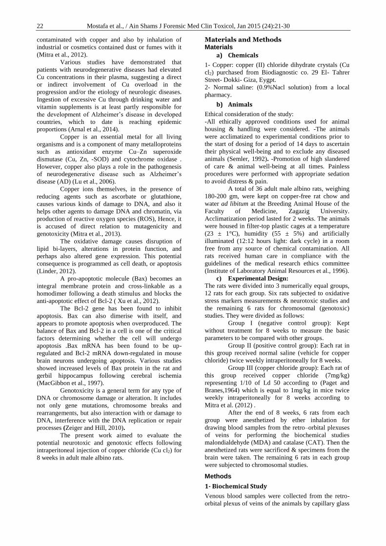
2- Histopathological result:
Gross inspection of the brain revealed normal
appearance in treated and control groups.
Examination of sections from the brain tissue
of control groups (group I & II) showed normal
neuronal structure formed of nerve cells with pale
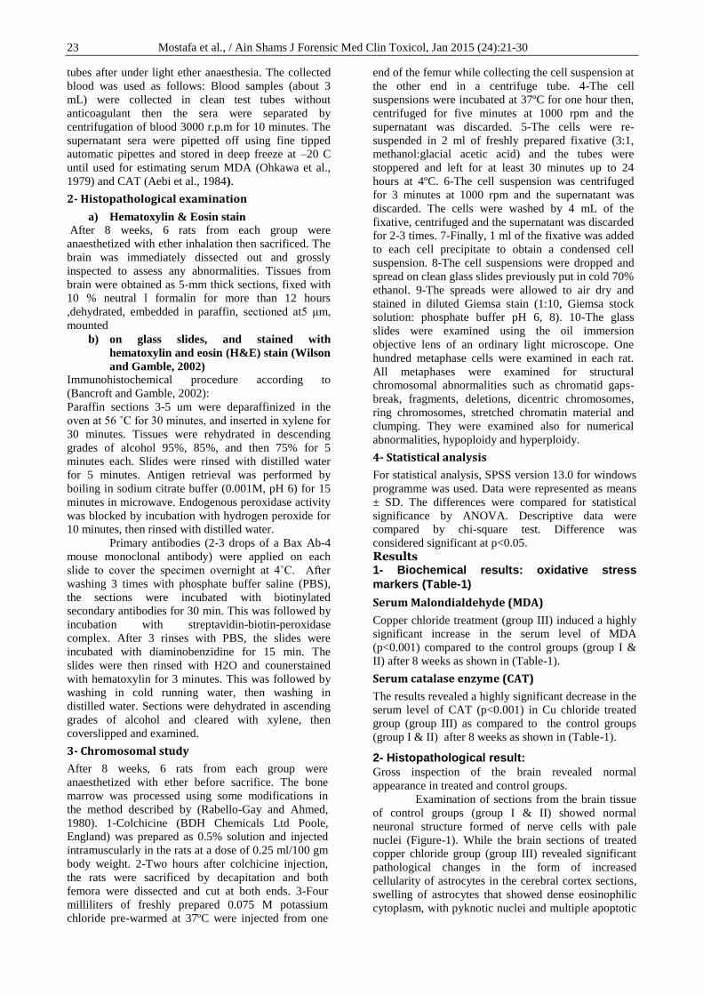
nuclei (Figure-1). While the brain sections of treated
copper chloride group (group III) revealed significant
pathological changes in the form of increased
cellularity of astrocytes in the cerebral cortex sections,
swelling of astrocytes that showed dense eosinophilic
cytoplasm, with pyknotic nuclei and multiple apoptotic
24 Mostafa et al., / Ain Shams J Forensic Med Clin Toxicol, Jan 2015 (24):21-30
bodies. Also, there were degenerated neurones with
deep eosinophilc cytoplasm (Figures- 2, 3&4).
3- Immunohistochemical results:
The brains of the both control groups (groups I & II)
showed no abnormal BaX protein expression (Figure-
5) while in the Cu chloride treated group (group III),
they showed increased BaX protein expression which
appeared as brownish cytoplasmic staining (Figure-6).
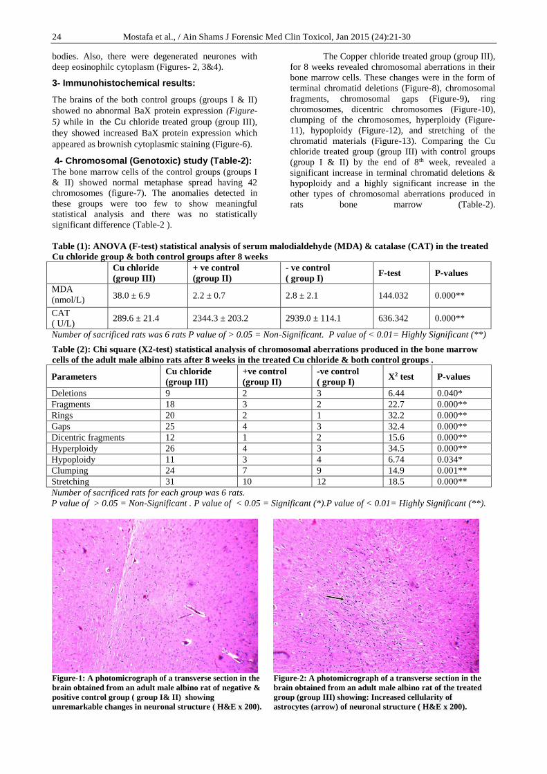
4- Chromosomal (Genotoxic) study (Table-2):
The bone marrow cells of the control groups (groups I
& II) showed normal metaphase spread having 42
chromosomes (figure-7). The anomalies detected in
these groups were too few to show meaningful
statistical analysis and there was no statistically
significant difference (Table-2 ).
The Copper chloride treated group (group III),
for 8 weeks revealed chromosomal aberrations in their
bone marrow cells. These changes were in the form of
terminal chromatid deletions (Figure-8), chromosomal
fragments, chromosomal gaps (Figure-9), ring
chromosomes, dicentric chromosomes (Figure-10),
clumping of the chromosomes, hyperploidy (Figure-
11), hypoploidy (Figure-12), and stretching of the
chromatid materials (Figure-13). Comparing the Cu
chloride treated group (group III) with control groups
(group I & II) by the end of 8th week, revealed a
significant increase in terminal chromatid deletions &
hypoploidy and a highly significant increase in the
other types of chromosomal aberrations produced in
rats bone marrow (Table-2).
Table (1): ANOVA (F-test) statistical analysis of serum malodialdehyde (MDA) & catalase (CAT) in the treated
Cu chloride group & both control groups after 8 weeks
Cu chloride
(group III)
+ ve control
(group II)
- ve control
( group I) F-test P-values
MDA
(nmol/L) 38.0 ± 6.9 2.2 ± 0.7 2.8 ± 2.1 144.032 0.000**
CAT
( U/L) 289.6 ± 21.4 2344.3 ± 203.2 2939.0 ± 114.1 636.342 0.000**
Number of sacrificed rats was 6 rats P value of > 0.05 = Non-Significant. P value of < 0.01= Highly Significant (**)
Table (2): Chi square (X2-test) statistical analysis of chromosomal aberrations produced in the bone marrow
cells of the adult male albino rats after 8 weeks in the treated Cu chloride & both control groups .
Parameters Cu chloride
(group III)
+ve control
(group II)
-ve control
( group I) X2 test P-values
Deletions 9 2 3 6.44 0.040*
Fragments 18 3 2 22.7 0.000**
Rings 20 2 1 32.2 0.000**
Gaps 25 4 3 32.4 0.000**
Dicentric fragments 12 1 2 15.6 0.000**
Hyperploidy 26 4 3 34.5 0.000**
Hypoploidy 11 3 4 6.74 0.034*
Clumping 24 7 9 14.9 0.001**
Stretching 31 10 12 18.5 0.000**
Number of sacrificed rats for each group was 6 rats.
P value of > 0.05 = Non-Significant . P value of < 0.05 = Significant (*).P value of < 0.01= Highly Significant (**).
Figure-1: A photomicrograph of a transverse section in the
brain obtained from an adult male albino rat of negative &
positive control group ( group I& II) showing
unremarkable changes in neuronal structure ( H&E x 200).
Figure-2: A photomicrograph of a transverse section in the
brain obtained from an adult male albino rat of the treated
group (group III) showing: Increased cellularity of
astrocytes (arrow) of neuronal structure ( H&E x 200).
25 Mostafa et al., / Ain Shams J Forensic Med Clin Toxicol, Jan 2015 (24):21-30
Figure-3: A photomicrograph of a transverse section in the
brain obtained from an adult male albino rat of the treated
group (group III) showing: degenerated neurones with deep
eosinophilc cytoplasm (arrow) and astrocytes with
pyknotic nuclei (double arrows) ( H&Ex 400).
Figure-4: A photomicrograph of a transverse section in the
brain obtained from an adult male albino rat of the treated
group (group III) showing: Astrocytes with multiple
apoptotic bodies (arrow) ( H&Ex 400).
Figure- 5: A photomicrograph of a transverse section of the
brain obtained from adult male albino rat
immunohistologically stained for Bax. Negative & positive
control groups (group I & II) showed no remarkable
immunostaining of Bax ( immunostain x400).
Figure- 6: A photomicrograph of a transverse section in the
brain obtained from adult male albino rat
immunohistologically stained for Bax of the treated group
(group III) showing: Astocytes with cytoplasmic
immunostaining of Bax ( immunostain x 400).
Figure-7: A photomicrograph of a metaphase spread
prepared from the bone marrow cells of an adult
male albino rat of negative & positive control group
(group I&II) showing no remarkable chromosomal
changes (Geimsa stain x 1000).
Figure-8: A photomicrograph of a metaphase spread
prepared from the bone marrow cells of an adult male
albino rat of Cu chloride treated group (group III)
showing terminal chromatid deletion ( arrow) (Geimsa
stain x 1000).
26 Mostafa et al., / Ain Shams J Forensic Med Clin Toxicol, Jan 2015 (24):21-30
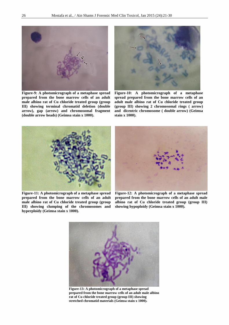
Figure-9: A photomicrograph of a metaphase spread
prepared from the bone marrow cells of an adult
male albino rat of Cu chloride treated group (group
III) showing terminal chromatid deletion (double
arrow), gap (arrow) and chromosomal fragment
(double arrow heads) (Geimsa stain x 1000).
Figure-10: A photomicrograph of a metaphase
spread prepared from the bone marrow cells of an
adult male albino rat of Cu chloride treated group
(group III) showing 2 chromosomal rings ( arrow)
and dicentric chromosome ( double arrow) (Geimsa
stain x 1000).
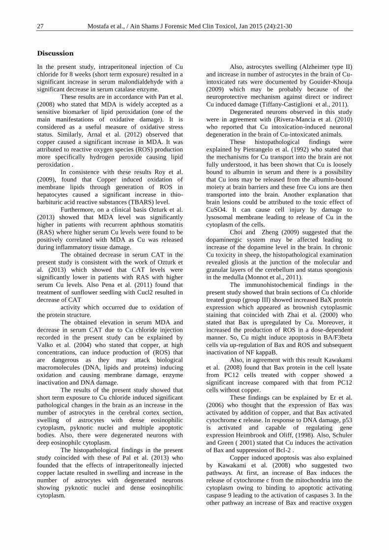
Figure-11: A photomicrograph of a metaphase spread
prepared from the bone marrow cells of an adult
male albino rat of Cu chloride treated group (group
III) showing clumping of the chromosomes and
hyperploidy (Geimsa stain x 1000).
Figure-12: A photomicrograph of a metaphase spread
prepared from the bone marrow cells of an adult male
albino rat of Cu chloride treated group (group III)
showing hypoploidy (Geimsa stain x 1000).
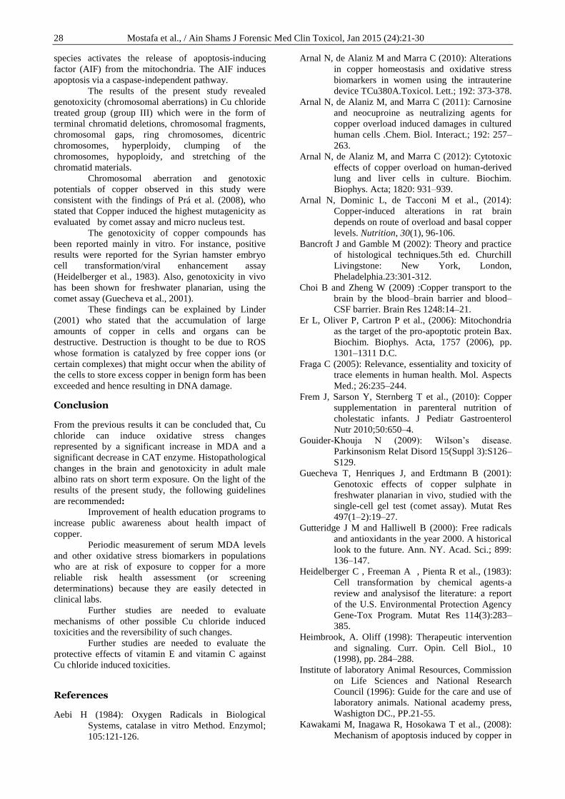
Figure-13: A photomicrograph of a metaphase spread
prepared from the bone marrow cells of an adult male albino
rat of Cu chloride treated group (group III) showing
stretched chromatid materials (Geimsa stain x 1000).
27 Mostafa et al., / Ain Shams J Forensic Med Clin Toxicol, Jan 2015 (24):21-30
Discussion
In the present study, intraperitoneal injection of Cu
chloride for 8 weeks (short term exposure) resulted in a
significant increase in serum malondialdehyde with a
significant decrease in serum catalase enzyme.
These results are in accordance with Pan et al.
(2008) who stated that MDA is widely accepted as a
sensitive biomarker of lipid peroxidation (one of the
main manifestations of oxidative damage). It is
considered as a useful measure of oxidative stress
status. Similarly, Arnal et al. (2012) observed that
copper caused a significant increase in MDA. It was
attributed to reactive oxygen species (ROS) production
more specifically hydrogen peroxide causing lipid
peroxidation .
In consistence with these results Roy et al.
(2009), found that Copper induced oxidation of
membrane lipids through generation of ROS in
hepatocytes caused a significant increase in thio-
barbituric acid reactive substances (TBARS) level.
Furthermore, on a clinical basis Ozturk et al.
(2013) showed that MDA level was significantly
higher in patients with recurrent aphthous stomatitis
(RAS) where higher serum Cu levels were found to be
positively correlated with MDA as Cu was released
during inflammatory tissue damage.
The obtained decrease in serum CAT in the
present study is consistent with the work of Ozturk et
al. (2013) which showed that CAT levels were
significantly lower in patients with RAS with higher
serum Cu levels. Also Pena et al. (2011) found that
treatment of sunflower seedling with Cucl2 resulted in
decrease of CAT
activity which occurred due to oxidation of
the protein structure.
The obtained elevation in serum MDA and
decrease in serum CAT due to Cu chloride injection
recorded in the present study can be explained by
Valko et al. (2004) who stated that copper, at high
concentrations, can induce production of (ROS) that
are dangerous as they may attack biological
macromolecules (DNA, lipids and proteins) inducing
oxidation and causing membrane damage, enzyme
inactivation and DNA damage.
The results of the present study showed that
short term exposure to Cu chloride induced significant
pathological changes in the brain as an increase in the
number of astrocytes in the cerebral cortex section,
swelling of astrocytes with dense eosinophilic
cytoplasm, pyknotic nuclei and multiple apoptotic
bodies. Also, there were degenerated neurons with
deep eosinophilc cytoplasm.
The histopathological findings in the present
study coincided with these of Pal et al. (2013) who
founded that the effects of intraperitoneally injected
copper lactate resulted in swelling and increase in the
number of astrocytes with degenerated neurons
showing pyknotic nuclei and dense eosinophilic
cytoplasm.
Also, astrocytes swelling (Alzheimer type II)
and increase in number of astrocytes in the brain of Cu-
intoxicated rats were documented by Gouider-Khouja
(2009) which may be probably because of the
neuroprotective mechanism against direct or indirect
Cu induced damage (Tiffany-Castiglioni et al., 2011).
Degenerated neurons observed in this study
were in agreement with (Rivera-Mancia et al. (2010)
who reported that Cu intoxication-induced neuronal
degeneration in the brain of Cu-intoxicated animals.
These histopathological findings were
explained by Pietrangelo et al. (1992) who stated that
the mechanisms for Cu transport into the brain are not
fully understood, it has been shown that Cu is loosely
bound to albumin in serum and there is a possibility
that Cu ions may be released from the albumin-bound
moiety at brain barriers and these free Cu ions are then
transported into the brain. Another explanation that
brain lesions could be attributed to the toxic effect of
CuSO4. It can cause cell injury by damage to
lysosomal membrane leading to release of Cu in the
cytoplasm of the cells.
Choi and Zheng (2009) suggested that the
dopaminergic system may be affected leading to
increase of the dopamine level in the brain. In chronic
Cu toxicity in sheep, the histopathological examination
revealed gliosis at the junction of the molecular and
granular layers of the cerebellum and status spongiosis
in the medulla (Monnot et al., 2011).
The immunohistochemical findings in the
present study showed that brain sections of Cu chloride
treated group (group III) showed increased BaX protein
expression which appeared as brownish cytoplasmic
staining that coincided with Zhai et al. (2000) who
stated that Bax is upregulated by Cu. Moreover, it
increased the production of ROS in a dose-dependent
manner. So, Cu might induce apoptosis in BA/F3beta
cells via up-regulation of Bax and ROS and subsequent
inactivation of NF kappaB.
Also, in agreement with this result Kawakami
et al. (2008) found that Bax protein in the cell lysate
from PC12 cells treated with copper showed a
significant increase compared with that from PC12
cells without copper.
These findings can be explained by Er et al.
(2006) who thought that the expression of Bax was
activated by addition of copper, and that Bax activated
cytochrome c release. In response to DNA damage, p53
is activated and capable of regulating gene
expression Heimbrook and Oliff, (1998). Also, Schuler
and Green ( 2001) stated that Cu induces the activation
of Bax and suppression of Bcl-2 .
Copper induced apoptosis was also explained
by Kawakami et al. (2008) who suggested two
pathways. At first, an increase of Bax induces the
release of cytochrome c from the mitochondria into the
cytoplasm owing to binding to apoptotic activating
caspase 9 leading to the activation of caspases 3. In the
other pathway an increase of Bax and reactive oxygen
28 Mostafa et al., / Ain Shams J Forensic Med Clin Toxicol, Jan 2015 (24):21-30
species activates the release of apoptosis-inducing
factor (AIF) from the mitochondria. The AIF induces
apoptosis via a caspase-independent pathway.
The results of the present study revealed
genotoxicity (chromosomal aberrations) in Cu chloride
treated group (group III) which were in the form of
terminal chromatid deletions, chromosomal fragments,
chromosomal gaps, ring chromosomes, dicentric
chromosomes, hyperploidy, clumping of the
chromosomes, hypoploidy, and stretching of the
chromatid materials.
Chromosomal aberration and genotoxic
potentials of copper observed in this study were
consistent with the findings of Prá et al. (2008), who
stated that Copper induced the highest mutagenicity as
evaluated by comet assay and micro nucleus test.
The genotoxicity of copper compounds has
been reported mainly in vitro. For instance, positive
results were reported for the Syrian hamster embryo
cell transformation/viral enhancement assay
(Heidelberger et al., 1983). Also, genotoxicity in vivo
has been shown for freshwater planarian, using the
comet assay (Guecheva et al., 2001).
These findings can be explained by Linder
(2001) who stated that the accumulation of large
amounts of copper in cells and organs can be
destructive. Destruction is thought to be due to ROS
whose formation is catalyzed by free copper ions (or
certain complexes) that might occur when the ability of
the cells to store excess copper in benign form has been
exceeded and hence resulting in DNA damage.
Conclusion
From the previous results it can be concluded that, Cu
chloride can induce oxidative stress changes
represented by a significant increase in MDA and a
significant decrease in CAT enzyme. Histopathological
changes in the brain and genotoxicity in adult male
albino rats on short term exposure. On the light of the
results of the present study, the following guidelines
are recommended:
Improvement of health education programs to
increase public awareness about health impact of
copper.
Periodic measurement of serum MDA levels
and other oxidative stress biomarkers in populations
who are at risk of exposure to copper for a more
reliable risk health assessment (or screening
determinations) because they are easily detected in
clinical labs.
Further studies are needed to evaluate
mechanisms of other possible Cu chloride induced
toxicities and the reversibility of such changes.
Further studies are needed to evaluate the
protective effects of vitamin E and vitamin C against
Cu chloride induced toxicities.
References
Aebi H (1984): Oxygen Radicals in Biological
Systems, catalase in vitro Method. Enzymol;
105:121-126.
Arnal N, de Alaniz M and Marra C (2010): Alterations
in copper homeostasis and oxidative stress
biomarkers in women using the intrauterine
device TCu380A.Toxicol. Lett.; 192: 373-378.
Arnal N, de Alaniz M, and Marra C (2011): Carnosine
and neocuproine as neutralizing agents for
copper overload induced damages in cultured
human cells .Chem. Biol. Interact.; 192: 257–
263.
Arnal N, de Alaniz M, and Marra C (2012): Cytotoxic
effects of copper overload on human-derived
lung and liver cells in culture. Biochim.
Biophys. Acta; 1820: 931–939.
Arnal N, Dominic L, de Tacconi M et al., (2014):
Copper-induced alterations in rat brain
depends on route of overload and basal copper
levels. Nutrition, 30(1), 96-106.
Bancroft J and Gamble M (2002): Theory and practice
of histological techniques.5th ed. Churchill
Livingstone: New York, London,
Pheladelphia.23:301-312.
Choi B and Zheng W (2009) :Copper transport to the
brain by the blood–brain barrier and blood–
CSF barrier. Brain Res 1248:14–21.
Er L, Oliver P, Cartron P et al., (2006): Mitochondria
as the target of the pro-apoptotic protein Bax.
Biochim. Biophys. Acta, 1757 (2006), pp.
1301–1311 D.C.
Fraga C (2005): Relevance, essentiality and toxicity of
trace elements in human health. Mol. Aspects
Med.; 26:235–244.
Frem J, Sarson Y, Sternberg T et al., (2010): Copper
supplementation in parenteral nutrition of
cholestatic infants. J Pediatr Gastroenterol
Nutr 2010;50:650–4.
Gouider-Khouja N (2009): Wilson’s disease.
Parkinsonism Relat Disord 15(Suppl 3):S126–
S129.
Guecheva T, Henriques J, and Erdtmann B (2001):
Genotoxic effects of copper sulphate in
freshwater planarian in vivo, studied with the
single-cell gel test (comet assay). Mutat Res
497(1–2):19–27.
Gutteridge J M and Halliwell B (2000): Free radicals
and antioxidants in the year 2000. A historical
look to the future. Ann. NY. Acad. Sci.; 899:
136–147.
Heidelberger C , Freeman A , Pienta R et al., (1983):
Cell transformation by chemical agents-a
review and analysisof the literature: a report
of the U.S. Environmental Protection Agency
Gene-Tox Program. Mutat Res 114(3):283–
385.
Heimbrook, A. Oliff (1998): Therapeutic intervention
and signaling. Curr. Opin. Cell Biol., 10
(1998), pp. 284–288.
Institute of laboratory Animal Resources, Commission
on Life Sciences and National Research
Council (1996): Guide for the care and use of
laboratory animals. National academy press,
Washigton DC., PP.21-55.
Kawakami M, Inagawa R, Hosokawa T et al., (2008):
Mechanism of apoptosis induced by copper in
29 Mostafa et al., / Ain Shams J Forensic Med Clin Toxicol, Jan 2015 (24):21-30
PC12 cells. Food and chemical
toxicology, 46(6), 2157-2164.
Kozlowski H, Janicka-Klos A, Brasun J et al., (2009):
Copper, iron, and zinc homesotasis and their
role in neurodegenerative disorders (metal
uptake, transport, distribution andregulation).
Coord Chem Rev 2009;253:2665–85.
Linder C (2001): Copper and genomic stability in
mammals. Mutat Res 475(1–2):141–152
Linder C (2012): The relationship of copper to DNA
damage and damage prevention in humans.
Mut. Res.; 733: 83– 91.
Lu J , Zheng Y , Wu D et al., (2006): Trace amounts of
copper induce neurotoxicity in the cholesterol-
fed mice through apoptosis. FEBS letters,
580(28), 6730-6740.
MacGibbon G, Lawlor P, Sirimanne E et al., (1997):
Bax expression in mammalian neurons
undergoing apoptosis, and in Alzheimer's
disease hippocampus. Brain research, 750(1),
223-234.
Mitra S, Keswani T, Dey M et al., (2012): Copper-
induced immunotoxicity involves cell cycle
arrest and cell death in the spleen and thymus.
Toxicol.; 293: 78–88.
Mitra S, Keswani T, Ghosh N et al., (2013): Copper
induced immunotoxicity promote differential
apoptotic pathways in spleen and thymus
.TOX. 5; 1143: 1–11.
Monnot A, Behl M and Ho S (2011): Regulation of
brain copper homeostasis by the brain barrier
systems: effects of Fe overload and Fe-
deficiency. Toxicol Appl Pharmacol
256(3):249–257.
Ohkawa H, Ohishi N and Yagi K (1979): Assay for
lipid peroxides in animal tissues by
thiobarbituric acid reaction. Anal. Biochem.;
95 (2): 351- 358.
Ozturk P, Kurutas B, Ataseven A et al., (2013):
Copper/zinc and copper/selenium ratios, and
oxidative stress as biochemical markers in
recurrent aphthous stomatitis. J. Tr. Elem.
Med. Biol.; 27: 312–316.
Paget L and Branes S (1964): Evaluation of drug
activities, pharmacokinetics. Vol.1. academic
press.
Pal A, Badyal R , Vasishta R et al., (2013).
Biochemical, histological, and memory
impairment effects of chronic copper toxicity:
a model for non-Wilsonian brain copper
toxicosis in wistar rat. Biological trace
element research, 153(1-3), 257-268.
Pan H, Zhang H, Chang D et al., (2008): The change
of oxidative stress products in diabetes
mellitus and diabetic retinopathy. Br. J.
Ophthalmol.; 92(4):548–551.
Pena L, Azpilicueta C, and Gallego S et al., (2011):
Sunflower cotyledons cope with copper stress
by inducing catalase subunits less sensitive to
oxidation. J. Tr. Elem. Med. Biol.; 25(3):
125–129.
Pietrangelo A, Panduro A, Chowdhury J et al., (1992):
Albumin gene expression is down-regulated
by albumin or macromolecule infusion in the
rat. J Clin Invest 89(6):1755–1760.
Rabello-Gay M and Ahmed A (1980): Acrylonitrile: In
vivo cytogenetic studies in mice and rats.
Mutat Res/Genet Toxicol; 79(3): 249-255.
Rivera-Mancia S, Perez-Neri I, Rios C et al., (2010)
:The transition metals copper and iron in
neurodegenerative diseases. Chem Biol
Interact 186(2):184–199.
Roy N, Mandal S, Sen G et al., (2009) : Superoxide
anion mediated mitochondrial dysfunction
leads to hepatocyte apoptosis preferentially in
the periportal region during copper toxicity in
rats. Chem. Biol. Interactions; 182: 136–147.
Prá D , Franke S, Giulian R et al., (2008): Genotoxicity
and mutagenicity of iron and copper in mice.
Biometals, 21(3), 289-297.
Schuler M and Green D (2001):Mechanisms of p53-
dependent apoptosis .Biochem. Soc. Trans.,
29 (2001), pp. 684–688.
Semler D (1992): The rat toxicology in: animal model
in toxicology. Gad, S.C., and Chenglis, C..P
(eds). Marcel Dekker, inc. New York ,Basel
and Honkong, ch.2; 21-75.
Tiffany-Castiglioni E, Hong S, and Qian Y (2011):
Copper handling by astrocytes: insights into
neurodegenerative diseases. Int J Dev
Neurosci 29(8):811–818.
Valko M, Izakovic M, Mazur C et al., (2004): Role of
oxygen radicals in DNA damage and cancer
incidence. Mol. Cell Biochem. ;266: 37–56.
Wilson I and Gamble M (2002): The hematoxylins and
eosins, In: Theory and practice of histological
techniques, Bancroft JD and Gamble M (eds.),
5th ed., Churchill Livingston, Elservier
Science Limited, London, UK, pp. 125-38.
Xu C, Wang Q , Sun L et al., (2012): Asiaticoside:
attenuation of neurotoxicity induced by MPTP
in a rat model of Parkinsonism via
maintaining redox balance and up-regulating
the ratio of Bcl-2/Bax. Pharmacology
Biochemistry and Behavior, 100(3), 413-418.
Zeiger E and Hill C (2010): Genetic toxicology
testing. Elsevier Ltd; 139-158.
Zhai Q , Ji H , Zheng Z et al., (2000): Copper induces
apoptosis in BA/F3beta cells: Bax, reactive
oxygen species, and NF kappaB are involved.
J Cell Physiol. 2000 Aug;184(2):161-70.
30 Mostafa et al., / Ain Shams J Forensic Med Clin Toxicol, Jan 2015 (24):21-30
الملخص العربى
للنحاس لفترة زمنية قصيرة المدى على ذكور السمية العصبية و الجينية المحتمله نتيجة التعرض الجرذان البيضاء البالغة
و جينية( مناعية هستوكيميائية ة وهستوباثولوجي و حيوية دراسة كيميائية (
1ايمان أحمد علاء الدين و نهال سليم أبوهاشم وهبة السيد مصطفى
يعتبر النحاس من العناصر الهامة التي تدخل في العديد من العمليات الحيوية و التي المقدمه : .لا غنى عنها لاستمرار الحياة و لكن من الممكن ان يكون ساما اذا استخدم بكثرة
من الذكور البيضاء البالغة تم أبيض استة و ثلاثون جرذ المواد و الطرق المستخدمة :و المجموعة تقسيمهم إلى ثلات مجموعات متساوية كالأتي : المجموعة الأولى )مجموعة ضابطة سالبة(
الثانية ) مجموعة ضابطة موجبة( تم اعطاء الجرذان محلول الملح و المجموعة الثالثة تم اعطاء الجرذان في هذه مجم / كجم مذاب في محلول الملح عن طريق الحقن البروتيني مرتين 7ئي المجموعة كلوريد النحاس الثنا
ثم .و الكاتاليز المالوندايالدهيدأسابيع وفي نهاية الدراسة تم سحب عينات الدم لقياس 8أسبوعيا لمدة مناعية هيستوكيمائية دراسة عمل وتمتم ذبح الجرذان و استخراج المخ لعمل دراسة هيستوباثولوجية
و تم فحص نخاع عظم .المبرمج الخلايا لموت باكس المسبب عن بروتين المخ للكشف من لقطاعات .الفخذ للجرذان بعد استخراجه للكشف عن التشوهات الكروموسومية
( لوحظ زيادة ذات دلالة كلوريد النحاس الثنائيفي المجموعة الثالثة )المجوعة المعالجة ب النتائج:في مصل الدم, تغيرات باثولوجية مثل زيادة وإنخفاض الكاتاليز المالوندايالدهيداحصائية في مستوى
عدد الخلايا النجمية ذات السيتوبلازم الايوسينى الكثيف وانتفاخها , مع وجود انوية ضامرة و ظهور جود العديد من الأجسام المعبرة عن موت الخلية المبرمج و انكماش وتفتت فى الخلايا العصبيه مع و
كما لوحظ زيادة بينما أظهر بروتين الباكس تفاعلا مناعيا قويا بالمخ. . السيتوبلازم الايوسينى الكثيفو قد حدثت هذه التغيرات .نخاع العظام ذات دلالة احصائية في أنواع التشوهات الكروموسومية في
أسابيع من الحقن البروتيني بكلوريد النحاس الثنائي. 8بعد مرور من النتائج السابقة, يمكن استنتاج ان التعرض لكلوريد النحاس الثنائي يؤدي الى ستنتاج:الأ
و تغيرات هستوباثولوجية . وإنخفاض الكاتاليز اجهاد تأكسدي ممثلا في ارتفاع مستوى المالوندايالدهيداس لفترة زمنية قصيرة للنح التعرضفي ذكور الجرذان البيضاء البالغة عند في المخ و ظهور السمية الجينية
.المدى
جامعة الزقازيق -كلية الطب البشري - أقسام الطب الشرعي و السموم الإكلينيكية والبا ثولوجي 1
Recommended