OPEN ACCESS ATLAS OF OTOLARYNGOLOGY, HEAD &
NECK OPERATIVE SURGERY
LOCAL AND REGIONAL ANAESTHESIA TECHNIQUES FOR OTOLOGIC (EAR)
SURGERY Alexander Bien, Richard Wagner, Eric Wilkinson
The logistics of performing otologic (ear)
surgery in developing countries and in
humanitarian settings are challenging. Im-
plementing the use of local anaesthesia to
perform middle ear and mastoid surgery in
such situations has many advantages.
This article will outline the rationale for
local anaesthesia in otologic surgery as well
as educate the reader about local anesthetic
agents and the anatomy of the ear that
allows local anaesthesia to be an effective
means under which to perform otologic
procedures.
Rationale for Local Anaesthesia
Performing otologic procedures under local
anaesthesia - as opposed to general anaes-
thesia - has many advantages in a humani-
tarian setting. Depending on the specific
setting, the main impetus for performing a
procedure under local anaesthesia may be
the lack of trained anaesthesia support. In
the absence of trained anaesthesia staff in
the form of anaesthetists or nurse anaes-
thetists, local anaesthesia is the safest op-
tion. Thus, safety is a primary advantage.
Another reason is the potential for the lack
of adequate and/or functional anaesthesia
machines and monitoring equipment. Not
only must there be a functioning anaes-
thesia machine that can deliver inhaled
anaesthetic agents and assume ventilatory
functions, but there must also be other
equipment to ensure adequate and monitor-
ed delivery of these functions. This inclu-
des a reliable pulse-oximeter, EKG machi-
ne, blood pressure monitor, and end-tidal
CO2 monitor. Even if inhaled agents were
not used and adequate anaesthesia was
attained with injectable agents alone, such a
propofol, most of these “ancillary” monitor-
ing devices would still be needed. Again,
this returns to the issue of safety.
Another reason is that of recovery time and
turnover; the ability to perform more cases
in a shorter amount of time. Time is of the
essence in the humanitarian setting, even
more so than in a Western medical setting.
A humanitarian mission may be limited to a
certain number of days or even daylight
hours. The capacity to perform even one
additional case in any given day may trans-
late into the benefit of many - depending on
the duration of the outreach - more patients.
No time needs to be allotted for the reversal
of anaesthesia and the monitoring needs
during recovery are minimal - limited
primarily to observation. Most, if not all, of
these concerns are eliminated with the use
of purely local anaesthetic.
Neurovascular Anatomy of the Ear
The ear is complex in terms of its neurovas-
cular composition, but an understanding of
these elements is crucial in the successful
application of local anaesthesia in otologic
surgery. With that in mind, it bears mention
that the goal of local anaesthesia (with
vasoconstrictive agents), especially in ear
surgery, is not simply anaesthesia so that the
patient feels no pain, but also haemostasis
so the surgeon feels no pain! For this
reason, a discussion of the relevant anatomy
for the practice of local anaesthesia in
otologic surgery must include not only the
neural innervation of the ear but also its
vascular supply.
Vascular supply
As evidenced by its brisk bleeding if
lacerated and its ability to heal after injury,
the auricle has a robust blood supply.
2
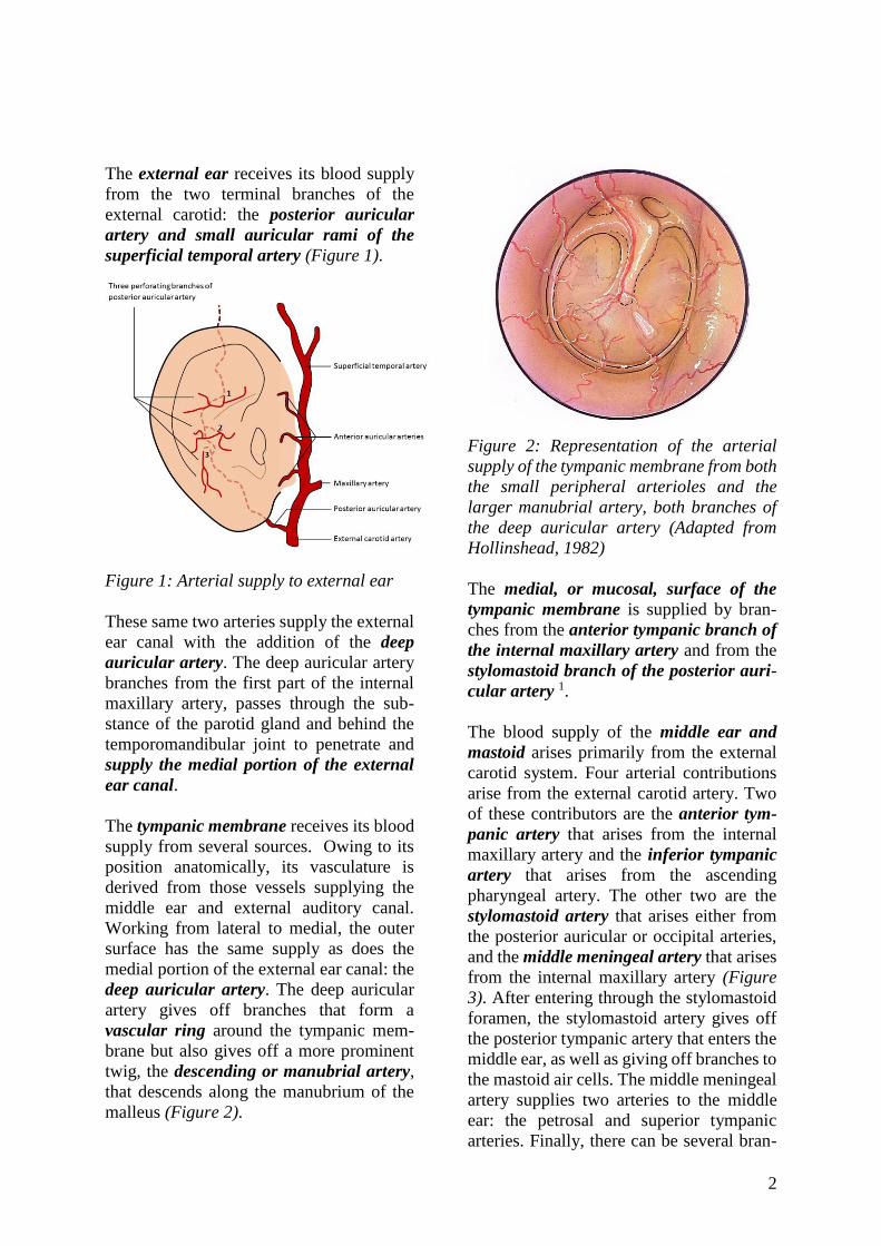
The external ear receives its blood supply
from the two terminal branches of the
external carotid: the posterior auricular
artery and small auricular rami of the
superficial temporal artery (Figure 1).
Figure 1: Arterial supply to external ear
These same two arteries supply the external
ear canal with the addition of the deep
auricular artery. The deep auricular artery
branches from the first part of the internal
maxillary artery, passes through the sub-
stance of the parotid gland and behind the
temporomandibular joint to penetrate and
supply the medial portion of the external
ear canal.
The tympanic membrane receives its blood
supply from several sources. Owing to its
position anatomically, its vasculature is
derived from those vessels supplying the
middle ear and external auditory canal.
Working from lateral to medial, the outer
surface has the same supply as does the
medial portion of the external ear canal: the
deep auricular artery. The deep auricular
artery gives off branches that form a
vascular ring around the tympanic mem-
brane but also gives off a more prominent
twig, the descending or manubrial artery,
that descends along the manubrium of the
malleus (Figure 2).
Figure 2: Representation of the arterial
supply of the tympanic membrane from both
the small peripheral arterioles and the
larger manubrial artery, both branches of
the deep auricular artery (Adapted from
Hollinshead, 1982)
The medial, or mucosal, surface of the
tympanic membrane is supplied by bran-
ches from the anterior tympanic branch of
the internal maxillary artery and from the
stylomastoid branch of the posterior auri-
cular artery 1.
The blood supply of the middle ear and
mastoid arises primarily from the external
carotid system. Four arterial contributions
arise from the external carotid artery. Two
of these contributors are the anterior tym-
panic artery that arises from the internal
maxillary artery and the inferior tympanic
artery that arises from the ascending
pharyngeal artery. The other two are the
stylomastoid artery that arises either from
the posterior auricular or occipital arteries,
and the middle meningeal artery that arises
from the internal maxillary artery (Figure
3). After entering through the stylomastoid
foramen, the stylomastoid artery gives off
the posterior tympanic artery that enters the
middle ear, as well as giving off branches to
the mastoid air cells. The middle meningeal
artery supplies two arteries to the middle
ear: the petrosal and superior tympanic
arteries. Finally, there can be several bran-

3
ches off of the internal carotid artery that
enter through the caroticotympanic canal to
supply the middle ear.
Figure 3: Contributions to the arterial
supply of the middle ear (Adapted from
Hollinshead, 1982)
Venous drainage of the external ear is via
the superficial temporal and posterior auri-
cular veins into the retromandibular and the
external jugular veins respectively (Figure
4). Ultimately, the retromandibular vein
splits to drain into the internal and external
jugular veins; on occasion, the posterior
auricular vein will drain directly to the
sigmoid sinus via the mastoid emissary
vein1.
Figure 4: Venous drainage of external ear
Nerve supply
Innervation of the auricle has been ex-
plained in exquisite detail elsewhere 2, but
an overview of the main contributing nerves
will be undertaken here. Sensation to the
external ear is provided by several cuta-
neous branches of cranial nerves as well as
by cutaneous branches of the cervical
plexus. This is reflective of the origin of
auricular skin that originates from both
brachial and post-brachial components 1.
There is invariably some overlap in the
innervation from person-to-person, but the
contributions are relatively constant
(Figures 5a, b).
A branch of the mandibular division of the
trigeminal nerve (V3), the auriculotemporal
nerve, supplies the anterior portion of the
external ear, the crus of the helix, and the
tragus. This same nerve supplies the ante-
rior and superior walls of the external audi-
tory canal 1, 3. A majority of the remaining
lateral, posterior, and medial portions of the
ear - excluding the concha - are supplied by
cutaneous branches of C2 and C3 via the
great auricular nerve. The C2 and C3 roots
of the cervical plexus also supply the skin
overlying the mastoid via the lesser
occipital nerve 1.
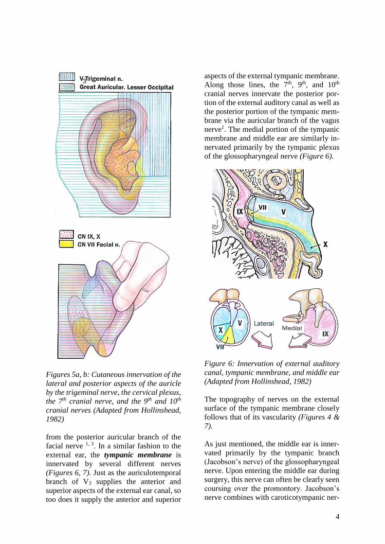
The concha of the external ear has simi-
larly complex and overlapping sensory in-
nervations. Branches of the 7th (facial), 9th
(glossopharyngeal), and 10th (vagus) cra-
nial nerves supply this area. At least two of
these cranial nerves (7th and 10th), and pos-
sibly the 9th, also supply the posterior exter-
nal auditory canal. All three of these nerves
reach their ultimate points of innervation
via the auricular branch (Arnold’s nerve) of
the vagus nerve. Arnold’s nerve arises from
the superior jugular ganglion and eventually
emerges through the tympanomastoid
fissure. Along its course, it picks up the
auricular branch (not the tympanic branch)
from the 9th along with a branch
4
Figures 5a, b: Cutaneous innervation of the
lateral and posterior aspects of the auricle
by the trigeminal nerve, the cervical plexus,
the 7th cranial nerve, and the 9th and 10th
cranial nerves (Adapted from Hollinshead,
1982)
from the posterior auricular branch of the
facial nerve 1, 3. In a similar fashion to the
external ear, the tympanic membrane is
innervated by several different nerves
(Figures 6, 7). Just as the auriculotemporal
branch of V3 supplies the anterior and
superior aspects of the external ear canal, so
too does it supply the anterior and superior
aspects of the external tympanic membrane.
Along those lines, the 7th, 9th, and 10th
cranial nerves innervate the posterior por-
tion of the external auditory canal as well as
the posterior portion of the tympanic mem-
brane via the auricular branch of the vagus
nerve1. The medial portion of the tympanic
membrane and middle ear are similarly in-
nervated primarily by the tympanic plexus
of the glossopharyngeal nerve (Figure 6).
Figure 6: Innervation of external auditory
canal, tympanic membrane, and middle ear
(Adapted from Hollinshead, 1982)
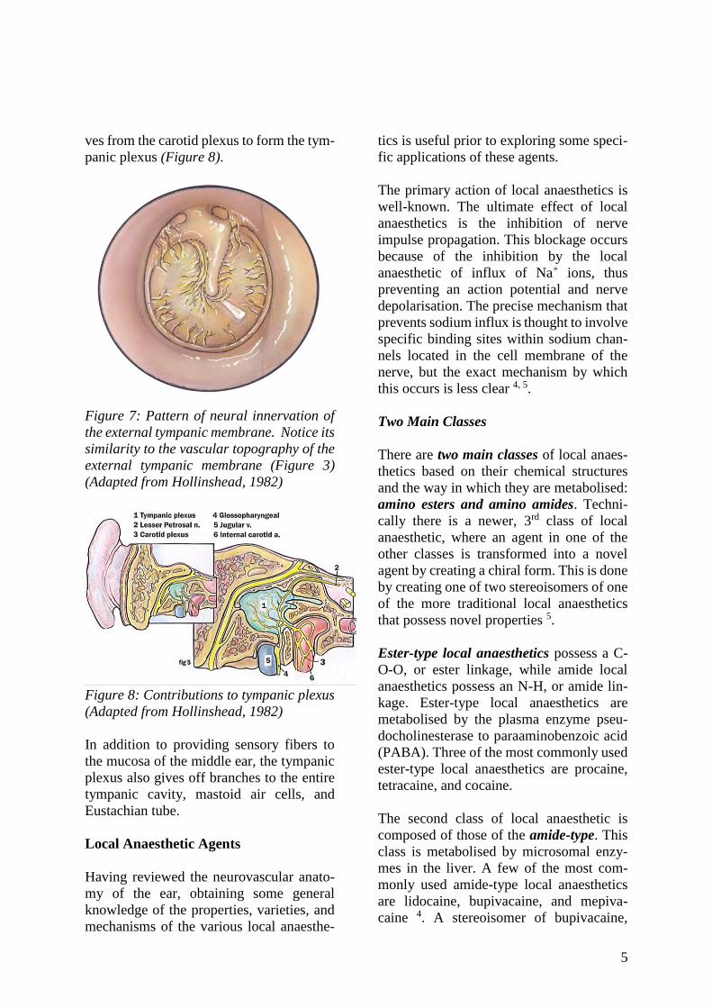
The topography of nerves on the external
surface of the tympanic membrane closely
follows that of its vascularity (Figures 4 &
7).
As just mentioned, the middle ear is inner-
vated primarily by the tympanic branch
(Jacobson’s nerve) of the glossopharyngeal
nerve. Upon entering the middle ear during
surgery, this nerve can often be clearly seen
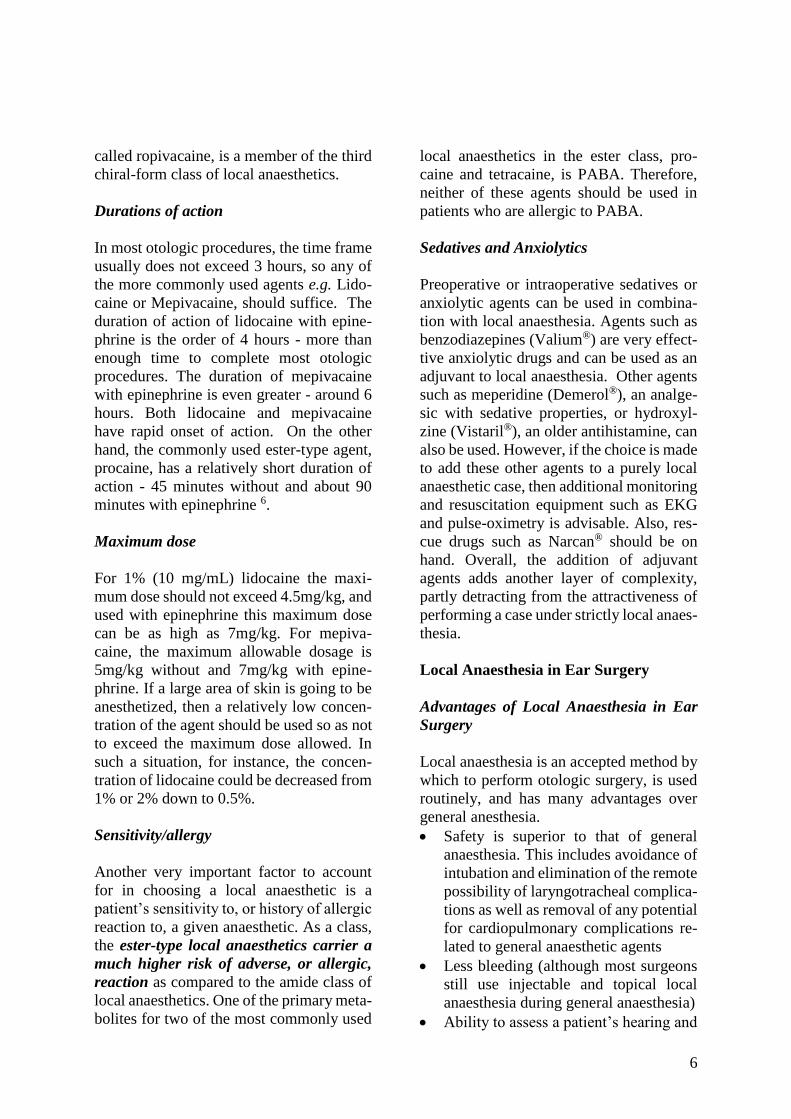
coursing over the promontory. Jacobson’s
nerve combines with caroticotympanic ner-
5
ves from the carotid plexus to form the tym-
panic plexus (Figure 8).
Figure 7: Pattern of neural innervation of
the external tympanic membrane. Notice its
similarity to the vascular topography of the
external tympanic membrane (Figure 3)
(Adapted from Hollinshead, 1982)
Figure 8: Contributions to tympanic plexus
(Adapted from Hollinshead, 1982)
In addition to providing sensory fibers to
the mucosa of the middle ear, the tympanic
plexus also gives off branches to the entire
tympanic cavity, mastoid air cells, and
Eustachian tube.
Local Anaesthetic Agents
Having reviewed the neurovascular anato-
my of the ear, obtaining some general
knowledge of the properties, varieties, and
mechanisms of the various local anaesthe-
tics is useful prior to exploring some speci-
fic applications of these agents.
The primary action of local anaesthetics is
well-known. The ultimate effect of local
anaesthetics is the inhibition of nerve
impulse propagation. This blockage occurs
because of the inhibition by the local
anaesthetic of influx of Na+ ions, thus
preventing an action potential and nerve
depolarisation. The precise mechanism that
prevents sodium influx is thought to involve
specific binding sites within sodium chan-
nels located in the cell membrane of the
nerve, but the exact mechanism by which
this occurs is less clear 4, 5.
Two Main Classes
There are two main classes of local anaes-
thetics based on their chemical structures
and the way in which they are metabolised:
amino esters and amino amides. Techni-
cally there is a newer, 3rd class of local
anaesthetic, where an agent in one of the
other classes is transformed into a novel
agent by creating a chiral form. This is done
by creating one of two stereoisomers of one
of the more traditional local anaesthetics
that possess novel properties 5.
Ester-type local anaesthetics possess a C-
O-O, or ester linkage, while amide local
anaesthetics possess an N-H, or amide lin-
kage. Ester-type local anaesthetics are
metabolised by the plasma enzyme pseu-
docholinesterase to paraaminobenzoic acid
(PABA). Three of the most commonly used
ester-type local anaesthetics are procaine,
tetracaine, and cocaine.
The second class of local anaesthetic is
composed of those of the amide-type. This
class is metabolised by microsomal enzy-
mes in the liver. A few of the most com-
monly used amide-type local anaesthetics
are lidocaine, bupivacaine, and mepiva-
caine 4. A stereoisomer of bupivacaine,
6
called ropivacaine, is a member of the third
chiral-form class of local anaesthetics.
Durations of action
In most otologic procedures, the time frame
usually does not exceed 3 hours, so any of
the more commonly used agents e.g. Lido-
caine or Mepivacaine, should suffice. The
duration of action of lidocaine with epine-
phrine is the order of 4 hours - more than
enough time to complete most otologic
procedures. The duration of mepivacaine
with epinephrine is even greater - around 6
hours. Both lidocaine and mepivacaine
have rapid onset of action. On the other
hand, the commonly used ester-type agent,
procaine, has a relatively short duration of
action - 45 minutes without and about 90
minutes with epinephrine 6.
Maximum dose
For 1% (10 mg/mL) lidocaine the maxi-
mum dose should not exceed 4.5mg/kg, and
used with epinephrine this maximum dose
can be as high as 7mg/kg. For mepiva-
caine, the maximum allowable dosage is
5mg/kg without and 7mg/kg with epine-
phrine. If a large area of skin is going to be
anesthetized, then a relatively low concen-
tration of the agent should be used so as not
to exceed the maximum dose allowed. In
such a situation, for instance, the concen-
tration of lidocaine could be decreased from
1% or 2% down to 0.5%.
Sensitivity/allergy
Another very important factor to account
for in choosing a local anaesthetic is a
patient’s sensitivity to, or history of allergic
reaction to, a given anaesthetic. As a class,
the ester-type local anaesthetics carrier a
much higher risk of adverse, or allergic,
reaction as compared to the amide class of
local anaesthetics. One of the primary meta-
bolites for two of the most commonly used
local anaesthetics in the ester class, pro-
caine and tetracaine, is PABA. Therefore,
neither of these agents should be used in
patients who are allergic to PABA.
Sedatives and Anxiolytics
Preoperative or intraoperative sedatives or
anxiolytic agents can be used in combina-
tion with local anaesthesia. Agents such as
benzodiazepines (Valium®) are very effect-
tive anxiolytic drugs and can be used as an
adjuvant to local anaesthesia. Other agents
such as meperidine (Demerol®), an analge-
sic with sedative properties, or hydroxyl-
zine (Vistaril®), an older antihistamine, can
also be used. However, if the choice is made
to add these other agents to a purely local
anaesthetic case, then additional monitoring
and resuscitation equipment such as EKG
and pulse-oximetry is advisable. Also, res-
cue drugs such as Narcan® should be on
hand. Overall, the addition of adjuvant
agents adds another layer of complexity,
partly detracting from the attractiveness of
performing a case under strictly local anaes-
thesia.
Local Anaesthesia in Ear Surgery
Advantages of Local Anaesthesia in Ear
Surgery
Local anaesthesia is an accepted method by
which to perform otologic surgery, is used
routinely, and has many advantages over
general anesthesia.
• Safety is superior to that of general
anaesthesia. This includes avoidance of
intubation and elimination of the remote
possibility of laryngotracheal complica-
tions as well as removal of any potential
for cardiopulmonary complications re-
lated to general anaesthetic agents
• Less bleeding (although most surgeons
still use injectable and topical local
anaesthesia during general anaesthesia)
• Ability to assess a patient’s hearing and

7
detect any vertigo during surgery (es-
pecially valuable during stapedectomy)
• Avoidance of a potentially lengthy and/
or disruptive emergence from anaesthe-
sia (advantageous in stapes surgery and
ossicular reconstruction procedures)
• Less postoperative nausea and vomi-
ting
• Less expensive 3
Auricular Injection Techniques
Several techniques have been advocated,
and are generally “variations on a theme”.
Hence only a few methods will be address-
ed in detail.
Plester injection technique (Figures 9-12)
• Step 1: Inject the region of the post-
auricular fold (Figure 9)
Figure 9: Postauricular injection tech-
nique proposed by Plester (From Yung
1996)
• Steps 2-4: Without removing the need-
le in Step 1, advance the needle in 3
vectors: directly toward the posterior
external ear canal, superior to the exter-
nal ear canal and inferior to the external
ear canal (Figure 10)
Figure 10: Postauricular injection
Steps 2-4 of Plester technique (From
Yung 1996)
• Steps 5 - 8: Inject the 4 quadrants of the
ear canal in a stepwise fashion (Figures
11 & 12) 7. This anaesthetises the exter-
nal ear canal while at the same time
achieving haemostasis of the skin of the
ear canal and tympanic membrane.
Take care during each of the canal injec-
tion steps to avoid the formation of
haematomas or vesicles that could im-
pair healing or obscure the tympanic
membrane during surgery 7
Figure 11: Canal injection steps 5-8 of
Plester technique (From Yung 1996)

8
Figure 12: Axial view of canal inject-
tion of Plester technique (From Yung
1996)
Fisch injection technique
Fisch’s technique uses fewer injection sites
and the sites are differently placed 5.
• Insert the needle about 1cm behind the
postauricular crease at a point halfway
between the mastoid tip and the top of
the ear
• Pass the needle anteriorly and inferior-
ly toward the tympanomastoid sulcus
and inject local anaesthetic lateral to,
but overlying the stylomastoid foramen
(Figure 13)
• Direct a 2nd needle pass anterosuperior-
ly toward the incisura and inject more
local anaesthetic (Figure 13)
• Wait 10 minutes 3 (In general, this is ad-
visable when working under local an-
aesthesia also to allow more time for
vasoconstriction to occur
• Inject the external ear canal; Fisch’s
technique differs from that of Plester in
that only one canal injection is per-
formed initially. This initial injection is
placed superiorly in the region of the
tympanosquamous suture line. After
this initial canal injection, the other
areas of the canal are tested for sensiti-
vity and the other quadrants are inject-
ed only if needed
Figure 13: Fisch postauricular inject-
tion technique (from Lancer and Fisch
1988)
A drawback of the Fisch technique is a high
risk of temporary facial nerve paresis due to
injection in the region of the stylomastoid
foramen; this can be very distressing for the
patient and surgeon alike. Using this inject-
tion technique in a series of 32 patients,
Fisch reported that 97% developed a tem-
porary postoperative facial weakness. For
this reason we recommend that no injec-
tions be administered below the level of the
external auditory canal.
Additional Intraoperative Analgesia
More local anaesthetic can be administer-
ed in any of these local anaesthetic techni-
ques if the patient feels pain during the
surgery. However the surgeon should en-

9
sure that a running total of the amount of
local anaesthetic agent is kept so as not to
exceed the maximum allowable dose.
Jacobson’s nerve (supplies the middle ear
mucosa) can be anaesthetised if a patient
experiences pain during the middle ear part
of the procedure, by the placing a cotton
ball or gelfoam soaked with 1% lidocaine or
4% tetracaine 7 on the promontory.
Field blocks
Field block is a form of regional anaes-
thesia that takes advantage of the subcu-
taneous course of somatosensory cuta-
neous nerve branches. A larger area can be
anaesthetized with fewer injections by
infiltrating the region of the course of any
given nerve. Ideally, this injection is done
in an area more proximal than the area in
which an incision is going to be made so as
to provide anaesthesia distal to the injection
site. It requires less anaesthetic agent to be
used as well as keeping the incision site
undistorted 5. Field blocks require an under-
standing of the anatomy of auricular inner-
vation. Nerves amenable to a field block for
ear surgery are the auriculotemporal,
greater auricular, and the lesser occipital
nerves (Figure 14).
Auriculotemporal nerve field block
The auriculotemporal nerve exits from the
parotid gland in front of the ear (Figure 15).
The area to infiltrate is located by palpating
the superficial temporal artery as it passes
over the zygoma (Figure 14). The injection
is placed in between this point and the
incisura - near the root of the zygoma.
About 2-3 ml of local anaesthetic is used to
block the nerve 5.
V3 nerve regional block
A more complete, truly regional auriculo-
temporal nerve block can be achieved by
Figure 14: Nerves suitable for field block:
greater auricular, auriculotemporal and
lesser (“smaller”) occipital nerves; note
superficial temporal artery
performing a V3 nerve block. This block
provides excellent anaesthesia over the bulk
of the cheek along with the upper pre-
auricular and auriculotemporal hair covered
regions. The anaesthetised area abuts the
region supplied by the great auricular nerve
more posteriorly 8.
• Palpate the sigmoid notch of the man-
dible as an initial landmark. The notch
is located inferior to the zygomatic arch
about 2.5 cm anterior to the tragus
• While placing a finger in the area of the
notch and asking the patient to open the
mouth; the mandibular condyle is felt
sliding under the examiner’s finger
• Ask the patient to close the mouth; the
examining finger will stay in the notch
10
• Mark this point with a marking pen
• Inject a small volume of local anaes-
thetic in this area before proceeding to
perform the block
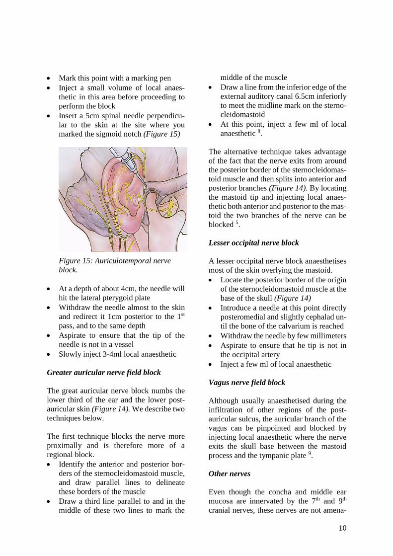
• Insert a 5cm spinal needle perpendicu-
lar to the skin at the site where you
marked the sigmoid notch (Figure 15)
Figure 15: Auriculotemporal nerve
block.
• At a depth of about 4cm, the needle will
hit the lateral pterygoid plate
• Withdraw the needle almost to the skin
and redirect it 1cm posterior to the 1st
pass, and to the same depth
• Aspirate to ensure that the tip of the
needle is not in a vessel
• Slowly inject 3-4ml local anaesthetic
Greater auricular nerve field block
The great auricular nerve block numbs the
lower third of the ear and the lower post-
auricular skin (Figure 14). We describe two
techniques below.
The first technique blocks the nerve more
proximally and is therefore more of a
regional block.
• Identify the anterior and posterior bor-
ders of the sternocleidomastoid muscle,
and draw parallel lines to delineate
these borders of the muscle
• Draw a third line parallel to and in the
middle of these two lines to mark the
middle of the muscle
• Draw a line from the inferior edge of the
external auditory canal 6.5cm inferiorly
to meet the midline mark on the sterno-
cleidomastoid
• At this point, inject a few ml of local
anaesthetic 8.
The alternative technique takes advantage
of the fact that the nerve exits from around
the posterior border of the sternocleidomas-
toid muscle and then splits into anterior and
posterior branches (Figure 14). By locating
the mastoid tip and injecting local anaes-
thetic both anterior and posterior to the mas-
toid the two branches of the nerve can be
blocked 5.
Lesser occipital nerve block
A lesser occipital nerve block anaesthetises
most of the skin overlying the mastoid.
• Locate the posterior border of the origin
of the sternocleidomastoid muscle at the
base of the skull (Figure 14)
• Introduce a needle at this point directly
posteromedial and slightly cephalad un-
til the bone of the calvarium is reached
• Withdraw the needle by few millimeters
• Aspirate to ensure that he tip is not in
the occipital artery
• Inject a few ml of local anaesthetic
Vagus nerve field block
Although usually anaesthetised during the
infiltration of other regions of the post-
auricular sulcus, the auricular branch of the
vagus can be pinpointed and blocked by
injecting local anaesthetic where the nerve
exits the skull base between the mastoid
process and the tympanic plate 9.
Other nerves
Even though the concha and middle ear
mucosa are innervated by the 7th and 9th
cranial nerves, these nerves are not amena-
11
ble to a regional block. Anaesthesia in the
distribution of these nerve can be achieved
by local infiltration of the external ear canal
and topical anaesthesia to the middle ear.
Effectiveness of Local Anesthesia for
Middle Ear Surgery
Several authors have commented on the
effectiveness of local anaesthesia for otolo-
gic surgery. Caner reported on 100 conse-
cutive patients undergoing various middle
ear procedures under local anaesthesia with
IV sedation, including mastoidectomy 10. In
this paper, 96% of patients who underwent
stapes surgery or tympanoplasty alone said
that they had no pain during surgery; of all
patients 22% said that pain was distressing.
The most distressing experiences were
anxiety (44%) and noise created by the pro-
cedure (33%). However 73% of patients in
this study said they would have a similar
operation done again under local anaes-
thesia. Furthermore, only one patient had a
transient facial weakness. In a similar paper
by Yung, 108 patients that underwent
various otologic procedures including mas-
toidectomy reported similarly favourable
results 7. The most frequent complaints
were that of noise during the operation
(30%) and anxiety (24%). Interestingly,
otalgia was reported as the lowest specified
discomfort (2%). As in Caner’s paper, a
high percentage (89%) reported that they
would prefer local anaesthesia for a similar
procedure. Lancer and Fisch also reported
a high success rate with local anaesthesia
and that both patients and surgeons were
highly satisfied with local anaesthesia that
there had been no adverse effects 3. One
major concern of the latter paper was the
97% rate of transient (partial to total) facial
nerve paralysis. Although transient, it was
still unpleasant for a significant number
(55%) of patients. These few studies corro-
borate the notion that otologic surgery done
under local ansesthesia is not only and
effective technique, but was acceptable to
both patient and surgeon. It should be noted,
however, that in all these studies adjuvant
sedation was used in addition to local
anaesthesia.
References
1. Hollinshead WH. Anatomy for sur
geons: Vol 1. The head and neck.
Philadelphia: Lippincott Williams &
Wilkins; 1982
2. Peuker ET, Filler TJ. The nerve supply
of the human auricle. Clin Anat 2002;
15:35-7
3. Lancer JM, Fisch U. Local anesthesia
for middle ear surgery. Clin Otolaryn
gol 1988;13:367-74
4. Auletta MJ, Grekin RC. Local anesthe-
sia for dermatologic surgery. New
York: Churchill Livingstone; 1991
5. Raj PP. Textbook of regional anesthe-
sia. New York: Churchill Livingstone;
2002
6. McLeod IK, Gallagher DJ III, Revis
DR, Seagle MB. “Local Anesthetics.”
eMedicine. July 22, 2008. May 28,
2010
http://emedicine.medscape.com/article/
873879-overview
7. Yung MW. Local anesthesia in middle
ear surgery: survey of patients and
surgeons. Clin Otolaryngol 1996;21:
404-8
8. Zide BM, Swift R. How to block and
tackle the face. Plast Reconstr Surg
1998;101:840-51
9. Cousins MJ, Bridenbaugh PO. Neural
blockade in clinical anesthesia and
management of pain, 3rd Ed. Philadel-
phia: Lippincott-Raven; 1998
10. Caner G, Olgun L, Gültekin G, Aydar L.
Local anesthesia for middle ear surgery.
Otolaryngol Head Neck Surg 2005;
295-7

12
Relevant Open Access chapter: Otology
outreach surgery in developing countries
under local and regional anaesthesia:
Techniques and pitfalls
https://vula.uct.ac.za/access/content/group/
ba5fb1bd-be95-48e5-81be-
586fbaeba29d/Otology%20outreach%20su
rgery%20techniques%20under%20local%
20and%20regional%20anaesthesia.pdf
Authors
Alexander G. Bien, M.D
St. Louis
Missouri, USA
Richard Wagner, M.D., F.A.C.S.
Director
Global ENT Outreach
Coupeville, WA, 98239, USA
Eric P. Wilkinson, M.D., F.A.C.S.
House Clinic
2100 W. Third Street, #111
Los Angeles, CA 900 USA
Editor
Johan Fagan MBChB, FCS(ORL), MMed
Professor and Chairman
Division of Otolaryngology
University of Cape Town
Cape Town, South Africa
THE OPEN ACCESS ATLAS OF
OTOLARYNGOLOGY, HEAD &
NECK OPERATIVE SURGERY www.entdev.uct.ac.za
The Open Access Atlas of Otolaryngology, Head & Neck Operative Surgery by Johan Fagan (Editor) [email protected] is licensed under a Creative Commons Attribution - Non-Commercial 3.0 Unported License
Recommended