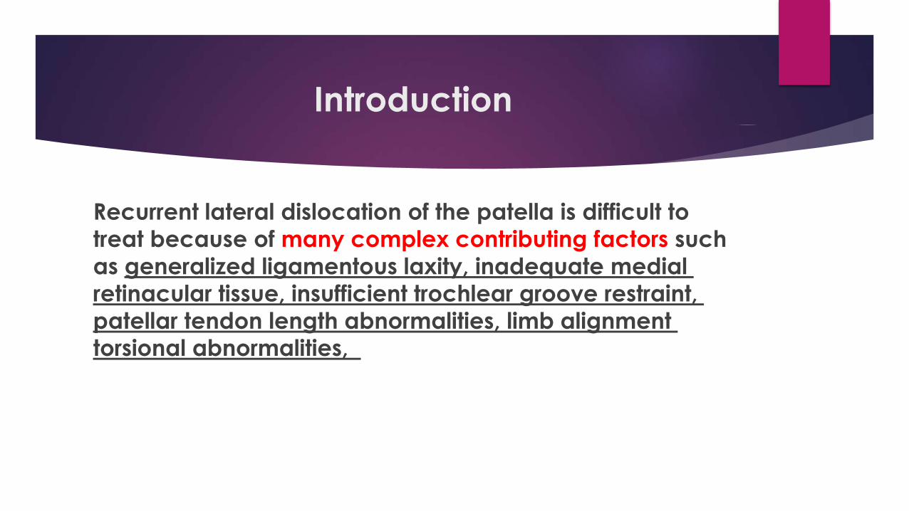
Introduction
Recurrent lateral dislocation of the patella is difficult to
treat because of many complex contributing factors such
as generalized ligamentous laxity, inadequate medial
retinacular tissue, insufficient trochlear groove restraint,
patellar tendon length abnormalities, limb alignment torsional abnormalities,
Over 130 different surgical methods have been described
which can be classified as proximal realignment, distal
realignment, proximal and distal realignment, lateral retinacular release, and medial retinacular plication

Many authors have described the
method of making the patellar tunnel
and using the wire anchors to fix the
side of the patella. However, drilling
tunnels transversely across the patella
creates a stress riser and can lead to
fracture. The use of wire anchors
increase the economic burden of
patients and also the patellar bone may be too soft for a secure anchor in
WHY THE MPFL RECONSTRUCTIONIS IS THE CONCERN IN TREATMENT OF THE PATELLAR DISLOCATION
The medial patellofemoral ligament (MPFL) was first described by Kaplan [1].
Since Warren and Marshall [2] introduced the concept of a three-layered pattern of the
medial capsular ligament of the knee and described the fibers of the MPFL existing
within layer II, and biomechanical studies had demonstrated that the MPFL accounts for
approximately 50% to 60% of the total lateral restraint; thus being the primary medial
stabilizer of the patella, MPFL reconstruction has became an accepted technique to treat
patellofemoral instability .
The aim of the study
Evaluation the results of MPFL reconstruction with
bone-fascia tunnel fixation at the medial margin of
the patella after a 6-year-minimum follow-up

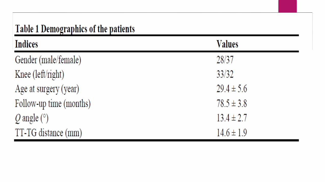
Sixty-five patients who underwent a primary MPFL reconstruction were included in the study
Methods
Inclusion criteria
Diagnosis of recurrent patellar
dislocation , confirmed with history
and physical and radiographic
examinations
Exclusion criteria
History of previous knee surgery
Multiple ligament injury
Significant patellofemoral articular
cartilage degenerative changes
Significant patellar mal-alignment
Severe trochlear dysplasia

Evaluation and Rating scales
Demographic data, physical examination, Kujala scale, Lysholm score, and
Tegner activity score were completed preoperatively and at each follow-up
evaluation.
Clinical data were prospectively collected preoperatively and at 3, 6, 12, 24, 36,
48, 60, and 72 months after surgery.

Surgical technique
Examination under anesthesia is performed on both knees
to assess for a tight lateral retinaculum. A lateral release
was performed if the patella was unable to be everted to
neutral.
A tourniquet is applied to the thigh and then diagnostic
arthroscopy is performed before reconstruction of the
MPFL. The cartilaginous situation, tracking of the patella
through a range of flexion, and trochlear shape are assessed

A longitudinal incision (2–3 cm) is performed on the
anteromedial side of the patella, then the soft tissue is
separated to the bone surface and the periosteum is
peeled back.
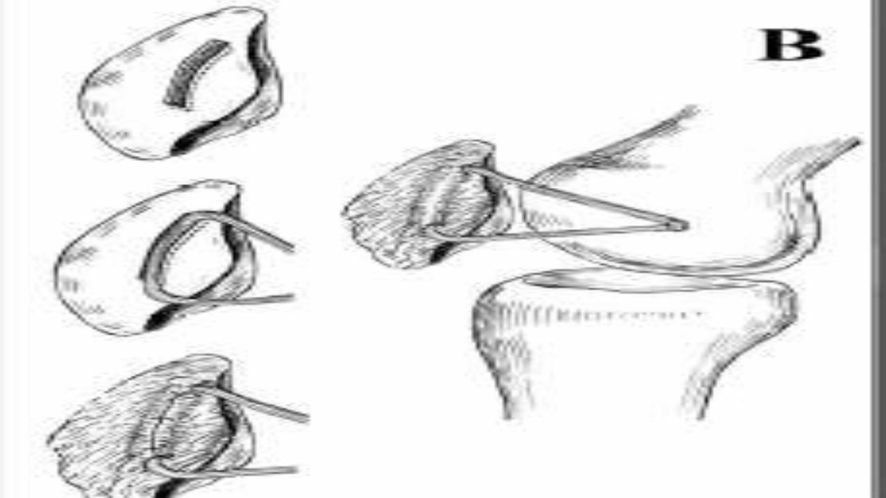
A bone groove which extends approximately from the
superomedial corner of the patella to the midpoint of
the medial margin of the patella is created using the
rongeur .
The groove must be deep enough, about 3 mm, so that
the allograft can be completely embedded.
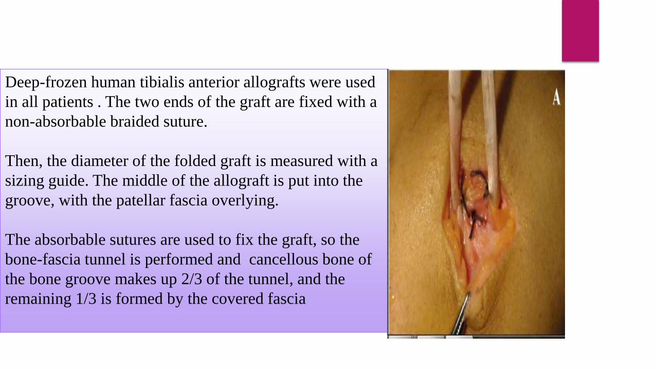
Deep-frozen human tibialis anterior allografts were used
in all patients . The two ends of the graft are fixed with a
non-absorbable braided suture.
Then, the diameter of the folded graft is measured with a
sizing guide. The middle of the allograft is put into the
groove, with the patellar fascia overlying.
The absorbable sutures are used to fix the graft, so the
bone-fascia tunnel is performed and cancellous bone of
the bone groove makes up 2/3 of the tunnel, and the
remaining 1/3 is formed by the covered fascia
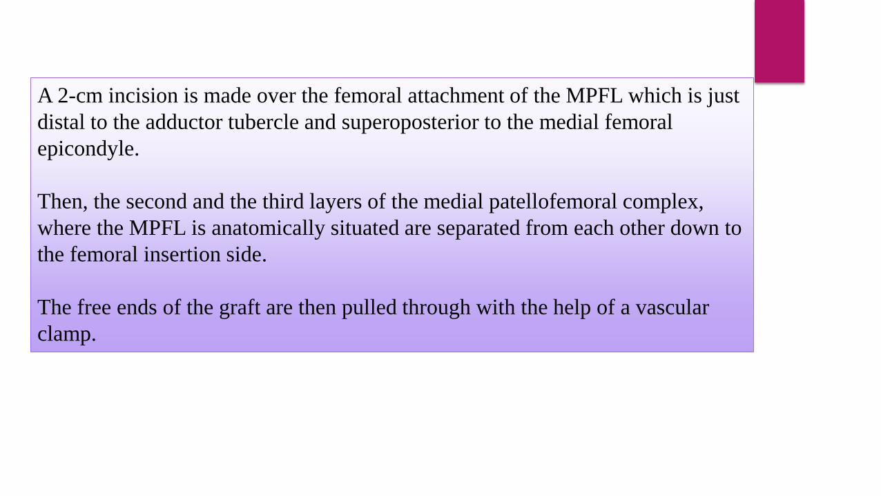
A 2-cm incision is made over the femoral attachment of the MPFL which is just
distal to the adductor tubercle and superoposterior to the medial femoral
epicondyle.
Then, the second and the third layers of the medial patellofemoral complex,
where the MPFL is anatomically situated are separated from each other down to
the femoral insertion side.
The free ends of the graft are then pulled through with the help of a vascular
clamp.
The femoral attachment of the graft is created by drilling a femoral tunnel
using a guide pin with an eyelet is placed at the anatomical femoral insertion and
cannulated acorn
The two graft strands are whipstitched with a No. 2 non-absorbable suture. The
sutures are then placed through the eyelet of the guide pin which is then
advanced out the
contralateral cortex of the distal femur. Next, the grafts are pulled into the
femoral tunnel,
The knee is cycled for several times from full flexion to full extension with the
graft under tension.
Insufficient tension will result in a lack of correction of the lateral instability,
whereas excessive tension will cause increased pressure in the patellofemoral
joint and may restrict knee range of motion. Therefore, it is essential to find a
tolerated tension.
Once sufficient tension has been obtained, the femoral attachment of the graft is
fixed with a bioabsorbable interference screw of the same size as the drill which
is used in 30° of knee flexion.
Postoperative rehabilitation
Postoperatively, all patients followed the same rehabilitation protocol after MPFL
reconstruction.
the patients were partially weight bearing in full extension with a knee brace. Two weeks
after surgery, the patients were allowed to progress from partial to full weight bearing
Patients typically returned to normal daily activities at 4 months postoperatively.
Furthermore, the function of athletes was acceptable after 6 years.

Results
Discussion
The graft of the femoral side is almost secured in the femoral tunnel
with an absorbable interference screw, but for the fixation of the
patellar side, it includes a few techniques for the reconstruction of this
ligament, making the patellar tunnel; and using the wire anchors is
described by many authors.

In 2003, Nomura and Inoue [4] described the patellar tunnel which was drilled
from the proximal third of the medial margin of the patella to the center of the
anterior aspect of the patella.
In 2007, Carmont and Maffulli [5] described a similar technique of drilling the
bone tunnels that traverse the entire patella.
.in 2009, Papp and Cosgarea [6] described the blind patellar tunnel which was
drilled from medial to lateral at the midpoint of the MPFL insertion

.
Disadvantages of the transverse patellar tunnels technique
Gomes [7] warned against damaging the cartilage surface of the patella by
creating a bone tunnel
Dobbs et al. [8] was able to show that drilling tunnels transversely across the
patella creates a stress riser and can lead to fracture
Advantages of this technique
1. eliminates the risks of patellar fracture and violation of the patellar articular
surface
2. allows us to place the graft at the anatomical insertion and recreate the double-
bundle structure of the MPFL and a sail-like triangular shape of the graft, which is
comparable to the original anatomy This seems to provide a higher stability during
flexion and decreases the patella rotation in contrast to a single-bundle isometric
technique
3. increase the surface area for graft-to-bone and the tendon to the bone contact is
pressure contact both provide good healing of the graft .

Conclusion
This method for MPFL reconstruction is a relatively easy
and safe procedure that provides enough graft strength
comparable to previous techniques. This is a simple
technique where the MPFL is reconstructed safely to avoid
patella fracture, anatomically to restore physiological
kinematics and stability, and economically to reduce
costs with bone-fascia tunnel fixation at the medial margin of the patella..
References
1- Kaplan EB: Factors responsible for the stability of the knee joint. Bull Hosp Joint Dis1957, 18:51–59.
2-Warren LF, Marshall JL: The supporting structures and layers on the medial side of the knee: an
anatomical study. J Bone Joint Surg Am 1979, 61:56–62.
3-Nomura E, Inoue M: Technical note: surgical technique and rationale for medial patellofemoral
ligament reconstruction for recurrent patellar dislocation. Arthroscopy 2003, 19:E47.
4. Carmont MR, Maffulli N: Medial patellofemoral ligament reconstruction: a new technique. BMC
Musculoskelet Disord 2007, 8:22.
5. Papp DF, Cosgarea AJ: Medial patellofemoral ligament reconstruction for recurrent patellar instability.
Tech Knee Surg 2009, 8:187–193.
6. Gomes JL: Medial patellofemoral ligament reconstruction for recurrent dislocation of the patella: a
preliminary report. Arthroscopy 1992, 8:335–340.
7. Dobbs RE, Greis PE, Burks RT: Medial patellofemoral ligament reconstruction. Tech Knee Surg 2007,
6:29–36.
Recommended