10/1/2014
1
MANAGEMENT OF SOLID ORGAN INJURIESJoseph Cuschieri, MD FACS
Professor of Surgery, University of Washington
Director of Surgical Critical Care, Harborview Medical Center
Introduction
• Solid organ injury is a leading cause of significantmorbidity and mortality following injury.
• Identification of serious solid organ injury may bechallenging.
• Many injuries, however, manifest during the initialassessment and treatment period. Thus, earlyidentification is essential.
Epidemiology
• Trauma remains leading cause of mortality under the age of 44.
• Solid organ injuries are a leading cause of mortality following closed head injury.
• Estimates indicate that by 2020, 8.4 million individuals will die annually as a result of trauma in the United States.
10/1/2014
2
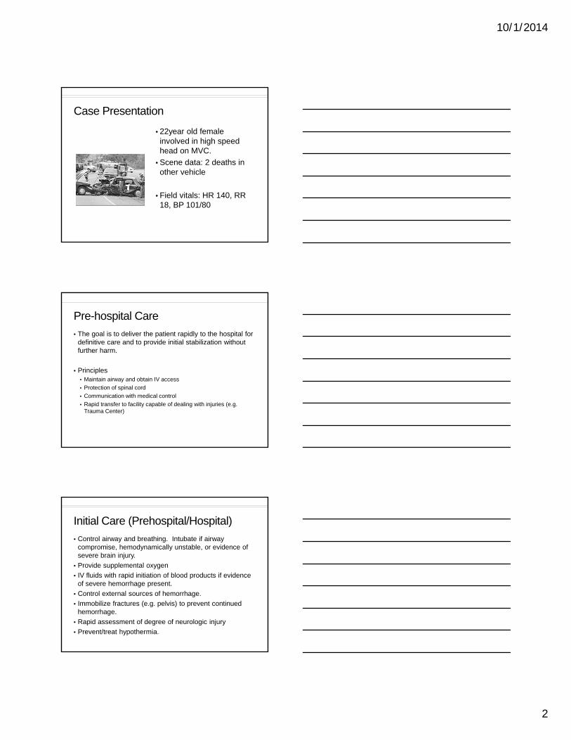
Case Presentation
• 22year old female involved in high speed head on MVC.
• Scene data: 2 deaths in other vehicle
• Field vitals: HR 140, RR 18, BP 101/80
Pre-hospital Care
• The goal is to deliver the patient rapidly to the hospital for definitive care and to provide initial stabilization without further harm.
• Principles• Maintain airway and obtain IV access
• Protection of spinal cord
• Communication with medical control
• Rapid transfer to facility capable of dealing with injuries (e.g. Trauma Center)
Initial Care (Prehospital/Hospital)
• Control airway and breathing. Intubate if airway compromise, hemodynamically unstable, or evidence of severe brain injury.
• Provide supplemental oxygen
• IV fluids with rapid initiation of blood products if evidence of severe hemorrhage present.
• Control external sources of hemorrhage.
• Immobilize fractures (e.g. pelvis) to prevent continued hemorrhage.
• Rapid assessment of degree of neurologic injury
• Prevent/treat hypothermia.
10/1/2014
3
Initial Assessment and Resuscitation
• Primary Survey: Identification and treatment of life threatening injuries.
• Airway with cervical spine precautions
• Breathing
• Circulation
• Disability
• Exposure
Patient Scenerio
• Vital signs: HR 140, BP 90/80, RR 28, Temp 35
• Airway: Speaking rapidly and anxious
• Breathing: Tachypneic and equal
• Circulation: 18 guage catheters bilateral upper extremities, 2 units PRBC transfused
• Disability: Moves all extremities, GCS 15
• Exposure: Pelvic instability, left lower extremity internally rotated and shortened. Blankets applied.
Abdominal assessment
• Vital Signs and Physical Exam
• Investigational Studies• FAST
• DPA/DPL
• CT Abdomen/Pelvis
10/1/2014
4
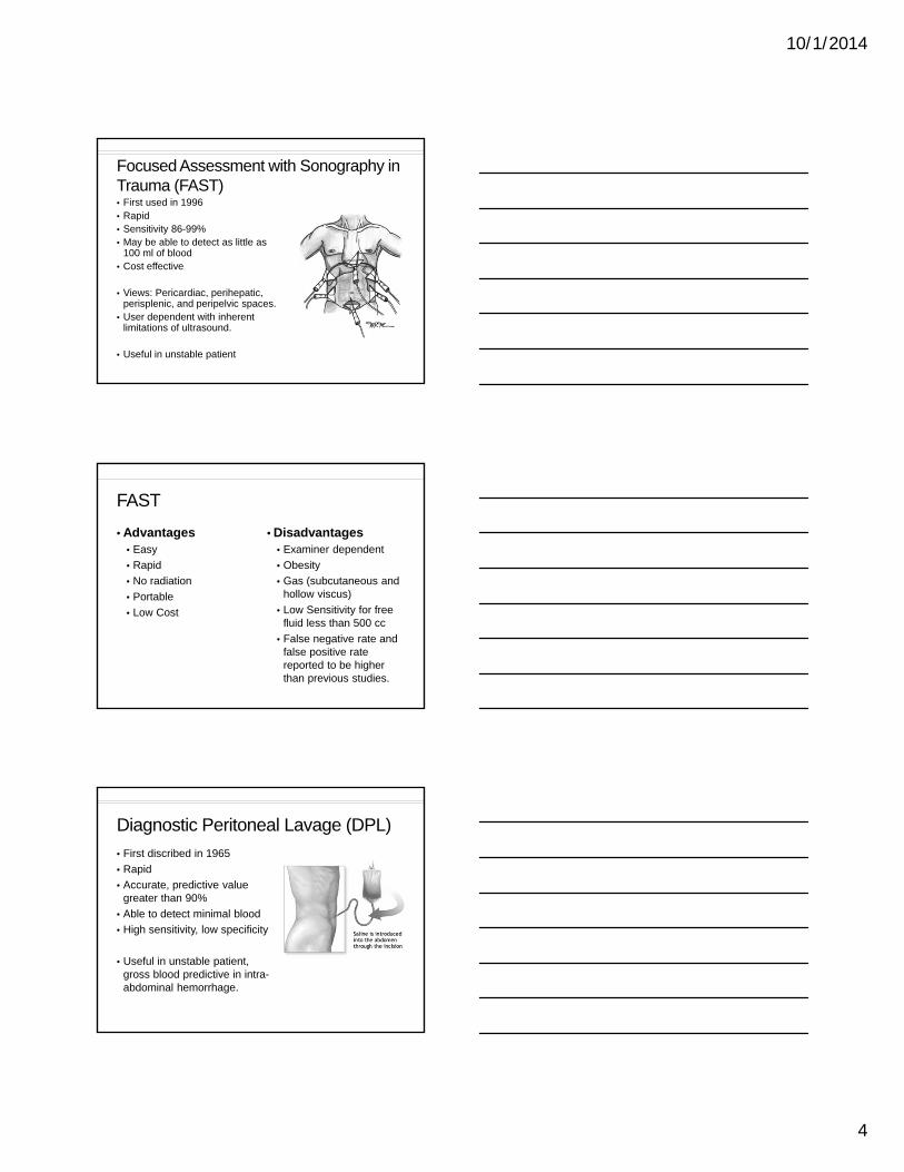
Focused Assessment with Sonography in Trauma (FAST)• First used in 1996• Rapid• Sensitivity 86-99%• May be able to detect as little as
100 ml of blood• Cost effective
• Views: Pericardiac, perihepatic, perisplenic, and peripelvic spaces.
• User dependent with inherent limitations of ultrasound.
• Useful in unstable patient
FAST
• Advantages• Easy
• Rapid
• No radiation
• Portable
• Low Cost
• Disadvantages• Examiner dependent
• Obesity
• Gas (subcutaneous and hollow viscus)
• Low Sensitivity for free fluid less than 500 cc
• False negative rate and false positive rate reported to be higher than previous studies.
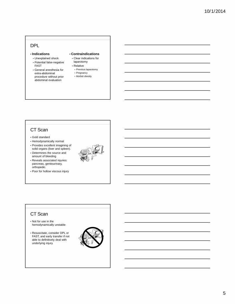
Diagnostic Peritoneal Lavage (DPL)
• First discribed in 1965
• Rapid
• Accurate, predictive value greater than 90%
• Able to detect minimal blood
• High sensitivity, low specificity
• Useful in unstable patient, gross blood predictive in intra-abdominal hemorrhage.
10/1/2014
5
DPL
• Indications• Unexplained shock
• Potential false-negative FAST
• General anesthesia for extra-abdominal procedure without prior abdominal evaluation
• Contraindications• Clear indications for
laparotomy
• Relative• Previous laparotomy
• Pregnancy
• Morbid obesity
CT Scan
• Gold standard
• Hemodynamically normal
• Provides excellent imagining of solid organs (liver and spleen).
• Determines the source and amount of bleeding
• Reveals associated injuries: pancreas, genitourinary, orthopedic.
• Poor for hollow viscous injury
CT Scan
• Not for use in the hemodynamically unstable
• Resuscitate, consider DPL or FAST, and early transfer if not able to definitively deal with underlying injury.
10/1/2014
6
Case Presentation
Case Presentation
Case Presentation
10/1/2014
7
Solid Organ Injuries
• Difficult to diagnose on physical exam
• May lead to significant blood loss
• Grading of solid organs dependent on degree of hematoma, laceration, or avulsion.
• Injuries may present late, leading to further difficulty in assessment and management.
• The most common solid organs injured spleen and liver.
Grading of Splenic Injury
Grade Type of Injury
Description
I Hematoma Subcapsular, <10%
Laceration Capsular tear, <1cm in depth
II Hematoma Subcapsular, 10-50%, <5 cm diameter
Laceration Capsular tear, 1-3 cm in depth
III Hematoma Subcapsular, >50%, ruptured; intraparenchymal hematoma > 5cm
Laceration >3cm in parenchymal depth or involving trabecular vessel
IV Laceration Segmental or hilar vessels, major devascularization (>25%)
V Laceration Completely shattered spleen
Vascular Hilar vascular injury that devascularizes the spleen
Key Principles
• Hemodynamically unstable patients require immediate laparotomy.• Splenectomy
• Non-operative management is an option in the hemodynamically stable patient ONLY.
• No patient should die as a consequence of non-operative management.
10/1/2014
8
Changes in Approach to Splenic Injury• Initial thought that the spleen has no purpose• Cellular and humoral immunity: IgM production, opsonization
of bacteria, tuftsin production, immune response to bloodborne antigens, hematopoesis
• Splenectomy has no consequences • Development of OPSI
• The spleen cannot heal• The spleen does heal with return of tensil strength within 6
weeks of injury.
• Non-operative management of splenic injury routinely results in bleeding at some point
• Bleeding occurs in less than 10% of individuals that initial undergo no-operative management of splenic injury.
• Lifelong risk for Overwhelming Post-splenectomy infection (OPSI)• Caused by pneumococcus, meningococcus, Haemophilus
influenzae• Initial Symptoms: fever, chills, muscle aches, headache, vomiting,
diarrhea, and abdominal pain• Progressive symptoms: bacteremic septic shock, extremity
gangrene, convulsions, and coma• Mortality rate of 50-80%
• from onset of initial symptoms, 68% of those deaths occur within 24 hours and 80% occur within 48 hours
• Prevention: routine vaccinations and prophylactic antibiotics
Immunologic consequences of splenectomy: OPSI
• Routine non-operative management- very high mortality
• 1920s—Due to high mortality of non-operative management, splenectomy performed for all splenic injuries. Mortality significantly improves as a result.
• 1980s Splenic preservation by splenorrhaphy
• 1990s—Non-operative evaluation becomes routine in pediatric population. Non-operative management in adults not well defined.
• 2000s– Non-operative management extended to adults with hemodynamic stability with good results.
Evolution of Splenic Management

10/1/2014
9
• Nonoperative management of blunt injury to the spleenand liver
• Class II data support non-operative management ofinjuries to the liver or spleen
• Severity of grade of injury to the liver or the spleen isnot a contraindication to non-operative management• Contrary to observations by Buntain 1988; Resciniti 1988;
Powell 1997; Cathay 1998; Bee, 2001
East Guidelines (2003)
• Hypotheses:
• Degree of patient injury based on ISS andhemodynamics will correlate with frequency of operation
• AAST Grade of splenic injury will predict frequency ofoperation
• Quantity of hemoperitoneum will correlate withfrequency of laparotomy
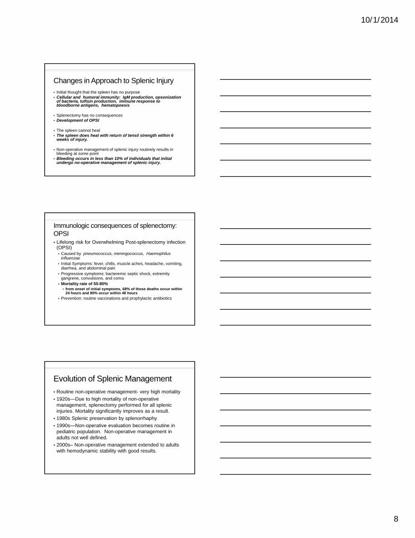
Blunt splenic injury in adults: EAST multi-institutional study Peitzman et al, J Trauma, 2000
• 1455 patients admitted to 15 different Level I trauma centers.
• 38.5% of patients underwent immediate operative intervention 61.5% of patients Admitted with planned non-operative management; of this group
• 10.8% failed non-operative management and underwent laparotomy
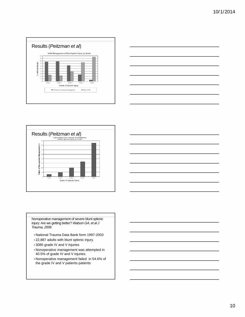
Results (Peitzman et al)
10/1/2014
10
Results (Peitzman et al)
Results (Peitzman et al)
• National Trauma Data Bank form 1997-2003
• 22,887 adults with blunt splenic injury.
• 3085 grade IV and V injuries
• Nonoperative management was attempted in 40.5% of grade IV and V injuries.
• Nonoperative management failed in 54.6% of the grade IV and V patients patients
Nonoperative management of severe blunt splenic injury: Are we getting better? Watson GA, et al J Trauma, 2006

10/1/2014
11
• National Trauma Data Bank from 1999-2004.
• 23,532 adults with blunt splenic injury.
• Conclusion…”We conclude that at least 80% of blunt splenic injury can be managed successfully nonoperatively, and that patients should be monitored from 3 to 5 days postinjury.”
Nonoperative management of severe blunt splenic injury: Are we getting better? Smith J, et al J Trauma, 2008
Harborview Experience 2001-2011
Harborview Experience 2001-2011 Requiring Splenectomy following Initial Non-Operative Managment
10/1/2014
12
Harborview Experience 2001-2011
• Any hypotension (<90 mmHg) associated with any grade of splenic injury was associated with an 80% failure of non-operative management.
• Arterial extravesation associated with 4 fold increase rate of non-operative failure.
• Shock index greater than 0.9 associated wit an 8 fold increase of non-operative failure.
• Combination of blush and elevated shock index associated with 16 fold increase rate of non-operative failure.
• Most non-operative failures occur with 24 hours 72%, but may occur up to 10-14 days later.
• Any evidence of elevated SI mandates continued monitoring until resolution for at least 24 hours. This management strategy would miss only 2% of non-operative failures.
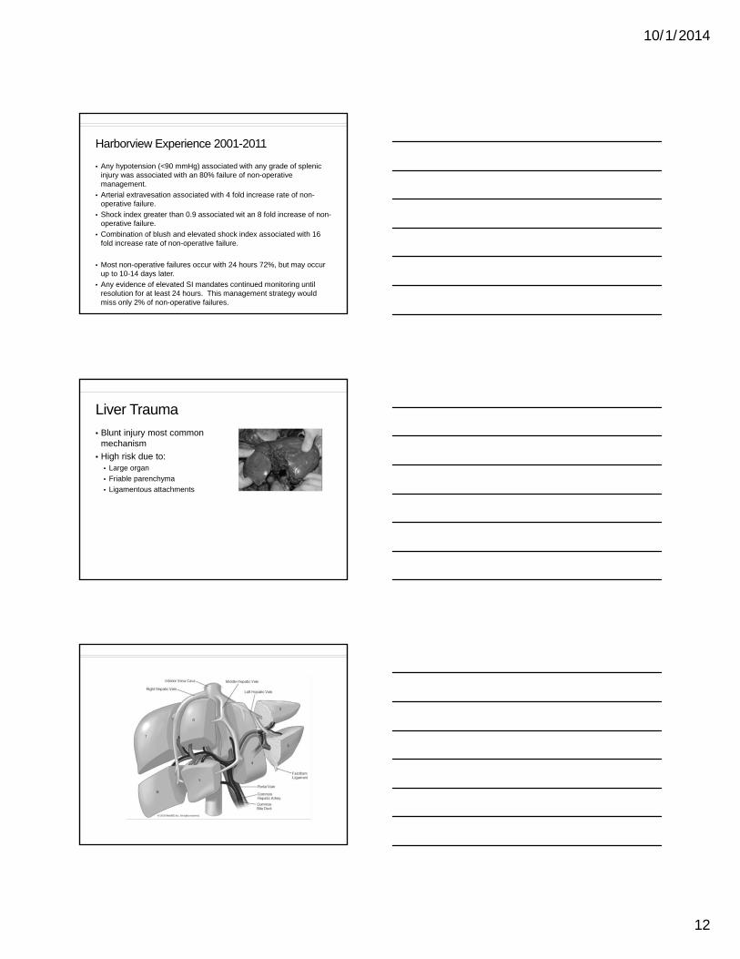
Liver Trauma
• Blunt injury most common mechanism
• High risk due to:• Large organ
• Friable parenchyma
• Ligamentous attachments
10/1/2014
13
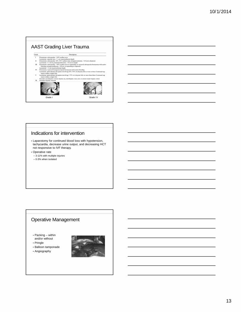
AAST Grading Liver Trauma
Grade I Grade IV
Indications for intervention
• Laparotomy for continued blood loss with hypotension, tachycardia, decrease urine output, and decreasing HCT not responsive to IVF therapy.
• Operative rate• 3-11% with multiple injuries
• 0-3% when isolated
Operative Management
• Packing – within and/or without
• Pringle
• Balloon tamponade
• Angiography
10/1/2014
14
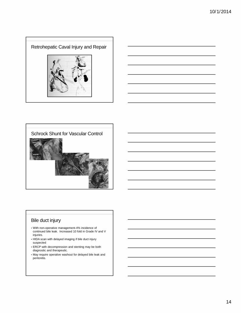
Retrohepatic Caval Injury and Repair
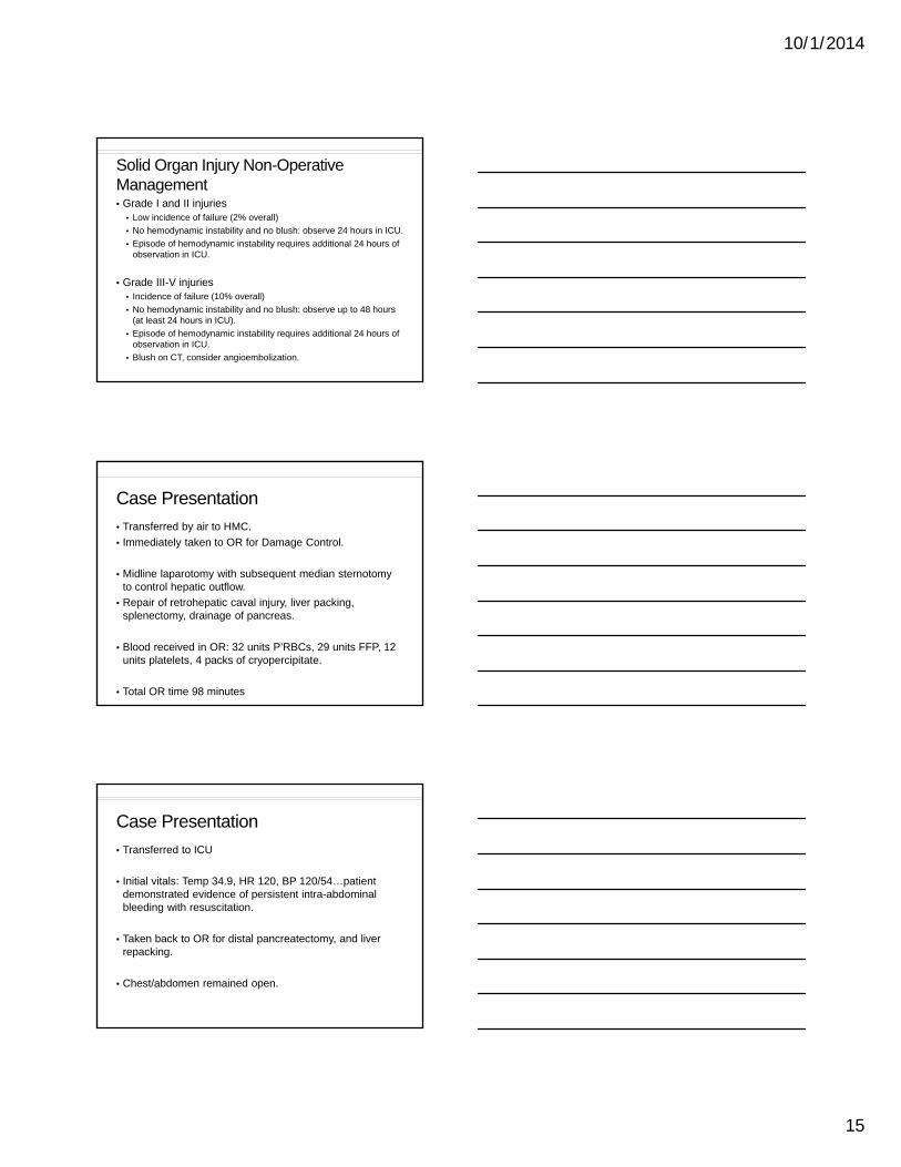
Schrock Shunt for Vascular Control
Bile duct injury
• With non-operative management-4% incidence of continued bile leak. Increased 10 fold in Grade IV and V injuries.
• HIDA scan with delayed imaging if bile duct injury suspected
• ERCP with decompression and stenting may be both diagnostic and therapeutic.
• May require operative washout for delayed bile leak and peritonitis.
10/1/2014
15
Solid Organ Injury Non-Operative Management• Grade I and II injuries
• Low incidence of failure (2% overall)
• No hemodynamic instability and no blush: observe 24 hours in ICU.
• Episode of hemodynamic instability requires additional 24 hours of observation in ICU.
• Grade III-V injuries• Incidence of failure (10% overall)
• No hemodynamic instability and no blush: observe up to 48 hours (at least 24 hours in ICU).
• Episode of hemodynamic instability requires additional 24 hours of observation in ICU.
• Blush on CT, consider angioembolization.
Case Presentation
• Transferred by air to HMC.
• Immediately taken to OR for Damage Control.
• Midline laparotomy with subsequent median sternotomy to control hepatic outflow.
• Repair of retrohepatic caval injury, liver packing, splenectomy, drainage of pancreas.
• Blood received in OR: 32 units P’RBCs, 29 units FFP, 12 units platelets, 4 packs of cryopercipitate.
• Total OR time 98 minutes
Case Presentation
• Transferred to ICU
• Initial vitals: Temp 34.9, HR 120, BP 120/54…patient demonstrated evidence of persistent intra-abdominal bleeding with resuscitation.
• Taken back to OR for distal pancreatectomy, and liver repacking.
• Chest/abdomen remained open.

10/1/2014
16
Case Presentation
• 2 days later underwent liver pack removal, temporary abdominal closure, and sternal closure.
• Subsequent underwent abdominal closure, C2 fracture stabilization, and pelvic fixation.
• Discharged 81 days later to rehab facility.
• Currently 2 years post injury, back to work and doing well.
QUESTIONS
Recommended


![Laparoscopy in Abdominal Trauma - Springer · dard of care in blunt abdominal solid organ injuries [37, 38], its implementation in penetrating abdominal trauma has been considerably](https://img.pdfslide.net/doc/110x75/5ca43df888c99355658be93f/laparoscopy-in-abdominal-trauma-springer-dard-of-care-in-blunt-abdominal-solid.jpg)