Katedra botaniky Univerzita Karlova v Praze - Přírodovědecká fakulta
Phylogenetic diversity and generic concept in the family Radiococcaceae, Chlorophyta
(Druhový koncept a molekulární diverzita čeledi
Radiococcaceae, Chlorophyta)
Marie Pažoutová
Diplomová práce Praha 2008
Vedoucí diplomové práce: Pavel Škaloud

Prohlašuji, že jsem předkládanou práci vypracovala samostatně s použitím citované literatury. Marie Pažoutová

This work was fully supported by the grant GA UK 149207/2007.

Můj dík patří na prvním místě mému školiteli Pavlovi Škaloudovi bez jehož nadšení, důvěry a nekonečné trpělivosti by se tato práce neobešla. Děkuji Pavlovi Přibylovi za velkorysou nabídku pomoci s kultivacemi v zázemí třeboňské sbírky a inspirativní klučičí přístup. Děkuji Tomášovi Hauerovi za všechno: za pomoc, za trpělivost, za přátelství. Děkuji Janici za přátelství a specifický vztah k Ottonovi. Děkuji všem kolegům v pražské laborce, že jsem se těch několik let mohla těšit z jejich společnosti a učit se a Jiřímu za zajímavé nápady. Děkuji kolegům z Budějc za vlídné přijetí.

Abstract:
The family Radiococcaceae, defined broadly as coccoid green algae with mucilaginous
cover reproducing only by autospores, is one of the most taxonomically problematic
groups among green algae. Radiococcaceae are common organisms of freshwater as well as
terrestrial habitats worldwide and they have been studied for more than 100 years, yet
their taxonomy remains unclear. There was never stable generic concept for this group.
Some of the traditional morphological traits, like the presence of mucilage itself, proved to
be unreliable.
I examined the phylogenetic position of 25 strains of Radiococcaceae from several
culture collections representing different traditional species with different morphology.
According to the analysis of the 18S rRNA gene the strains are placed within two classes,
10 in Chlorophyceae and 15 in Trebouxiophyceae. I distinguished 7 distinct clades in the
former and 5 in the latter and found new well suported phylogenetic lineages of green
algae. The morphology and reproduction strategies of strains were studied in different
culture conditions. These characters were compared with the results of phylogenetic
analysis. The relevance of morphological criteria is discussed and taxonomical revisions
concerning the strains are proposed.

1. Introduction.................................................................................................................................................... 6 1.1 Algae and Mucilage.................................................................................................................................. 6 1.2 Radiococcaceae......................................................................................................................................... 6
1.2.1 Why Radiococcaceae...................................................................................................................... 6 1.2.2 Definition........................................................................................................................................ 7 1.2.3 Histories of Genera ......................................................................................................................... 8
1.3 Morphological criteria ........................................................................................................................... 16 1.4 Aim of this work .................................................................................................................................... 18
2. Materials and Methods................................................................................................................................. 19 2.1 Strains ..................................................................................................................................................... 19 2.2 Isolation .................................................................................................................................................. 19 2.3 Cultivation.............................................................................................................................................. 19 2.4 Observation, Documentation, Staining................................................................................................. 20 2.5 Molecular methods ................................................................................................................................ 20 2.6 Analysis of molecular data..................................................................................................................... 21
3. Results ........................................................................................................................................................... 23 3.1 Molecular analysis of 18S rRNA............................................................................................................ 23 3.2 Phylogenetic position of strains ............................................................................................................ 26
3.2.1 Chlorophyceae .............................................................................................................................. 26 3.2.2 Trebouxiophyceae ........................................................................................................................ 27
3.3 Morphology ............................................................................................................................................ 28 4. Discussion ..................................................................................................................................................... 38
4.1 molecular and morphological approach in taxonomy and determination .......................................... 38 4.2 evaluation of morphological criteria ..................................................................................................... 40 4.3 taxonomical conclucions ....................................................................................................................... 49 4.4 what to do with Radiococcaceae ........................................................................................................... 58 4.5 Conclusion:............................................................................................................................................. 59
5. References..................................................................................................................................................... 60 6. Appendices.................................................................................................................................................... 67
5

1. Introduction
1.1 Algae and Mucilage
From the beginning of studies on algae, the appearance of the organisms, its
morphology, bore great importance. The arrangement of algal body – the thallus – was for
more than a century perhaps the primary criterion to take into account when one was to
distinguish different entities, different taxons (Silva 2007).
The simplest form of algal body, a singe immobile cell without flagella or rhizopodia is
called “coccoid”. Sometimes simple cells envelop themselves within a mucilaginous cover.
Then it is often referred to as a capsal thallus. Another term in use for quite a similar form
is “palmelloid stage”. This stands for a more or less temporary capsal stage in a more
complex life cycle (like for example in Chlamydomonas).
For many species and genera, the mucilaginous sheats have been a distinguishing
criterion. Thus, for anyone involved in taxonomy of green algae, the presence of mucilage
has been a thing to take into account.
1.2 Radiococcaceae
1.2.1 Why Radiococcaceae There were many genera of green algae described with mucilaginous sheaths around
cells. Some of them bore conspicuous traits that helped to place them in (more or less)
well defined taxonomical units (like for example Dictyosphaerium, where the cells are
joined together by equally branched mucilaginous stalks originating from the old mother
cell wall; Nägeli 1849). Other genera, simple green balls, were hard to sort out. These
organisms were usually to be found in various families like Palmellaceae, Tetrasporaceae,
Oocystaceae, Chlorelaceae, Coccomyxaceaeor Protococcaceae.
To solve out the problem of simple-shaped capsal algae and to make the system
workable, the family Radiococcaceae was erected (Fott 1959) to accomodate all the genera
of unstable position into one common group. The idea was nice, but in the practice it
6

never worked out. The generic concept kept changing from author to author and failed to
give a reliable tool for everyday determining routine.
1.2.2 Definition The name Radiococcaceae was first given by Fott in a german translation of his
textbook (Algenkunde, 1959). Unfortunately, there was no description, neither in latin
nor in german, so it was not published validly according to the International Code of
Botanical Nomenclature (the Code; version in use at that time was probably that by
Lanjouw et al. 1954).
The name Radiococcaceae was correctly validised by Komárek (1979) with this
description:
Cellulae sphaericae, globosae, ovoideae, ellipsoideae vel fusiformes, plus minusve
asymmetricae, in colonias mucosas plus minusve irregulariter dispositae, non conjunctae.
Chloroplastum parietale, cum vel sine pyrenoideo. Propagatio autosporibus, zoosporae vel
hemizoosporae absunt.
(Cells spherical, globose, ovoidal, ellipsoidal or fusiform, more or less asymmetrical,
arranged more or less irregularly /irreg or not/ in mucilaginous colonies, not connected
together. Chloroplast parietal, with or without pyrenoid. Reproduction through
autospores, zoospores or hemizoospores absent.)
As one can see from the description, Komárek (1979) stressed the absence of
zoospores, not following the studies of Fott (1959), Fott (1974) and Hindák (1977), but
rather adhering to Koršikov’s point of view (Koršikov 1953).
Komárek’s taxonomical concept of Radiococcaceae was then adopted in all relevant
works (e.g. Komárek & Fott 1983, Ettl & Gärtner 1994, Kostikov et al. 2002).
The most up-todate definition was given by Kostikov et al. 2002 as follows: “colonial
autospore-producing green algae with spherical, regularly or irregularly ellipsoid cells
with a smooth cell wall, lacking vegetative cell division, lying in a more or less thick and
more or less strong mucilage.”
7

In the next part, I present a brief outline of history of the genera and generic concepts
connected with the family Radiococcacae. I will follow the description by Komárek (1979)
and discuss the autosporic genera preferably, with few important zoosporic genera, too. I
will omit genera that do not fit the condition “cells free, not connected together” (e. g.
subfam. Dictyochlorelloideae in Fott 1974 and Komárek & Fott 1983).
1.2.3 Histories of Genera First descriptions of the genera of capsal green algae date the 19th century. Here, the
works of Küetzing (1843, 1845, 1849) and Nägeli (1849) are the most important – and
within them mainly genera Palmella, Palmogloea, Tetraspora, Gloeocystis, Palmodictyon
and Palmodactylon. Obviously, the depth of the descriptions – the information provided
and the quality of the drawings – is often not sufficient for a taxonomist today. Some of
the early genera were rather a mixture of unrelated organisms: for example several species
of Palmella and Palmogloea were later recognized as cyanobacteria and placed to the
genus Aphanothece, two members of Palmogloea were moved to genus Mesotaenium,
Zygnematophyceae. On the contrary, few species later recognized as members of
Radiococcaceae were described in the cyanobacterial genus Gloeocapsa (e.g. Coccomyxa
confluens, Gloeocystis polydermatica).
The main feature of Palmella LYNGBYE 1819, probably the oldest of genera of interest,
is an indeterminate, shapeless mass of mucilage, in which the cells are embedded.
Although Nägeli (1849) stated there was no evidence of motile stages, probably all
following authors congruently regarded the genus as zoosporic. Chodat (1902) made an
emendation of the genus, accepting only single “well characterized” species (the type
species of the emended genus, P. miniata).
Another capsal genus Tetraspora LINK 1820 was characterized by the presence of two
“gelatinous flagella”, later called pseudocilliae. Although it was often put in a relationship
with Gloeocystis and other radiococcacean genera, Tetraspora species themselves were
not confused with Radiococcaceae.
8

The description of Palmogloea KÜTZING 1843 was quite brief (original in Latin see in
the picture XYZ) and accompanied by no picture. Usually Palmogloea was taken as
somewhat similar to Palmella, but without zoospores. The genus was re-established by
Fott and Nováková (1971) and subsequently rejected by Hindák (1978) (as mentioned
further on).
When establishing Gloeocystis NÄGELI 1849, the author put emphasis on the form of
multilayered mucilaginous envelopes. The morphology of this genus strongly resembles
that of palmelloid stages of Chlamydomonas, though in original Nägeli’s description the
organism lacks motile cells. However, there is particular similarity of some characters as
well as drawings with Palmella. This will be dicussed in more detail in this work.
The genera Palmodictyon KÜTZING 1845 and Palmodactylon NÄGELI 1849 are
different from the others by a specific feature: the cells are lying within a mucilaginous
tube, often quite long and branched (the colony is not indeterminate or rounded). The
difference between these two genera is a structureless mucilage (Palmodactylon) versus
stratified mucilage with envelopes around individual cells or small groups of cells
(Palmodictyon). For West (1904), it was a reason to place these taxa into two different
subfamilies. On the contrary, the fine distinction was not accepted by Lemmermann
(1915), who united the two genera in one under the older name Palmodictyon.
Dactylothece LAGERHEIM 1883 resembles the genus Gleocystis in the layered form of
mucilaginous colony, but there are some intristing differences: cells are more ellipsoid and
West (1904) also points out plate-like chloroplast occupying only about 2/3 of the cell and
the cell division taking place only in one direction. While Gloeocystis resembles much of
palmelloid stages of Chlamydomonas (Chlorophyceae), the characteristics of Dactylothece
rather reflect that the type material was a specimen of Trebouxiophyceae. The genus has
been often mistaken (or synonymized) either with Coccomyxa or Gloeocystis and
encompassed in Palmogloea according to Drouet & Daily (1956) and Fott & Nováková
(1971). On the other hand, the arrangement of cells (more or less in rows with small
distance to each other) was a reason for Komárek & Fott (1983) to keep this genus and
encompass it in the subfamily Palmodictyoideae.
9

Sphaerocystis CHODAT 1897 has globose cells aggregated together in groups of 1-16
cells in a spherical mass of mucilage; daughter colonies are embedded within the mother
colony until it breaks and sets them free. It was described having biciliate zoospores, but
was later emended as autosporic genus by Koršikov (1953). Not all authors accepted the
emendation.
Under a name Coccomyxa, SCHMIDLE 1901 green alga with following characters was
described: cells single or in groups of two or four, enlongated, longer than wide,
assymetrical („unevenly curved sides“), with rounded or narrowed ends, with parietal
chloroplast lying by one side of the cell, lacking pyrenoid, that divides by transversally
usually forming four autospores. Contrary to Nägeli (1849), the layered form of mucilage
was not emphasized and some later species (e.g. Coccomyxa subglobosa), it was not
structured at all.
Radiococcus (DE WILDEMAN) SCHMIDLE 1902 was a new name for Pleurococcus, later
Tetracoccus nimbatus. Its characteristics according to Schmidle (1902): freshwater algae
forming microscopic colonies with cells arranged strictly in fours (not less, not more) and
embedded within ray-like structured mucilaginous envelope; has chloroplast that covers
only part of the cell volume, with one pyrenoid. Produces four autospores that are
released after the sporangial cell wall breaks, than usually stay in tetrads surrounded by
irregularly distributed fragments of the mother cell wall. The authenticity of ray-like
mucilage was later doubted by Fott (1974), whose emendation of the genus placed
emphasis on tetraedrical arrangement of both vegetative cells and autospores.
Pseudotetraspora WILLE 1906 is probably the first radiococcacean genus from marine
habitat. The main character different from all the previous genera is that the mucilaginous
colony is flat, plate-like with only one layer of cells. Usually consists of daughter colonies
and the cells, oval to spherical and sometimes slightly asymmetrical, group in twos or
fours. Chloroplast shape is of interes here: lobate to stellate, with pyrenoid. Reproduces by
four or eight autospores.
10

Dispora PRINTZ 1914 is another genus with plate-like colonies of more faint and
homogenous mucilage; its cells group in fours and reproduce by four autospores,
chloroplast is cup-shaped, without pyrenoid.
According to some authors (e.g. Hindák 1988) the description of Eutetramorus
WALTON 1918 was quite weak. Its main characteristic is the arrangement of collony, four
groups of four cells lying on the perifery of the mucilaginous sphere; cells posses central
pyrenoid. The author supposed this specimen was related to Coelastrum, he did not
discuss the difference between the new genus and for example Radiococcus. The main
difference from this older taxon would be the lack of ray-like structure in the mucilage
and probably the position of cells on the perifery of the mucilaginous sphere (not in the
centre).
Planktosphaeria G. M. SMITH 1918 has spherical cells with one central and later (in
mature cells) many peripheral chloroplasts, each possessing one pyrenoid. It was
originally described from the freshwater habitat and supposed to reproduce by autospores.
However, later a production of zoospores was reported from soil isolates (Starr 1954). The
zoosporic form was moved to genus Follicularia (Lukešová 1994) and the authority of both
of the names is still in question.
Sporotetras BUTCHER 1932 was described with emphasis on the overall shape of colony
as epilithic, attached specimen „obviously related to Tetraspora“. In early stages it
resembles genera Pseudotetraspora and Dispora in the flat form of colonies (but with more
cell layers), than the colony develops into a rounded mass with individually enveloped
cells in groups of four or eight on the mucilage surface. The shape of the cells is somewhat
pyriform, with apices towards the centre of colony, chloroplast lobed with large and
distinct pyrenoid.
The placement of Thorakochloris PASCHER 1932 into Radiococcaceae is questionable,
this taxon was not included in latest revisions. The reason is that, although it reproduces
through „successive division of protoplast“ that results in immobile daughter cells, the
young spores possess contractile vacuoles and sometimes also a stigma. This indicates a
close affinity to chlamydomonads. Cells of Thorakochloris group in 16 or less often in
11

four, characteristically arranged in layered mucilage, also typical is the placement of
fragments of sporangial cell wall. Chloroplast is massive, without pyrenoids.
Phacomyxa SKUJA 1956 falls into the group of genera with flattened colonies. The cells
are of diverse shape, embedded in more or less layered mucilage, in one plane, or in
packet-like or irregular groups. Each cell posses several parietal chloroplasts without
pyrenoid, but with small starch grains. It reproduces by two or four (occasionally eight)
autospores that are released by mucilaginisation of the sporangial cell wall.
Pascher’s first edition of Süsswasser-flora Deutschlands, Österreichs und der Schweiz
could be considered as a milestone in the 20th centrury phycology. In chapters on
Chlorophyceae the author kept the traditional family Palmellaceae with, among others,
genera Gloeocystis and Palmodictyon (regarded as zoosporic, despite of original
descriptions; Lemmermann 1915). Radiococcus was placed in Chlorellaceae, whilst
Coccomyxa and Dactylothece in the provisional group of uncertain taxonomical position
(Lemmermann 1915, Pascher 1915).
Smith (1950) brought in the family Coccomyxaceae, which, remarkably, he excluded
from Chlorococcales because of „their multiplication by vegetative cell division“. Here,
Dactylothece, Coccomyxa and Dispora were placed. Smith also retained the zoosporic
family Palmellaceae with Gloeocystis, Palmodictyon and Sphaerocystis. Radiococcus and
Planktosphaeria were included in Oocystaceae.
An intrinsic progress in the knowledge of capsal green algae was brought by O. A.
Koršikov (1953). Although his monograph was concerned solely with algae found in
Ukraine, it added a lot of new information on the diversity and morphology of green algae
in general. Koršikov described a great portion of the (later) radiococcacean taxa. Most of
these he placed in the family Protococcaceae, where, except Protococcus itself, all genera
were of capsal thalus. He included only autosporic algae in this group. In this context, he
emended the genus Sphaerocystis as autospore producing (as was already mentioned, not
all authors respected the emendation).
12

Because of the author’s tragical death, his work was never finished and the
monograph was issued incomplete. Apart from some minor cavities in the data, there are
no latin diagnoses and no typification of the genera. However, this does not violate the
validity of description according to the Code (McNeill 2006), and Koršikov’s new
descriptions were widely accepted (e.g. Fott 1959, Hindák 1977).
Koršikov added four new genera of autosporic capsal green algae, three newly
described, one only a new name. The most delimiting feature was for him the
arrangement of cells in colony.
The cells of Coenochloris KORŠIKOV 1953 should be arranged in tight accumulations in
the centre of the colony. However, for authours of later papers, rather the oval or globose
cell shape and the breakage of mother cell wall was of importance. According to the
author, the species may or not posses a pyrenoid.
The characteristic of Coenocystis KORŠIKOV 1953 is: cells not globose, arranged in
fours or eights, with pyrenoid and showing remnants of the mother cell wall for a period
of time (shorter than in case of Coenochloris).
Coenococcus KORŠIKOV 1953 was defined by spherical cells grouped in fours,
production of four autospores and a complete gelatinisation of the mother cell wall. This
genus brought rather controversy, being not clearly delimited in relation to two older
genera, Radiococcus and Eutetramorus. It was synonymized with Eutetramorus (Bourrelly
1966, Komárek & Fott 1983, Kostikov et al. 2002) and with Radiococcus (Fott 1974).
Schizochlamydella KORŠIKOV 1953 was a new name for Schizochlamys delicatula. The
older genus was placed near Tetraspora and was supposed to have pseudociliae, which are
lacking in S. delicatula. According to Koršikov‘s diagnosis, here cells are scattered in a
structureless mucilage without any particular arangement, cells altering with empty
sporangia that rupture and stay in one piece. Cells have cup-shaped chloroplast with or
without pyrenoid (this trait was not discernible in the original description of S.
delicatula). Reproduction takes place by two autospores.
13

After Koršikov (1953) most important events were the efforts to encompass all
supposingly related capsal genera into a consistent family.
Fott’s first edition of phycological textbook (in Czech, 1956), rather mirrored the old
system of Pascher (Lemmermann 1915, Pascher 1915) and Smith (1950), with Gloeocystis
in Palmellaceae and Radiococcus in Oocystace and many other (more problematic) genera
simply omitting. (Koršikov’s new taxons were not included yet.) But the second version of
the textbook (in German, 1959) came with a new concept and a group of genera under the
name Radiococcaceae. As was mentioned before, the propre description of the family was
not given. Fott included folowing genera into the new family: Radiococcus, Coenococcus,
Coenocystis, Schizochlamydella and Thorakochloris. Radiococcaceae were characterized
by the lack of zoospores in contrary to otherwise rather similar algae in Gloeocystidaceae.
The system of Bourrelly (1966) seems to combine main concepts of previous
authorities. He adopted the new family Radiococcace, which he understood in much
broader sense and added to it few more genera (often with unknown mode of
reproduction and three even without pigmentation). Apart from this family, he included
some more „radiococcacean“ genera in Coccomyxaceae (Coccomyxa, Dactylothece,
Dispora), Gloeocystaceae (not Gloeocystidaceae; Gloeocystis), Hormotilaceae
(Palmodictyon) and Chlorococcaceae (Planktosphaeria).
Inspired by Drouet and Daily (1956), Fott and Nováková (1971) later revised the
taxonomy of aerophytic mucilaginous genera Palmogloea and Gloecystis. They
synonymized these two taxa together, with some members of two other genera,
Dactylothece and Coccomyxa, and concluded that the name Palmogloea, the oldest,
should hold the priority. In following review of the whole group Fott (1974) left the name
Radioccaceae and used a new name Palmogloeaceae. Again, the delimitation of the family
was broader than in Fott’s Algenkunde (1959), combining autosporic as well as zoosporic
genera together. A new subfamily Dictyochlorelloideae was added (cells in mucilaginous
colonies conected by mucilaginous strands, the connectives do not originate from
sporangial cell wall).
14

In 1977, Hindák published first of his monumental works on green algae (Biologické
práce) with a chapter of studies on Radiococcaceae. He adhered to first Fott’s concept of
Radiococcaceae, but mixed zoosporic and autosporic taxa together. He also omitted
Thorakochloris and added Sphaerocystis and Planctococcus.
In the same publication Hindák described a new genus Catenococcus HINDÁK 1977 in
the family Hormotilaceae, which was later transferred to Radiococcaceae, subfam.
Palmodictyoideae (Komárek & Fott 1983) and finaly synonymized with Radiococcus
(Kostikov et al. 2002).
Regarding Palmogloea and Gloeocystis, Hindák (1978) had different opinion than Fott
and Nováková (1971). According to the Article 70 of the actual edition of the Code
(Stafleu et al. 1978; this article is not present in late versions), Hindák rejected the genus
Palmogloea, becouse the description did not allow to be interpreted unambiguously. On
the contrary he accepted the genus Gloeocystis.
Ten years later Hindák established two new genera, Neocystis HINDÁK 1988 for
former Coenochloris species with oval cells and no pyrenoid and Sphaerochloris HINDÁK
1988 for those species of Coenochloris that showed special way of autospore development
and release, with young spores arranged in one layer in sporangium and only later after
their liberation shifting to tetrahedric position (Hindák 1988).
In 2000 Garhundacystis KOSTIKOV et HOFFMANN 2000 was erected for specimens that
lack spherical cells, have parietal chloroplast with pyrenoid and reproduce by two
autospores that are released after the rupture of mother cell wall.
Kostikov et al. (2002) made the last extensive review of the family (excluding subfam.
Dictyochlorelloideae), where they also discussed the use and credibility of traditional
morphological characteristics. Their aim was to establish firm and logical system, where
the criteria have always the same relevance. In that intent they made a lot of taxonomical
changes among genera, and added ten new generic names: Coenobotrys, Coenodispora,
Diplosphaeropsis, Korshikoviobispora, Palmococcus, Planktococcomyxa, Schizochloris,
Sphaerochlamydella, Sphaerococcomyxa and Sphaeroneocystis.
15

Nowadays it seems almost surprising to approach to taxonomical issues without the
employment of phylogeny. In case of Radiococcaceae, however, only single work with
molecular analysis on several strains has been published so far (Wolf et al. 2003). With
one sequence of Planktosphaeria gelatinosa, Sphaerochlamydella capsulata and three of
Radiococcus polycoccus, they clearly showed polyphyly of the group (with representants
in both Chlorophyceae and Trebouxiophyceae) and called for furter studies. Apart from
this work sequences of several Coccomyxa species and Coenocystis inconstans (authentic
strain that has been lost in the collection) are available, accompanied by too short
fragments of unknown organisms labelled Gloeocystis spp.
1.3 Morphological criteria
The morphological traits used as taxonomical criteria in the taxonomy of
Radiococcaceae were discussed in detail mainly in Kostikov (2002) and works of Hindák
(e.g. Hindák 1984). Here I put a brief list with examples how the criteria were applied in
Radiococcaceae. Characters that were observed during my work are further discussed in
chapter four in this thesis.
• shape of the whole colony – important on the generic level since the early
taxonomy, distinguished indeterminate colonies (Palmella, Palmogloea), spherical
colonies (e.g. Sphaerocystis, Radiococcus, Eutetramorus), tubular colonies (Palmodictyon,
Palmodactylon, Catenococcus) or flat plate-like colonies (subfamily Disporoideae:
Dispora, Pseudotetraspora, Phacomyxa and Sporotetras; plate-like with mucilaginous
thorns in Crucigloea).
• arragement of cells in the colony – for some genera there is no visible arrangement
of cells in the mucilage, but there are many other possibilities: aggregated group of cells
(1-16 or more), lying towards the edge of the colony (Sphaerocystis); tetrads (Radiococcus,
Eutetramorus, Coenococcus); groups of four or eight (Coenocystis); tight accumulations of
cells in the centre of colony (Coenochloris), uniseriate chain of cells (Catenococcus) etc.
16

• form of mucilage – in case of most of the genera the mucilage is weak, sometimes
diffluent without distinct margin, on the other side thick layered mucilaginous envelopes
discriminate the genera Gloeocystis, Dactylothece and Coccomyxa and according to few
older authors also Palmodactylon.
As summarized by Kostikov et al. (2002), the mucilage could be of different
consistency, strong or diffluent or sometimes even disappearing; originating from
secretion or from gelatinisation of the mother cell wall.
• mode of reproduction – in older taxonomical systems, autosporic and zoosporic
organisms were by some authors put together, since Komárek’s validization of the family
Radiococcaceae, only autosporic reproduction was comprised by definition. Hindák (1982)
distinguished between autospore production and true vegetative division, where the
mother cell wall takes part in the newly built daughter cell (applies to genera that produce
only two spores).
• number of autospores – after succesive divisions, the cells usually produce 2, 4, 8, 16
or even 32 autospores, the lower numbers being more common. For some genera only one
possibility was given (for exapmle strictly four autospores in the descritption of
Radiococcus), others could produce different number in spores.
• position of autospores – tetrahedrical, parallel or serial arrangement of spores was
used in Komárek & Fott (1983); this trait was doubted by Hindák (1984) and Kostikov et
al. (2002). This character was not mentioned in most of the generic descriptions.
• way of autospore release – basically, the mother cel wall can either rupture or
gelatinize. In can rupture with one crack and stay undivided (Schizochlamydella) or break
into several pieces (Thorakochloris), regularly or irregularly, or it first enlarges and than
dissolves, or ruptures and later the fragments dissolve (Coenocystis); the sporangium cell
wall can also dissolve as a whole or only on one side etc. The complete mucilaginisation
leads to layered form of Gloeocapsa-like clusters in Gloeocystis, Dactylothece or
Coccomyxa. The evidence of sporangium rupture was taken from presence of the cell wall
remnants in the sample.
17

• cell shape – usually spherical and ellipsoidal, sometimes elongated cells are
distingushed; this marker being used partly on the generic level (e.g. Eutetramorus,
Coenococcus), in other cases on the species level (Gloeocystis vesiculosa and Gloeocystis
polydermatica; genus Coenochloris); rather exceptional is pyriform cell shape in
Sporotetras; species Coenocystis obtusa with unusually prolonged cells was later moved to
Kirchneriella.
• number of chloroplasts – the majority of radiococcacean taxa have only one
chloroplast per cell, the exceptions are only few: genus Planktosphaeria, Phacomyxa
sphagnophila and Palmodictyon varium.
• type of chloroplast – this trait was commonly used as a diagnostic feature at the
genus level for non motile green algae, but usually not in case of Radiococcaceae, where
simple parietal chloroplast was – with the exception of e.g.., Phacomyxa spahgnophila
and Palmodictyon varium ...and some zoosporic algae in the older systems
• presence or absence pyrenoid – another common marker that was used on different
taxonomical level, either generic or species (genera without pyrenoids – Coccomyxa,
Neocystis; with pyrenoids – Eutetramorus, with and without together – Coenochloris in
the original Koršikov’s conception); more than one pyrenoid was reported form
Sphaerocystis schroeteri or Radiococcus polycoccus (described as Sphaerocystis).
1.4 Aim of this work
The aim of this work was to obtain more molecular data (sequences of 18S rRNA
gene) from members of Radiococcaceae, to explore their phylogenetic diversity and in the
same time to characterize all the sequenced strains with traditional morphological tools.
The focus of this work lies in the comparison of molecular, morphological and life cycle
data. Evaluation of the traditionally used taxonomical criteria and an application of
polyphasic approach in the taxonomy, of the studied group should be the result, so that
the morphological criteria respect the framework of phylogeny.
18

2. Materials and Methods
2.1 Strains
The strains of algae investigated in this work were kindly provided by the following
culture collections: The Culture Collection of Algae of Charles University of Prague
(CAUP), The Culture Collection of Algal Laboratory in Třeboň (CCALA), The Culture
Collection of Algae and Protozoa (CCAP), The Culture Collection of Algae at the
University of Göttingen (SAG) and The Culture Collection of Algae at The University of
Texas at Austin (UTEX). The list of strains with more information is shown in the tab. 3.
2.2 Isolation
In addition, I isolated two new strains (Gloeocystis polydermatica NP 20-02 and
Gloeocystis polydermatica NP 20-04) from the sandstone rock in the Bohemian
Switzerland National Park. The material was aseptically scratched down from the rock
surface, placed in the sterile microtube and processed in the laboratory within few days.
For the isolation, part of the sample was mixed with distilled water and a small amount of
glass bullets (0,5mm in diameter, Sigma) and moved to agar plates with BBM medium
(Bischoff & Bold 1963).
2.3 Cultivation
The algal cultures were maintained in test tubes on BBM medium (Bischoff & Bold
1963) solidified by 1,5 % agar. The tubes were provided with constant source of light of 5-
15 μmol.m-2.s-1 and kept at a temperature of 15 °C. To compare the morphology under
different conditions, the strains were also cultivated in aerated liquid cultures. In this case
medium ½ SŠ was used (Zachleder & Šetlík 1982) and the temperature was higher,
19
approximately 25 °C After five days the algae in liquid medium were provided with 2 %
CO2. On selected strains I also tried the cultivation in liquid medium without aeration, but
with constant movement of the medium. Here Erlanmayer flasks with ½ BBM medium
were placed on a rocking platform (Rocker 25, Labnet, 100rpm) in 22 °C.
The chlorophycean strains were treated to induce zoospore production. This was done
during the cultivation in aerated liquid medium by cutting the light off in the exponential
phase; the method is described in Přibyl & Cepák (2007).
2.4 Observation, Documentation, Staining
Morphological observations were done with the Olympus BX 51 light microscope
equipped with Nomarski DIC optics and Olympus DP 71 digital camera or Olympus
Camedia C-5060WZ microphotographic equipment. To detect the mucilage, the algae
were stained with methylene blue and Indian ink.
2.5 Molecular methods
The total genomic DNA was isolated from either fresh or lyofilized biomass following
the Invisorb Spin Plant Mini Kit protocol (Invitek). Obtained DNA was amplified by
polymerase chain reaction (PCR) with universal of algal-specific (Vivi) 18S rRNA primers
(see tab. 1) and with Jump Start Red Taq Polymerase (Sigma). It was processed on the XP
thermal cycler (Bioer) using following cycle: initial denaturation 95°C, 5min –
[denaturation 95°C, 1 min – annealing 54°C, 3 min] – elongation 72°C, 1 min – final
elongation 72°C, 10 min; the cycle of denaturation and annealing being performed 35x.
The amplified fragments were visualised stained with ethidium bromide by
electrophoresis in 1% agarose gel. Than the PCR product was purified either with the
JetQuick PCR Purification Kit (Genomed), when there was clean algal PCR-product, or
with the QIAquick Gel Extraction Kit (Quiagen), when also contaminating foreign DNA
was amplified. Than, for part of strains, a 1/4 sequencing reaction and purification with
20

ethanol/ sodium acetate precipitation was performed using the ABI Prism Big-Dye
terminator cycle sequencing ready reaction kit (Applied Biosystems) and the product
proccessed on the ABI 3100 Avant automated sequencer (Applied Biosystems). The rest of
strains was sent to the Macrogen company in the form of purified PCR product. The
primers used in sequencing reaction are presented in tab 1.
Tab. 1: Primers used in PCR and seguencing reactions.
(o. = orientation: forward/reverse)
primer name
o. sequence PCR seq. citation
Katana F F AACCTGGTTGATCCTGCCAGT Katana et al. (2001)
34F F GTCTCAAAGATTAAGCCATGC Friedl (unpubl.)
NS1 F GTAGTCATATGCTTGTCT Hamby et al. (1988)
402-23F F GCTACCACATCCAAGGAAGGCA Katana et al. (2001)
1122F F GGCTGAAACTTAAAGGAATTG Friedl (unpubl.)
370R R AGGCTCCCTCTCCGGAATCRAACCC Friedl (unpubl.)
1263R R GAACGGCCATGCACCACC Friedl (unpubl.)
vivi (1650)
R TCACCAGCACACCCAAT Kipp (2004)
Katana R R TGATCCTTCTGCAGGTTCACCTACG Katana et al. (2001)
18L R CACCTACGGAAACCTTGTTACGACTT Hamby et al. (1988)
2.6 Analysis of molecular data
Sequence data reads were assebled together to complete sequences with Seqassem
(Hepperle 2004). For each strain, a hundred of most similar sequences was searched using
Blast algorythm (http://blast.ncbi.nlm.nih.gov/Blast.cgi, Basic Local Alignment Search
21

Tool; Altschul et al. 1990). All other sequences of representants of green algae were
obtained form the on-line NCBI sequence database.
Sequences were edited using programs BioEdit (Hall 1999) and Mega (Tamura et al. 2007)
and aligned with Muscle (Edgar & Robert 2004), Mega and Clustal X (Thompson et al.
1997). Phylogenetic analysis were computed with Paup, version 4.0b10 (Swofford 2000),
PhyML (Guindon & Gascuel 2003) and Mr Bayes (Huelsenbeck & Ronquist 2001) the
program MrMt Gui (Posada & Crandall 1998) was used to choose the appropriate
substitution model. The topology of the final tree was taken from the Maximum
Likelihood analysis made with Paup, bootstrap support was computed in PhyML
(Maximum Likelihood) and Paup (Maximum Parsimony), and posterior probabilities in
MrBayes. The GTR+Γ+I model was chosen as the best.
22

3. Results
3.1 Molecular analysis of 18S rRNA
In total, rRNA sequences of 23 strains were obtained. The lenght of the sequences
spans from 1299 base pairs to 2144 base pairs, with the exception of the strain
Coenochloris koshikovii CAUP, where only partial sequence of 543 bp was gained so far.
In the final alignment, data sets of 1792 (Chlorophyceae) and 1569 (Trebouxiophyceae)
nucleotide positions were analyzed (from these 323 and 316 were parsimony-
informative).
The phylogeny as revealed by Maximum Likelihood analysis, is shown in figs. 1 and 2.
Representants of Radiococcaceae are distributed within two classes of green algae,
Chlorophyceae (10 strains) and Trebouxiophyceae (13 strains). Within these two groups
the strains are scattered among a number of different phylogenetic lineages. I
distinguished 7 clades in the first and 5 clades in the second class. The bootstrap values
supporting the lineages vary from no or weak support to quite reliable numbers and are
discussed individually for each lineage.
Regarding the sequences previously published and available from the NCBI database,
no strain is related to Coccomyxa species in Trebouxiphyceae. The Neocystis-clade was
put in close relativity to Coenocystis inconstans by some analysis, but with very poor
bootstrap support, so that this relationship is not reliable. Coenochloris planoconvexa
CAUP 5502 lies in Oocystaceae, as does Schizochlamydella capsulata, but they are not
sister to each other. Not a lot can be assumed regarding the position of Radiococcus
polycoccus Kr 98/4. It does not cluster with strains of Radiococcus polycoccus from SAG
analyzed here.
23

Fig. 1: Maximum likelihood tree of Chlorophyceae inferred by partial 18S rRNA sequences. ML/MP
bootstrap values greater than 50% and Bayesian posterior probabilities greater than 0.5 are indicated.
24

Fig. 2: Maximum likelihood tree of Trebouxiophyceae inferred by partial 18S rRNA sequences. ML/MP
bootstrap values greater than 50% and Bayesian posterior probabilities greater than 0.5 are indicated.
25

3.2 Phylogenetic position of strains
3.2.1 Chlorophyceae
(Planktosphaeria-clade.)
Planktosphaeria gelatinosa CAUP H 1401 pairs with Planktosphaeria gelatinosa SAG
262-1b (data published by Wolf et al. 2003). The position of this pair within
Chlorophyceae is unclear and there is no other sequence in close relationship to them.
(Sphaerocystis -clade.)
Strains of Coenochloris polycocca SAG 217-1a, SAG 217-1b, Sphaerocystis schroeteri
CCALA 483 and Coenochloris polycocca 217-1c sequenced as Radiococcus by Wolf et al.
(2003) cluster together constituting a new, well supproted clade within Chlorophyceae.
There is not a relevant support for any group sister to them.
(Selenastraceae.)
Coenochloris korsikovii CAUP H 5503 is with high bootstrap support placed in the
group Selenastraceae. Somewhat distant from other members of the clade, its closest
relatives are Monoraphidium pusillum and Monoraphidium contortum.
(Scenedesmaceae.)
Coenochloris pyrenoidosa CCALA 324 lies with 100 % bootstrap values within
Scenedesmaceae. In its closest neighbourhood Coelastrum microporum and Pectodictyon
pyramidale is placed, but the bootstrap values inside the cluster are quite poor.
(Chlamydomonas-clade.)
There are four strains marked as Gloeocystis spp. lying within the CW group of
Chlamydomonas s.l. However, these strains do not constitute a single phylogenetic
lineage.
a. Gloeocystis vesiculosa Kašpárková 2004/3 clusters with Chlorococcum cf. tatrense,
Gloeocystis sp. and Chlamydocapsa sp.
b. Gloeocystis vesiculosa CCAP 31/3 clusters together with three unknown
chlamydomonad species sequenced from environmental samples.
26
c. Gloeocystis ampla and Gloeocystis gigas are placed within the clade of „true
chlamydomonas“ (Pröschold et al. 2001). G. ampla pairs with Chlamydomonas
zebra in the neighbourhood of C. reinhardtii and Volvox carteri. The closest
relative of G. gigas may be Chloromonas oogama or Chlamydomonas debaryana,
but there is no bootstrap support for either of the two.
3.2.2 Trebouxiophyceae
(Oocystaceae.)
Coenochloris planoconvexa CAUP H 5502, together with previously sequenced
Schizochlamydella capsulata CCMP 245 are members of the well delimited group of
Oocystaceae. The two strains do not cluster together, however. The closest organism to C.
planoconvexa is uncultured species of Oocystis from environmental sample. S. capsulata
groups with Amphikrikos sp. and another unrecognized organism.
(Sphaerochlamydella-clade.)
Schizochlamydella minutissima forms a tight group with unknown „pico-“ coccoid
organisms (Nannochloris sp.) from environmental samples. This clade is probably a sister
to Chlorella protothecoides var. acidicola.
(Neocystis-clade.)
A new clade of soil trebouxiophytes is formed by four strains, Neocystis brevis CAUP
D 802, Neocystis sp. CAUP D 801, Sphaerocystis bilobata CCALA 482 and Sphaerocystis
oleifera Elster 1998/26. In some analysis Coenocystis inconstans, another radiococcacean
strain, lies as a sister taxon to this group, but this topology has not a robust bootstrap
values and is not supported by Maximum Parsimony. The two Neocystis strains group in a
sufficiently supported pair together.
(„polydermatica“-clade.)
Another new clade consists of seven analyzed strains: Gloeocystis polydermatica
CAUP H 6701, Gloeocystis polydermatica CCAP 31/5, Eutetramorus cf. fottii CAUP H
7701, Coenocystis oleifera CCAP 176/2, Coenocystis signiensis CCAP 176/3 and two new
27

isolates of Gloeocystis polydermatica (NP 20-02, 20-04). It is a sister group of the
Pseudochlorella clade. The inter-relationships within the group were not sufficiently
solved, the strains are genetically very close.
(Trebouxiophytes of uncertain position.)
The position of Sphaerochlamydella minutissima CAUP H 7501 and Coenochloris
bilobata CCAP 176/1 remains unclear. In the consensus tree computed with Maximum
Parsimony the two strains clustered together, but the pair did not have a strong support
and it was not featured in Maximum Likelihood tree either,
3.3 Morphology
In the following text, I summarize the information on morphology for all the strains
analyzed. The strains are listed according to the order in previous chapter, with
phylogenetically related strains grouped together. For authentic strains the remarks on
morphological data from the original description (o.d.) are added. Taxonomical changes
done in recent revisions are mentioned.
The morphology is definitely not the same in Chlorophyceae and Trebouxiophyceae.
Though it is sometimes not possible to draw the strict line („always/never“), the different
tendencies are summarized in tab. 2.
Tab. 2. Differences in morphology between Chlorophyceae and Trebouxiophyceae.
Chlorophyceae Tebouxiophyceae
cells generally bigger cells generally smaller vegetative cells always spherical (lack of ellipsoidal stages; except monadoid cells in Chlamydomonas-clade or zoospores)
spherical or ellipsoidal or, quite often, both
chloroplast massive, complex, structured, occasionally many peripheral chloroplasts
chloroplast smooth, parietal, cup-shaped, band-shaped or trough shaped
if present, pyrenoid large and prominent, in some cases more than one per cell
if present, pyrenoid smaller and simple, with smooth margin; only one
zoospores and motile stages observed in some cases
no motile stages
28
Planktosphaeria gelatinosa CAUP H 1401
→ Follicularia starii (Lukešová 1993)
• no regular arrangement of cells, mucilage not visible in agar culture, fine mucilage
observed in aerated culture after stainig with Indian ink
• cells spherical
• cell diameter 5–16 μm; extremities 25-27 μm
• chloroplast massive, structured, parietal, with one or more prominent pyrenoids, in old
cells many chloroplast distributed on the periphery of the cell
• reproduction: probably 2+4+8+16 autospores – depends on culture conditions; 4–8 and
only sporadically 16 autospores observed by Hindák (1984); zoospores observed when
grown in aerated culture without light (confirmation of data of Hindák 1984)
• remnants of mother cell wall present
• thick cell walls, chlorophyl-free central area with nucleus
Coenochloris polycocca SAG 217-1a
→ Radiococcus polycoccus (Kostikov et al. 2002)
• cells envelopped in massive mucilagious covers, lying in a common mucilage, either
solitary or in groups of 16 or more (or less) cells – several generations together
• cells spherical, huge
• cell diameter (12)21–36(42) μm
• chloroplast massive, filling the whole cell, with one or several pyrenoids
• reproduction by 16 or more autospores
• remnants of mother cell wall not observed
Coenochloris polycocca SAG 217-1b
→ Radiococcus polycoccus (Kostikov et al. 2002)
• morphology similar to the strain Coenochloris polycocca SAG 217-1a
29

Sphaerocystis schroeteri CCALA 483
→ genus zoosporic, not a member of Radiococcaceae (Kostikov et al. 2002)
• cells not arranged in liquid culture, on agar somewhat distributed in a mucilage
• cells spherical
• cell diameter (5,5–)8–19(–45) μm
• chloroplast parietal, filling the whole cell, with 1 or more (up to 9) massive pyrenoids
• polysporic sporangia of (8) 16 or 32 cells
• empty sporangia present
• old cultures bright orange
Coenochloris korsikovii CAUP H 5503 – Authentic Strain
• no regular arrangement of cells, mucilage not visible in agar culture, traces of mucilage
present in aerated liquid culture
• cells spherical, or almost spherical
• cell breadth 5–13 μm; cell length 6–14 μm; smaller (about 7 μm) in liquid culture
• chloroplast massive, structured, parietal, with no visible pyrenoid but conspicuous
chlorophyl-free central area
• reproduction: 2 or 4 or 8 autospores, polysporic sporangia (aplanosporangia?) in aerated
liquid culture
• remnants of mother cell wall not observed
Coenochloris pyrenoidosa CCALA 324
• no regular arrangement of cells, mucilage not visible, neither in agar nor in aerated
culture
• cells spherical
• cell diameter 5–12–(16) μm
• chloroplast massive, structured, parietal, in old cells many chloroplast attached to the
cell wall; with one or two pyrenoids
30
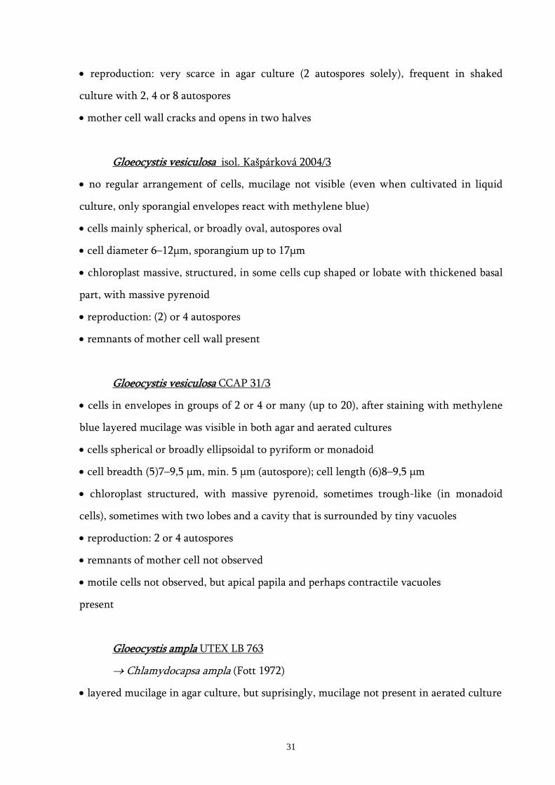
• reproduction: very scarce in agar culture (2 autospores solely), frequent in shaked
culture with 2, 4 or 8 autospores
• mother cell wall cracks and opens in two halves
Gloeocystis vesiculosa isol. Kašpárková 2004/3
• no regular arrangement of cells, mucilage not visible (even when cultivated in liquid
culture, only sporangial envelopes react with methylene blue)
• cells mainly spherical, or broadly oval, autospores oval
• cell diameter 6–12μm, sporangium up to 17μm
• chloroplast massive, structured, in some cells cup shaped or lobate with thickened basal
part, with massive pyrenoid
• reproduction: (2) or 4 autospores
• remnants of mother cell wall present
Gloeocystis vesiculosa CCAP 31/3
• cells in envelopes in groups of 2 or 4 or many (up to 20), after staining with methylene
blue layered mucilage was visible in both agar and aerated cultures
• cells spherical or broadly ellipsoidal to pyriform or monadoid
• cell breadth (5)7–9,5 μm, min. 5 μm (autospore); cell length (6)8–9,5 μm
• chloroplast structured, with massive pyrenoid, sometimes trough-like (in monadoid
cells), sometimes with two lobes and a cavity that is surrounded by tiny vacuoles
• reproduction: 2 or 4 autospores
• remnants of mother cell not observed
• motile cells not observed, but apical papila and perhaps contractile vacuoles
present
Gloeocystis ampla UTEX LB 763
→ Chlamydocapsa ampla (Fott 1972)
• layered mucilage in agar culture, but suprisingly, mucilage not present in aerated culture
31

• cells spherical or monadoid, motile monadoid cells present (in both agar/aerated c.)
• cell breadth 6–10 μm; cell lenght 7–9 μm (spherical cells) to 9–11 μm (monadoid cells),
sporangium 7,5×11 μm
• chloroplast parietal, cup-shaped with massive pyrenoid in the thickened basal part
• reproduction: division in two or four, in all directions
• remnants of broken mother cell not observed, but sometimes an empty cell wall as
undivided shell was visible
Gloeocystis gigas UTEX LB 291
• no regular arrangement of cells, mucilage not visible in agar culture
• cells spherical or monadoid
• cell diameter 9–11–12 μm (sphaecial cells), 8×11 μm (monads)
• chloroplast parietal with a massive pyrenoid in the basal part (huge starch grains)
• one or two vacuoles near the apex of the monadoid cell
• reproduction unknown (probably 2 autospores)
• remnants of mother cell not observed
Coenochloris planoconvexa CAUP H 5502 – Authentic Strain
• no regular arrangement of cells, mucilage not visible in agar culture, but unusual ray-
like shaped mucilaginous cover observed in aerated culture
• cells mostly ellipsoid to broadly oval, oocystis-like, often assymetrical
• cell breadth 4–6–(8) μm; cell length (4,5) –8–9 μm
• chloroplast parietal, trough-shaped with smooth, faint but clearly visible pyrenoid
surrounded by small starch grains
• reproduction: only scarcely 2 autospores observed; 4 autospores according to o.d.,
ocasional production of 2 or 8 autospore noted by later observation of the author (Hindák
1980)
• remnants of mother cell wall present, mcw splits and into two or several parts (Hindák
1977)
32

Schizochlamydella minutissima CCAP 57/1 – Authentic Strain
→ Sphaerochlamydella minutissima (Kostikov et al. 2002)
• no regular arrangement of cells, mucilage not visible, neither in agar nor in aerated
culture
• cells strictly spherical, very small
• cell diameter 2–3,5–6 μm; (exceptional cell 9 μm)
• chloroplast parietal, bilobate, often with a single large vacuole between the lobes
• tiny vacuoles on the surface of the cell (almost regularly)
• reproduction: autospores 4 or 8, scarcely observed; usually 4, but also 2 and 8 autospores
according to o.d.
• remnants of mother cell wall not observed
Neocystis sp. CAUP D 801
• no regular arrangement of cells, mucilage not visible in agar culture, fine mucilage
observed in aerated culture after stainig with Indian ink
• cells mostly oval, some broadly oval or spherical
• cell breadth 2–6 μm; cell length 4–8 μm;
• chloroplast parietal, lobate
• reproduction: 2–(4) autospores; ellipsoidal sporangia
• remnants of mother cell wall present
Neocystis brevis CAUP D 802 – Authentic Strain
• no regular arrangement of cells, mucilage not visible in agar culture, fine mucilage
observed in aerated culture after stainig with Indian ink
• cells elongate, ellipsoid, broadly oval to spherical
• cell breadth 2–5,8 μm; cell length 3,5–8 μm
• chloroplast parietal, bilobate
• reproduction: (2)–4–(8) autospores, broadly ellipsoidal sporangia
33

• remnants of mother cell wall present
Sphaerocystis bilobata CCALA 482
→ Radiococcus bilobatus (Kostikov et al. 2002)
• no regular arrangement of cells, mucilage not visible in agar culture
• cells spherical or broadly oval
• cell breadth 3–8 μm; cell length 4,5–8,5 μm
• chloroplast smooth, parietal, cup-shaped, with a pyrenoid surrounded by a ring of starch
grains
• reproduction by (2), 4 and 8 autospores
• remnants of mother cell not observed
Sphaerocystis oleifera strain Elster 98/26
→ Coenochloris oleifera (Kostikov et al. 2002)
• no regular arrangement of cells, mucilage not visible
• small cells ellipsoidal, almost oocystoid, big cells spherical
• cell breadth 3,5–7 μm; cell length 5–9 μm
• chloroplast parietal, bilobate, pyrenoid hardly visible
• reproduction: 4 or 8 autospores
• remnants of mother cell present
Gloeocystis polydermatica CAUP H 6701 – Authentic Strain
→ Sporotetras polydermatica (Kostikov et al. 2002)
• no regular arrangement of cells, mucilage not visible in agar culture
• cells ellipsoidal, broadly ellipsoidal to spherical
• cell breadth 3,5–7 μm; cell length 6–9 μm
• chloroplast smooth, parietal, band shaped with fine pyrenoid
• reproduction: 2 or 4 autospores, ellipsoid sporangia
• remnants of mother cell wall not observed
34
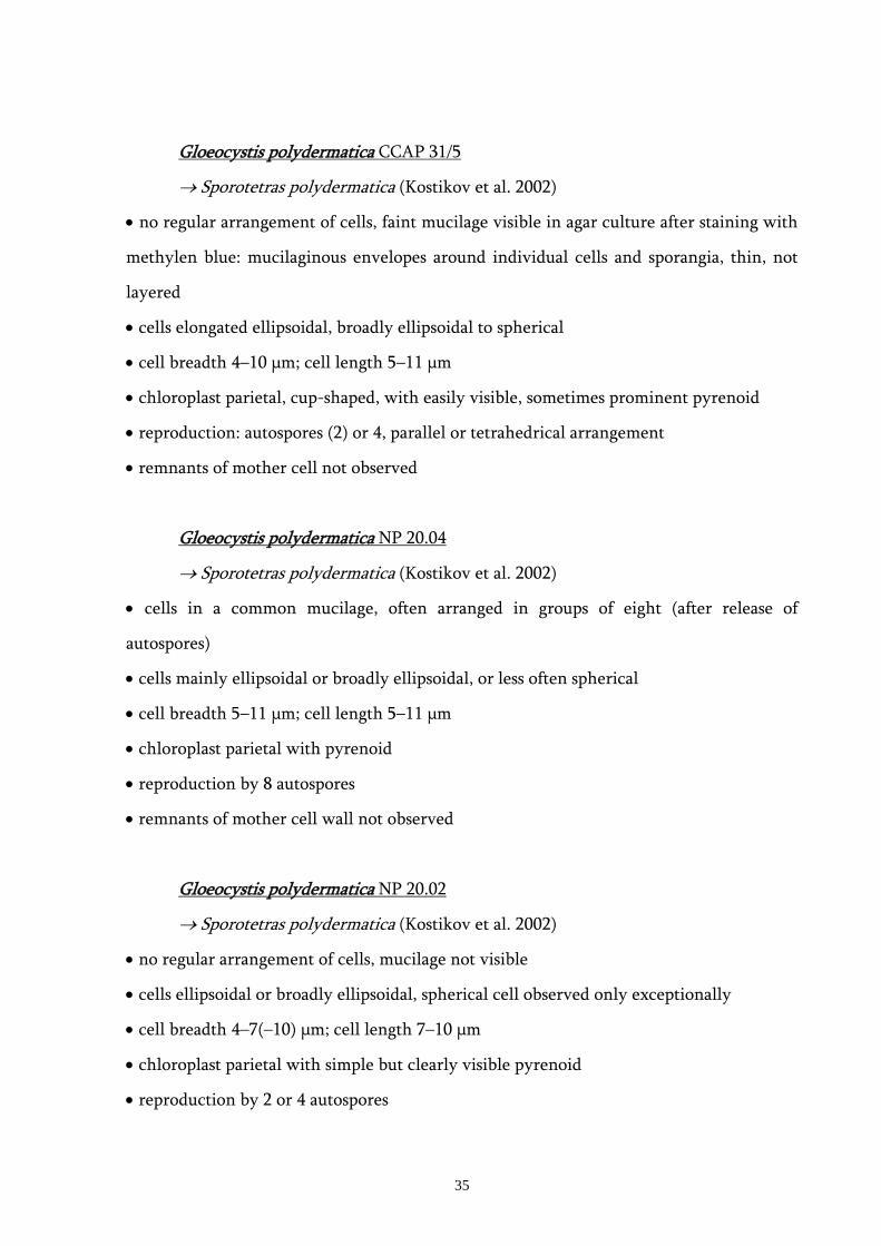
Gloeocystis polydermatica CCAP 31/5
→ Sporotetras polydermatica (Kostikov et al. 2002)
• no regular arrangement of cells, faint mucilage visible in agar culture after staining with
methylen blue: mucilaginous envelopes around individual cells and sporangia, thin, not
layered
• cells elongated ellipsoidal, broadly ellipsoidal to spherical
• cell breadth 4–10 μm; cell length 5–11 μm
• chloroplast parietal, cup-shaped, with easily visible, sometimes prominent pyrenoid
• reproduction: autospores (2) or 4, parallel or tetrahedrical arrangement
• remnants of mother cell not observed
Gloeocystis polydermatica NP 20.04
→ Sporotetras polydermatica (Kostikov et al. 2002)
• cells in a common mucilage, often arranged in groups of eight (after release of
autospores)
• cells mainly ellipsoidal or broadly ellipsoidal, or less often spherical
• cell breadth 5–11 μm; cell length 5–11 μm
• chloroplast parietal with pyrenoid
• reproduction by 8 autospores
• remnants of mother cell wall not observed
Gloeocystis polydermatica NP 20.02
→ Sporotetras polydermatica (Kostikov et al. 2002)
• no regular arrangement of cells, mucilage not visible
• cells ellipsoidal or broadly ellipsoidal, spherical cell observed only exceptionally
• cell breadth 4–7(–10) μm; cell length 7–10 μm
• chloroplast parietal with simple but clearly visible pyrenoid
• reproduction by 2 or 4 autospores
35
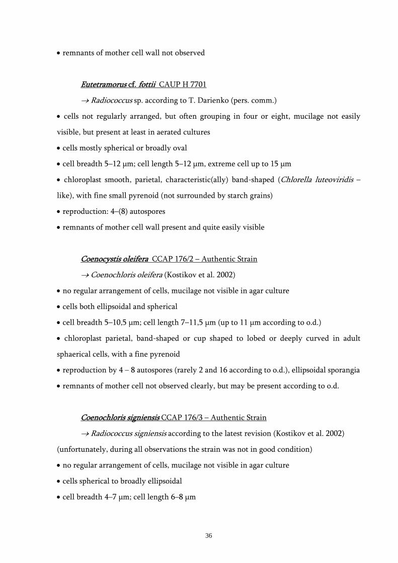
• remnants of mother cell wall not observed
Eutetramorus cf. fottii CAUP H 7701
→ Radiococcus sp. according to T. Darienko (pers. comm.)
• cells not regularly arranged, but often grouping in four or eight, mucilage not easily
visible, but present at least in aerated cultures
• cells mostly spherical or broadly oval
• cell breadth 5–12 μm; cell length 5–12 μm, extreme cell up to 15 μm
• chloroplast smooth, parietal, characteristic(ally) band-shaped (Chlorella luteoviridis –
like), with fine small pyrenoid (not surrounded by starch grains)
• reproduction: 4–(8) autospores
• remnants of mother cell wall present and quite easily visible
Coenocystis oleifera CCAP 176/2 – Authentic Strain
→ Coenochloris oleifera (Kostikov et al. 2002)
• no regular arrangement of cells, mucilage not visible in agar culture
• cells both ellipsoidal and spherical
• cell breadth 5–10,5 μm; cell length 7–11,5 μm (up to 11 μm according to o.d.)
• chloroplast parietal, band-shaped or cup shaped to lobed or deeply curved in adult
sphaerical cells, with a fine pyrenoid
• reproduction by 4 – 8 autospores (rarely 2 and 16 according to o.d.), ellipsoidal sporangia
• remnants of mother cell not observed clearly, but may be present according to o.d.
Coenochloris signiensis CCAP 176/3 – Authentic Strain
→ Radiococcus signiensis according to the latest revision (Kostikov et al. 2002)
(unfortunately, during all observations the strain was not in good condition)
• no regular arrangement of cells, mucilage not visible in agar culture
• cells spherical to broadly ellipsoidal
• cell breadth 4–7 μm; cell length 6–8 μm
36

• chloroplast parietal with a simple pyrenoid, may be surrounded by starch grains
• reproduction: only 2 autospores observed; 2 or 4 or rarely 8 according to o.d.
• remnants of mother cell present
Sphaerochlamydella minutissima CAUP H 7501
• cells regullarly arranged in pairs or in fours in a common mucilage, with ± similar
distances among them, sometimes with individual envelopes around groups of 2 or 4;
suprisingly different appearance in aerated culture: not regular, tight clusters of at least 8
cells)
• cells ellipsoidal to spherical, strikingly small
• cell breadth 2–5 μm; cell length 3–6 μm
• chloroplast parietal, cup-shaped, trough-shaped or bilobed
• reproduction: 2 or 4, rarely 8 autospores
• remnants of mother cell wall not observed so far
Coenochloris bilobata CCAP 176/1 – Authentic Strain
→ Radiococcus bilobatus (Kostikov et al. 2002)
• no regular arrangement of cells, mucilage not visible in agar culture
• young cells elongated, adult cells spherical
• cell breadth 4–9(–11) μm; cell length 6–12 μm; (usually 6 μm and up to 8,5 μm according
to o.d.)
• chloroplast parietal, often thin and deeply bilobed, pyrenoid not observed in this study
but should be present according to the original description
• reproduction by 4 + 8 autospores
• remnants of mother cell not observed but should be present acording to o.d.
37

4. Discussion
The primary aim of this work was to assess the phylogenetic diversity of representants
of the traditional family Radiococcacae. The need of molecular data in taxonomy of green
algae was stressed elsewhere (e.g. Pröschold & Leliaert 2007) and in our particular case by
Kostikov et al. (2002). It was already shown by Wolf et al. (2003) that the family is
polyphyletic. With more molecular data available, this work bears a robust evidence, that
the group Radiococcaceae is rather a mixture of many unrelated taxa. New lineages of
trebouxiophyte and chlorophyte algae were also discovered.
The taxonomy of the family Radiococcaceae was based on common morphological
characters – in the same way as the majority of green algal taxa was classified (Komárek &
Fott 1983, Ettl & Gärtner 1994). With the introduction of molecular techniques, since
about 20 years ago, the traditional taxonomical concept was challenged. New analysis
discovered for example fragmentation of genera into different classes (e.g. Chlorella, Huss
et al. 1999, Botryococcus, Senousy et al. 2004, Muriella, Hanagata 1998, Pediastrum,
Buchheim et al. 2005), or gathered morphologically dissimilar taxa into a single lineage of
close relatives (e.g. Micractinium and Chlorella, Krienitz et al. 2004, Luo et al. 2006). This
work bears new examples of both cases – distributing the species within Chlorophyceae
and Trebouxiophyceae and unifying strains of different labels in well supported clades on
the other hand – and follows the line of revealing hidden diversity among green algae
(Fawley et al. 2004, Lewis & Flechtner 2004, Vanormelingen et al. 2007).
4.1 molecular and morphological approach in taxonomy and determination
The unexpected number of clades involved in this analysis of Radiococcaceae supports
the strong need of revision of the point of view we used to take when dealing with the
taxonomy of green algae. For example, in our case, a use of mucilage production as a
taxonomical criterion on the level of family is untenable.
38

It is more than evident that the family Radiococcaceae as it was understood does not
exist. It is therefore necessary to find a new concept to sort out the former radiococcacean
taxa. This cannot be done without molecular tools. Isolation of more strains, covering as
much morphological variability as possible, together with phylogenetic analysis of the
strains, seems necessary to allow a general revision of the group. This process would
comprise two steps: first, we need to sort out the specimens of Chlorophyceae and
Trebouxiophyceae and secondly to define each new clade by specific combination of
characters.
I am convinced that for the use in every-day practice (at least before handy all-in-
minute barcoding machine is invented and widely available), we should keep the
traditional tools of determination , i. e. a simple system based on morphological characters
with the support of ecology or perhaps physiology – but the criteria should be revised and
updated according to latest progress in molecular taxonomy.
Not only for every-day determination the morphology is important. Thorough
morphological observations and studies of life cycle should not be replaced by sequencing
even in taxonomy. Molecular taxonomists often use strains from culture collection
without revising its identity, which could lead to misinterpretation of molecularr data.
For example, Senousy et al. 2004 discussed the species concept in Botryococcus, where the
species B. sudeticus was by some authors recognized as Botryosphaera or
Botryosphaerella. While molecular data clearly showed that the B. sudetica is totally
unrelated to most representants of Botryococcus, the issue was left open and no taxonomic
conclusion was made. The strain and its sequence are still labeled as Botryococcus which
could possibly cause confusion in other studies. Further evidence was than added by
Přibyl & Cepák (2007) who reported zoospore production in the same strain of B. sudetica.
On the basis of that observation they finally removed B.sudetica from the autosporic
genus Botryococcus.
39

4.2 evaluation of morphological criteria
The most serious issue in the generic system of Radiococcaceae is the stability of
morphological traits when comparing fresh material with cultivated one.
In general, the applicability of a particular morphological trait on a particular
taxonomical level, is limited by its variability on that level. Only markers of certain
stability can be chosen as taxonomical criteria (for example, when the number of
pyrenoids, hypothetically, spans from two to four, no one would chose the character
„three pyrenoids“ as a relevant marker). In case of Radiococcaceae, however, the
morphology often changes significantly when the alga is removed from natural sample to
culture and also when different cultivation conditions are applied (like for example the
shape of colony and form of mucilage, as will be discussed later).
Most descriptions of the radiococcacean genera were based on observation of fresh
material (e.g. Koršikov 1953), only few authentic strains are available from more recent
studies (Kostikov & Hoffmann 2000; Broady 1976, 1982; Hindák 1977, 1978,1984). But to
describe the morphology thoroughly – to see the shape of the chloroplast or to observe the
whole life cycle for example – we usually need the cultivation and a long-term
observation. Because of the lack of authentic cultures, for most of the radiococcacean taxa
it is impossible to verify the morphology of the type species and to amend the eventual
cavities in the description.
Moreover, the morphology of authentic strains does not necessarily correspond with
the original description after decades of cultivation. This can be a result of either
mutations or adaptations to distinct environmental conditions in cultivation (more
constant, free of predators, free of water movement etc.). The AFLP data obtained by
Muller et al. (2005) showed no differences in genetic material of several clones kept in
different collections for many years: it seems, supprisingly, that strains in culture
collections remain (relatively) genetically stable. Than the possibility of adaptive
morphological changes should be examined.
40
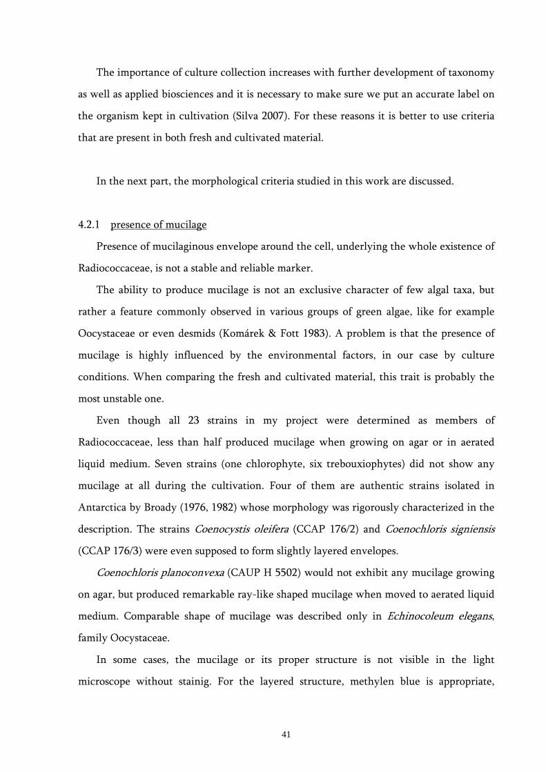
The importance of culture collection increases with further development of taxonomy
as well as applied biosciences and it is necessary to make sure we put an accurate label on
the organism kept in cultivation (Silva 2007). For these reasons it is better to use criteria
that are present in both fresh and cultivated material.
In the next part, the morphological criteria studied in this work are discussed.
4.2.1 presence of mucilage
Presence of mucilaginous envelope around the cell, underlying the whole existence of
Radiococcaceae, is not a stable and reliable marker.
The ability to produce mucilage is not an exclusive character of few algal taxa, but
rather a feature commonly observed in various groups of green algae, like for example
Oocystaceae or even desmids (Komárek & Fott 1983). A problem is that the presence of
mucilage is highly influenced by the environmental factors, in our case by culture
conditions. When comparing the fresh and cultivated material, this trait is probably the
most unstable one.
Even though all 23 strains in my project were determined as members of
Radiococcaceae, less than half produced mucilage when growing on agar or in aerated
liquid medium. Seven strains (one chlorophyte, six trebouxiophytes) did not show any
mucilage at all during the cultivation. Four of them are authentic strains isolated in
Antarctica by Broady (1976, 1982) whose morphology was rigorously characterized in the
description. The strains Coenocystis oleifera (CCAP 176/2) and Coenochloris signiensis
(CCAP 176/3) were even supposed to form slightly layered envelopes.
Coenochloris planoconvexa (CAUP H 5502) would not exhibit any mucilage growing
on agar, but produced remarkable ray-like shaped mucilage when moved to aerated liquid
medium. Comparable shape of mucilage was described only in Echinocoleum elegans,
family Oocystaceae.
In some cases, the mucilage or its proper structure is not visible in the light
microscope without stainig. For the layered structure, methylen blue is appropriate,
41

whilst for simple detection of a fine diffluent envelope, Indian ink works better. It is
probable that without staining the mucilage would not be detected in many algae. This
could lead to ambiguous determination, when a simple coccal cell is labelled for example
either as Chlorella (when the mucilage was not detected) or as Coenochloris.
As was pointed out by Kostikov et al. (2002), we deal with different kind of
mucilaginous material: thick or thin, strong or weak, distinct or diffluent, spherical or of
various shapes or shapeless... This fact too was often negliged in the taxonomy. (For
example, the envelope of the three Coenochloris polycocca strains from SAG reached
about 2-3μm in thickness, was layered and prominent regardless the cultivation. This
surely is not the same character like the unstable ray-like structure seen on Coenochloris
planoconvexa or faint pall of mucilage around Neocystis spp. visible only after staining
with Indian ink.)
The layered structure of mucilage, characterictic for Gloeocystis and some other
genera, was only scarcely observed on cultured material (hints in Gloeocystis
polydermatica CCAP 31/5), and never so distinct like in the fresh sample. This is not the
case of chlamydomonad species, where the envelopes are often conspicuously layered, but
here the structure is rather a layered sporangium cell wall which may eventually
gelatinize. The change of mucilage character during cultivation of Planktosphaeria
gelatinosa mentioned also Hindák (1984).
There was also a special form of radially structured mucilage described in the genus
Radiococcus. Nothing like it I observed on the analyzed strains. As I mentioned in the
introduction, the ray-like structure was doubted by Komárek 1974 and Hindák 1984.
Without robust evidence by thorough microscopic analysis, their hypothesis that the ray-
like structure was probably caused by associated rod-like bacteria is to be accepted.
Few years ago, Parachlorela beijerinckii Krienitz et al. (2004) was described as a new
species in a sister group of Chlorella. It was a simple mucilage producing coccoid green
alga, but because the description was strongly based on molecular data, its classification
within Radiococcaceae was not considered.
42

4.2.2 shape of the colony and arrangement of cells
Because all the mophological observations were done on a cultured material, it was
not possible to see the apperance of the taxa in the natural state. This refers especially to
the shape of the whole mucilaginous colony, that should be spherical in many planctic
taxa (e.g. genera Radiococcus, Eutetramorus, Sphaerocystis, Coenochloris). Unfortunately,
a determined shape of colony was not seen in any agar or liquid culture in this work. For
strains in culture collections it could be difficult or even impossible to obtain original
morphological data on the material from which the strain was isolated. The use of such a
marker, observable only on fresh material, is questionable.
The arrangement of cells is also an issue. Cells grouped in two, four or more within a
sporangial wall were present in chlamydomonad strains, but this does not correspond to
the character of arrangement as it was understood for example by Koršikov (1953). Groups
of four or eight after autospore release were observed in some strains from the
„polydermatica“ clade. Cells of strains form the Sphaerocystis clade were in groups of 16
or more, several generations together. The most distinct arrangement was observed in
agar culture of Sphaerochlamydella minutissima (CAUP H 7501), nice pairs of cells
distributed in more or less even distance to each other. But this appearance changed
dramatically when the strain was transferred to aerated liquid culture: the cells grouped in
eights and were haphasardly assembled in clusters in the medium.
4.2.3 sporangial cell wall behaviour
Various ways how the autospores are released from the mother cell were thoroughly
described by Kostikov et al. (2002). Basically, there are two possibilities: rupture (which
leaves remnants of the cell wall around the young daughter cells) or gelatinisation (the
mother cell wall turns in gelatinous sheath around the daughter cell). This model has
many modifications, for example rupture of the mother cell wall (mcw) and later
gelatinisation of the fragments. Some authors (Kostikov 1953, Komárek & Fott 1983)
distinguished the „late gelatinization“, Kostikov et al. (2002) relied only on the presence of
mother cell wall fragments in the sample. Again, it brings the question of applicability of
43
the marker. It is obvious that one can easily misjudge this character based on a single
observation.
During my observations, the question of mcw behaviour was not always succesfully
solved and in few cases, for example only once a single fragment was observed. In the
Neocystis-clade, the mother cell wall splits and its fragments are scattered around the cells
in all strains. In the „polydermatica“-clade the remnants were not observed except
Eutetramorus cf. fottii (CAUP H 7701) and only once a ruptured empty cell wall was seen
in Coenocystis signiensis (CCAP 176/3). According to Broady (1976) the remnants should
be visible in C. signiensis (CCAP 176/3) and C. oleifera (CCAP 176/2). Thus in this case,
mcw behaviour is not specific for the phylogenetic lineage. An exceptionally clear form of
cleavage was observed in culture of Coenochloris pyrenoidosa (CCALA 324). Here the
sporangium wall splits in two more or less adequate portions.
As a good illustration, the strain Eutetramorus cf. fottii (CAUP H 7701) can be
mentioned. It was isolated and subject to thorough microscopy analysis by Škaloud (2004)
without an observation of mcw fragmentation. Few years later, the strain was revised by
T. Darienko (unpubl.) and determined as Radiococcus sp. because of the presence of mcw
remnants. During this study, mcw remnants were also clearly visible. However, this
feature was not evident in the fresh material and in newly established culture (Škaloud
2004).
4.2.4 vegetative cell shape
In the group of Radiococcaceae sphaerical or ellipsoid vegetative cells were
traditionally recognized. According to Kostikov et al. (2002), three types of morphology
are to be seen: cells always spherical, cells sometimes spherical and cells never spherical.
The first two groups were put together in the latest revision of the system (Kostikov et al.
2002). I suppose that this decision was not appropriate, leading to the underestimation of
the general appearance of the organism. In almost all ellipsoid-celled taxa in this study it
was possible to observe at least few spherical cells, or there were mainly ellipsoidal with
the minority of spherical cells.
44

The best results were obtained when this particular marker was used on the level of
classes. Only spherical cells were present in all chlorophyte strains (with the exception of
the chlamydomonas group). The trebouxiophytes can have morphology of all three types,
i. e. cells spherical (Schizochlamydella minutissima CCAP 57/1) or ellipsoidal
(Coenochloris planoconvexa CAUP H 5502, more or less also Gloeocystis polydermatica
NP 20-02 and 20-04) or both (most of trebouxiophyte strains). The cell shape give a good
information when correlated with cell size (see below).
4.2.5 cell size
The vegetative cell size was not used on generic level in any relevant review /systems/
of Radiococcaceae (Koršikov 1953, Bourrelly 1966, Fott 1974, Komárek and Fott 1983,
Kostikov et al. 2002), (sometimes it was used to distinguish species). However, the cell size
corresponds more or less with the distinction of two classes, Chlorophyceae and
Trebouxiophyceae.
4.2.6 pyrenoid
The presence/absence of pyrenoid was applied either on the generic or species level in
green algae and in Radiococcaceae too (Hindák 1977, Komárek & Fott 1983). This
ambiguous use was criticised by Kostikov et al. (2002), who established a system with
pyrenoid as generic marker. There are several issues concening such a use.
Not every microscopic observation would detect the presence of pyrenoid. For
example, in the description of Coenocystis inconstans, the authors state that „a pyrenoid is
present, but hardly distinguished from the chloroplast stroma“ (Hanagata & Chihara
1999). In case of Cenochloris korsikovii (CAUP H 5503) in this study it was not possible to
detect a pyrenoid with only the light microscopy, but the isolator and autor of the
description stated the alga possessed a nude pyrenoid (Hindák 1984).
Nozaki et al. (1998) bore evidence that the precence of pyrenoid can depend on
environmental conditions: 17 of 23 strains of Chlorogonium (all commonly with
pyrenoids) lost or showed only reduced pyrenoids in medium with organic compounds.
45

Molecular data can even advocate the use of pyrenoid presence on the generic level –
for example, in traditinal genus Chlorella s.l., the presence of pyrenoid was characteristic
for about half of the species (Fott & Nováková 1969). After the revisions based on
phylogenetic analysis, all species of „true chlorellas“ have cells with pyrenoid (Krienitz et
al. 2004). However, this was not the case of the Neocystis-clade in this work. On the other
hand, the same marker has no taxonomical significance on the generic and even species
level in Chlamydomonas and Chloromonas. The study of Pröschold et al. (2001) showed
that species of both genera, that were traditionally recognized according to the presence
resp. absence of pyrenoid, are mixed together in several phylogenetic lineages and the lost
of pyrenoid occured more than once in the evolution of chlamydomonad species.
During my observations the pyrenoid was present in all but one chlorophyceae, in
several strains more than one per cell (up to nine pyrenoids in Sphaerocystis schroeteri
CCALA 483). In trebouxiphyceae the presence was either characteristic for the whole
clade (e.g. the „polydermatica“-clade) or only for some members of the phylogenetic
lineage (not observed in both Neocystis strains, hardly visible but present in Sphaerocystis
oleifera isol. Elster 1998/26, present and surrounded by little starch grains in S. bilobata
CCALA 482). These results advocate that the presence of pyrenoid should not be used as
taxonomical criterion without the justification by molecular data.
What I find important is the form of pyrenoid. Among Trebouxiophyceae, the
pyrenoid was always single, rather small and simple, without prominent margin,
sometimes surrounded by little rounded starch grains, sometimes without. Among
Chlorophyceae the pyrenoid was massive and bordered by prominent envelope.
4.2.7 number of chloroplasts
Usually, there is only one chloroplast per cell in Radiococcaceae. The only exceptions
are Planktosphaeria gelatinosa, Phacomyxa sphagnophila and Palmodictyon varium.
Among the 23 strains analyzed, numerous chloroplasts were observed only in adult cells
of Planktosphaeria gelatinosa (CAUP H 1401). A similar feature was found also in
Coenochloris pyrenoidosa (CCALA 324), but it is not sure yet whether the stage was an
46

adult cell or an early development of sporangium. Cells with numerous chloroplasts are
unknown among Scenedesmaceae, where the strain belongs to (Komárek & Fott 1983).
4.2.8 shape of chloroplast
This marker was only occasionally used to separate few genera of distinct chloroplast
shape, e.g. Chondrosphaera and Pseudotetraspora with stellate chloroplast in Fott (1974)
and Bourrelly (1966), respectively. On the species level the shape of chloroplast was used
by Komárek & Fott (1983) to separate between Coenochloris mucosa and C. sphagnicola
and between Palmodictyon species.
Here very different appearance of chloroplast was found when comparing
Chlorophyceae and Trebouxiophyceae. In the first class, the chloroplast was (almost)
always massive and not smooth, filling the whole volume of cell except central area with
nucleotide. In two strains it divided later into many small periferal chloroplast (see
above). In the second class, the chloroplast was smooth and often comprised only part of
the cell. Four distinct types of chloroplast were found among Trebouxiophyceae: cup-
shaped, band-shaped, bilobed and through-like. Unfortunately, usually it was not possible
to use a particular shape of chloroplast as a diacritical trait for whole clade. Through-
shaped chloroplast, common among Oocystaceae, was found in Coenochloris
planoconvexa (CAUP H 5502). Bilobed type was typical for Schizochlamydella
minutissima (CAUP 57/1), Coenochloris bilobata (CAUP 176/1) and the Neocystis-clade,
where, unfortunatedly, was not always apparent in S. bilobata (CCALA 482) and S.
oleifera (isol. Elster 1998/26). Band shaped chloroplast was pronounced in some strains in
the „polydermatica“-clade.
Recently, taxonomical stadies using polyphasic approach advocated use of chloroplast
shape in taxonomy (Pröschold et al. 2001). In case of chlorophycean strains, the use of
confocal microscopy could bear interesting results too.
47
4.2.9 number of autospores and mode of reproduction
The use of this autospore number on the generic level was given weight by Kostikov
et al. (2002). In works of other authors (Koršikov 1953, Fott 1974, Hindák 1977, Komárek
& Fott 1983) it was more a complementary criterion, or eventually was not used at all
(Bourrelly 1966).
Kostikov distinguished two main groups of species: those producing „a small number,
generally two“ autospores and those producing generally four or eight. (The word
„generally“ means that exceptions were accepted). But for example in Hindák (1977),
Radiococcus and Coenochloris differed in production of „always four“ or „four or eight“
autospores. According to Kostikov’s concept, these two genera were in the same group.
Kostikov also admitted, that the number of autospores may depend on culture
conditions (based on unpublished data) and „the stability of this feature needs further
investigations“ (Kostikov et al. 2002). This assumption was fully supported by my
observations: the number of autospores proved to be the most variable morphological trait
in different conditions of cultivation. In general, whilst some strains do not change at all
(mostly Trebouxiophyceae), for others the number of autospores increases considerably
when the strain is transferred from agar to liguid culture (mostly Chlorophyceae).
This could correspond with the finding of Hindák (1984) when the fresh material of
Planktosphaeria gelatinosa produced commonly 16 or 32 autospores and the cultivated 4
or 8 autospores. It is possible that the aerated liguid culture simulates better the natural
environment and so the number of autospores is closer to the state in situ.
Trebouxiophyte strains tend to be more stable, with the only exception of
Sphaerochlamydella minutissima (CAUP 57/1) that produced much more eight-celled
sporangia in liguid culture than on agar. Polysporic sporangia were not observed in any
member of Trebouxiophyceae. On the other hand the number of autospores in
Chlorophyceae was highly variable, spanning from two (on agar) to 32 (liquid culture for
Coenochloris hindakii or any cultivation for the Sphaerocystis-clade) and was often
limited by culture conditions.
48
The position of autospores was usually tetraedrical, only occasionaly parallel (S.
bilobata isol. Elster 1998/26), but both types were observed in the same strain, so to this
character no taxonomic weight is given, as was already done by Kostikov et al. (2002).
All chlorophyte strains were treated for zoospore production. The effort was succesful
only in case of Planktosphaeria gelatinosa (CAUP H 1401), only supporting similar
observation by Starr (1954) and Hindák (1984). The strain Coenochloris hindakii (CAUP
H 5503) produced in the same conditions polysporic sporangia (aplanosporangia?). No
other motile stages were observed except chlamydomonad strains G. ampla (UTEX LB
763) and G. gigas (UTEX LB 291).
4.3 taxonomical conclucions
Planktosphaeria gelatinosa CAUP H 1401
Under the name Planktosphaeria gelatinosa Smith (1915) a planctic organism showing
no motile stages was described. The strain sequenced here was isolated by R. C. Starr in
1952 (deposited 1954). Based on his own isolates from soil, Starr reported the observation
of zoospores in this species (Starr 1954). Some authors than understood the taxon as
zoosporic, but others respected the original description and regarded Starr’s zoosporic
isolates as different organisms (Lukešová 1993, Ettl & Gärtner 1994, Kostikov et al. 2002).
According to A. Lukešová (unpubl.), zoosporic soil specimens commonly determined
as Planktosphaeria probably all belong to the revised genus Follicularia MILLER 1924. This
hypothesis needs to be verified by molecular data, however. In this work sequence of a
freshwater and soil isolate cluster together. More confusingly, zoospore production was
reported in both soil and planctic material by Hindák (1984).
Authentic strains of the type species of Planktosphaeria and Follicularia are not
available, but authentic strains of other species of Follicularia are being analyzed by T.
Pröschold (unpubl.). If P. gelatinosa sequenced here is related with Follicularia strains,
than all the specimens should bear the name Follicularia. Or they could be all transferred
to Planktosphaeria, with the emendation of the genus. If the two strains od P. gelatinosa
49

(CAUP H 1401 = UTEX B 124 and SAG 262-1b) lie in a distinct lineage, not with
Follicularia, a new generic name should be applied.
Coenochloris pyrenoidosa SAG 217–1a, SAG 217–1b, SAG 217–1c and Sphaerocystis
schroeteri CCALA 483
Although bearing different names, the strains SAG 217-1a, SAG 217-1b, SAG 217-1c
and CCALA 483 show substantial similarity: relatively big sphaerical cells, massive
chloroplast with prominent pyrenoid(s), thick gelatinizing layered cell walls, reproduction
by numerous autospores. Being also phylogenetically related, these four strains should
belong to a single genus.
The three SAG strains have different names in the collections of SAG and UTEX and
according to Kostikov et al. (2002) and Wolf et al. (2003): Coenochloris, Coelastrum and
Radiococcus. According to Koršikov (1953) the genus Coenochloris is characterized by
oval cells in groups of four or eight (rarely 16) tightly accumulated in the centre of a
structureless mucilage (the genus contained species either with or without pyrenoid).
Athough it is not possible to observe the proper arrangement of cells in strains kept in
agar cultures, I suppose that on the basis of cell size, number of pyrenoids, higher number
of autospores and layered mucilaginizing cell wall, the name Coenochloris shold not be
applied to the three strains discussed. The genus Radiococcus was firstly recognized by a
radial structure in its mucilage, a feature that was discussed elsewhere (Fott 1974, Hindák
1984, above in this work), and after emendation (Fott 1974) by arrangement of cells in
tetrads and reproduction by (strictly) four autospores (not (2–) 4–8 (–16) according to
Kostikov et al. 2002). Therefore this name cannot be applied either. Coelastrum clearly
has different morphology, here, for example, adult cells are not connected in coenobia,
also sometimes more than one pyrenoid is present.
The four strains seem to fit the best within the delimitation of the genus
Sphaerocystis: cells are spherical and tend to stay in groups after release of autospores,
embedded within a substantial mucilage, with parietal chloroplast and one or more
pyrenoids. Only the number of autospores commonly observed (usually 32 or 16) exceeds
50
the number presented in literature (4, 8 or 16) (Koršikov 1953) and the cell wall is rather
thick. I suppose that in this situation, it is not reasonable to establish a new genus,
omitting the traditionaly used, though poorly defined name. Because there is not an
authentic culture of a type species, Sphaerocystis schroeterii, and because the genus was
already emended, anyway, I suggest to establish the strain Sphaerocystis schroeterii as an
epitype of the emended genus Sphaerocystis and to move the three strains of Coenochloris
polycocca into this genus.
Coenochloris korsikovii CAUP H 5503
The authentic strain of Coenochloris korsikovii (CAUP H 5503) lies within the family
Selenastraceae, most close to Monoraphidium spp., but not in close relationship to any
particular isolate. The simple morphology is quite exceptional within the group, where
species with elongated, sometimes curved or twisted cells are mostly present. The 18S
rRNA data analysis itself was for Krienitz et al. (2004) a reason to establish a new taxon.
Here the morphological as well as phylogenetic distinction of the strain Coenochloris
korsikovii (CAUP H 5503) among Selenastraceae bears arguments enough to establish a
new genus for the strain CAUP 5503. Because the alga did not commonly exhibit
mucilaginous envelopes and fragments of broken mother cell wall and produced
polysporic sporangia (probably aplanosporangia) under certain conditions, I do not find
adequate to call it Coenochloris.
Coenochloris pyrenoidosa CCALA 324
The isolate from little pool named Coenochloris pyrenoidosa falls within the family
Scenedesmaceae (its position in more detail is shown on fig. 3). It is probably a sister to
Coelastrum microporum, isolate Kr1980/11 (Krienitz, unpublished). The sphaerical cell
shape may be surprising within this family, but cell shape variability is not unknown
among Scenedesmaceae. From the morphological point of view, two other specimens are
interesting: Pectodictyon pyramidale was comprised in Radiococcaceae, subfam.
Dictyochlorelloideae according to Komárek & Fott (1983). It is excluded from „true
51

Radiococcaceae“, because its cells are connected with mucilaginous strands (not free). This
alga is more or less close to a specimen that almost does not produce mucilage at all. The
second one is Scenedesmus rotundus, whose position within the clade is not clear. It is a
recently described species of Scenedesmus (s.l.) from desert soil. It possesses very simple
sphaerical cells and resembles the strain studied here.
To make final taxonomic decisions, thorough polyphasic study of the Coenochloris
pyrenoidosa and Coelastrum microporum and possibly the two other strains together
would be reasonable.
Fig. 3: Phylogeny of the family Scenedesmaceae, NJ, Tamura-Nei.
52

Gloeocystis vesiculosa CCAP 31/3 and isol. Kašpárková 2004/3, Gloeocystis ampla
UTEX LB 763, Gloeocystis gigas UTEX LB 291
Four strains of Gloeocystis spp. are members of the CW group of Chlorophyceae
(Chlamydomonas-clade in broad sense). Their position within the clade, each strain lying
within its distinct lineage, impairs seriously the validity of the Gloeocystis concept. It also
shows evidence that the ability to form mucilaginous palmelloid stages is common among
chlamydomonad algae.
The description of Gloeocystis, type spec. Gloeocystis vesiculosa Nägeli (1849), gives
not sufficient information on the genus that would allow unambiguous interpretation.
Although Fott & Nováková (1971) rejected this genus, Hindák (1978) was of different
opinion and many authors kept his concept more or less similar till today (Komárek &
Fott 1983, Ettl & Gärtner 1994, Kostikov et al. 2002). The name Gloeocystis should be
applied (according to these works) to autosporic algae of either spherical (G. vesiculosa) or
ellipsoidal (G. polydermatica) cells lying within concentric layers of mucilage.
Although Nägeli (1849) observed no motile stages, I am convinced that the specimen
originaly described as Gloeocystis vesiculosa was in fact a palmelloid stage of some
chlamydomonad alga. It could be a chlamydomonas-like organism who lost the ability to
produce motile stages or where immotile form is the prevailing one. There are several
points to support this assumption: (1) cells in the original drawing by Nägeli are quite
similar to those in drawing of Tetraspora and Palmella by the same author; (2) the
appearance of mucilaginous envelopes is very close to that observed on chlamydomonad
strains in this work, and they are slightly different from the mucilage around Gloeocystis
polydermatica that surely is not a chlamydomonad organism; (3) all spherical Gloeocystis
spp. strains sequenced in this work are chlamydomonad organisms; (4) the „colourless
area“ (or lumen in chloroplast) adjacent to sister-cell, as described by Nägeli for genus
Gloeocystis but also for Palmella (1849), is a common feature in chlamydomonads (s.l.)
and it was also observed in chlamydomonad strains during this study. On the other hand,
this feature is not present in trebouxiophyte Gloeocystis polydermatica.
Basically, there are three ways to solve the problem of Gloeocystis:
53

a) to conclude that the genus was not approprietly described and the type species
referres to a stage in life-cycle of another organism and the name should be rejected.
b) to isolate more representants of Gloeocystis vesiculosa with corresponding
morphology and ecology (the strains analyzed here were of different origin, often
freshwater, whilst the original material was aerophytic, growing on a stone) and subject
these strains to further phylogenetic analysis. If the aerophytic algae form a uniform
cluster within chlamydomonadales and none of them produces motile stages in any
observable condition, than the original Nägeli’s name can be applied to such organisms,
aerophytic chlamydomonads (in broad sense) that lost the ability to form motile stages.
c) to prove that the type Gloeocystis vesiculosa was not approprietly delimited and all
data about this species in literature were based on mistaken observations (all organisms
determined as G. vesiculosa were stages of chlamydomonads). Because the name is
traditionally used for aerophytic green algae with layered mucilage, Gloeocystis
polydermatica might be conserved as a new type for the (trebouxiophyte) genus
Gloeocystis.
Coenochloris planoconvexa CAUP H 5502
The new species Coenochloris planoconvexa was established mainly because its
assymetrical, planoconvex cell shape (Hindák 1977), eventhough this feature was not
present in all isolates of the same specimen (Hindák 1980, 1984). By the morphology itself,
mainly based on the cell shape and presence of polar thickenings of the cell wall, the
strain can be assigned to Oocystaceae. The same result was obtained here by analysis of
18S rRNA. The genus Oocystis, however, is not monophyletic (Hepperle et al. 2000).
Without a revision of the genus, and when the strain CAUP H 5502 is not closely related
to any of three sequenced Oocystis species, we can hardly assign Coenochloris
planoconvexa to Oocystis.
The isolate CAUP 5502 exhibits an extraordinary shape of mucilaginous envelope,
that could possibly serve as a morphological criterion to delineate a new genus, with the
awareness that this particular trait may not be pronounced in some cultivations. Similar
54

feature was described earlier in the genus Echinocoleum JAO et LEE 1947. For comparison,
strain Echinocoleum elegans SAG 37.93 was studied. The shape of mucilaginous envelope
was essentially different, with only three or four massive mucilaginous arms comparing to
many little rays in Coenochloris planoconvexa. Nor the 18S rRNA data support a close
affinity of these two strains: Echinocoleum elegans is a sister to Amphikrikos sp. and
Schizochlamydella capsulata within Oocystaceae.
Schizochlamydella minutissima CCAP 57/1
The strain Schizochamydella minutissima CCAP 57/1 constitutes a well supported
lineage with few unknown Nannochloris sequences. It is placed in the neighbourhood
with true Nannochloris species and the Koliella spiculiformis-clade. So it lies in the group
where other algae with very small cell size are present, but suprisingly, this new clade
appears closer to the Koliella clade than to the small coccoid forms.
S. minutissima was established as a type species of new genus Sphaerochlamydella by
Kostikov et al. (2002). Because the strain sequenced here is a type strain isolated by
Broady (1982), its phylogenetic position assigns a position of the genus
Sphaerochlamydella. Other sequnces included in the clade represent undetermined
organisms. It is probable that for the small size of these organisms, their particular
morphology was simply omitted. This is not the case of the type strain, however, which
apart its significantly small size exhibits distinct morphology, with deeply bilobed
chloroplast without pyrenoid and usually a relatively big vacuole between the lobes.
I suppose that this particular characteristics also justify its removement from the
genus Schizochlamydella, that should encompass species with (a) cup-shaped chloroplast,
(b) two autospores (here commonly also 4 and 8), (c) persisting mother cell wall and (d)
no particular arrangement in the mass of mucilage.
Into the new genus Sphaerochlamydella also Catenococcus minutus was moved by
Kostikov et al. (2002). This organism should display a different arrangement of cells, with
cell size even smaller (1,2 – 2,8 μm) than S. minutissima (Komárek & Fott 1983). Without
55

sequence to compare, it is impossible to assume about its position within the
contemporary system.
Neocystis sp. CAUP D 801, Neocystis brevis CAUP D 802, Sphaerocystis bilobata
CCALA 482, Sphaerocystis oleifera isol. Elster 1998/26
A new clade of trebouxiphytes is here constituted. All species originate from soil
biotopes from different parts of the world. They have mostly oval or sometimes spherical
cells, do not produce a lot of mucilage and have prevailingly bilobed chloroplast (this was
not verified for S. bilobata, suprisingly, but the strain was determined and assigned to the
„bilobata“ species by an acknowledged algologist).
The old generic name Sphaerocystis is apparently not appropriate for two of the
species (see discussion on the chlorophycean Sphaerocystis clade above). Nor would be
Coenochloris (defined by the arrangement of cells) or Radiococcus, in which these species
were moved by Kostikov et al. (2002). The name Neocystis may be considered. This genus
was established by Hindák to accomodate non-spherical cells without pyrenoid and
Koršikov’s Coenochloris ovalis was established as a type species. Because the authentic
strain is not availabe, here we must rely on the original description and drawing, which,
unfortunately, does not show much. The usually periferal chloroplast could be cup-
shaped, but surely is not bilobate. Because of the deeply bilobed chloroplast and the
presence of pyrenoid in two of the strains, this clade should not bear the name Neocystis
and rather be described with a new generic name.
Gloeocystis polydermatica CAUP H 6701, CCAP 31/5, NP 20.02 and NP 20.04,
Eutetramorus cf. fottii CAUP H 7701, Coenocystis oleifera CCAP 176/2, Coenochloris
signiensis CCAP 176/3
Among this group algae with ellipsoidal, broadly ellipsoidal and spherical cells are
present, with smooth parietal, often band-shaped chloroplast with a pyrenoid. Three
autenthic strains are lying within this clade: Gloeocystis polydermatica Hindák
Coenocystis (Sphaerocystis) oleifera and C. signiensis Broady. Since none of these strains
56

is a generic type, it is not appropriate to assign this clade to one of the genera, at least
when a conservation under a new type species is not applied.
This clade does not exhibit any particular trait that would allow clear characterization
of this new taxon in Trebouxiphyceae. Some of the specimens, but not arguably all of
them, produced layered mucilaginous matrix in fresh state, eventhou this marker is hard
to trace in cultures. So there is reason for choosing the name Gloeocystis, that was
traditionally used for such an algae growing on rocks, wood etc. This question was already
discussed above. Another name used G. polydermatica-like algae was Dactylothece. This
genus was not thoroughly described either and resembles more the genus Coccomyxa.
Both of the names are not adequate for species with often almost spherical cells, that
sometimes tend to stay in groups of four or eight, have tetraedrically arranged autospores
and discernible pyrenoid.
Sphaerochlamydella minutissima CAUP H 7501, Coenochloris bilobata CCAP 176/1
Sphaerochlamydella minutissima CAUP H 7501 and Coenochloris bilobata CCAP
176/1 are not relatives to any sequenced trebouxiphyte. Because they do not represent
type species, new generic names shodl be proposed. Authentic strain of the type
Sphaerochlamydella minutissima (under an older name Schizochlamydella) was
sequenced in this work and discussed above. The authentic strain Coenochloris type
species (C. pyrenoidosa) is not available, however the morphology of that species is
apparently different and it probably belongs to Chlorophyceae.
The first specimen, S. minutissima, is spectacular when cultured on agar by its nice
regular arrangement of cells. This arrangement is lost, however, in liquid medium.
Coenochloris micrococca, described by Komárek, appears morphologically somewhat
close, but it should have cells even smaller, posses (though hardly visible) pyrenoid and
was found only in Cuba.
The culture of Coenochloris bilobata CCAP 176/1 was in very poor condition during
all cultivation, unfortunately, so it is not possible to add more information to the original
57

description of Broady (1976) or choose a criterion to set up a new taxon except the
sequence itself.
4.4 what to do with Radiococcaceae
As I discussed above, it is not appropriate to use common morphological characters
without comparing their validity with molecular data. Regarding the latest system of
Radiococcaceae, I suggest to abandon all new generic names, for which the authentic
strain is not avaliable. When it is clear that the morphology itself needs not essentially
reflect the real relationships of taxa, and more, that the characters in use are unstable, I
don’t find reasonable to confuse the more or less established, anyhow complicated system
with even more complicated names. More strains should be isolated, sequenced and
documented with thorough morphological data. Before new genera are defined, I would
call for handling the original delimitations of genera more properly.
58

4.5 Conclusion:
With 23 new sequences, this work investigated the phylogenetic diversity of a the
family Radiococcaceae and brought robust evidence that the group, as it was understood,
is polyphyletic. Molecular analysis presented here revealed the diversity that woud not be
apparent by morphological data themselves. Morphological data used as traditional
taxonomical criteria were evaluated. A proposal for taxonomical changes was made.
59

5. References Altschul, S. F., Gish, W., Miller, W., Myers, E.W. & Lipman, D.J. (1990): Basic Local
Alignment Search Tool. Journal of Molecular Biology 215: 403-410. Bourrelly, P. (1966): Les algues d’eau douce. I. Les algues vertes. 511 pp., Boubée & Cie,
Paris. Broady, P. A. (1976): Six new species of terrestrial algae from Signy Island, South Orkney
Islands, Antarctica. British Phycology 11: 387-405. Broady, P. A. (1982): New Records of Chlorophycean Micro-Algae Cultured from
Antarctic Terrestrial Habitats. Nova Hedwigia 36: 445-479. Buchheim, M., Buchheim, J., Carlson, T., Braband, A., Hepperle, D., Krienitz, L., Wolf, M.
& Hegewald, E. (2005): Phylogeny of the hydrodictyaceae (Chlorophyceae): Inferences from rDNA data. Journal of Phycology 41(5): 1039-1054.
Butcher, R. W. (1932): Notes on new and little-known algae from the beds of rivers. New
Phytologist 31(5): 289-309. Chodat, R. (1897): Études de biologie lacustre. Recherches sur les algues pélagiques de
quelques lacs suisses et français. Bulletin Herb Boiss 5(5): 289-309, Drouet, F. & Daily, W. A. (1956): Revision of the coccoid Myxophyceae. Butler Univ. bot.
Stud. 12: 1-218. Edgar, R. C. (2004): MUSCLE: multiple sequence alignment with high accuracy and high
throughput. Nucleic Acids Research 32(5): 1792-97. Ettl, H. & Gärtner, G. (1995): Syllabus der Boden-, Luft- und Flechtenalgen. 721 pp.,
Gustav Fischer Verlag, Stuttgart. Fott, B. (1956): Sinice a řasy. 520 pp.,Nakladatelství ČSAV, Praha. Fott, B. (1959): Algenkunde. 482 pp., Gustav Fischer Verlag, Jena. Fott, B. (1974): Taxonomie der palmelloiden Chlorococcales (Familie Palmogloeaceae).
Preslia 46: 1-31. Fott, B. & Nováková, M. (1969): A monograph of the genus Chlorella. The fresh-water
species. – In: Fott, B. (ed.): Studies in Phycology. p. 50-74, Academia, Praha.
60
Fott, B. & Nováková, M. (1971): Taxonomy of the Palmelloid Genera Gloeocystis NÄGELI and Palmogloea KÜTZING (Chlorophyceae). Archiv für Protistenkunde 113: 322-333.
Guindon, S. & Gascuel, O. (2003): A simple, fast, and accurate algorithm to estimate large
phylogenies by maximum likelihood. – Systematic Biology 52(5): 696-704. Hall, T. A. (1999): BioEdit: a user-friendly biological sequence alignment editor and
analysis program for Windows 95/98/NT. Nucleic Acids Symposium Series 41: 95-98.
Hanagata, N. (1998): Phylogeny of the subfamily Scotiellocystoideae (Chlorophyceae,
Chlorophyta) and related taxa inferred from 18S ribosomal RNA gene sequence data. Journal of Phycology 34(6): 1049-1054
Hanagata, N. & Chihara, M. (1999): Coenocystis inconstans, a New Species of Bark-
Inhabiting Green Algae (Chlorophyceae, Chlorophyta). Journal of Japanese Botany 74(4): 204-211.
Hepperle, D. (2004): SeqAssem©. A sequence analysis tool, contig assembler and trace
data visualization tool for molecular sequences. http://www.sequentix.de. Hepperle, D., Hegewald, E., Krienitz, L. (2000): Phylogenetic position of the Oocystaceae
(Chlorophyta). Journal of Phycology 36(3): 590-595. Hindák, F. (1977): Studies on the chlorococcal algae (Chlorophyceae) I. Biologické práce.
195 pp., Veda, Bratislava. Hindák, F. (1978): The genus Gloeocystis (Chlorococcales, Chlorophyceae). Preslia 50: 3-
11. Hindák, F. (1980): Studies on the chlorococcal algae (Chlorophyceae) II. Biologické práce.
196 pp., Veda, Bratislava. Hindák, F. (1982): Systematic position of some genera of green algae characterized by the
formation of mucilaginous or pseudofilamentous colonies. Preslia 54: 1-18. Hindák, F. (1984): Studies on the chlorococcal algae (Chlorophyceae) III. Biologické práce.
312 pp., Veda, Bratislava. Hindák, F. (1988): Studies on the chlorococcal algae (Chlorophyceae) IV. Biologické práce.
294 pp., Veda, Bratislava.
61

Huelsenbeck J.P. & Ronquist F. (2001): MRBAYES: Bayesian inference of phylogenetic trees. – Bioinformatics Application Note 17(8): 754-755
Huss, V.A.R., Frank, C., Hartmann, E.C., Hirmer, M., Kloboucek, A., Seidel, B.M.,
Wenzeler, P. & Kessler, E. (1999): Biochemical taxonomy and molecular phylogeny of the genus Chlorella sensu lato (Chlorophyta). Journal of Phycology 35(3): 587-598.
Katana, A., Kwiatowski, J., Spalik, K., Zakrys, B., Szalacha, E. & Szymanska, H.(2001):
Phylogenetic position of Koliella (chlorophyta) as inferred from nuclear and chloroplast small subunit rDNA. Journal of Phycology 37(3): 443-451
Krienitz, L., Hegewald, E.H., Hepperle, D., Huss, V.A.R., Rohrs, T. & Wolf, M. (2004):
Phylogenetic relationship of Chlorella and Parachlorella gen. nov (Chlorophyta, Trebouxiophyceae). Phycologia 43(5): 529-542.
Komárek, J. (1979): Änderungen in der Taxonomie der Chlorokokkalgen. Algological
Studies 24: 239-263. Komárek, J. & Fott, B. (1983): Chlorococcales. – In Huber-Pestalozzi, G. (ed.) Das
Phytoplankton des Süßwassers, Band 7. 1044 pp., Schweizerbart’sche Verlagsbuchhandlung, Stuttgart.
Koršikov, O. A. (1953): Pidklas Protokokovi (Protococcineae). In: Vyznachnik
prisnovodnych vodorostej Ukrainskoj RSR. Vydavatelstvo Akademie Nauk Ukrainskoi RSR Kijev.
Kostikov, I. & Hoffmann, L. (2000): Garhundacystis gen. nov., a new genus of the
Radiococcaceae (Chlorophyceae). Algological studies 96: 19-27. Kostikov, I., Darienko, T., Lukešová, A. & Hoffmann, L. (2002): Revision of the
classification system of Radiococcaceae Fott ex Komárek (except the subfamily Dictyochlorelloideae) (Chlorophyta). Algological studies 104: 23-58.
Krienitz, L., Hegewald, E.H., Hepperle, D., Huss, V.A.R., Rohrs, T. & Wolf, M. (2004):
Phylogenetic relationship of Chlorella and Parachlorella gen. nov (Chlorophyta, Trebouxiophyceae). Phycologia 43(5) 529-542.
Kützing, F. T. (1843): Phycologia generalis oder Anatomie, Physiologie und Systemkunde
der Tange. 458 pp., F.A. Brockhaus, Leipzig. Kützing, F. T. (1845): Phycologia germanica, di Deutschlands Algen in bündigen
Beschreibungen. 240 pp., Brockhaus,Nordhausen.
62
Kützing, F. T. (1849): Species algarum. 922 pp.,F.A. Brockhaus, Leipzig. Lemmermann, E. (1915): Tetrasporales. In: Pascher, A. (ed.): Die Süsswasserflora
Deutschlands, Österreichs und der Schweiz. Heft 5. Lagerheim, G. (1883): Beitrag zur Algenflora von Schweden. Öfv. Kgl. Vetensk. Akad.
Förh.1883 (3): 37-78. Lanjouw, J., Baehni, C., Robyns, W., Rollins, R. C., Ross, R., Rousseau, J., Schulze, G. M.,
Smith, A. C., Vilmorin, R. de & Stafleu, F. A. (1956): International code of botanical nomenclature, adopted by the Eighth International Botanical Congress, Paris, July 1954. Regnum vegetabile 46.
Lewis, L. A. & Flechtner, V. R. (2004): Cryptic species of Scenedesmus (Chlorophyta) from
desert soil communities of Western North America. Journal of Phycology 40(6): 1127-1137.
Lukešová, A. (1993): Revision of the genus Follicularia MILL. (Chlorophyceae). Archiv für
Protistenkunde 143: 87-100. Luo, W., Pflugmacher, S., Pröschold, T., Walz, N. & Krienitz, L. (2006): Genotype versus
phenotype variability in Chlorella and Micractinium (Chlorophyta, Trebouxiophyceae). Protist 157(3): 315-333.
Lyngbye, H.C. (1819): Tentamen hydrophytologiae danicae continens omnia hydrophyta
cryptogama Daniae, Holsatiae, Faeroae, Islandiae, Groenlandiae hucusque cognita, systematice disposita, descripta et iconibus illustrata, adjectis simul speciebus norvegicis. 248 pp., Hafniae, Kopenhagen
McNeill, J., Barrie, F.R., Burdet, H.M.,Demoulin, V, Hawksworth, D.L., Marhold, K., Nicolson, D.H, Prado, J., Silva, P.C., Skog, J.E., Wiersema J.H. & Turland, N.J. (2006): International Code of Botanical Nomenclature (Vienna Code). Regnum Vegetabile 146 A.R.G. Gantner Verlag KG.
Muller, J., Friedl, T., Hepperle, D., Lorenz, M. & Day, J.G. (2005): Distinction between
multiple isolates of Chlorella vulgaris (Chlorophyta, Trebouxiophyceae) and testing for conspecificity using amplified fragment length polymorphism and its rDNA sequences. Journal of Phycology 41(6): 1236-1247.
Nägeli (1849): Gattungen einzelliger Algen. 139 pp., Friedrich Schulthess, Zürich. Nozaki, H., Ohta, N., Morita, E. & Watanabe M. M. (1998): Toward a natural system of
species in Chlorogonium (Volvocales, Chlorophyta): A combined analysis of
63

morphological and rbcL gene sequence data. Journal of Phycology 34(6): 1024-1037.
Pascher, A. (1915): Einzellige Gattungen unsicherer Stellung. In: Pascher, A. (ed.): Die
Süsswasserflora Deutschlands, Österreichs und der Schweiz. Heft 5. 250 pp., Gustav Fischer Verlag, Jena
Pascher, A. (1932): Drei neue Protococcalengattungen. Archiv für Protistenkunde 76:
409-419. Posada, D. and Crandall, K. A. (1998): Modeltest: testing the model of DNA substitution.
Bioinformatics 14 (9): 817-818. Printz, H. (1914): Kristianiatraktens Protococcoider. Skrift. Videnskaps. Kristiania, Math.-
naturvid. Kl. 1913 (6): 1-123. Pröschold, T. & Leliaert, F. (2007): Systematics of the green algae: conflict of classic and
modern approaches. – In. Broadie, J. & Lewis, J. M. (eds.) Unravelling the algae: the past, present, and future of algal systematics. p. 124 – 148, Taylor and Francis, Boca Raton.
Pröschold, T., Marin, B., Schlosser, U. G. & Melkonian, M. (2001): Molecular phylogeny
and taxonomic revision of Chlamydomonas (Chlorophyta). I. Emendation of Chlamydomonas Ehrenberg and Chloromonas Gobi, and description of Oogamochlamys gen. nov. and Lobochlamys gen. nov. Protist 152(4): 265-300.
Přibyl, P. & Cepák, V. (2007): Evidence for sexual reproduction and zoospore formation in
Botryosphaerella sudetica UTEX 2629, previously assigned to the genus Botryococcus (Chlorophyceae, Chlorophyta). Nova Hedwigia 85: 63-71.
Schmidle, W. (1901): Über drei Algengenera. Ber. Deutsch. Bot. Gesel. 19: 10-24. Schmidle, W. (1902): Über die Gattung Radiococcus Schmidle n. gen. Allgem. Bot.
Zeitschr. 8: 41-42. Senousy, H. H., Beakes, G. W., Hack, E. (2004): Phylogenetic placement of Botryococcus
braunii (Trebouxiophyceae) and Botryococcus sudeticus isolate UTEX 2629 (Chlorophyceae). Journal of Phycology 40(2): 412-423.
Silva, P. C. (2007): Historical Review of Attempts to Decrease Subjectivity in Species
Identification, with Particular Regard to Algae. Protist 159(1): 153-161. Skuja, H. (1956): Taxonomische uber biologische Studien über das Phytoplankton
schwedischer Binnengewässer. Nova Acta Reg. Soc. Sci. Uppsal. 4(16): 164.
64

Smith, G. M. (1918): A second list of algae found in Wisconsin Lakes. Trans Wiscons.
Acad. Sci. Arts a. Lett 19: 614-654. Smith, G. M. (1950): The fresh-water algae of the United States. Ed 2. 716 pp., McGraw-
Hill Book Co., New York. Starr, R. (1954): Reproduction by zoospores in Planktosphaeria gelatinosa G. M. Smith.
Hydrobiologia 6: 392-397. Stafleu, F. A., Demoulin, V., Greuter, W., Hiepko, P., Linczevski, I.A., McVaugh, R.,
Meikle, R. D., Rollins, R. C., Ross, R., Schopf, J. M. & Voss, E. G. (1978): International code of botanical nomenclature, adopted by the Twelfth International Botanical Congress, Leningrad, July 1975. Regnum vegetabile 97: I-XIV, 1-457.
Swofford, D.L. (2000): PAUP*: Phylogenetic Analysis Using Parsimony (*and Other
Methods), Version 4.0b10, Sinauer Associates, Sunderland, MA. Škaloud, P. (2004): Aero-terestrické řasy vrcholových partií NPP Boreč. – Diploma thesis.
Department of botany, Charles University, Prague. Tamura, K., Dudley, J., Nei, M. & Kumar, S. (2007): MEGA4: Molecular Evolutionary
Genetics Analysis (MEGA) software version 4.0. Molecular Biology and Evolution 24: 1596-1599.
Thompson,J.D., Gibson,T.J., Plewniak,F., Jeanmougin,F. and Higgins,D.G. (1997): The
ClustalX windows interface: flexible strategies for multiple sequence alignment aided by quality analysis tools. Nucleic Acids Research, 24:4876-4882.
Walton, L. B. (1918): Eutetramorus globosus, a new genus and species of algae belonging
to the Protococcoideae. Ohio Journal of Sciences 1918: 126. West (1904): A treatise of the British freshwater algae. Cambridge University Press,
Cambridge. Wille, N. (1906): Algologische Untersuchungen an der biologischen Station in Drontheim
Kgl. Norske vid. selsk. Skr 1906(3): 5-39. Wolf, M., Hepperle, D. & Krienitz, L. (2003): On the phylogeny of Radiococcus,
Planktosphaeria and Schizochlamydella (Radiococcaceae, chlorophyta). Biologia 58 (4): 759-765.
65

Zachleder, V. & Šetlík, I. (1982): Effect of irradiance on the course of RNA synthesis in the cell cycle of Scenedesmus quadricauda. Biologia Plantarum 24: 341-353.
66

67
6. Appendices

Tab. 3: Strains sequenced and analyzed in this work. The strains names are put as they are labeled in the collections. Strain code Name Origin Isolated by Authentic?
CCAP 176/1
Coenochloris bilobata Wet moss and peat, Signy Island, South
Orkney Islands, Antarctica
Broady 1972
YES
CAUP H 5503 Coenochloris korsikovii Danube river, Bratislava-Železná
Studienka, Slovakia
Hindák, 1982/6
YES
CAUP H 5502 Coenochloris planoconvexa Morava river, Bratislava, Slovakia Hindák, 1975/129 YES
SAG 217-1a Coenochloris polycocca Garden pond, Cambridge, UK E.G. Pringsheim 1940
SAG 217-1b Coenochloris polycocca Freshwater, Sweden W. Rodhe
SAG 217-1c Coenochloris polycocca Freshwater, Cambridge, UK R. C. Starr 1951
CCALA 324 Coenochloris pyrenoidosa Fishpond, Prague - Břevnov, Czech Republic,
Řeháková 1960/13
CCAP 176/3 Coenochloris signiensis Soil and moss, Signy Island, South
Orkney Islands, Antarctica
Broady 1972
YES
CCAP 176/2 Coenocystis oleifera Soil and moss, Signy Island, South
Orkney Islands, Antarctica
Broady 1972
YES
CAUP H 7701 Eutetramorus cf. fottii Rock surface, ventarole, Boreč hill, Czech
Republic
Škaloud, 2003

Strain code Name Origin Isolated by Authentic?
LB 763 Gloeocystis ampla ?, Bloomington, Indiana, USA A. Wilbois Coleman 1965
LB 291 Gloeocystis gigas Pool, Loch na Neil; Mull, Ebbudes, UK R.A. Lewin, 1952(?)
CAUP H 6701 Gloeocystis polydermatica Wet sandstone rocks, Adršpach, Czech
Republic
Nováková, 1968/2
YES
CCAP 31/5 Gloeocystis polydermatica Domestic lawn, Windermere, England De Ville 1993
NP 20-02 Gloeocystis polydermatica Wet sandstone rock, Bohemian
Switzerland National Park, Czech
Republic
Pažoutová 2006
NP 20-04 Gloeocystis polydermatica Wet sandstone rock, Bohemian
Switzerland National Park, Czech
Republic
Pažoutová 2006
Kašpárková
2004/3
Gloeocystis vesiculosa
Stone, Dolomites Mts., Italy Kašpárková 2004/3
CCAP 31/3
Gloeocystis vesiculosa Freshwater; Greely's Pond, Connecticut,
USA
Lewin 1950
CAUP D 801 Neocystis sp. Soil, Antarctic Flint, 1964/3
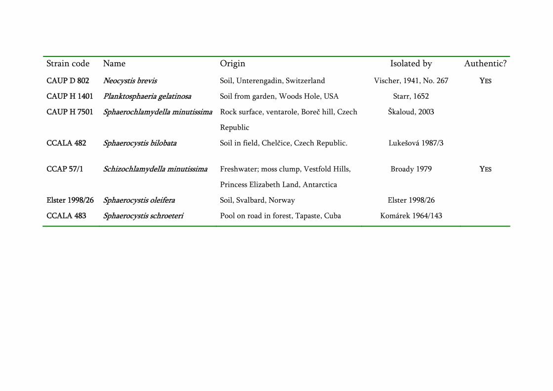
Strain code Name Origin Isolated by Authentic?
CAUP D 802 Neocystis brevis Soil, Unterengadin, Switzerland Vischer, 1941, No. 267 YES
CAUP H 1401 Planktosphaeria gelatinosa Soil from garden, Woods Hole, USA Starr, 1652
CAUP H 7501 Sphaerochlamydella minutissima Rock surface, ventarole, Boreč hill, Czech
Republic
Škaloud, 2003
CCALA 482 Sphaerocystis bilobata Soil in field, Chelčice, Czech Republic. Lukešová 1987/3
CCAP 57/1
Schizochlamydella minutissima Freshwater; moss clump, Vestfold Hills,
Princess Elizabeth Land, Antarctica
Broady 1979
YES
Elster 1998/26 Sphaerocystis oleifera Soil, Svalbard, Norway Elster 1998/26
CCALA 483 Sphaerocystis schroeteri Pool on road in forest, Tapaste, Cuba Komárek 1964/143
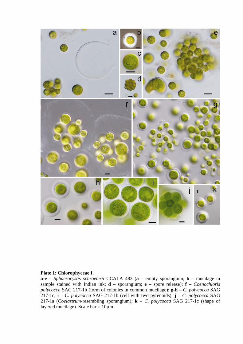
Plate 1: Chlorophyceae I. a-e – Sphaerocystis schroeterii CCALA 483 (a – empty sporangium; b – mucilage in sample stained with Indian ink; d – sporangium; e – spore release); f – Coenochloris polycocca SAG 217-1b (form of colonies in common mucilage); g-h – C. polycocca SAG 217-1c; i – C. polycocca SAG 217-1b (cell with two pyrenoids); j – C. polycocca SAG 217-1a (Coelastrum-resembling sporangium); k – C. polycocca SAG 217-1c (shape of layered mucilage). Scale bar = 10µm.
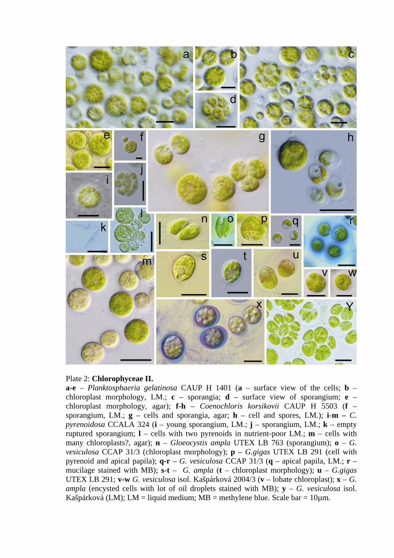
Plate 2: Chlorophyceae II. a-e – Planktosphaeria gelatinosa CAUP H 1401 (a – surface view of the cells; b – chloroplast morphology, LM.; c – sporangia; d – surface view of sporangium; e – chloroplast morphology, agar); f-h – Coenochloris korsikovii CAUP H 5503 (f –sporangium, LM.; g – cells and sporangia, agar; h – cell and spores, LM.); i-m – C. pyrenoidosa CCALA 324 (i – young sporangium, LM.; j – sporangium, LM.; k – empty ruptured sporangium; l – cells with two pyrenoids in nutrient-poor LM.; m – cells with many chloroplasts?, agar); n – Gloeocystis ampla UTEX LB 763 (sporangium); o – G. vesiculosa CCAP 31/3 (chloroplast morphology); p – G.gigas UTEX LB 291 (cell with pyrenoid and apical papila); q-r – G. vesiculosa CCAP 31/3 (q – apical papila, LM.; r – mucilage stained with MB); s-t – G. ampla (t – chloroplast morphology); u – G.gigas UTEX LB 291; v-w G. vesiculosa isol. Kašpárková 2004/3 (v – lobate chloroplast); x – G. ampla (encysted cells with lot of oil droplets stained with MB); y – G. vesiculosa isol. Kašpárková (LM); LM = liquid medium; MB = methylene blue. Scale bar = 10µm.

Plate 3: Trebouxiophyceae. a-b – Sphaerocystis bilobata CCALA 482 (cells and sporangia); c – G. polydermatica NP 20-04 (groups of cells and sporangium); d - Eutetramorus cf. fottii CAUP H 7701; e (not marked) – Neocystis brevis CAUP D 802 (sporangium); f – Neocystis sp. CAUP D 801; g – Sphaerocystis bilobata CCALA 482; h – N. brevis (bilobate chloroplast); i – Neocystis sp. (autospore release); j – G. polydermatica CCAP 31/5 (mucilage stained with MB); k – Coenocystis oleifera CCAP 176/2; l – Eutetramorus cf. fottii (autospores); m – N. brevis; n – Sphaerochlamydella minutissima CAUP H 7501 (groups of cells in LM, stained with methylene blue); q – Coenochloris bilobata CCAP 176/1; r – Sphaerochlamydella minutissima; s-t – Coenochloris planoconvexa CAUP H 5502 (s – cell with trough shaped chloroplast; t – cells with ray-like mucilage, LM, stained with methylene blue); u – Echinocoleum elegans SAG 37.93 (massive ray-like mucilage, LM, stained with methyleme blue); v-w – C. planoconvexa; x-y – Schizochlamydella minutissima CCAP 57/1 (x – cells with bilobate chloroplast and single vacuole; y – sporangium); z – Sphaerochlamydella minutissima (arrangement of cells); a’-b’ – specimens of „Radiococcaceae“ in fresh material from sandstone surface.
Recommended