International Conference on Clinical PET-CT and Molecular Imaging (IPET 2015 ): PET-CT in the era of multimodality imaging and image-guided therapy
October, 05-09, 2015, Vienna
Radiopharmaceuticals in Neurological and Psychiatric Disorders
Emilia Janevik Faculty of Medical Sciences Goce Delcev – Stip, Republic of Macedonia
Everything that healthcare providers do has a real, meaningful impact on human life Nuclear medicine is the only imaging modality that depend of the injected radiopharmaceutical Every radiopharmaceutical that is administrated holds far more than just a radionuclide Radiopharmaceuticals want it to deliver confidence, efficiency and a higher standard of excellence. And above all, renewed hope for each patient’s future Radiopharmaceuticals for planar imaging, (SPECT), (PET), PET-CT or SPECT-CT fusion imaging PET-MRI is currently being developed for clinical application
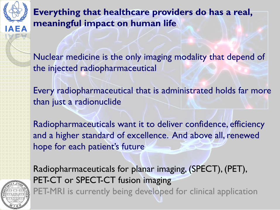
First contact…Projects related to International Atomic Energy Agency - IAEA: 1.(technical cooperation) 1995-1997 Preparation and QC od Technetium 99m Radiopharmaceuticals
99mTc - HMPAO
Understanding the utilization of radiopharmaceuticals for neurological and psychiatric disorders
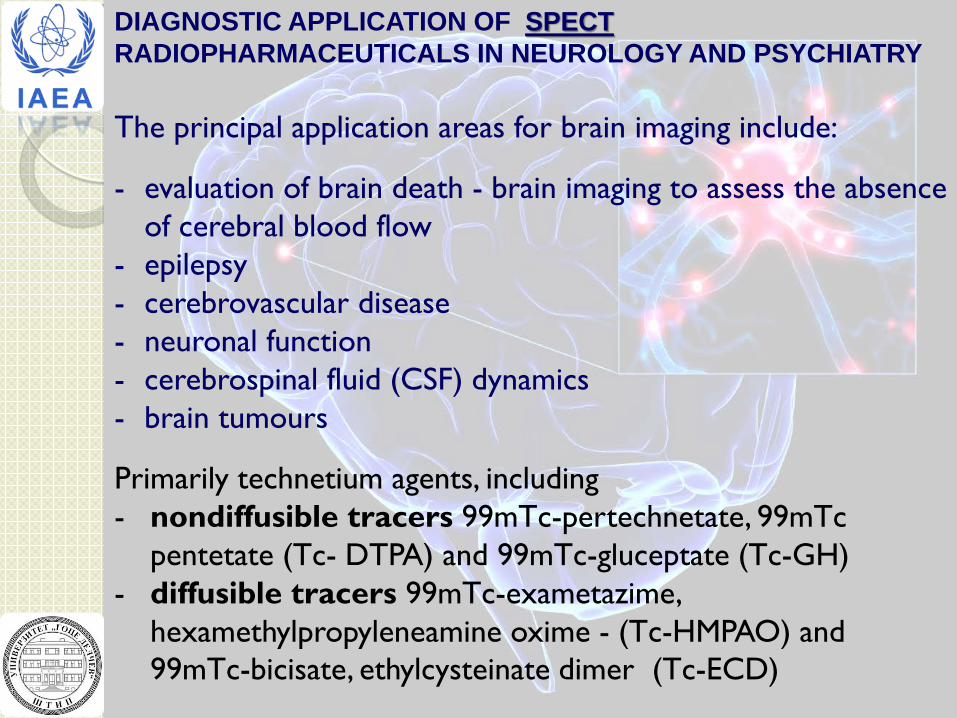
DIAGNOSTIC APPLICATION OF SPECT RADIOPHARMACEUTICALS IN NEUROLOGY AND PSYCHIATRY The principal application areas for brain imaging include:
- evaluation of brain death - brain imaging to assess the absence of cerebral blood flow
- epilepsy - cerebrovascular disease - neuronal function - cerebrospinal fluid (CSF) dynamics - brain tumours
Primarily technetium agents, including - nondiffusible tracers 99mTc-pertechnetate, 99mTc
pentetate (Tc- DTPA) and 99mTc-gluceptate (Tc-GH) - diffusible tracers 99mTc-exametazime,
hexamethylpropyleneamine oxime - (Tc-HMPAO) and 99mTc-bicisate, ethylcysteinate dimer (Tc-ECD)
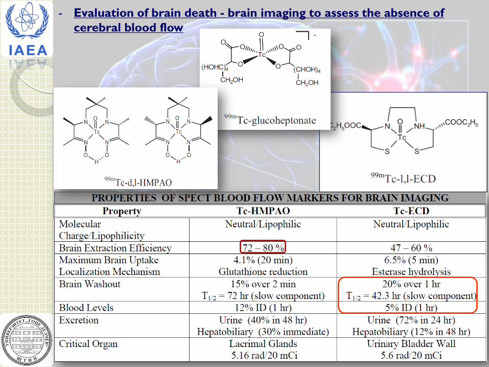
- Evaluation of brain death - brain imaging to assess the absence of cerebral blood flow

Cerebral delivery of radiotracer to the brain after i.v. administration
Major veins that drain blood from the brain
Major arteries that distribute blood in the brain
Normal posterior radionuclide cerebral angiogram (flow study). Images shown are acquired at 2 second intervals after IV injection of 20 mCi (740 MBq) of 99mTc-gluceptate Images show venous drainage through the lateral (transverse) venous sinuses and the jugular veins, which return blood to the heart.
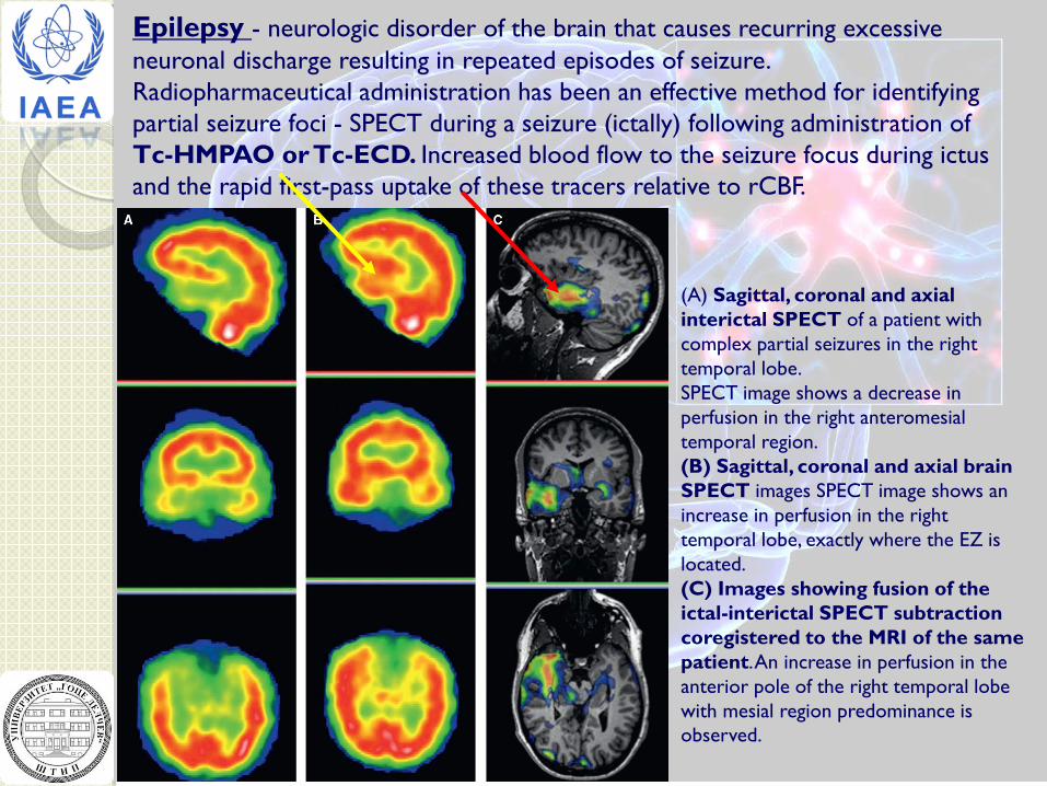
Epilepsy - neurologic disorder of the brain that causes recurring excessive neuronal discharge resulting in repeated episodes of seizure. Radiopharmaceutical administration has been an effective method for identifying partial seizure foci - SPECT during a seizure (ictally) following administration of Tc-HMPAO or Tc-ECD. Increased blood flow to the seizure focus during ictus and the rapid first-pass uptake of these tracers relative to rCBF.
(A) Sagittal, coronal and axial interictal SPECT of a patient with complex partial seizures in the right temporal lobe. SPECT image shows a decrease in perfusion in the right anteromesial temporal region. (B) Sagittal, coronal and axial brain SPECT images SPECT image shows an increase in perfusion in the right temporal lobe, exactly where the EZ is located. (C) Images showing fusion of the ictal-interictal SPECT subtraction coregistered to the MRI of the same patient. An increase in perfusion in the anterior pole of the right temporal lobe with mesial region predominance is observed.
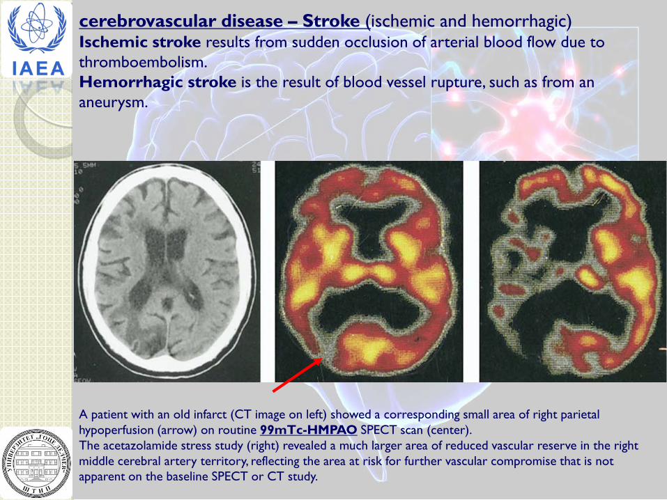
cerebrovascular disease – Stroke (ischemic and hemorrhagic) Ischemic stroke results from sudden occlusion of arterial blood flow due to thromboembolism. Hemorrhagic stroke is the result of blood vessel rupture, such as from an aneurysm.
A patient with an old infarct (CT image on left) showed a corresponding small area of right parietal hypoperfusion (arrow) on routine 99mTc-HMPAO SPECT scan (center). The acetazolamide stress study (right) revealed a much larger area of reduced vascular reserve in the right middle cerebral artery territory, reflecting the area at risk for further vascular compromise that is not apparent on the baseline SPECT or CT study.
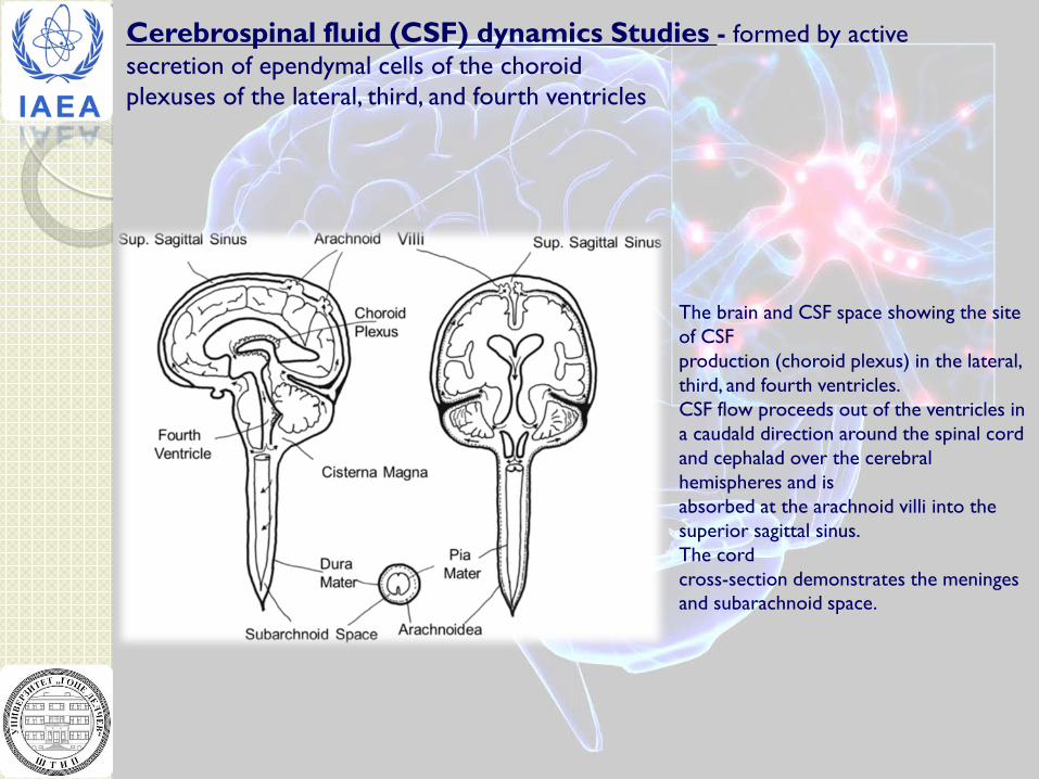
Cerebrospinal fluid (CSF) dynamics Studies - formed by active secretion of ependymal cells of the choroid plexuses of the lateral, third, and fourth ventricles
The brain and CSF space showing the site of CSF production (choroid plexus) in the lateral, third, and fourth ventricles. CSF flow proceeds out of the ventricles in a caudald direction around the spinal cord and cephalad over the cerebral hemispheres and is absorbed at the arachnoid villi into the superior sagittal sinus. The cord cross-section demonstrates the meninges and subarachnoid space.
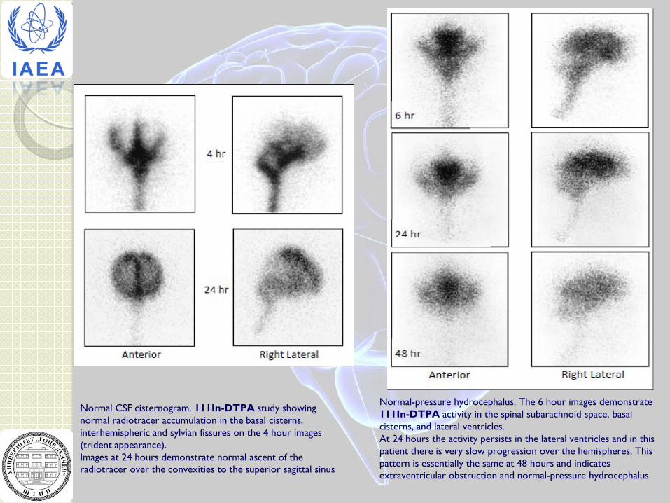
Normal CSF cisternogram. 111In-DTPA study showing normal radiotracer accumulation in the basal cisterns, interhemispheric and sylvian fissures on the 4 hour images (trident appearance). Images at 24 hours demonstrate normal ascent of the radiotracer over the convexities to the superior sagittal sinus
Normal-pressure hydrocephalus. The 6 hour images demonstrate 111In-DTPA activity in the spinal subarachnoid space, basal cisterns, and lateral ventricles. At 24 hours the activity persists in the lateral ventricles and in this patient there is very slow progression over the hemispheres. This pattern is essentially the same at 48 hours and indicates extraventricular obstruction and normal-pressure hydrocephalus
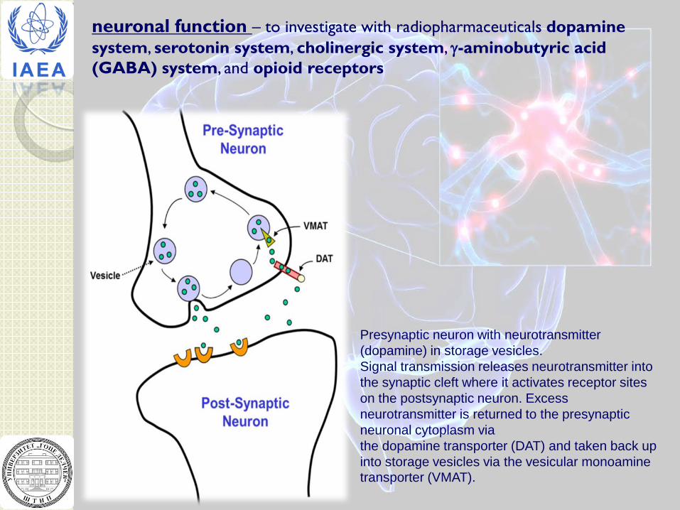
neuronal function – to investigate with radiopharmaceuticals dopamine system, serotonin system, cholinergic system, γ-aminobutyric acid (GABA) system, and opioid receptors
Presynaptic neuron with neurotransmitter (dopamine) in storage vesicles. Signal transmission releases neurotransmitter into the synaptic cleft where it activates receptor sites on the postsynaptic neuron. Excess neurotransmitter is returned to the presynaptic neuronal cytoplasm via the dopamine transporter (DAT) and taken back up into storage vesicles via the vesicular monoamine transporter (VMAT).
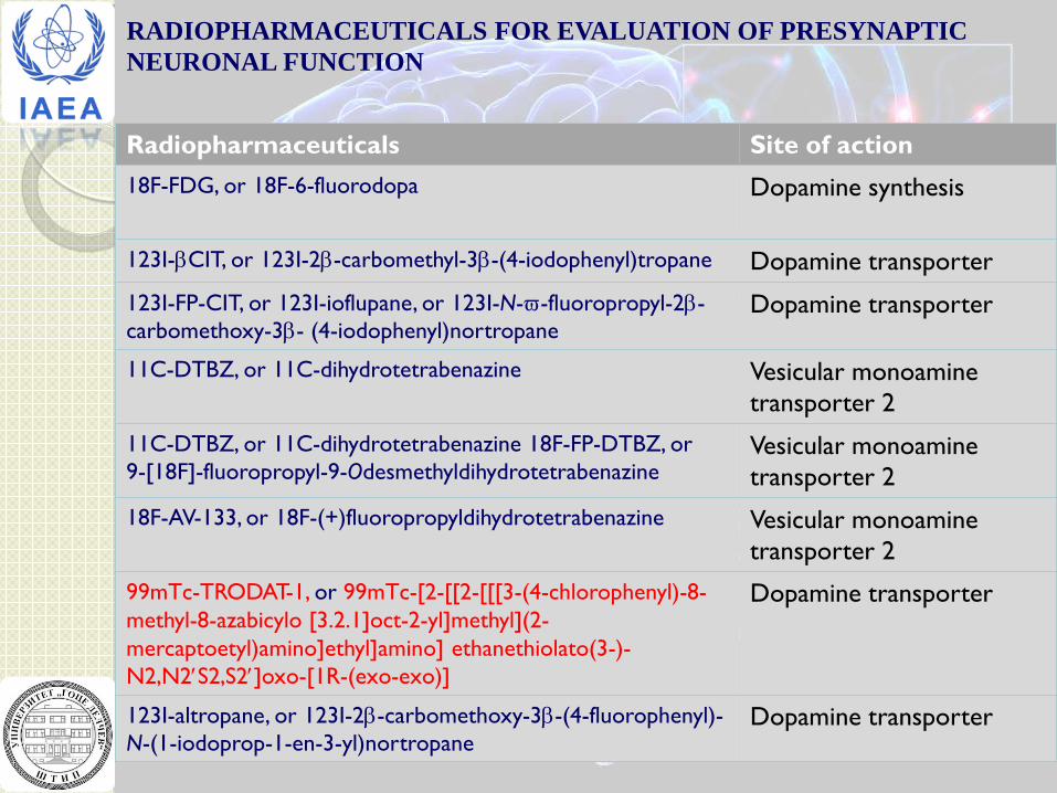
RADIOPHARMACEUTICALS FOR EVALUATION OF PRESYNAPTIC NEURONAL FUNCTION
Radiopharmaceuticals Site of action 18F-FDG, or 18F-6-fluorodopa Dopamine synthesis
123I-βCIT, or 123I-2β-carbomethyl-3β-(4-iodophenyl)tropane Dopamine transporter
123I-FP-CIT, or 123I-ioflupane, or 123I-N-ϖ-fluoropropyl-2β- carbomethoxy-3β- (4-iodophenyl)nortropane
Dopamine transporter
11C-DTBZ, or 11C-dihydrotetrabenazine Vesicular monoamine transporter 2
11C-DTBZ, or 11C-dihydrotetrabenazine 18F-FP-DTBZ, or 9-[18F]-fluoropropyl-9-Odesmethyldihydrotetrabenazine
Vesicular monoamine transporter 2
18F-AV-133, or 18F-(+)fluoropropyldihydrotetrabenazine Vesicular monoamine transporter 2
99mTc-TRODAT-1, or 99mTc-[2-[[2-[[[3-(4-chlorophenyl)-8- methyl-8-azabicylo [3.2.1]oct-2-yl]methyl](2- mercaptoetyl)amino]ethyl]amino] ethanethiolato(3-)- N2,N2′S2,S2′]oxo-[1R-(exo-exo)]
Dopamine transporter
123I-altropane, or 123I-2β-carbomethoxy-3β-(4-fluorophenyl)- N-(1-iodoprop-1-en-3-yl)nortropane
Dopamine transporter

BASIC REQUIREMENTS FOR A CNS RECEPTOR RADIOPHARMACEUTICALS
1. Ability to cross the BBB (neutral, MW < 700, log P 1.0–3.0) 2. In vivo stability 3. High and selective binding affinity for the target receptor
(IC50 < 10 nM) 4. High uptake in brain 5. Localized in target sites 6. Formation (if any) of non-binding
radiometabolites
Schematic representation of a receptor specific radiopharmaceutical
chelate linker Receptor Targeting Molecule
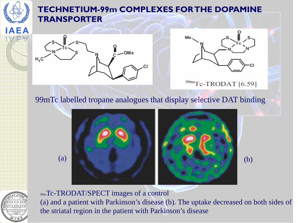
TECHNETIUM-99m COMPLEXES FOR THE DOPAMINE TRANSPORTER
99mTc labelled tropane analogues that display selective DAT binding
99mTc-TRODAT/SPECT images of a control (a) and a patient with Parkinson’s disease (b). The uptake decreased on both sides of the striatal region in the patient with Parkinson’s disease
(a) (b)
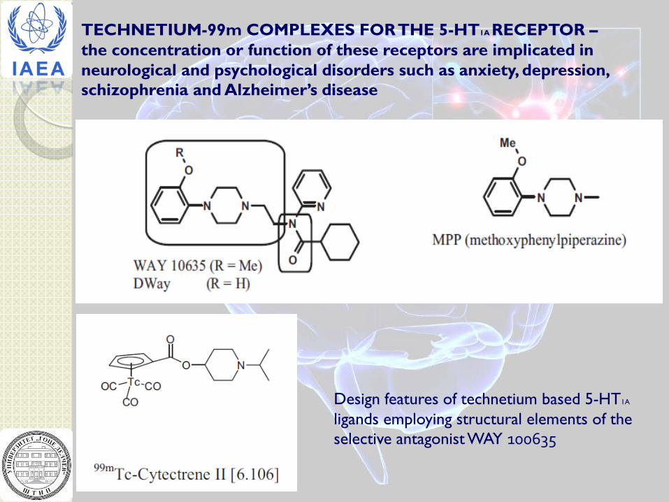
TECHNETIUM-99m COMPLEXES FOR THE 5-HT1A RECEPTOR – the concentration or function of these receptors are implicated in neurological and psychological disorders such as anxiety, depression, schizophrenia and Alzheimer’s disease
Design features of technetium based 5-HT1A
ligands employing structural elements of the selective antagonist WAY 100635
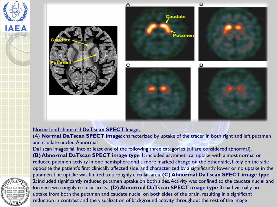
Normal and abnormal DaTscan SPECT Images. (A) Normal DaTscan SPECT image: characterized by uptake of the tracer in both right and left putamen and caudate nuclei.. Abnormal DaTscan images fall into at least one of the following three categories (all are considered abnormal). (B) Abnormal DaTscan SPECT image type 1: included asymmetrical uptake with almost normal or reduced putamen activity in one hemisphere and a more marked change on the other side, likely on the side opposite the patient's first clinically affected side, and characterized by a significantly lower or no uptake in the putamen. The uptake was limited to a roughly circular area. (C) Abnormal DaTscan SPECT image type 2: included significantly reduced putamen uptake on both sides. Activity was confined to the caudate nuclei and formed two roughly circular areas. (D) Abnormal DaTscan SPECT image type 3: had virtually no uptake from both the putamen and caudate nuclei on both sides of the brain, resulting in a significant reduction in contrast and the visualization of background activity throughout the rest of the image
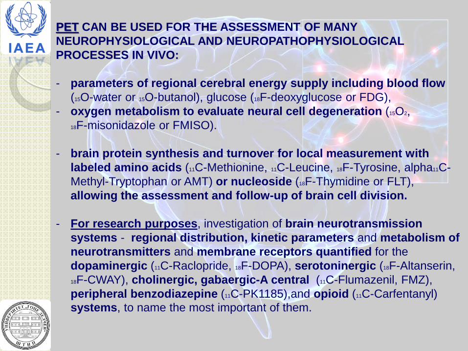
PET CAN BE USED FOR THE ASSESSMENT OF MANY NEUROPHYSIOLOGICAL AND NEUROPATHOPHYSIOLOGICAL PROCESSES IN VIVO: - parameters of regional cerebral energy supply including blood flow
(15O-water or 15O-butanol), glucose (18F-deoxyglucose or FDG), - oxygen metabolism to evaluate neural cell degeneration (15O2, 18F-misonidazole or FMISO).
- brain protein synthesis and turnover for local measurement with
labeled amino acids (11C-Methionine, 11C-Leucine, 18F-Tyrosine, alpha11C-Methyl-Tryptophan or AMT) or nucleoside (18F-Thymidine or FLT), allowing the assessment and follow-up of brain cell division.
- For research purposes, investigation of brain neurotransmission
systems - regional distribution, kinetic parameters and metabolism of neurotransmitters and membrane receptors quantified for the dopaminergic (11C-Raclopride, 18F-DOPA), serotoninergic (18F-Altanserin, 18F-CWAY), cholinergic, gabaergic-A central (11C-Flumazenil, FMZ), peripheral benzodiazepine (11C-PK1185),and opioid (11C-Carfentanyl) systems, to name the most important of them.
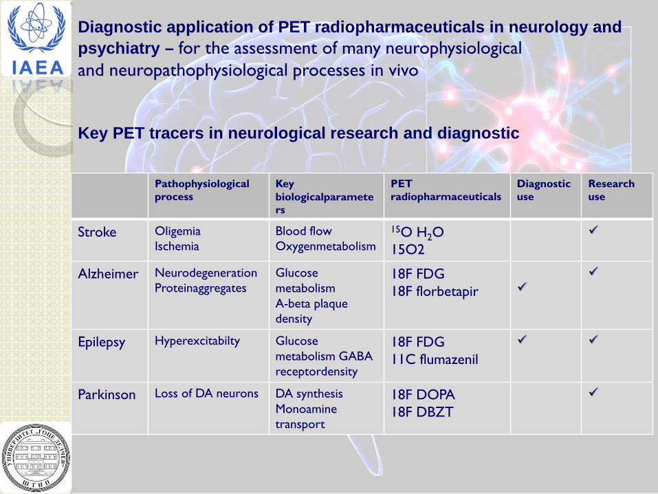
Diagnostic application of PET radiopharmaceuticals in neurology and psychiatry – for the assessment of many neurophysiological and neuropathophysiological processes in vivo
Pathophysiological process
Key biologicalparameters
PET radiopharmaceuticals
Diagnostic use
Research use
Stroke Oligemia Ischemia
Blood flow Oxygenmetabolism
15O H2O 15O2
Alzheimer Neurodegeneration Proteinaggregates
Glucose metabolism A-beta plaque density
18F FDG 18F florbetapir
Epilepsy Hyperexcitabilty Glucose metabolism GABA receptordensity
18F FDG 11C flumazenil
Parkinson Loss of DA neurons DA synthesis Monoamine transport
18F DOPA 18F DBZT
Key PET tracers in neurological research and diagnostic
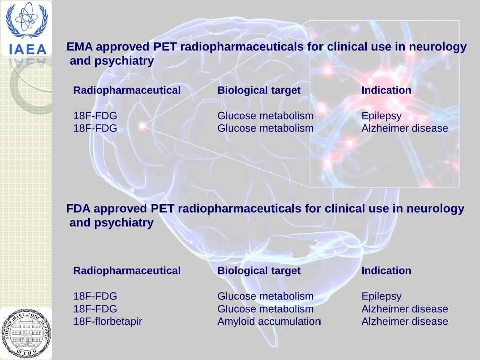
EMA approved PET radiopharmaceuticals for clinical use in neurology and psychiatry
Radiopharmaceutical Biological target Indication 18F-FDG Glucose metabolism Epilepsy 18F-FDG Glucose metabolism Alzheimer disease
FDA approved PET radiopharmaceuticals for clinical use in neurology and psychiatry
Radiopharmaceutical Biological target Indication 18F-FDG Glucose metabolism Epilepsy 18F-FDG Glucose metabolism Alzheimer disease 18F-florbetapir Amyloid accumulation Alzheimer disease
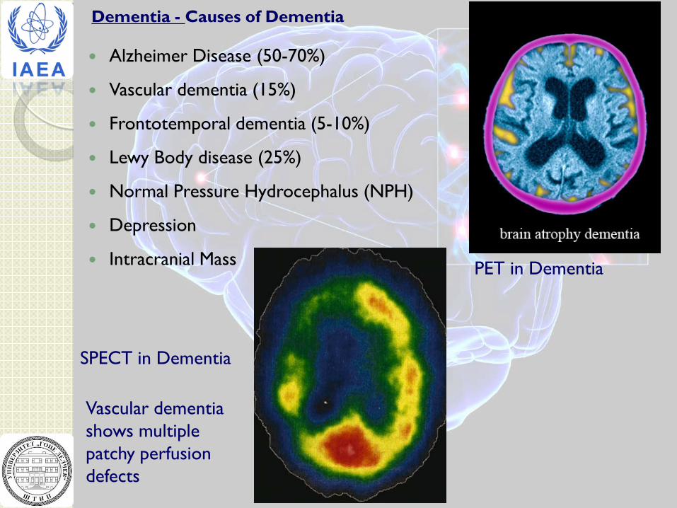
Dementia - Causes of Dementia Alzheimer Disease (50-70%)
Vascular dementia (15%)
Frontotemporal dementia (5-10%)
Lewy Body disease (25%)
Normal Pressure Hydrocephalus (NPH)
Depression
Intracranial Mass
Vascular dementia shows multiple patchy perfusion defects
SPECT in Dementia
PET in Dementia
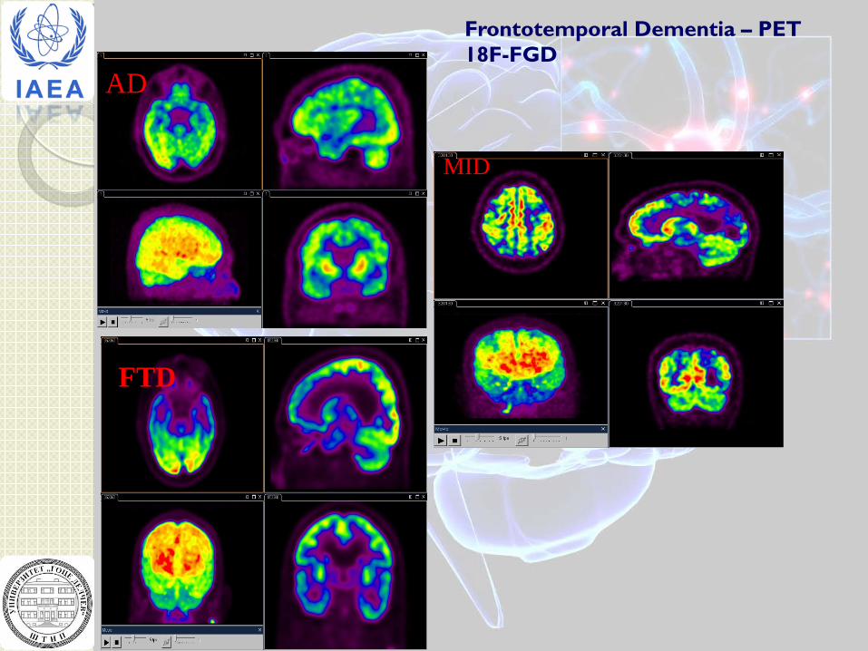
Frontotemporal Dementia – PET 18F-FGD
AD
FTD
MID
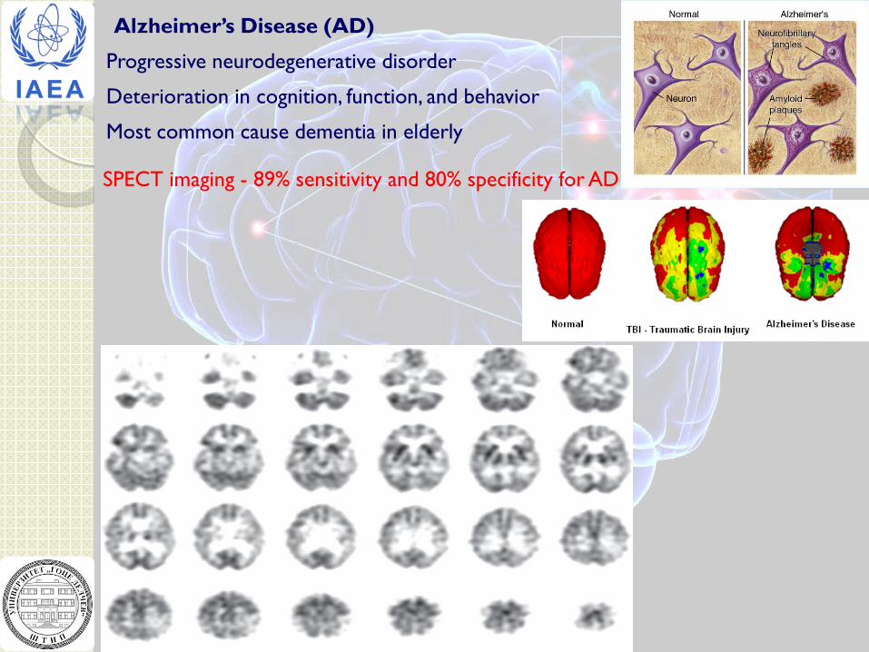
Alzheimer’s Disease (AD)
Progressive neurodegenerative disorder
Deterioration in cognition, function, and behavior
Most common cause dementia in elderly
SPECT imaging - 89% sensitivity and 80% specificity for AD
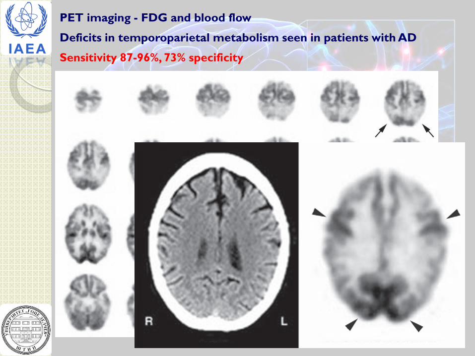
PET imaging - FDG and blood flow
Deficits in temporoparietal metabolism seen in patients with AD
Sensitivity 87-96%, 73% specificity
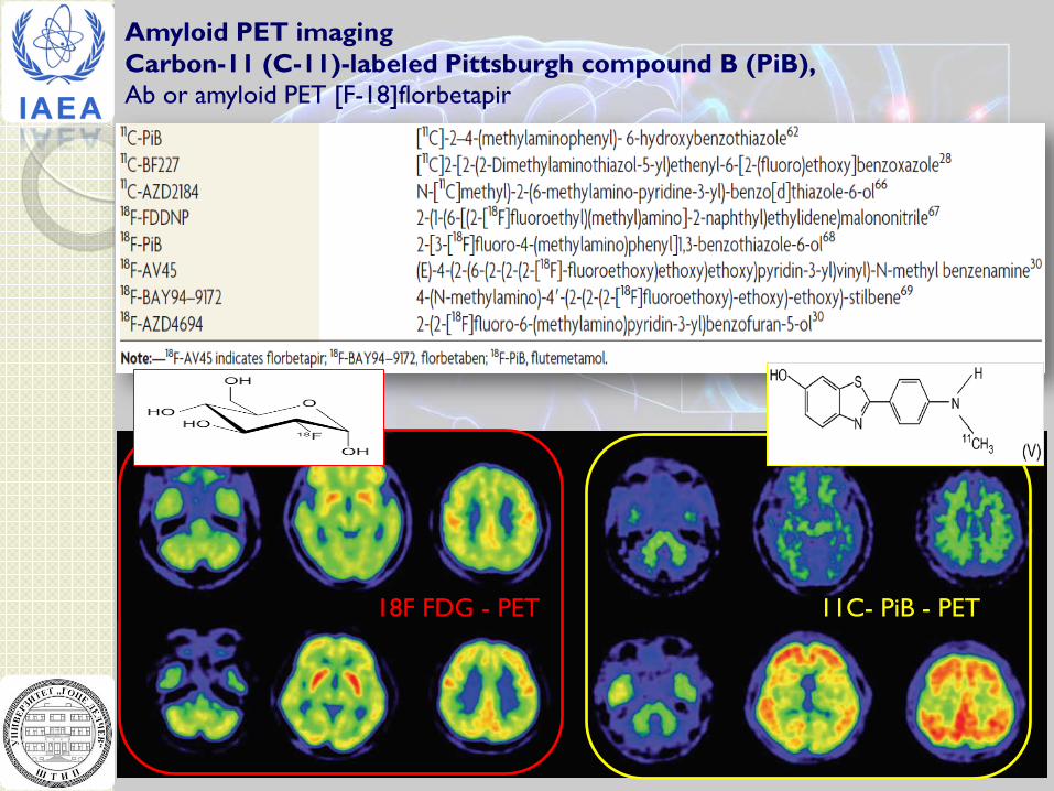
Amyloid PET imaging Carbon-11 (C-11)-labeled Pittsburgh compound B (PiB), Ab or amyloid PET [F-18]florbetapir
18F FDG - PET 11C- PiB - PET
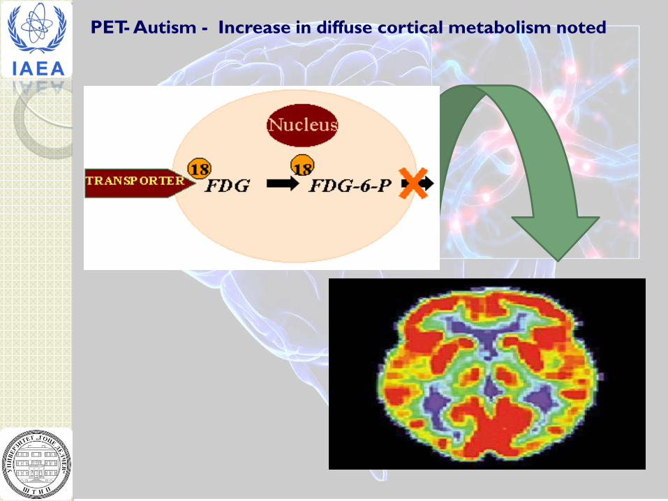
PET- Autism - Increase in diffuse cortical metabolism noted
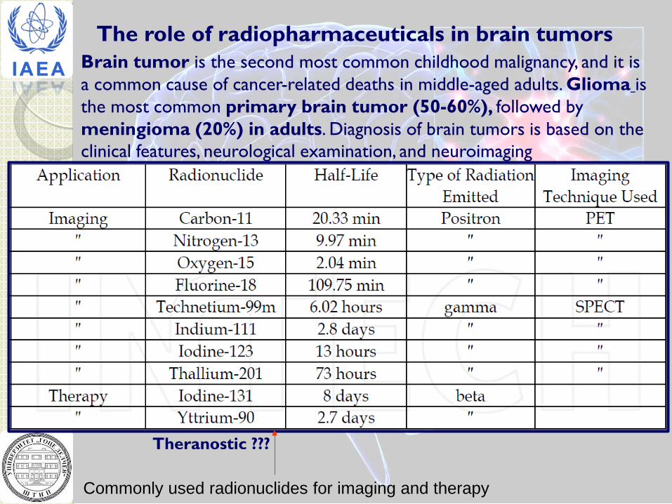
The role of radiopharmaceuticals in brain tumors Brain tumor is the second most common childhood malignancy, and it is a common cause of cancer-related deaths in middle-aged adults. Glioma is the most common primary brain tumor (50-60%), followed by meningioma (20%) in adults. Diagnosis of brain tumors is based on the clinical features, neurological examination, and neuroimaging
Commonly used radionuclides for imaging and therapy
Theranostic ???
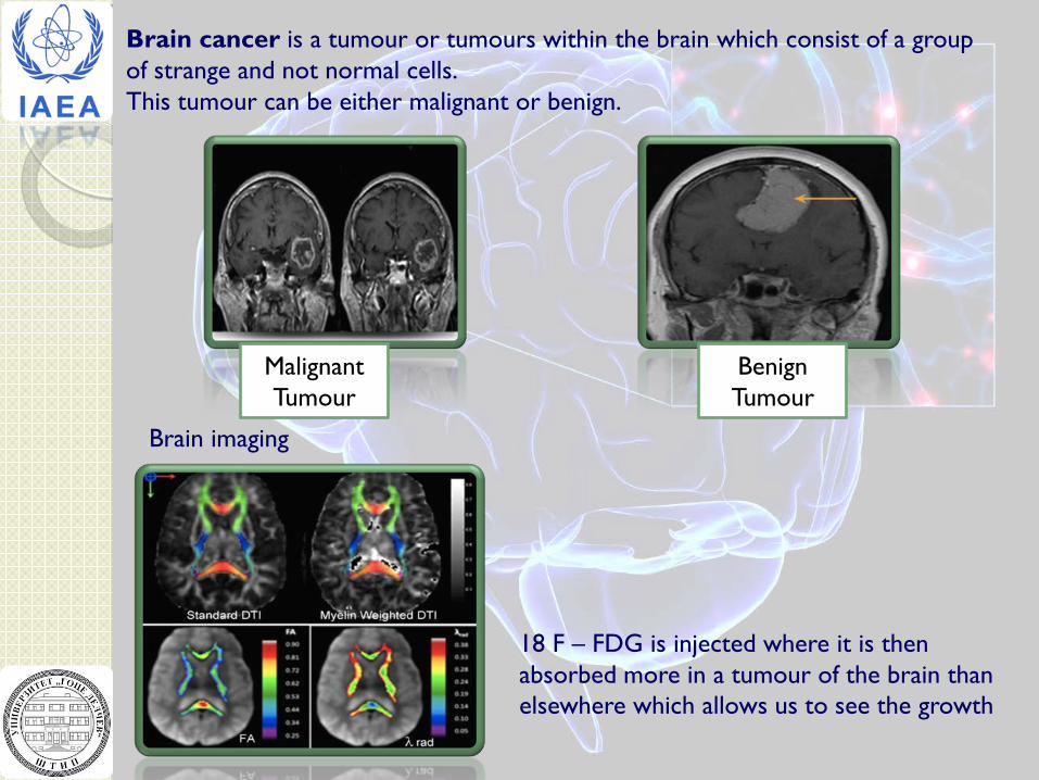
Brain cancer is a tumour or tumours within the brain which consist of a group of strange and not normal cells. This tumour can be either malignant or benign.
Malignant Tumour
Benign Tumour
Brain imaging
18 F – FDG is injected where it is then absorbed more in a tumour of the brain than elsewhere which allows us to see the growth
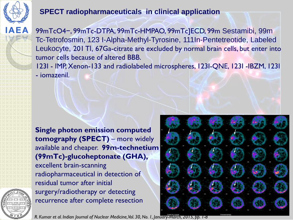
Single photon emission computed tomography (SPECT) – more widely available and cheaper. 99m-technetium (99mTc)-glucoheptonate (GHA), excellent brain-scanning radiopharmaceutical in detection of residual tumor after initial surgery/radiotherapy or detecting recurrence after complete resection
R. Kumar et al. Indian Journal of Nuclear Medicine, Vol. 30, No. 1, January-March, 2015, pp. 1-8
99mTcO4−, 99mTc-DTPA, 99mTc-HMPAO, 99mTc]ECD, 99m Sestamibi, 99m Tc-Tetrofosmin, 123 I-Alpha-Methyl-Tyrosine, 111In-Pentetreotide, Labeled Leukocyte, 201 Tl, 67Ga-citrate are excluded by normal brain cells, but enter into tumor cells because of altered BBB. 123I - IMP, Xenon-133 and radiolabeled microspheres, 123I-QNE, 123I -IBZM, 123I - iomazenil.
SPECT radiopharmaceuticals in clinical application
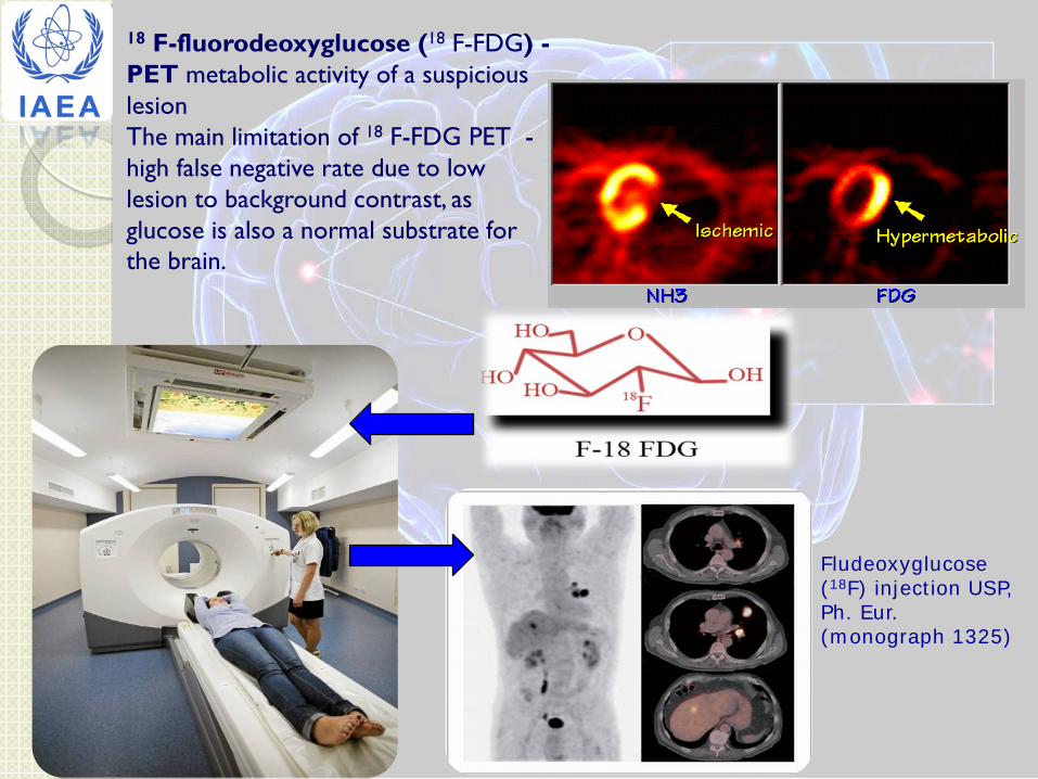
18 F-fluorodeoxyglucose (18 F-FDG) - PET metabolic activity of a suspicious lesion The main limitation of 18 F-FDG PET - high false negative rate due to low lesion to background contrast, as glucose is also a normal substrate for the brain.
Fludeoxyglucose (18F) injection USP, Ph. Eur. (monograph 1325)
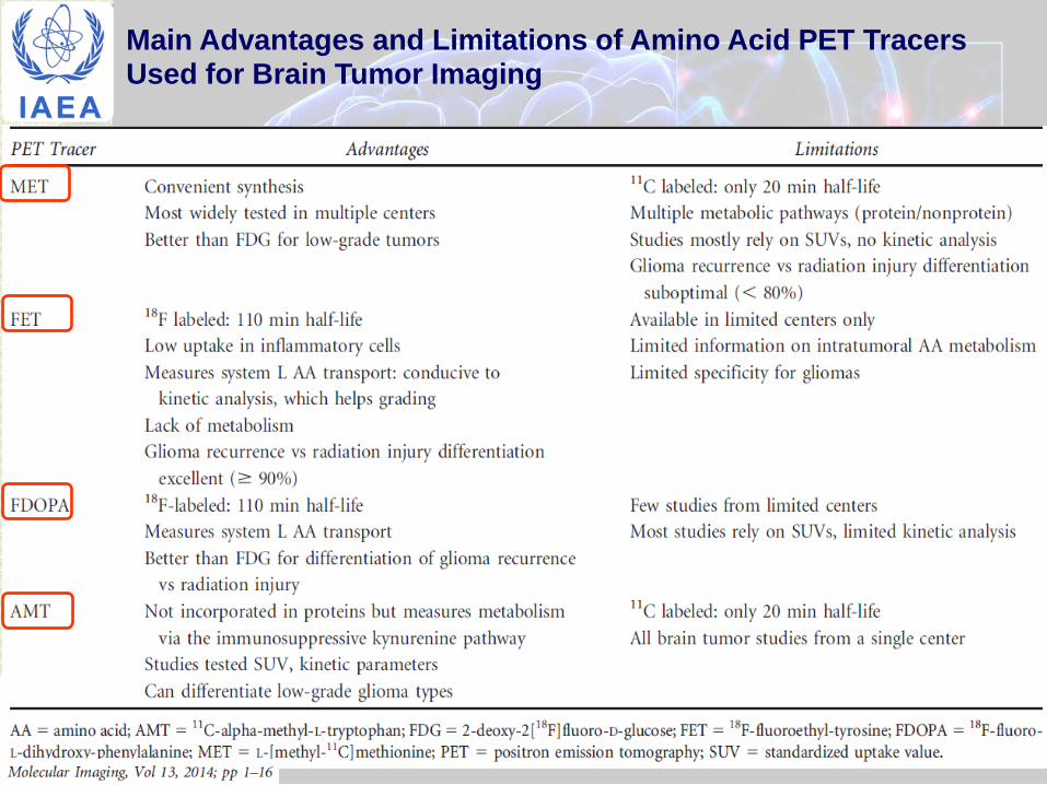
Main Advantages and Limitations of Amino Acid PET Tracers Used for Brain Tumor Imaging
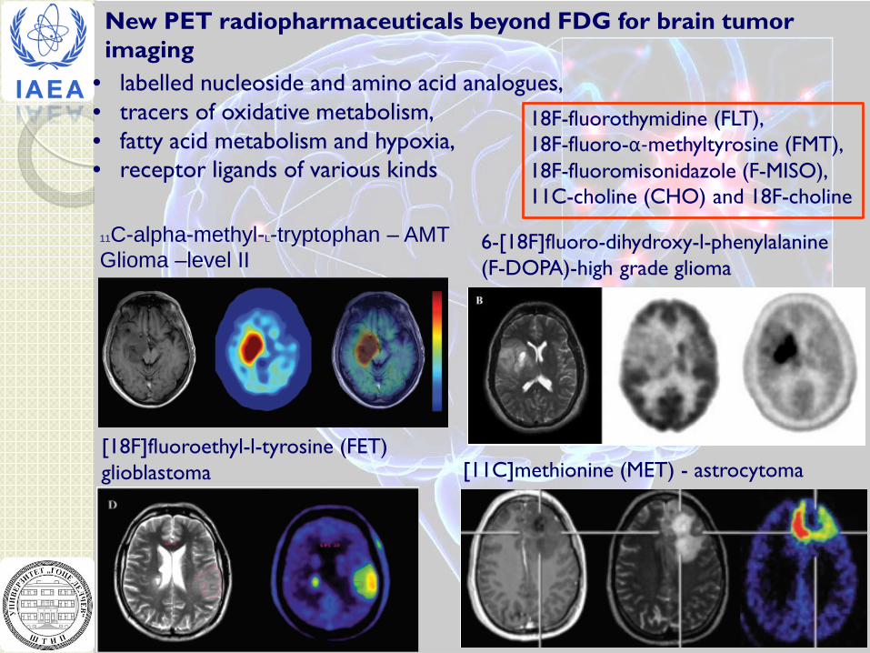
New PET radiopharmaceuticals beyond FDG for brain tumor imaging
• labelled nucleoside and amino acid analogues, • tracers of oxidative metabolism, • fatty acid metabolism and hypoxia, • receptor ligands of various kinds
18F-fluorothymidine (FLT), 18F-fluoro-α-methyltyrosine (FMT), 18F-fluoromisonidazole (F-MISO), 11C-choline (CHO) and 18F-choline
[18F]fluoroethyl-l-tyrosine (FET) glioblastoma [11C]methionine (MET) - astrocytoma
6-[18F]fluoro-dihydroxy-l-phenylalanine (F-DOPA)-high grade glioma
11C-alpha-methyl-L-tryptophan – AMT Glioma –level II

Radiopharmaceuticals for brain tumor treatment………… Or for plaining brain tumor treatment ??? Drug development … Schematic Representation of a Drug for Imaging and Targeted Therapy
Molecular Address • Antibodies, their
fragments and modifications
• Regulatory peptides and analogs thereof
• Amino Acids
Target • Antigens
(CD20, HER2)
• GPCRs
• Transporters
pharmacokinetic modifier
Linker Ligand Chelator
Reporting Unit • 99mTc, 111In, 67Ga • 64Cu, 68Ga • Gd3+
Cytotoxic Unit • 90Y, 177Lu, 213Bi • 105Rh, 67Cu,
186,188Re H.R. Maecke
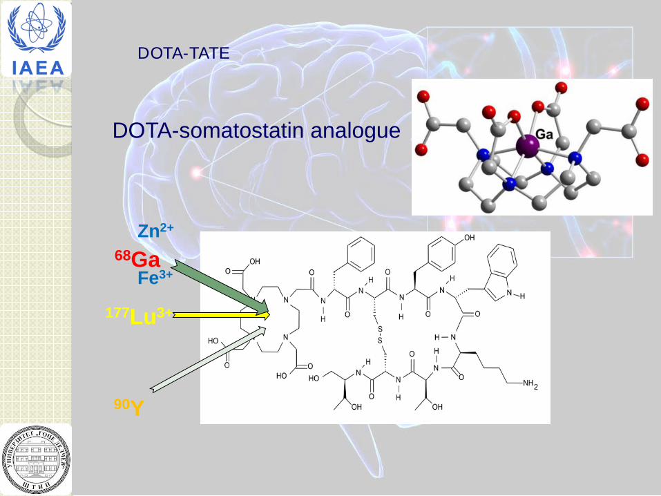
DOTA-somatostatin analogue
177Lu3+
Zn2+
Fe3+ 68Ga
90Y
DOTA-TATE
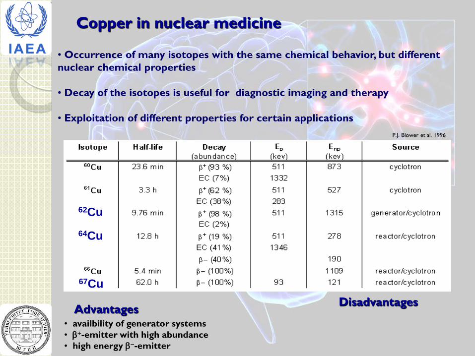
Copper in nuclear medicine
• Occurrence of many isotopes with the same chemical behavior, but different nuclear chemical properties • Decay of the isotopes is useful for diagnostic imaging and therapy • Exploitation of different properties for certain applications
• availbility of generator systems • β+-emitter with high abundance • high energy β−-emitter
Advantages Disadvantages
P.J. Blower et al. 1996
62Cu 64Cu
67Cu
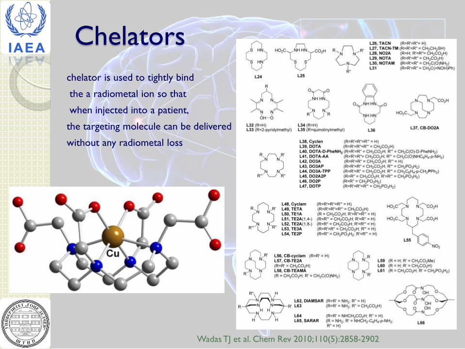
Chelators chelator is used to tightly bind
the a radiometal ion so that
when injected into a patient,
the targeting molecule can be delivered
without any radiometal loss
Wadas TJ et al. Chem Rev 2010;110(5):2858-2902
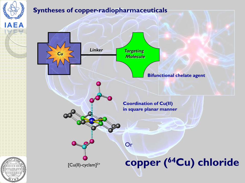
Cu Targeting Molecule
Linker
Bifunctional chelate agent
Coordination of Cu(II) in square planar manner
[Cu(II)-cyclam]2+
Syntheses of copper-radiopharmaceuticals
Or
copper (64Cu) chloride
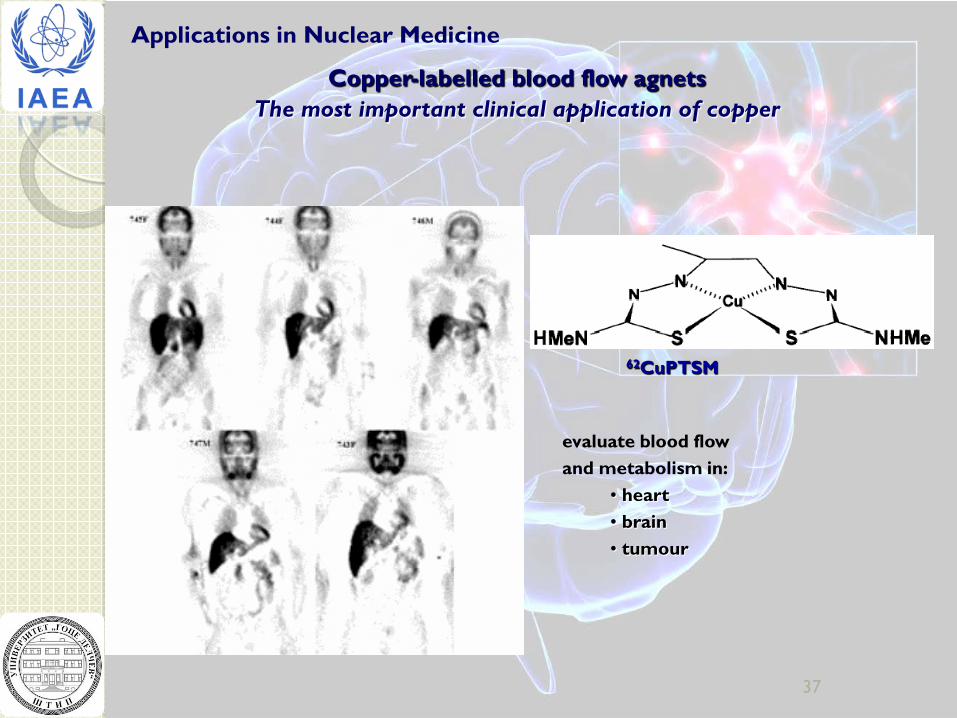
Applications in Nuclear Medicine
37
Copper-labelled blood flow agnets The most important clinical application of copper
62CuPTSM
evaluate blood flow and metabolism in:
• heart • brain • tumour

Copper-labeled hypoxia imaging agents
62CuATSM
Detection of ischemia of the: • brain • heart • hypoxic tumor
64CuATSM
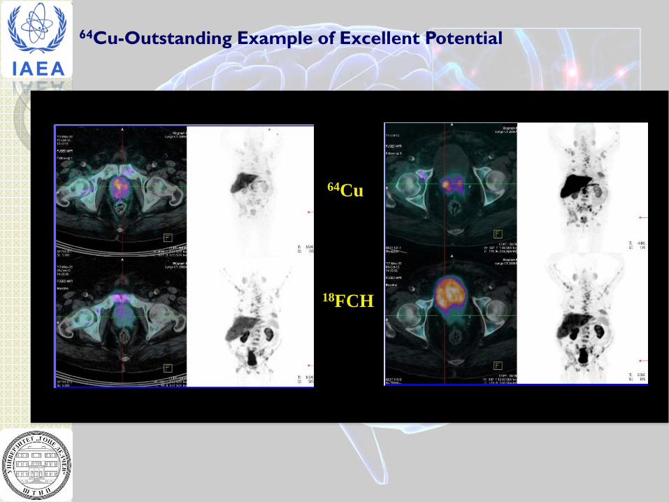
64Cu
18FCH
64Cu-Outstanding Example of Excellent Potential
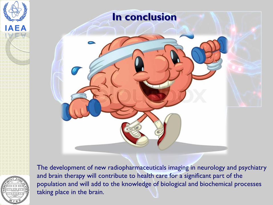
In conclusion
The development of new radiopharmaceuticals imaging in neurology and psychiatry and brain therapy will contribute to health care for a significant part of the population and will add to the knowledge of biological and biochemical processes taking place in the brain.
Thank you
Recommended