Immunoglobulins
RAKESH SHARDA
Department of Veterinary Microbiology
NDVSU College of Veterinary Science & A.H.,
MHOW

Structure and Functions
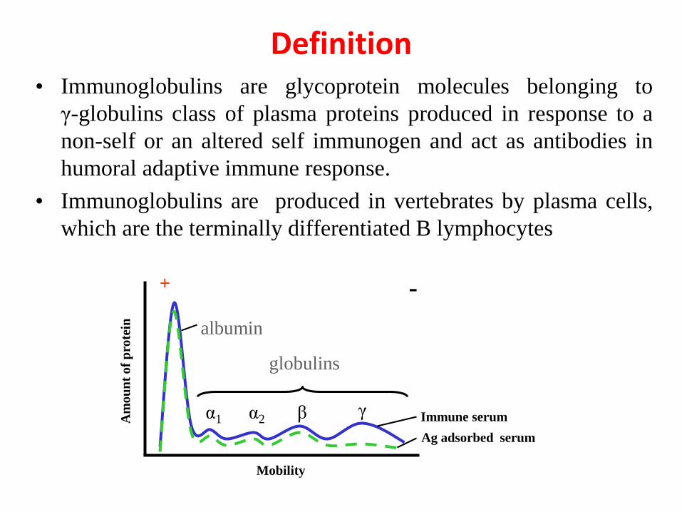
Definition• Immunoglobulins are glycoprotein molecules belonging to
γ-globulins class of plasma proteins produced in response to a
non-self or an altered self immunogen and act as antibodies in
humoral adaptive immune response.
• Immunoglobulins are produced in vertebrates by plasma cells,
which are the terminally differentiated B lymphocytes
Immune serum
Ag adsorbed serum
α1 α2 β γ
+ -
albumin
globulins
Mobility
Am
ou
nt
of
pro
tein
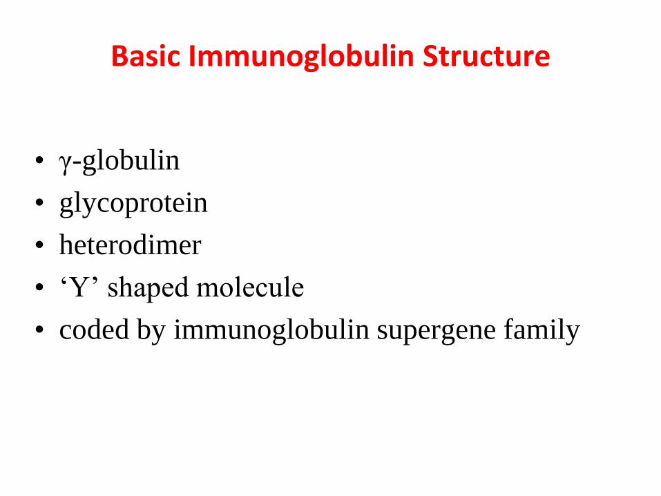
Basic Immunoglobulin Structure
• γ-globulin
• glycoprotein
• heterodimer
• ‘Y’ shaped molecule
• coded by immunoglobulin supergene family
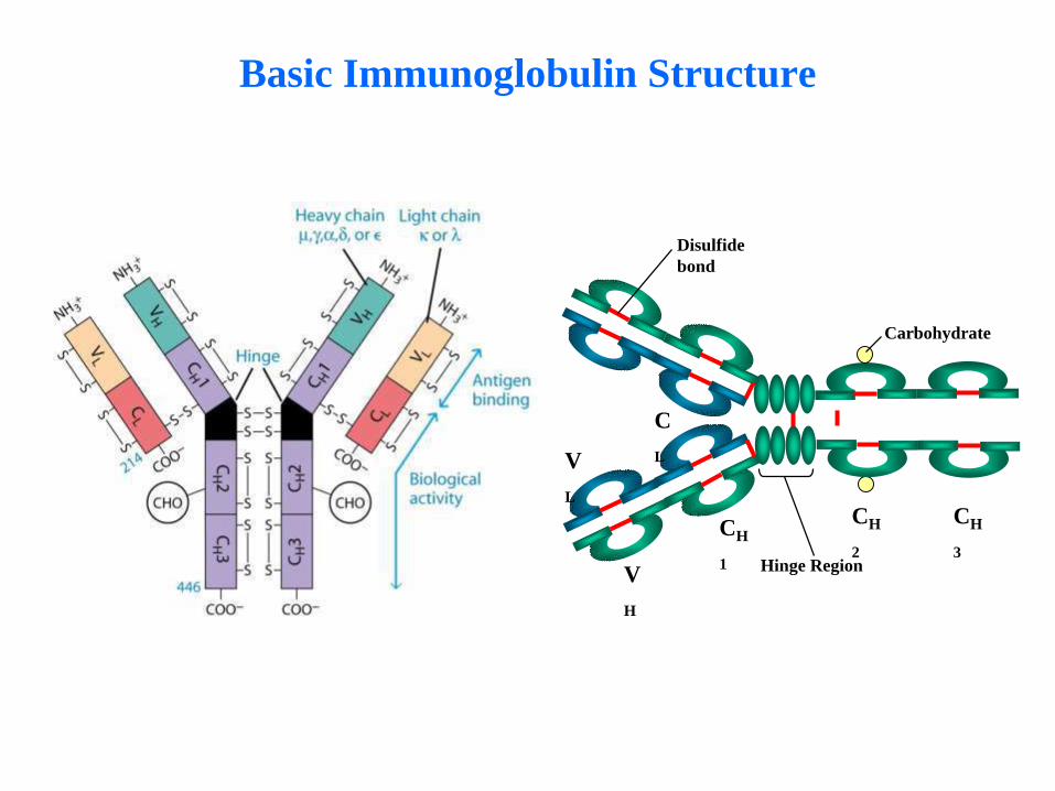
Basic Immunoglobulin Structure
CH
1
V
L
C
L
V
H
CH
2
CH
3Hinge Region
Carbohydrate
Disulfide
bond
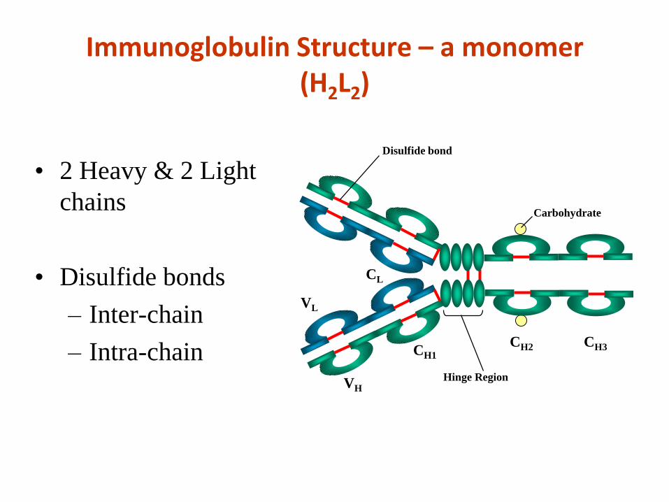
Immunoglobulin Structure – a monomer (H2L2)
• 2 Heavy & 2 Light
chains
• Disulfide bonds
– Inter-chain
– Intra-chain CH1
VL
CL
VH
CH2 CH3
Hinge Region
Carbohydrate
Disulfide bond
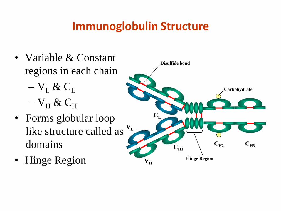
Immunoglobulin Structure
• Variable & Constant
regions in each chain
– VL & CL
– VH & CH
• Forms globular loop
like structure called as
domains
• Hinge Region
CH1
VL
CL
VH
CH2 CH3
Hinge Region
Carbohydrate
Disulfide bond
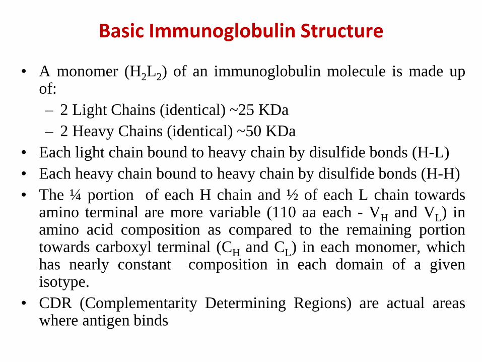
• A monomer (H2L2) of an immunoglobulin molecule is made upof:
– 2 Light Chains (identical) ~25 KDa
– 2 Heavy Chains (identical) ~50 KDa
• Each light chain bound to heavy chain by disulfide bonds (H-L)
• Each heavy chain bound to heavy chain by disulfide bonds (H-H)
• The ¼ portion of each H chain and ½ of each L chain towardsamino terminal are more variable (110 aa each - VH and VL) inamino acid composition as compared to the remaining portiontowards carboxyl terminal (CH and CL) in each monomer, whichhas nearly constant composition in each domain of a givenisotype.
• CDR (Complementarity Determining Regions) are actual areaswhere antigen binds
Basic Immunoglobulin Structure
Basic Immunoglobulin Structure
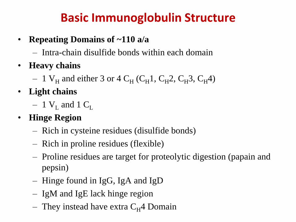
• Repeating Domains of ~110 a/a
– Intra-chain disulfide bonds within each domain
• Heavy chains
– 1 VH and either 3 or 4 CH (CH1, CH2, CH3, CH4)
• Light chains
– 1 VL and 1 CL
• Hinge Region
– Rich in cysteine residues (disulfide bonds)
– Rich in proline residues (flexible)
– Proline residues are target for proteolytic digestion (papain and
pepsin)
– Hinge found in IgG, IgA and IgD
– IgM and IgE lack hinge region
– They instead have extra CH4 Domain
Basic Immunoglobulin Structure
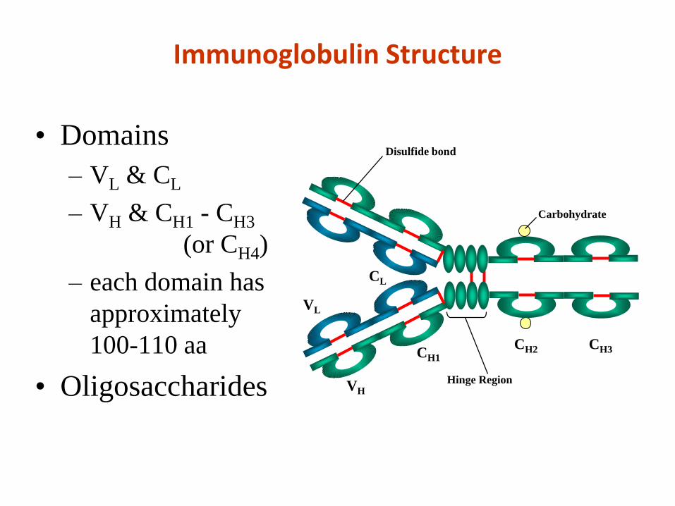
Immunoglobulin Structure
• Domains
– VL & CL
– VH & CH1 - CH3
(or CH4)
– each domain has
approximately
100-110 aa
• Oligosaccharides
CH1
VL
CL
VH
CH2 CH3
Hinge Region
Carbohydrate
Disulfide bond
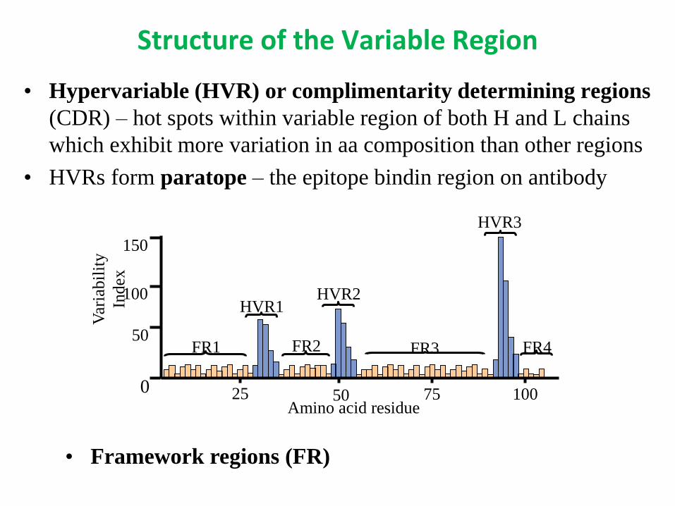
Structure of the Variable Region
• Hypervariable (HVR) or complimentarity determining regions
(CDR) – hot spots within variable region of both H and L chains
which exhibit more variation in aa composition than other regions
• HVRs form paratope – the epitope bindin region on antibody
HVR3
FR1 FR2 FR3 FR4
HVR1HVR2
Var
iabil
ity
Index
25 7550 100Amino acid residue
150
100
50
0
• Framework regions (FR)
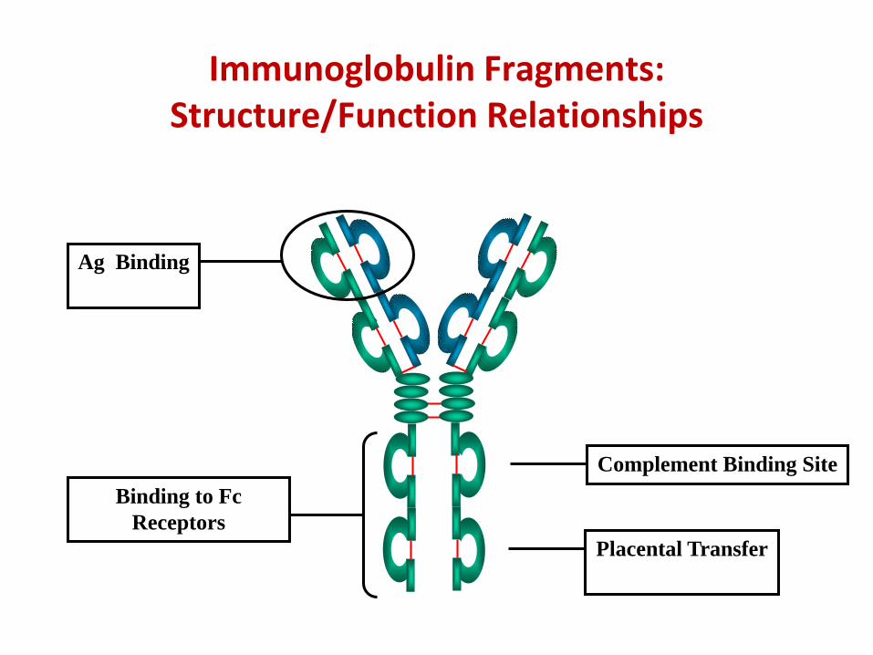
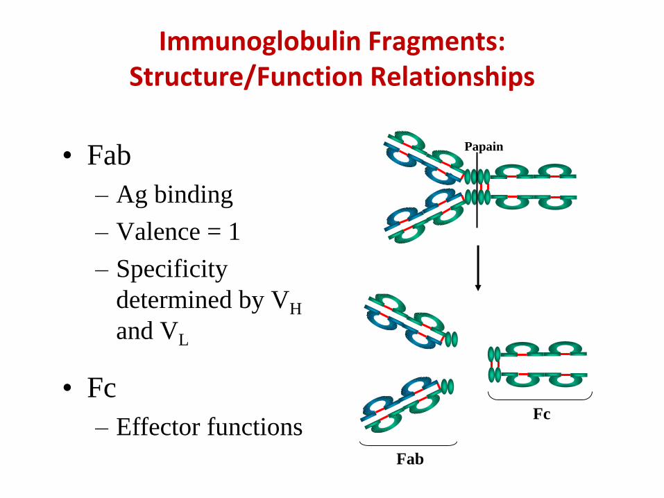
Immunoglobulin Fragments: Structure/Function Relationships
Ag Binding
Complement Binding Site
Placental Transfer
Binding to Fc
Receptors
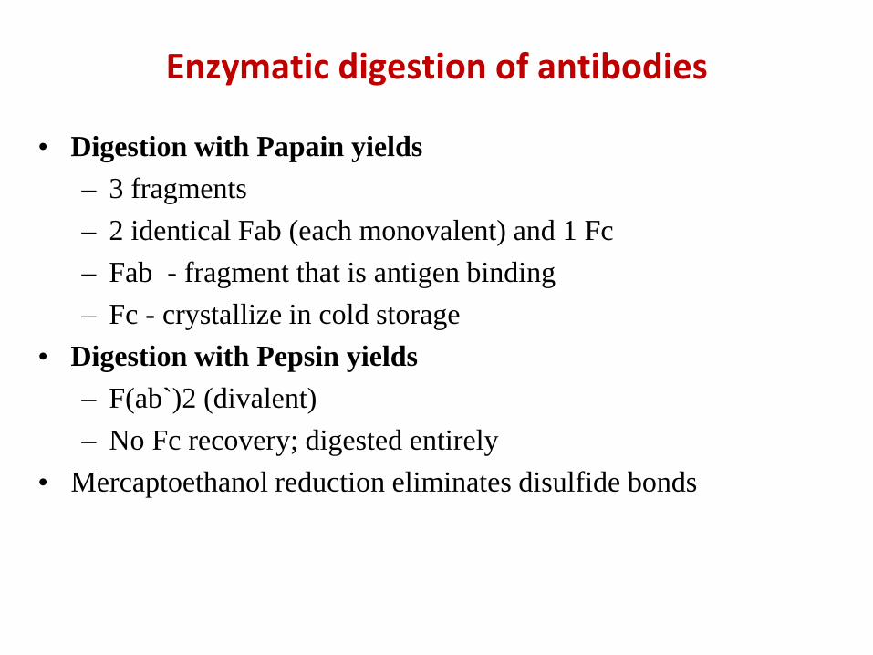
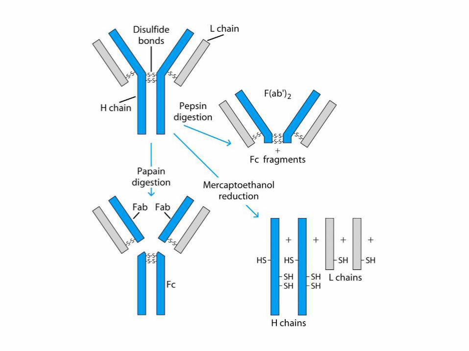
• Digestion with Papain yields
– 3 fragments
– 2 identical Fab (each monovalent) and 1 Fc
– Fab - fragment that is antigen binding
– Fc - crystallize in cold storage
• Digestion with Pepsin yields
– F(ab`)2 (divalent)
– No Fc recovery; digested entirely
• Mercaptoethanol reduction eliminates disulfide bonds
Enzymatic digestion of antibodies
Immunoglobulin Fragments: Structure/Function Relationships
• Fab
– Ag binding
– Valence = 1
– Specificity
determined by VH
and VL
Papain
Fc
Fab
• Fc
– Effector functions
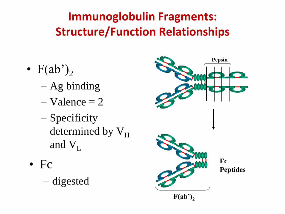
Immunoglobulin Fragments: Structure/Function Relationships
• F(ab’)2
– Ag binding
– Valence = 2
– Specificity
determined by VH
and VL
Pepsin
Fc
Peptides
F(ab’)2
• Fc
– digested
• Sequencing of heavy chains of several immunoglobulins in
human beings and mice revealed:
– A highly variable (V) region of 100-110 amino acids at amino
terminus of each H chain
– Five basic amino acid sequence patterns in remaining constant
(C) region of H chains which differ between H chains of each
pattern, but not in all H chains of a given pattern
– ,, , , types of heavy chains
– IgA, IgG, IgD, IgE and IgM classes of immunoglobulins
– The above classes are called isotype named on basis of type
of heavy chain
– or light chains; each class can have either of these
– Minor differences led to sub-classes
Immunoglobulin Classes
Human Immunoglobulin Heavy chain Subclasses
• IgG Subclasses
– IgG1 - Gamma 1 (γ1) heavy chains
– IgG2 - Gamma 2 (γ2) heavy chains
– IgG3 - Gamma 3 (γ3) heavy chains
– IgG4 - Gamma 4 (γ4) heavy chains
• IgA subclasses
– IgA1 - Alpha 1 (α1) heavy chains
– IgA2 - Alpha 2 (α2) heavy chains
Human Immunoglobulin Light chain Subclasses
• Kappa (κ)
– No subclass
• Lambda (λ)
– Lambda 1 (λ1)
– Lambda 2 (λ2)
– Lambda 3 (λ3)
– Lambda 4 (λ4)
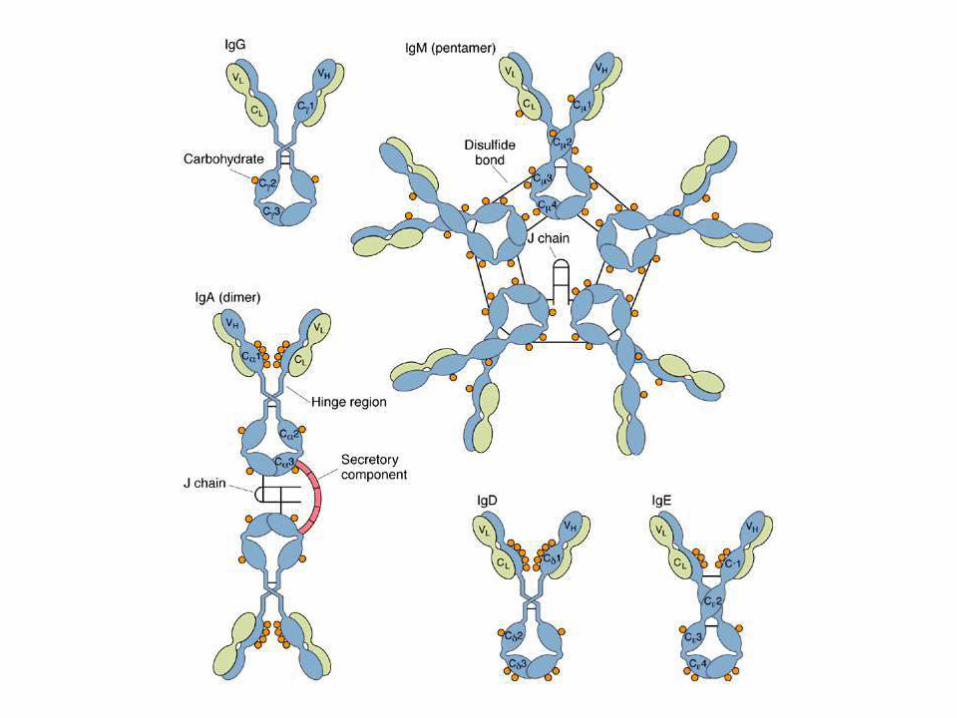
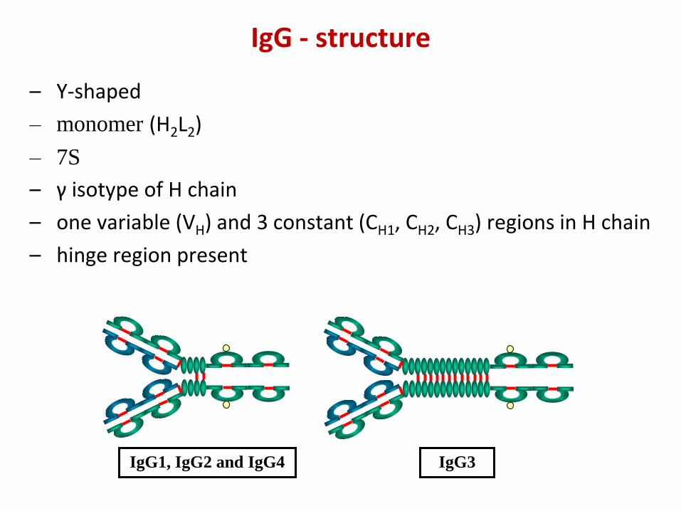
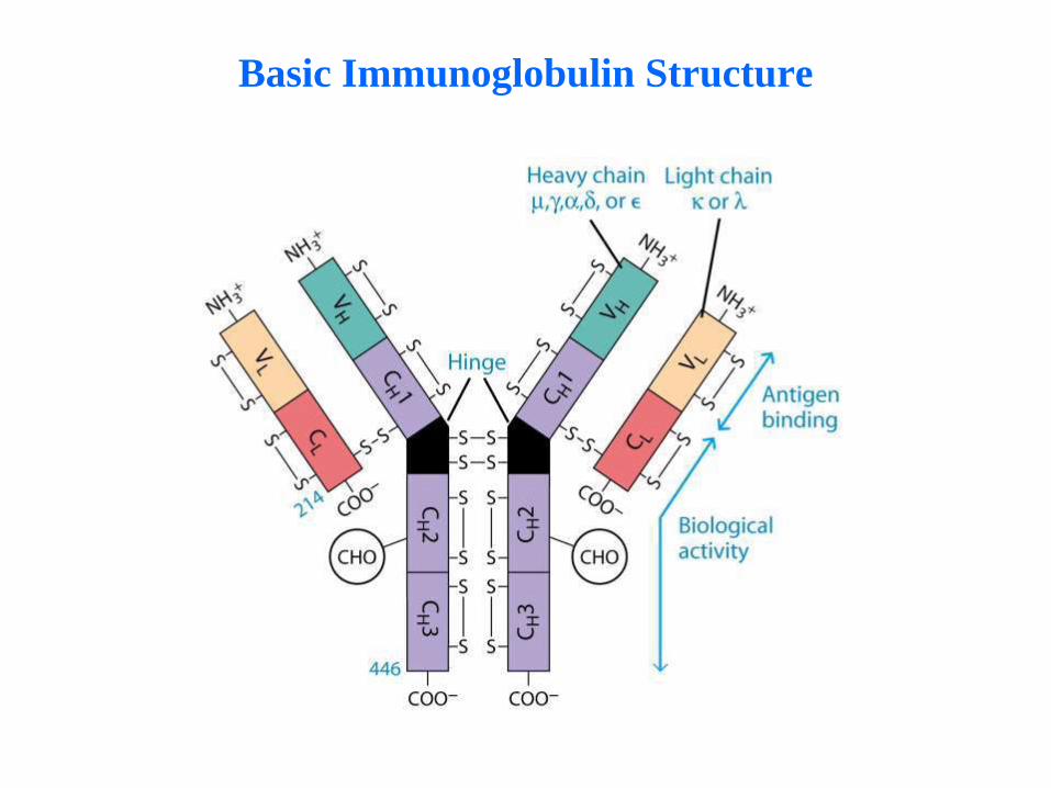
IgG - structure
– Y-shaped
– monomer (H2L2)
– 7S
– γ isotype of H chain
– one variable (VH) and 3 constant (CH1, CH2, CH3) regions in H chain
– hinge region present
IgG1, IgG2 and IgG4 IgG3
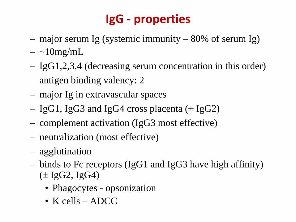
IgG - properties
– major serum Ig (systemic immunity – 80% of serum Ig)
– ~10mg/mL
– IgG1,2,3,4 (decreasing serum concentration in this order)
– antigen binding valency: 2
– major Ig in extravascular spaces
– IgG1, IgG3 and IgG4 cross placenta (± IgG2)
– complement activation (IgG3 most effective)
– neutralization (most effective)
– agglutination
– binds to Fc receptors (IgG1 and IgG3 have high affinity) (± IgG2, IgG4)
• Phagocytes - opsonization
• K cells – ADCC
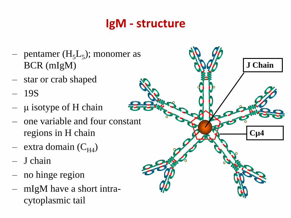
IgM - structure
– pentamer (H5L5); monomer as
BCR (mIgM)
– star or crab shaped
– 19S
– μ isotype of H chain
– one variable and four constant
regions in H chain
– extra domain (CH4)
– J chain
– no hinge region
– mIgM have a short intra-
cytoplasmic tail
Cµ4
J Chain
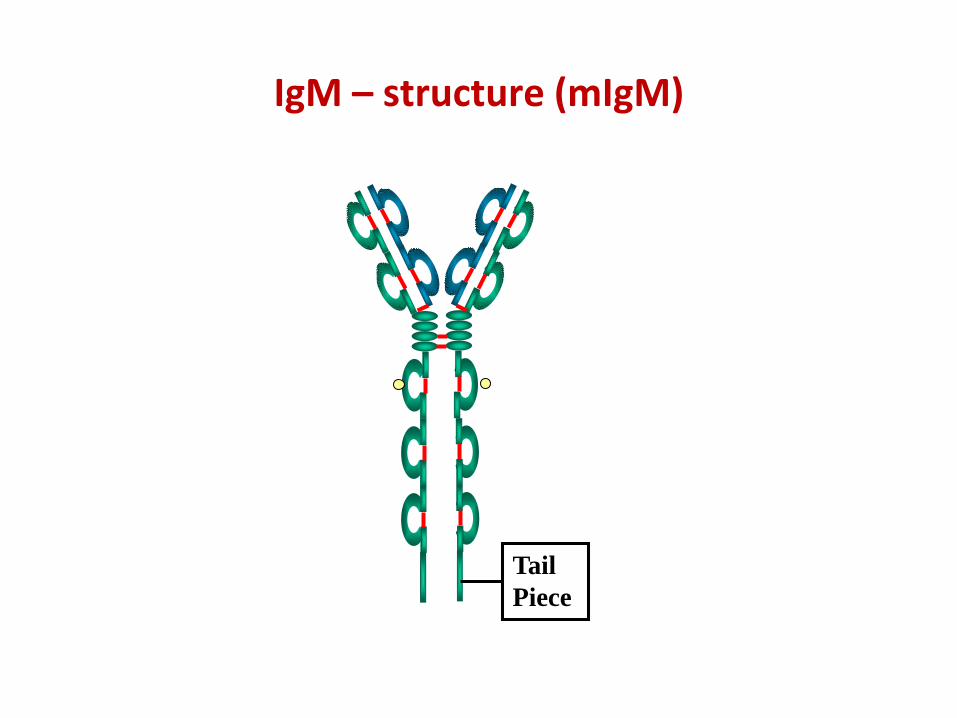
IgM – structure (mIgM)
Tail
Piece
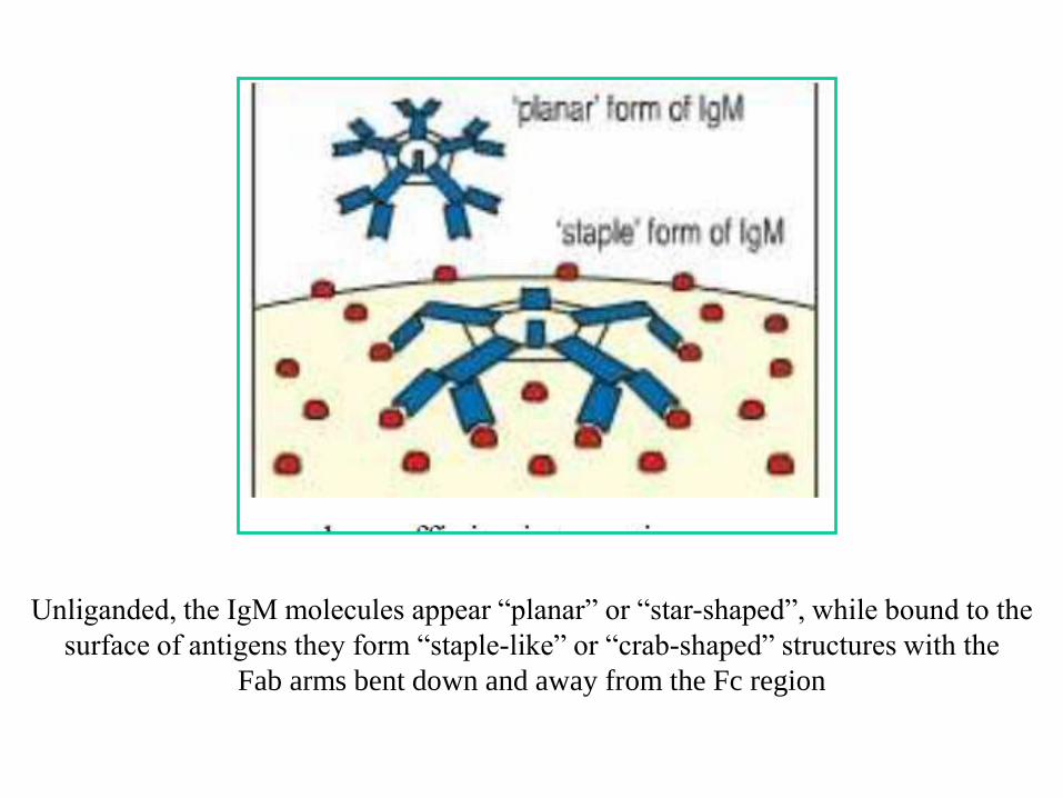
Unliganded, the IgM molecules appear “planar” or “star-shaped”, while bound to the
surface of antigens they form “staple-like” or “crab-shaped” structures with the
Fab arms bent down and away from the Fc region
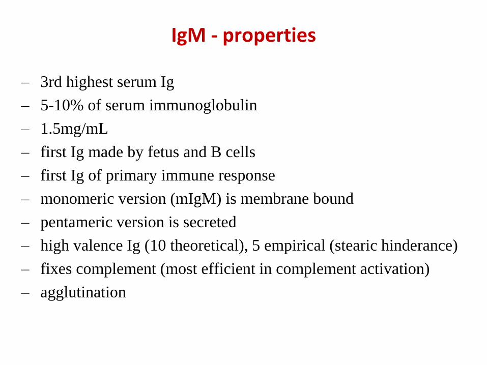
IgM - properties
– 3rd highest serum Ig
– 5-10% of serum immunoglobulin
– 1.5mg/mL
– first Ig made by fetus and B cells
– first Ig of primary immune response
– monomeric version (mIgM) is membrane bound
– pentameric version is secreted
– high valence Ig (10 theoretical), 5 empirical (stearic hinderance)
– fixes complement (most efficient in complement activation)
– agglutination

Fixation of C1 by IgG and IgM Abs
No activation Activation
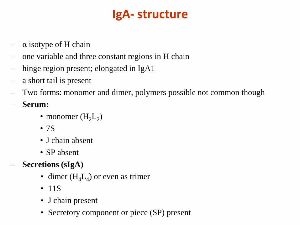
IgA- structure
– α isotype of H chain
– one variable and three constant regions in H chain
– hinge region present; elongated in IgA1
– a short tail is present
– Two forms: monomer and dimer, polymers possible not common though
– Serum:
• monomer (H2L2)
• 7S
• J chain absent
• SP absent
– Secretions (sIgA)
• dimer (H4L4) or even as trimer
• 11S
• J chain present
• Secretory component or piece (SP) present
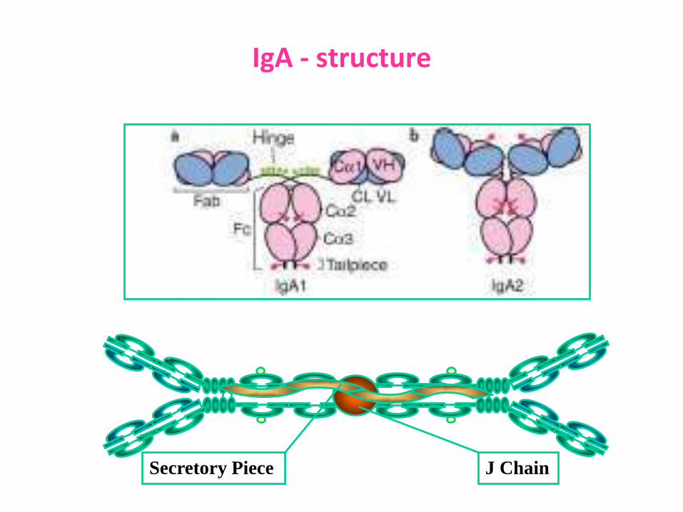
IgA - structure
J ChainSecretory Piece
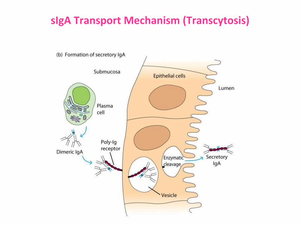
sIgA Transport Mechanism (Transcytosis)
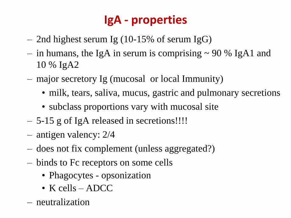
IgA - properties
– 2nd highest serum Ig (10-15% of serum IgG)
– in humans, the IgA in serum is comprising ~ 90 % IgA1 and
10 % IgA2
– major secretory Ig (mucosal or local Immunity)
• milk, tears, saliva, mucus, gastric and pulmonary secretions
• subclass proportions vary with mucosal site
– 5-15 g of IgA released in secretions!!!!
– antigen valency: 2/4
– does not fix complement (unless aggregated?)
– binds to Fc receptors on some cells
• Phagocytes - opsonization
• K cells – ADCC
– neutralization
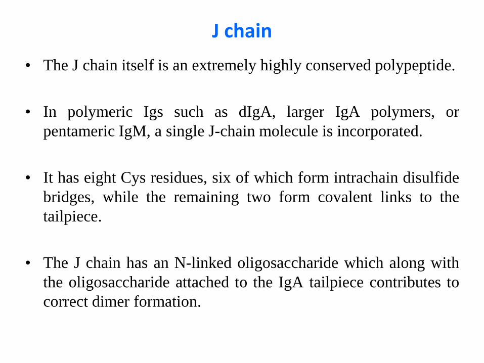
J chain
• The J chain itself is an extremely highly conserved polypeptide.
• In polymeric Igs such as dIgA, larger IgA polymers, or
pentameric IgM, a single J-chain molecule is incorporated.
• It has eight Cys residues, six of which form intrachain disulfide
bridges, while the remaining two form covalent links to the
tailpiece.
• The J chain has an N-linked oligosaccharide which along with
the oligosaccharide attached to the IgA tailpiece contributes to
correct dimer formation.
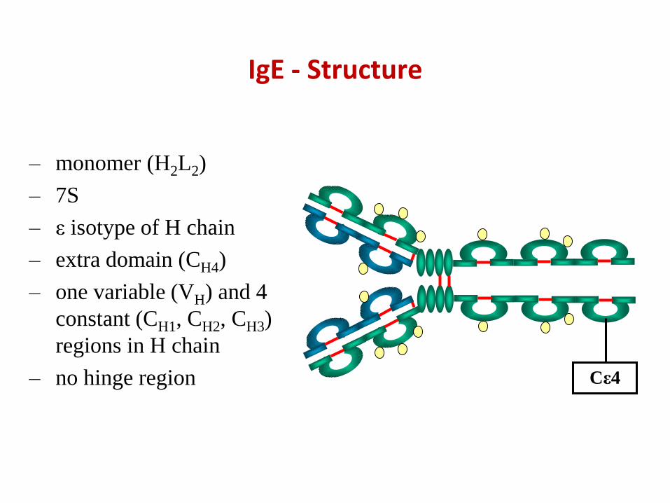
IgE - Structure
– monomer (H2L2)
– 7S
– ε isotype of H chain
– extra domain (CH4)
– one variable (VH) and 4
constant (CH1, CH2, CH3)
regions in H chain
– no hinge region Cε4
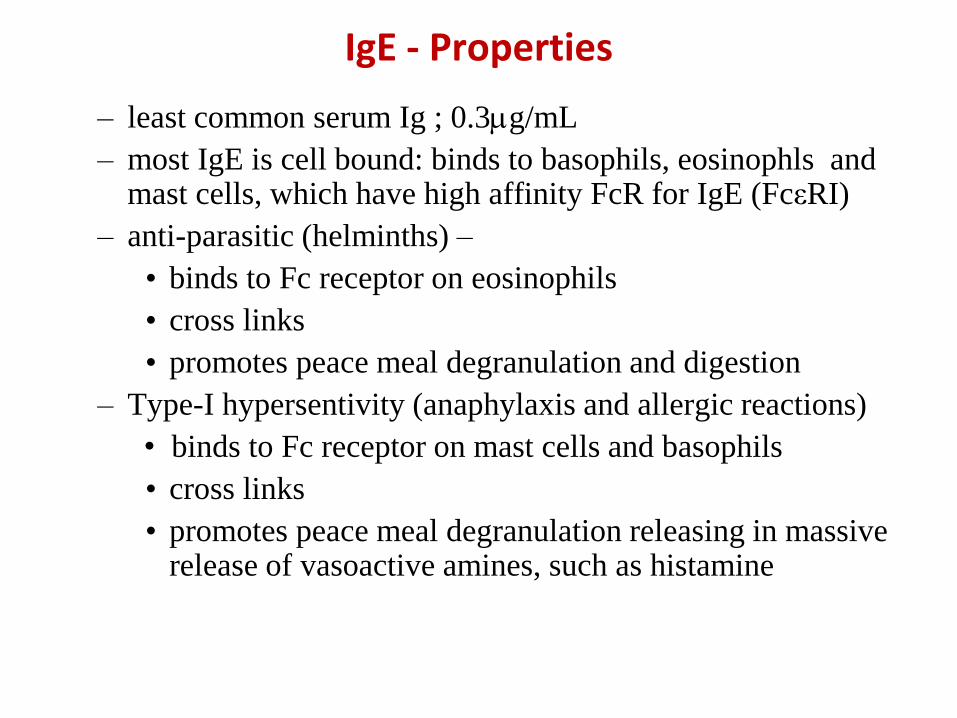
IgE - Properties
– least common serum Ig ; 0.3g/mL
– most IgE is cell bound: binds to basophils, eosinophls and mast cells, which have high affinity FcR for IgE (FcεRI)
– anti-parasitic (helminths) –
• binds to Fc receptor on eosinophils
• cross links
• promotes peace meal degranulation and digestion
– Type-I hypersentivity (anaphylaxis and allergic reactions)
• binds to Fc receptor on mast cells and basophils
• cross links
• promotes peace meal degranulation releasing in massive release of vasoactive amines, such as histamine
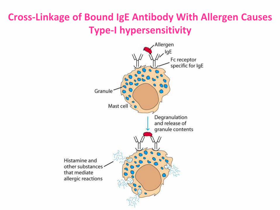
Cross-Linkage of Bound IgE Antibody With Allergen Causes Type-I hypersensitivity
IgD- structure
– Y-shaped
– monomer (H2L2)
– 7S
– δ isotype of H chain
– one variable (VH) and
3 constant (CH1, CH2,
CH3) regions in H
chain
– hinge region present
– tail piece present
Tail Piece
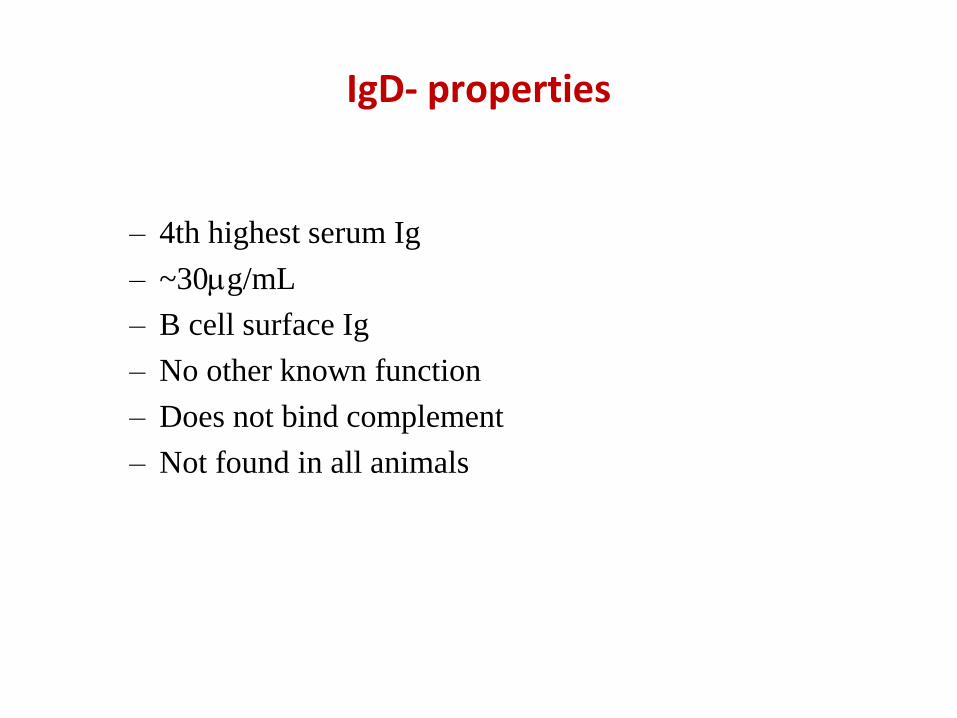
IgD- properties
– 4th highest serum Ig
– ~30g/mL
– B cell surface Ig
– No other known function
– Does not bind complement
– Not found in all animals
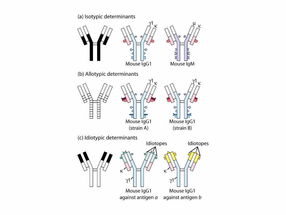
Antigenic determinants on immunoglobulins
• Immunoglobulins, being protein in nature, arethemselves immunogenic for other individuals ofsame or different species, i.e. Igs also have epitopes,
• Antigenic Determinants on Abs are of three types:
– Isotypic
– Allotypic
– Idiotypic
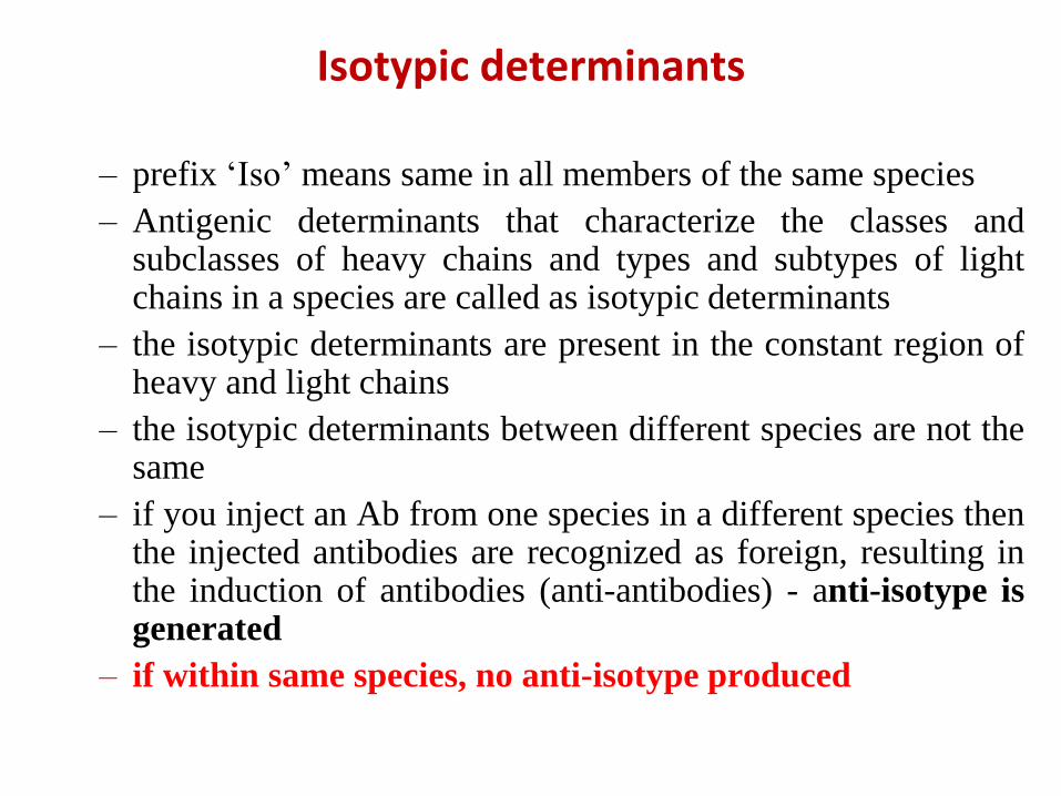
– prefix ‘Iso’ means same in all members of the same species
– Antigenic determinants that characterize the classes andsubclasses of heavy chains and types and subtypes of lightchains in a species are called as isotypic determinants
– the isotypic determinants are present in the constant region ofheavy and light chains
– the isotypic determinants between different species are not thesame
– if you inject an Ab from one species in a different species thenthe injected antibodies are recognized as foreign, resulting inthe induction of antibodies (anti-antibodies) - anti-isotype isgenerated
– if within same species, no anti-isotype produced
Isotypic determinants
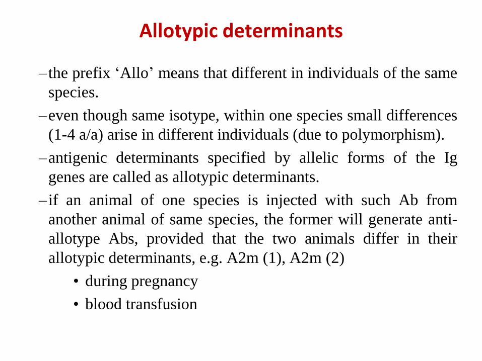
– the prefix ‘Allo’ means that different in individuals of the same
species.
–even though same isotype, within one species small differences
(1-4 a/a) arise in different individuals (due to polymorphism).
–antigenic determinants specified by allelic forms of the Ig
genes are called as allotypic determinants.
– if an animal of one species is injected with such Ab from
another animal of same species, the former will generate anti-
allotype Abs, provided that the two animals differ in their
allotypic determinants, e.g. A2m (1), A2m (2)
• during pregnancy
• blood transfusion
Allotypic determinants
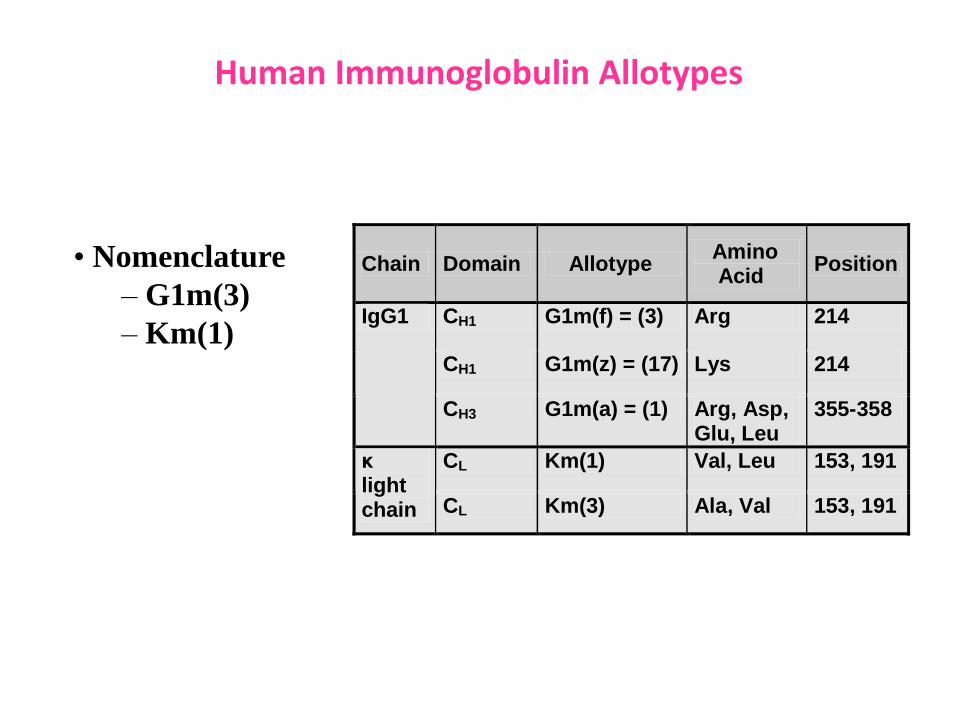
Human Immunoglobulin Allotypes
• Nomenclature
– G1m(3)
– Km(1)
Chain Domain Allotype Amino Acid
Position
CH1 G1m(f) = (3) Arg 214
CH1 G1m(z) = (17) Lys 214
IgG1
CH3 G1m(a) = (1) Arg, Asp, Glu, Leu
355-358
CL Km(1) Val, Leu 153, 191 κ light chain CL Km(3) Ala, Val 153, 191
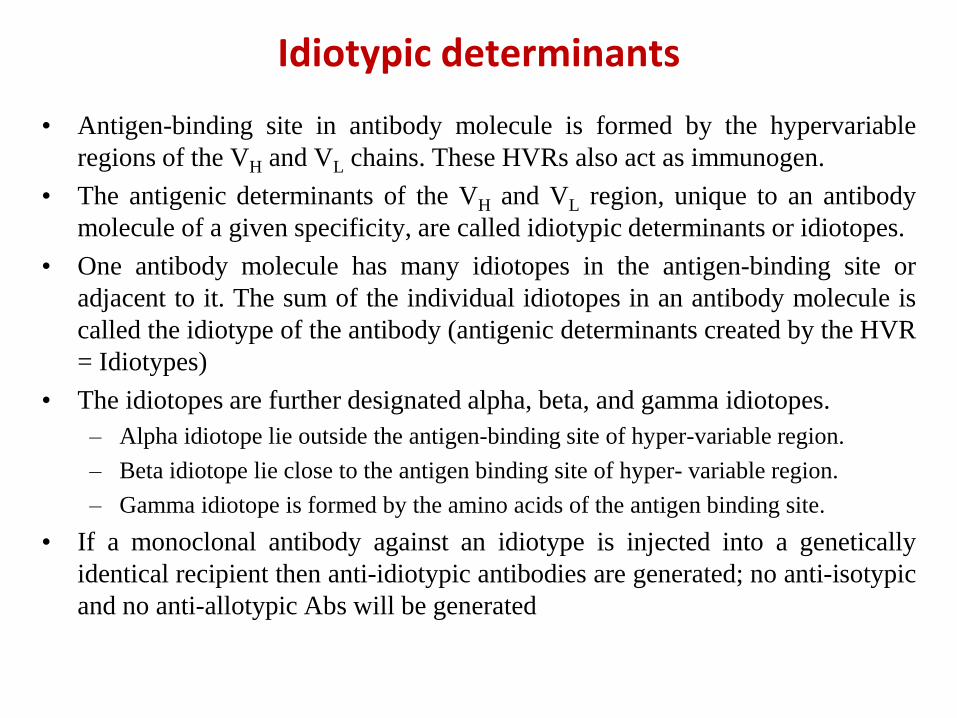
• Antigen-binding site in antibody molecule is formed by the hypervariable
regions of the VH and VL chains. These HVRs also act as immunogen.
• The antigenic determinants of the VH and VL region, unique to an antibody
molecule of a given specificity, are called idiotypic determinants or idiotopes.
• One antibody molecule has many idiotopes in the antigen-binding site or
adjacent to it. The sum of the individual idiotopes in an antibody molecule is
called the idiotype of the antibody (antigenic determinants created by the HVR
= Idiotypes)
• The idiotopes are further designated alpha, beta, and gamma idiotopes.
– Alpha idiotope lie outside the antigen-binding site of hyper-variable region.
– Beta idiotope lie close to the antigen binding site of hyper- variable region.
– Gamma idiotope is formed by the amino acids of the antigen binding site.
• If a monoclonal antibody against an idiotype is injected into a genetically
identical recipient then anti-idiotypic antibodies are generated; no anti-isotypic
and no anti-allotypic Abs will be generated
Idiotypic determinants
Immunoglobulin Genetics(Antibody diversity)
Antibody diversity - Introduction
• Antibody diversity is defined as the phenomenon of immense variability
characteristic of antibodies, which enables the immune system to react
specifically against the essentially unlimited kinds of antigens it encounters.
• Antibody diversity in humans comes from several stages of immunoglobulin
development, including both pre-immune repertoire and the post-immune
repertoire
• In the pre-immune repertoire, there are six major sources of antibody
diversity in human beings, generating a potential pre-immune diversity of
>1016 different antibodies.
• In the post-immune repertoire following exposure to antigen there occurs
somatic hypermutation which results in affinity maturation of antibodies
with higher affinity to the targeted antigen epitope, resulting in more
effective binding and elimination of the antigen from circulation during the
secondary immune response.
• It has been demonstrated that during the affinity maturation process, the
average number of mutations in VH and VL are eight and five, respectively.
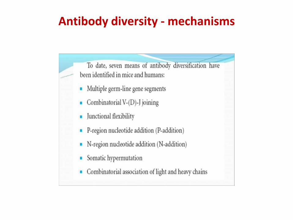
Antibody diversity - mechanisms
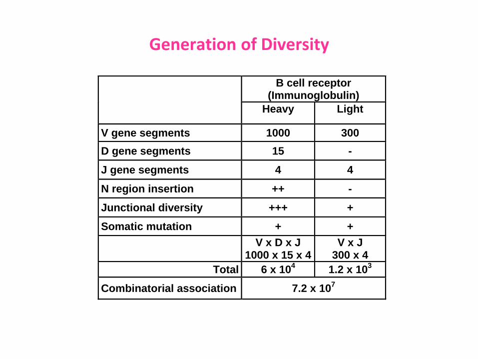
Generation of Diversity
B cell receptor (Immunoglobulin)
Heavy Light
V gene segments 1000 300
D gene segments 15 -
J gene segments 4 4
N region insertion ++ -
Junctional diversity +++ +
Somatic mutation + +
V x D x J
1000 x 15 x 4 V x J
300 x 4
Total 6 x 10⁴ 1.2 x 10
3
Combinatorial association 7.2 x 10⁷
Immunoglobulins of animals
Salient differential features
• There are five major immunoglobulin isotypes in mammals:IgM, IgD, IgG, IgA, and IgE.
• IgM is widely conserved throughout vertebrates, with theexception of the coelacanth.
• IgM, IgD, and IgA, or its analog, IgX, have been described innon-mammalian tetrapods.
• Birds, reptiles, and amphibians express IgY, a likelyevolutionary precursor to IgG and IgE.
• A unique immunoglobulin isotypes, such as IgX in amphibiansor IgW in sharks have been reported.
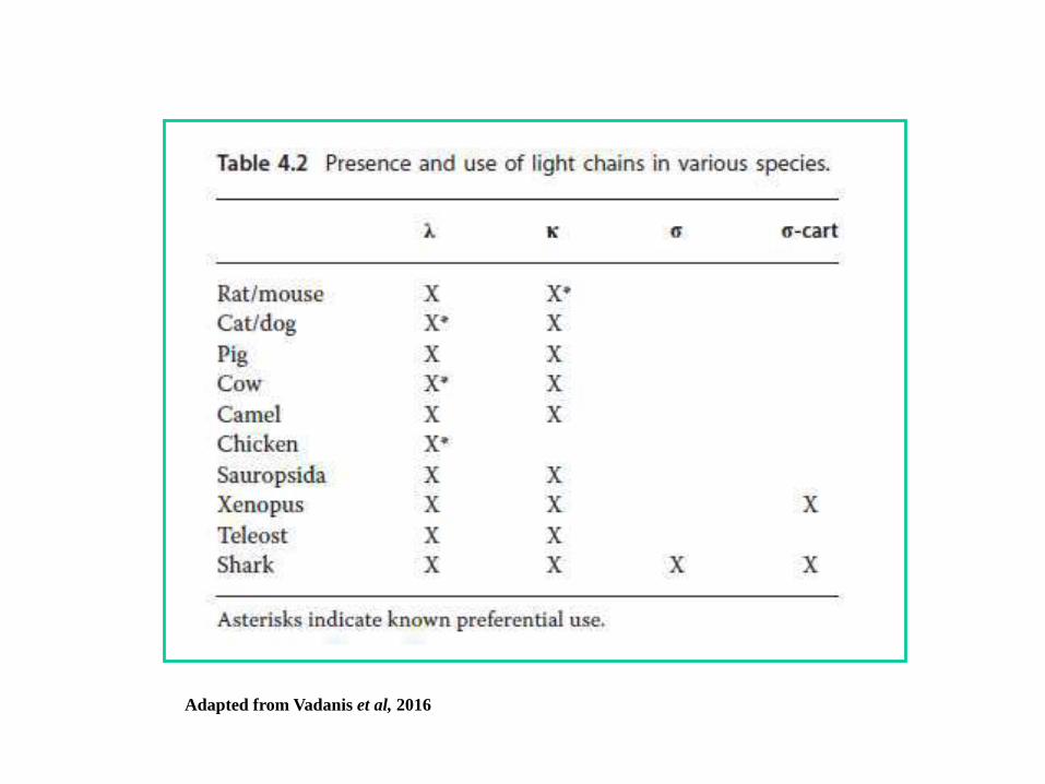
• Variability also exists in the type and usage frequencies of lightchains. In certain species, the κ and λ light chains are utilizedequally, while in others one or the other is preferred.
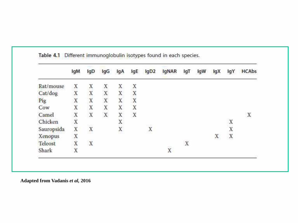
Adapted from Vadanis et al, 2016
Adapted from Vadanis et al, 2016
Salient differential features (contd.)
• The majority of maternal antibody transport in animals occursthrough the consumption of colostrum and milk after birth
• In the cats, IgA is present as a dimer both in serum andmucosa, while in most species IgA is present as a dimer only inits secreted form.
• Class-switch recombination (CSR) to IgD is possible in cattleand perhaps in porcine due to their unique IgD switch region,which is absent in humans and rodents.
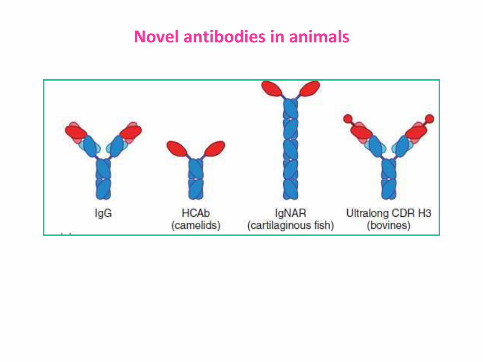
• Cows can have an unusually long complementarity-determining region 3 of the heavy chain (CDR H3), thebiological function of which remains unknown (recombinantmonoclonal humanized cow antibodies with ultra long CDR H3sare a potential candidate for immunotherapy of COVID-19)
Salient differential features (contd.)
• Camels have IgG type of antibodies (also called as“nanobodies”) which lack light chains and CH1 domain; onlyposess two H chains, each with one VH (referrd as VHH domain)and two CH domains (HCAbs).
• Alpacas and llamas also have HCAbs that are very similar tothose found in camels
• Llamas Abs are the a potential candidate for immunotherapy ofCOVID-19.
• Chickens have serum IgM, IgA, and IgY, the first two beinghomologs of their mammalian counterparts; however, they donot have IgE or IgD.
• IgY appears to be related to both mammalian IgG and IgE andmay be an evolutionary common ancestor to both
Novel antibodies in animals
Colostral antibodies in bovines
• In bovines colostrum is rich in IgG1type of antibody (human
colostrum has IgA), which is secreted into udder from dam’s
blood.
• Provides natural passive immunity
• Colostrum is rich in antibodies against pathogens to which dam
has been exposed either naturally or through vaccination
• Colostrum of dam or from foster mother of same form should be
used
• Clostrum feeding may cause loose faeces in calves, do not give
antibiotics.
• Anti-rotaviral antibodies appear after 3 days, hence colostrum
feeding should be continued for 5-6 days
• Vaccination of dams in third trimester of gestation against
pathogens which cause disease in neo natal claves is
recommended
Recommended