1/10 Asian J Agric & Biol. 2021(1).
Asian J Agric & Biol. 2021(1). DOI: 10.35495/ajab.2020.09.502
Recent advances in molecular characterization of Sarcocystis species in some meat producing animals: an updated review
Sara Omar Swar1, Bushra Hussain Shnawa2, 3* 1College of Agricultural Engineering Sciences, Salahaddin University, Kurdistan, Iraq 2Department of Biology, Faculty of Sciences, Soran University, Kurdistan, Iraq 3Scientific Research Center, Soran University, Kurdistan, Iraq
Abstract Sarcocystosis is a parasitic disease caused by Sarcocystis species that infect humans
and animals. It is prevalent in small ruminants like sheep and goats worldwide and
causing pathogenic impacts that lead to economic losses owing to carcass
condemnation, abortion, and death. Recently, several molecular and phylogenetic
analyses have been developed to differentiate Sarcocystis species including, the 18S
rRNA, 28S rRNA, 18S rDNA, and ITS-1 region. In recent years, the mitochondrial
cytochrome c oxidase subunit 1 (cox-1) was successfully used for this purpose. The
DNA barcoding using the cox1 gene is a reliable tool to distinguish and identify the
main Sarcocystis genotypes. Therefore, several studies confirmed that the cox1 gene is
a promising DNA marker for studying the genus Sarcocystis. The current review aims
to highlight the molecular methods that exist for the identification of Sarcocystis
species. The results showed that the Sarcocystis species of sheep and goats were
genetically close related and may be considered as sibling strains, as well as the cross-
infection may happen among them. Consequently, the host specificity of several
Sarcocystis species is questionable. The findings additional emphasized that
experimental transmission investigations within the proposed definitive host are
required to confirm the characteristics and host ranges of the Sarcocystis spp. in sheep
and goats. The current review represents updated knowledge about molecular
discrimination of Sarcocystis species in small ruminants by reviewing and analyzing
the recent articles in this aspect.
Keywords: Sarcocystis species, Small ruminants, Molecular identification, PCR
How to cite this:
Swar SO and Shnawa BH, 2021. Recent advances in molecular characterization of
Sarcocystis species in some meat producing animals: An updated review. Asian J.
Agric. Biol. 2021(1). DOI: https://doi.org/10.35495/ajab.2020.09.502
This is an Open Access article distributed under the terms of the Creative Commons Attribution 3.0 License.
(https://creativecommons.org/licenses/by/3.0), which permits unrestricted use, distribution, and reproduction in any medium, provided the
original work is properly cited.
Introduction
Sarcocystis is a coccidian parasite related to the
phylum Apicomplexa, class Sporozoasida, order
Eucoccidiorida, family Sarcocystidae and subfamily
Sarcocystina (Levine, 1986; Dubey et al., 2016).
Sarcocystis was firstly recorded by Friedrich
Miescher in 1843 as a white thread-like structure
within the skeletal muscular tissue of a deer mouse
from his house in Switzerland, therefore, it is called
Review Article
Received: September 26, 2020
Accepted: November 13, 2020
Online First:
December 22, 2020
Published:
January 30, 2021
*Corresponding author email: [email protected]
AJAB
Sara Omar Swar and Bushra Hussain Shnawa
2/10 Asian J Agric & Biol. 2021(1).
Miescher`s tubules. The name of Sarcocystis derived
from Greek, Sarkos means flesh and Kystis meaning
bladder, which refers to the encysted stage in
mammalian muscular tissue (Dubey et al., 2016).
Approximately 196 Sarcocystis species are valid, but
just 26 of them have a well-known life cycle.
Sarcocystis have a wide range of animals that can
serve as final hosts and the types of animals that can
act as intermediate hosts, which distinguish it from
Neospora caninum and Toxoplasma gondii (Dubey et
al., 2016; Lindsay and Dubey, 2020). Sarcocystosis is
a world-widely distributed protozoan disease that
infects different kinds of mammals, birds, and reptiles.
Several species of Sarcocystis are pathogenic that
result in a severe disease, which can lead to abortion
and carcasses condemnation in meat-producing
animals, also some species are described as zoonotic
(Dubey et al., 2015). Small ruminants as meat-
producing animals are one of the main divisions of the
food supply chain for humans in most countries and
infected with various economically significant
parasitic diseases, including sarcocystosis and
Eimeria spp. infection (Latif et al., 1999; Hassanen et
al., 2020).
Globally, a high prevalence of sarcocystosis was
recorded in sheep such as 96.1, 86.5, 81.5-90, 100 and
95.3% in Brazil, Izmir-Turkey, Saudi Arabia, Iran, and
Egypt, respectively (Beyazit et al., 2007; Dehaghi et al.,
2013; Al-Quraishy et al., 2014; Elmishmishy et al.,
2018; Minuzzi et al., 2019). Also, Morsy et al. (2011)
showed that 79.4% of goats were infected with
microsarcocysts in Egypt. In Iraq, Latif et al. (1999)
recorded a percentage of infection in sheep and goats as
97 and 97.4%, respectively. Also, in the north of Iraq, the
percentage of infection with microsarcocysts reached
97% in sheep and 100% in goats (Zangana and Hussein,
2017), whereas the infection with macrosarcosystis
recorded as 9.5 and 8.8% in both sheep and goats,
respectively (Swar and Shnawa, 2020).
Moreover, Barham et al. (2005) observed 97 and 34%
of goats were infected with microcysts and macrocysts
respectively, in Al-Sulaimany province-Iraq. Several
methods have been developed for the diagnosis of
sarcocystosis. These methods can be categorized into
the macroscopic and microscopic examination,
serology, and molecular. This review aims to address
the current situation of the recent publications that
achieved various molecular analyses to confirm the
characterization of Sarcocystis species and to
highlight the gap in the information regarding
sarcocystosis.
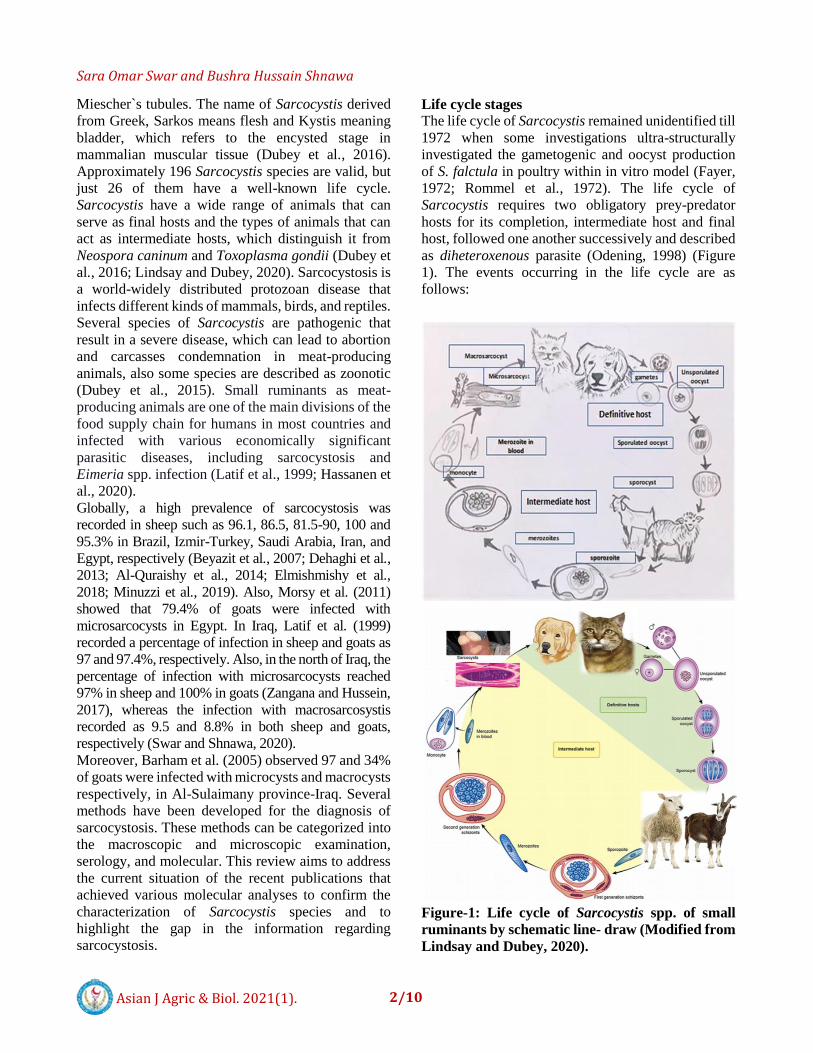
Life cycle stages
The life cycle of Sarcocystis remained unidentified till
1972 when some investigations ultra-structurally
investigated the gametogenic and oocyst production
of S. falctula in poultry within in vitro model (Fayer,
1972; Rommel et al., 1972). The life cycle of
Sarcocystis requires two obligatory prey-predator
hosts for its completion, intermediate host and final
host, followed one another successively and described
as diheteroxenous parasite (Odening, 1998) (Figure
1). The events occurring in the life cycle are as
follows:
Figure-1: Life cycle of Sarcocystis spp. of small
ruminants by schematic line- draw (Modified from
Lindsay and Dubey, 2020).

Sara Omar Swar and Bushra Hussain Shnawa
3/10 Asian J Agric & Biol. 2021(1).
Asexual stages
It is found only in the intermediate host, which is
mostly a prey animal. Infection begins when
Sarcocystis oocysts or sporocysts from feces of the
final host are ingested by these animals with
contaminated food or water. Sporozoites are liberated
from the oocysts under the effect of trypsin and bile.
These sporozoites invade the gut wall and lodge
primarily in the endothelial cells of the small arteries.
Four cycles of asexual development, called merogony
and schizogony are known (Fayer et al., 2015). During
the first three stages of development, sporozoites
undergo many nuclear divisions followed by
segmentation to generate merozoites, which are
motile and crescent in shape. Following these cycles
of schizonts, the Sarcocystis encysts in the muscles
and initially forming metrocytes and then transform to
bradyzoites. Sarcocysts having bradyzoites refer to
the last encysted stage in the skeletal, cardiac, and
smooth muscles of herbivores infected animals, which
is infectious for carnivorous animals as definitive
hosts (Fayer et al., 2015; Dubey, 2015; Dubey et al.,
2016; Khater et al., 2020). Two types of sarcocysts
can be found in sheep and goats, including
microsarocyst and macrosarcocyst, which related to
different species of Sarcocystis as shown, in Figure 2
and 3. Till now, there are seven Sarcocystis spp. have
recorded from domestic sheep, four of them (S.
tenella, S. arieticanis, S. microps, and S. mihoensis)
found to use dogs as definitive hosts. Besides the other
three (S.gigantea, S.medusiformis, and S. moulei) that
transmitted by felids (Al-Hoot et al., 2005; Kalantari
et al., 2016; Gjerde et al., 2020). Goats are
intermediate hosts for three common species of
Sarcocystis (S. capracanis, S. moulei, and
S.hircicanis) (Lindsay and Dubey, 2020). Moreover,
S. gigantea and S. tenella species that commonly
infect sheep have identified in goats also (Ghaffar et
al., 1989; Hong et al., 2016). All the mentioned
species of goats were able to form microsarcocysts
and utilize dogs as definitive hosts except S. moulei
and S gigantea form macrosarcocysts and use cats as
definitive hosts for completing their life cycles. These
findings suggest sheep and goats can probably serve
as alternative hosts for several closely related
Sarcocystis spp.
A and B: thick-walled sarcocyst with radial striations
of S. tenella in sheep esophagi, scale bar= 500 nm. C:
Heavily infected esophagus of goat showed three
different sizes of sarcocysts with inflammation, scale
bar = 5 μm. D, E, and F: morphologic features of
sarcocysts in the esophagus of goats. Showing a thick
wall sarcocysts related to S. capracanis. All sections
were stained with hematoxylin and eosin. D Scale bar
= 500, E= Scale bar = 2 μm, and F Scale bar = 500
nm. (Swar, 2020; unpublished results).
Figure-2: Microscopic sarcocysts in histological
section.
Figure-3: Macroscopic sarcocysts in esophagi of
sheep and goats (modified from Swar and Shnawa,
2020)
Sexual stages
The final host acquires the infection by ingesting
mature sarcocysts within the muscles of infected
animals (Lindsay and Dubey, 2020). The Sarcocysts
are digested in the digestive system of the definitive
host and release the bradyzoites, which invade the
mucosa of the small intestine. Then, they are changed
into both male and female gametes, namely,
microgametes and macrogametes, respectively. These
Sara Omar Swar and Bushra Hussain Shnawa
4/10 Asian J Agric & Biol. 2021(1).
gametes undergo fertilization to form a zygote, which
leads to the formation of the non-motile oocysts. The
sexual cycle and fertilization need to be completed
within one day. The oocysts of this parasite sporulate
in the small intestine. The sporulated oocysts are thin-
walled contain two sporocysts, each with four
sporozoites. Then, it ruptures, releasing the sporocysts
into the intestinal lumen that are excreted with the
feces. The prepatent and patent periods differ
according to Sarcocystis spp., but in most of them,
oocysts are first to shed in the feces after 7 to 14 days
post acquiring Sarcocysts (Lindsay and Dubey, 2020).
Species infecting sheep and goats
Several species of Sarcocystis infect sheep (Table 1),
some of them transmitted by canids and others by
feline (cat). The species transmitted by dogs are
mainly pathogenic and cause microsarcocyst in
skeletal and cardiac muscles of infected animals.
These species, like S. tenella, can cause pathologic
effects in sheep including, anorexia, anemia, weight
loss, abortion, neural symptoms, and death (Dubey et
al., 1988; Abdel-Baki et al., 2009). While the species
transmitted by cats, for example, S. gigantea, and S.
medusiformis produce macrosarcocyst in the muscular
tissue like the esophagus, tongue, and larynx also, they
are less pathogenic than the microsarcocysts (Collins
et al., 1979; Dubey et al., 2016). There are three
common species of this parasite in domestic goats: S.
caprafelis (synonym S .moulei), S. hiricanis, and S.
capracanis (Table1). Commonly, S. hiricanis and S.
capracanis appear as microscopic sarcocysts, whereas
S. moulei forms macroscopic cysts (Dubey et al.,
2016). Clinically, S. capracanis is more pathogenic
than other species (Collins and Charleston, 1979), the
infected goats show fever, weakness, anorexia, weight
loss, tremors, abortion, and also lead to death in heavy
infection (Dubey et al., 1981).
Nowadays, several publications pointed out that the
infection of sheep and goats with species of
Sarcocystis that are uncommon in these hosts, as the
infection of sheep with S. moulei that normally infect
goats in Saudi Arabia and Iran (Al-Hoot et al., 2005;
Kalantari et al., 2016). Also, the infection of goats with
S. gigantea that commonly infects sheep (Ghaffar et
al., 1989) and concluded that the goats can be a host of
three species of Sarcocystis that are previously
classified as S. moulei, including S. ovifelis (S.
gigantea). Also, Hong et al. (2016) proved the
infection of goats with S. tenella which is commonly
known as sheep specific by molecular and
ultrastructural investigation in Korea.
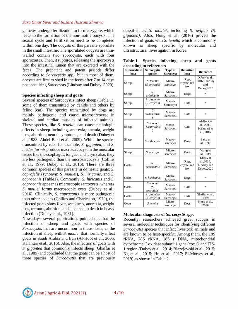
Table-1. Species infecting sheep and goats
according to references Intermediate
host
Sarcocystis
species
Type of
Sarcocyst
Definitive
host Reference
Sheep S. tenella
(S.ovicanis)
Micro-
sarcocyst
Dogs,
coyote, red fox
Dubey et al., 2016; Lindsay
and
Dubey,2020
Sheep S.
arieticanis Micro-
Sarcocyst Dogs =
Sheep
S. gigantea
(S .ovifelis)
Macro-
Sartcocyst Cats =
Sheep
S
medusiformi
s
Macro-Sarcocyst
Cats =
Sheep S. moulei
(S.caprafelis
)
Macro-
Sarcocyst Cats
Al-Hoot et
al., 2005;
Kalantari et al., 2016
Sheep
S. mihoensis Macro-
sarcocyst Dogs
Saito et al.,1997
Sheep S. microps Micro-
sarcocyst Dogs
Wang et
al.,1988
Goats S.
capracanis
Micro-
Sarcocyst
Dogs,
coyote, red fox
Dubey et
al.,2016;
Lindsay and Dubey,2020
Goats S. hircicanis Micro-
Sarcocyst Dogs =
Goats
S. moulei
(S.
caprafelis)
Macro-Sarcocyst
Cats =
Goats S. gigantea (S .ovifelis)
Macro-Sarcocyst
Cats Ghaffar et al.,
1989
Goats S.tenella Micro-
sarcocyst Dogs
Hong et al.,
2016
Molecular diagnosis of Sarcocystis spp.
Recently, researchers achieved great success in
several molecular techniques for identifying different
Sarcocystis species that infect livestock animals and
are known to be host-specific. Among them, the 18S
rRNA, 28S rRNA, 18S r DNA, mitochondrial
cytochrome C oxidase subunit 1 gene (cox1), and ITS-
1 region (Dubey et al., 2014; Blazejewski et al., 2015;
Ng et al., 2015; Hu et al., 2017; El-Morsey et al.,
2019) as shown in Table 2.

Sara Omar Swar and Bushra Hussain Shnawa
5/10 Asian J Agric & Biol. 2021(1).
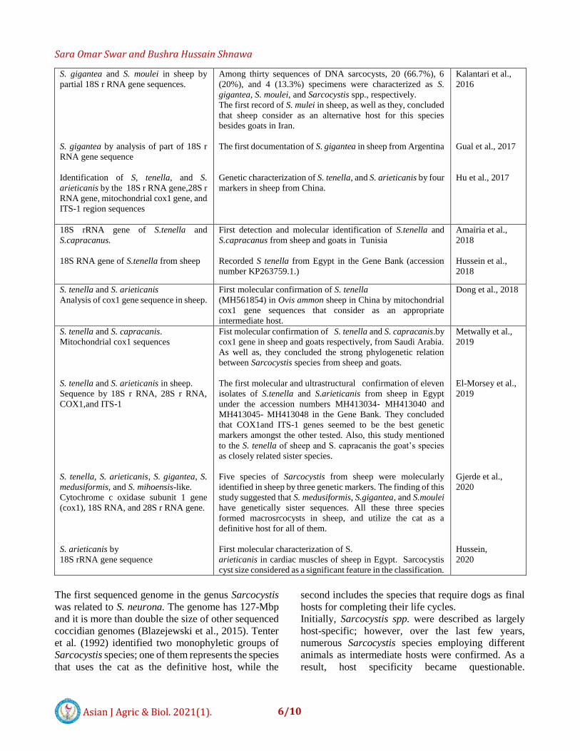
Table-2. Some recent publications in molecular diagnosis of Sarcocystis spp from sheep and goats
Sarcocystis species and method Molecular findings References
Six Sarcocystis spp.
Ribosomal RNA sequences.
S. tenella RFLP-PCR genotyping for the
18S rRNA
S. gigantea and S. tenella Mitochondrial
cytochrome c oxidase subunit I gene
(cox1) and the nuclear ssrRNA gene
sequences
S. tenella proved by 18S r RNA gene
sequence with PCR-RFLP technique.
S. gigantea and S. tenella in sheep by the
18s rRNA gene sequence
S. gigantea and S. medusiformis by the
PCR and RFLP techniques.
S. neurona strain SO SN1 RNA and
Genomic DNA Were sequenced
S. capracanis by 18S RNA sequences
S. tenella by
18S r RNA method.
S. arieticanis and S.capracanis in sheep
and goats respectively by18S ribosomal
DNA(r DNA)
S. tenella in goats
With 18S r DNA sequence
18S rRNA gene of S. tenella
Two groups of Sarcocystis spp. were identified molecularly.
The first group contains two species that need cats as definitive
hosts, and the second one has Sarcocystis spp. that the dogs act
definitive hosts for them. Also, Sarcocystis was separated from
Toxoplasma and the classification of them as two genera into
different subfamilies of the Sarcocystidae was refuted.
Twenty- two of 602 Brazilian sheep were positive for
Sarcocystis species. Identification of the 18S rRNA gene of S.
tenella (GenBank accession number L24383-1).
For the first time established cox1 as a new genetic marker for
the identification of Sarcocystis spp. Also, it presented the first
molecular characterization of S. gigantea (KC209733) besides
S. tenella of sheep in Norway. Results of ssrRNA gene
sequences showed that three of the four sequences of
microscopic sarcocysts isolated from sheep were
indistinguishable (KC209734–KC209736), one nucleotide was
incompatible with the fourth sequence (KC209737). Sequence
identity in BLAST showed sequences were most similar
(99.1% identity) to S. capracanis (L76472), whereas had
96.4% similarity with S. tenella (L24383).
RFLP-PCR analysis explained that microscopic cysts were S.
tenella in 70% of the tested specimens of sheep from Iran.
Genotyping of ten sarcocysts revealed that the pattern of
macrsarcocyst identified as S. gigantea and the microsarcocyst
is S. tenella in Iranian sheep.
The results proved that fat macrosarcocysts were S. gigantea as
29.31% and thin macro-sarcocysts were S. medusiformis in
7.52%.
Identified the first genome sequence of S. neurona. The
accession number of the nucleotide sequence was SRP052925
in the National Center for Biotechnology Information
(http://www.ncbi.nlm.nih.gov/bioproject/252030)
Microsarcocysts in goats skeletal muscles were characterized
as S. capracanis
Sequence findings of Sarcocystis spp. in sheep confirmed the
presence of S. tenella only in Italy.
Molecular and ultrastructural results proved the first
confirmation of S.tenella and S.arieticanis in sheep and
S.capracanis in goats in Brazil.
First molecular and ultrastructural documentation of S.tenella
in domestic goats in Korea
Molecular characterization and phylogeny of S.tenella in sheep
in Baghdad-Iraq.
Tenter et al.,1992
da Silva et al.,
2009
Gjerde, 2013a
Shahbazi et al.,
2013
Bahari et al.,
2014
Farhang-Pajuh et
al., 2014
Blazejewski et
al., 2015
Kutty et al., 2015
Bacci et al., 2016
Bittencourt et al.,
2016
Hong et al., 2016
Whaeeb and
Faraj, 2016
Sara Omar Swar and Bushra Hussain Shnawa
6/10 Asian J Agric & Biol. 2021(1).
S. gigantea and S. moulei in sheep by
partial 18S r RNA gene sequences.
S. gigantea by analysis of part of 18S r
RNA gene sequence
Identification of S, tenella, and S.
arieticanis by the 18S r RNA gene,28S r
RNA gene, mitochondrial cox1 gene, and
ITS-1 region sequences
Among thirty sequences of DNA sarcocysts, 20 (66.7%), 6
(20%), and 4 (13.3%) specimens were characterized as S.
gigantea, S. moulei, and Sarcocystis spp., respectively.
The first record of S. mulei in sheep, as well as they, concluded
that sheep consider as an alternative host for this species
besides goats in Iran.
The first documentation of S. gigantea in sheep from Argentina
Genetic characterization of S. tenella, and S. arieticanis by four
markers in sheep from China.
Kalantari et al.,
2016
Gual et al., 2017
Hu et al., 2017
18S rRNA gene of S.tenella and
S.capracanus.
18S RNA gene of S.tenella from sheep
First detection and molecular identification of S.tenella and
S.capracanus from sheep and goats in Tunisia
Recorded S tenella from Egypt in the Gene Bank (accession
number KP263759.1.)
Amairia et al.,
2018
Hussein et al.,
2018
S. tenella and S. arieticanis
Analysis of cox1 gene sequence in sheep.
First molecular confirmation of S. tenella
(MH561854) in Ovis ammon sheep in China by mitochondrial
cox1 gene sequences that consider as an appropriate
intermediate host.
Dong et al., 2018
S. tenella and S. capracanis.
Mitochondrial cox1 sequences
S. tenella and S. arieticanis in sheep.
Sequence by 18S r RNA, 28S r RNA,
COX1,and ITS-1
S. tenella, S. arieticanis, S. gigantea, S.
medusiformis, and S. mihoensis-like.
Cytochrome c oxidase subunit 1 gene
(cox1), 18S RNA, and 28S r RNA gene.
S. arieticanis by
18S rRNA gene sequence
Fist molecular confirmation of S. tenella and S. capracanis.by
cox1 gene in sheep and goats respectively, from Saudi Arabia.
As well as, they concluded the strong phylogenetic relation
between Sarcocystis species from sheep and goats.
The first molecular and ultrastructural confirmation of eleven
isolates of S.tenella and S.arieticanis from sheep in Egypt
under the accession numbers MH413034- MH413040 and
MH413045- MH413048 in the Gene Bank. They concluded
that COX1and ITS-1 genes seemed to be the best genetic
markers amongst the other tested. Also, this study mentioned
to the S. tenella of sheep and S. capracanis the goat’s species
as closely related sister species.
Five species of Sarcocystis from sheep were molecularly
identified in sheep by three genetic markers. The finding of this
study suggested that S. medusiformis, S.gigantea, and S.moulei
have genetically sister sequences. All these three species
formed macrosrcocysts in sheep, and utilize the cat as a
definitive host for all of them.
First molecular characterization of S.
arieticanis in cardiac muscles of sheep in Egypt. Sarcocystis
cyst size considered as a significant feature in the classification.
Metwally et al.,
2019
El-Morsey et al.,
2019
Gjerde et al.,
2020
Hussein,
2020
The first sequenced genome in the genus Sarcocystis
was related to S. neurona. The genome has 127-Mbp
and it is more than double the size of other sequenced
coccidian genomes (Blazejewski et al., 2015). Tenter
et al. (1992) identified two monophyletic groups of
Sarcocystis species; one of them represents the species
that uses the cat as the definitive host, while the
second includes the species that require dogs as final
hosts for completing their life cycles.
Initially, Sarcocystis spp. were described as largely
host-specific; however, over the last few years,
numerous Sarcocystis species employing different
animals as intermediate hosts were confirmed. As a
result, host specificity became questionable.

Sara Omar Swar and Bushra Hussain Shnawa
7/10 Asian J Agric & Biol. 2021(1).
Regarding this aspect, In Saudi Arabia, Al-Hoot et al.
(2005) identified S. moulei from the sheep by ultra-
structural study, the species which commonly infect
goats. Also, S. moulei was confirmed molecularly in
sheep by Kalantari et al. (2016) in Iran. In the same
regard, S. capracanis was recognized in the
cerebrospinal fluid of 2 sheep with
meningoencephalitis in the United Kingdom
(Formisano et al., 2013). Therefore, sheep can serve
as an alternative intermediate host for this species
besides goats. In addition, in a study regarding
sarcocystosis of sheep in Egypt, Elmishmishy et al.
(2018) recorded the complete similarity between the
isolate of S. gigantea from sheep and that of S. moulei
and suggested the cross-transmission of S. moulei
between sheep and goats and they are
phylogenetically close. Another study also
demonstrated that the S. tenella of sheep and S.
capracanis the goat’s species are genetically closely
related sister species by ultra-structural and
phylogenetic analysis using 18S r RNA, 28S r RNA,
COX1, and ITS-1 genetic markers (El-Morsey et al.,
2019). In Saudi Arabia, a recent study concluded a
strong phylogenetic correlation among Sarcocystis
species from sheep and goats (Metwally et al., 2019).
Yang et al. (2001) confirmed that morphologically
identical species from two different intermediate hosts
should be considered the same species. However,
many Sarcocystis species seem to have a wider
intermediate host option than previously known. Also,
more recent findings suggest that S. medusiformis, S.
gigantea, and S. moulei possess genetically sister
sequences. All these species proved to form
macrosrcocysts in sheep, and the cat is the definitive
host for them (Gjerde et al., 2020). This phenomenon
has also been observed in the infection of cattle and
water buffalo with uncommon Sarcocystis spp. As a
result, these investigations lead to the suggestion that
Sarcocystis species are non-specific to the host (Jehle
et al., 2009; Xiang et al., 2011; Gjerde et al., 2016;
Dakhil et al., 2017; El-kady et al., 2018).
Gjerde (2013a; 2013b) concluded that cox1 sequences
appear to explain better than the ssrRNA gene
sequences for characterization of the closely related
species of Sarcocystis, and recommended using this
novel genetic marker in future studies. Additionally,
Gjerde et al. (2016) suggested that the cox1 gene was
superior to both 18S and 28S rRNA genes. In the same
regard, El-Morsey et al. (2019) confirmed that
cox1and ITS-1 genes looked to be the best genetic
markers amongst the other tested in the differentiation
of the closely related Sarcocystis spp. within the
intermediate hosts because of their high divergence, as
shown in Table 2. Another investigation compared the
new sequences of four genetic markers (18S
rRNA, 28S rRNA, mitochondrial COX1, and ITS-1)
for S. tenella and S.arieticanis, and confirmed that
the ITS-1 region could be more useful for
distinguishing the closely related Sarcocystis spp.
owing to its high divergence (Hu et al., 2017). Besides,
the same was true for the genetic similarity of sheep
and goats infection with Eimeria spp., which was
recorded by phylogenetic analysis of the ITS-1
sequences of this parasite in Egypt. The sequence of
(ITS-1) region of E. ahsata was 100% similar to ovine
E. ahsata and clustered in a single clade with E.
cardinalis and E. faurei. On the other hand, E. arloingi
was 100% identical to E. arloingi of goat and clustered
with bovine E. ellipsoidalis (Hassanen et al., 2020). Conclusion In conclusion, the information about Sarcocystis spp.
is still not highly clarifying. However, several
publications related to the biological aspect of
Sarcocystosis have been achieved, whereas there are
numerous ongoing researches on molecular and
ultrastructural diagnosis still perform globally. The
18S rRNA ribosomal gene and mitochondrial
cytochrome c oxidase subunit I (cox1) genes were the
most analysis used for molecular characterization and
phylogenetic analysis of Sarcocystis spp. The results
of some reviewed articles showed that the Sarcocystis
species of sheep and goats were closely related and
may consider as sibling strains, as well as the cross-
infection, may happen among these animals.
Therefore, the host specificity of several Sarcocystis
species is questionable. The findings further
emphasize that experimental transmission
investigations within the proposed definitive host are
required to confirm the characteristics and host ranges
of the Sarcocystis spp. within these two ruminants.
Disclaimer: None.
Conflict of Interest: None.
Source of Funding: None.
References Abdel-Baki AA, Allam G, Sakran T and El-Malah EM,
2009. Lambs infected with UV-attenuated
Sara Omar Swar and Bushra Hussain Shnawa
8/10 Asian J Agric & Biol. 2021(1).
sporocysts of Sarcocystis ovicanis produced
abnormal sarcocysts and induced protective
immunity against a challenge infection. Korean J.
Parasitol. 47: 131-138.
Al-Hoot AS, Al-Qureishy SA, Al-Rasheid K and
Bashtar AR, 2005. Microscopic study on
Sarcocystis moulei from sheep and goats in Saudi
Arabia. J. Egypt. Soc. Parasitol. 35: 295-312.
Al-Quraishy S, Morsy K, Bashtar AR, Ghaffar FA and
Mehlhorn H, 2014. Sarcocystis arieticanis
(Apicomplexa: Sarcocystidae) infecting the heart
muscles of the domestic sheep, Ovis aries
(Artiodactyla: Bovidae), from K. S. A. on the basis
of light and electron microscopic data. Parasitol.
Res. 113: 3823–3831.
Amairia S, Amdouni Y, Rouatbi M, Rjeibi MR, Awadi
S and Gharbi M, 2018. First detection and
molecular identification of Sarcocystis spp. in
small ruminants in North-West Tunisia.
Transbound Emerg. Dis. 65: 441–446.
Bacci C, Vismarra A, Passeri B, Sciarrone F, Mangia
C, Genchi M, Fabbi M, Vicari N, Bruini I, Brindani
F and Kramer L, 2016. Detection of Toxoplasma
gondii and Sarcocystis tenella in indigenous
CornigLiese sheep in Italy using serological and
molecular methods. Small Rum. Res. 135: 13-16.
Bahari P, Salehi M, Seydabadi M and Mohammadi A,
2014. Molecular identification of macroscopic and
microscopic cysts of sarcocystis in sheep in north
Khorasan province, Iran. J. Mol. Cellul. Med. 3:
51-6.
Barham M, Stutzer H, Karanis P, Latif BM and Neiss
WF, 2005. Seasonal variation in sarcocystis
species infections in goat in northern Iraq.
Parasitol. 130: 151-156.
Beyazit A, Yazicioğlu O and Karaer Z, 2007. The
prevalence of ovine Sarcocystis species in Izmir
province in sheep, Ankara Univ. Vet. Fak. Derg.
54:111-116.
Bittencourt MV, Meneses IDS, Ribeiro-Andrade M, de
Jesus RF, de Araújo FR and Gondim LFP, 2016.
Sarcocystis spp. in sheep and goats: frequency of
infection and species identification by
morphological, ultrastructural, and molecular tests
in Bahia, Brazil. Parasitol. Res. 115: 1683–1689.
Blazejewski T, Nursimulu N, Pszenny V,
Dangoudoubiyam S, Namasivayam S, Chiasson M
A, Chessman K, Tonkin M and Swapna LS,
2015. Systems-Based analysis of the Sarcocystis
neurona genome identifies pathways that
contribute to a heteroxenous life cycle. mBio. 6: 1-
16.
Collins GH, Atkinson E and Charleston WAG, 1979.
Studies on Sarcocystis species III: the macrocystic
species of sheep. New Zealand Vet. J. 27: 204-206.
Collins GH and Charleston WAG, 1979. Studies on
Sarcocystis species IV: A species infecting dogs
and goats; development in goats. New Zealand Vet.
J. 27: 260-262.
Dakhil HG, Abdallah BH and Abdallah FA, 2017.
Molecular identification of sarcocystis fusiformis
and S. moulei infecting water buffaloes (bubalus
bubalis) in southern Iraq. World J. Pharm. Res. 6:
215-229.
Da Silva RC, Su C and Langoni H, 2009. First
identification of Sarcocystis tenella (Railliet, 1886)
Moulé, 1886 (Protozoa: Apicomplexa) by PCR in
naturally infected sheep from Brazil. Vet. Parasitol.
165: 332–336.
Dehaghi MM, Fallahi M, Masoud S and Radfar MH,
2013. Survey of Sarcocystis infection in
slaughtered sheep in Kerman Abattoir, Kerman,
Iran. Comp. Clin. Pathol. 22: 343–346.
Dong H, Su R, Wang Y Zhang L, Yang Y and Hu J,
2018. Sarcocystis species in wild and domestic
sheep (Ovis ammon and Ovis aries) from China.
BMC Vet. Res. 14: 377.
Dubey JP, Weisbrode SE, Speer CA and Sharma SP,
1981. Sarcocystosis in goats: clinical signs and
pathologic and hematologic findings. J. Am. Vet.
Med. Assoc. 178: 683-699.
Dubey JP, Lindsay DS, Speer CA, Fayer R and
Livingston CW, 1988. Sarcocystis arieticanis and
other Sarcocystis species in sheep in the United
States. J. Parasitol. 74: 1033-1038.
Dubey JP, 2015. Foodborne and waterborne zoonotic
sarcocystosis. Food Waterborne Parasitol. 1: 2–11.
Dubey JP, Calero BR, Rosenthal BM, Speer CA and
Fayer R, 2016. Sarcocystosis of animals and
humans. 2nd Ed. Boca Raton: CRC Press; Taylor
and Francis Group.
Dubey JP, Lane EP and van Wilpe E, 2014. Sarcocystis
cafferin. sp. (Protozoa: Apicomplexa) from the
African Buffalo (Syncerus caffer). J. Parasitol.
100: 817–827.
Dubey JP, van Wilpe E, Calero-Bernal R, Verma SK
and Fayer R, 2015. Sarcocystis heydorni n. sp.
(Apicomplexa: Sarcocystidae) with cattle (Bos
taurus) and human (Homo sapiens) cycle.
Parasitol. Res. 114: 4143–4147.
Sara Omar Swar and Bushra Hussain Shnawa
9/10 Asian J Agric & Biol. 2021(1).
El-kady AM, Hussein NM and Hassan AA, 2018. First
molecular characterization of Sarcocystis spp. in
cattle in Qena Governorate, Upper Egypt. J.
Parasit. Dis. 42: 114-121.
Elmishmishy B, Al-Araby M, Abbas I and Abu-Elwafa
S, 2018. Genetic variability within isolates of
Sarcocystis species infecting sheep from Egypt.
Vet. Parasitol: Reg. Stud. Rep. 13: 193–197.
El-Morsey A, Abdo W, Sultan K, Elhawary NM and
AbouZaid AA, 2019. Ultrastructural and
Molecular Identification of the sarcocysts of
Sarcocystis tenella and Sarcocystis arieticanis
Infecting Domestic Sheep (Ovisaries) from Egypt.
Acta Parasitologica. 64: 501–513.
Farhang-Pajuh F, Yakhchali M and Mardani K, 2014.
Molecular determination of abundance of infection
with Sarcocystis species in slaughtered sheep of
Urmia, Iran. Vet. Res. Forum. 5: 181–186.
Fayer R, 1972. Gametogony of Sarcocystis species in
cell culture. Sci. 175: 65-67.
Fayer R, Esposito DH and Dubey JP, 2015. Human
Infections with Sarcocystis Species. Clin.
Microbiol. Rev. 28: 295–311.
Formisano P, Aldridge B, Alony Y, Beekhuis L, Davies
E, Del Pozo J, Dunn K, English K, Morrison
L, Sargison N, Seguino A, Summers BA, Wilson
D, Milne E and Beard PM, 2013. Identification
of Sarcocystis capracanis in cerebrospinal fluid
from sheep with neurological disease. Vet.
Parasitol. 193: 252–255.
Ghaffar FA, Heydorn AO and Mehlhorn H, 1989. The
fine structure of cysts of Sarcocystis moulei from
goats. Parasitol. Res. 75: 416–418.
Gjerde B, 2013a. Phylogenetic relationships among
Sarcocystis species in cervids, cattle and sheep
inferred from the mitochondrial cytochrome c
oxidase subunit I gene. Int J Parasite. 43: 579–591.
Gjerde B, 2013b. Sarcocystis species in red deer
revisited: with a re-description of two known
species as Sarcocystis elongatan. sp. and
Sarcocystis truncata n. sp. based on mitochondrial
sequences. Parasitol. 141: 441–452.
Gjerde B, Hilali M and Abbas IE, 2016. Molecular
differentiation of Sarcocystis buffalonis and
Sarcocystis levinei in water buffaloes (Bubalus
bubalis) from Sarcocystis hirsuta and Sarcocystis
cruzi in cattle (Bos taurus). Parasitol. Res. 115:
2459–2471.
Gjerde B, de la Fuente C, Alunda JM and Luzón M,
2020. Molecular characterization of five
Sarcocystis species in domestic sheep (Ovis aries)
from Spain. Parasitol. Res. 119: 215-231.
Gual l, Bartiley PM, Katzer F, Innes EA, Canton G J
and Moore DP, 2017. Molecular confirmation of
Sarcocystis gigantea in a naturally infected sheep
in Argentina: A case report. Vet. Parasitol. 248:
25-27.
Hassanen EAA, Anter RGA, El-Neshwy WM and
Elsohaby I, 2020. Prevalence and phylogenetic
analysis of Eimeria species in sheep and goats in
Sharkia Governorate, Egypt. Pak. Vet. J.
http://dx.doi.org/10.29261/pakvetj/2020.064
Hong EJ, Sim C, Chae JS, Kim HC, Park J, Choi
KS, Yu DH, Park CH, Yoo JG and Park BK, 2016.
Ultrastructural and molecular identification of
Sarcocystis tenella (Protozoa, Apicomplexa) in
naturally infected Korean native goats. Vet. Med.
61: 374–381.
Hu JJ, Huang S, Wen T, Esch GW, Esch GW, Liang
Y and Li HL, 2017. Sarcocystis spp. in domestic
sheep in Kunming City, China: prevalence,
morphology, and molecular characteristics.
Parasitol. 24: 30.
Hussein NM, Hassan AA and Abd Ella OH, 2018.
Morphological, ultrastructural, and molecular
characterization of Sarcocystis tenella from sheep
in Qena governorate, upper Egypt. Egypt. Acad. J.
Biol. Sci. 10: 11-19.
Hussein NM, 2020. Morphological and molecular
characterization of Sarcocystis arieticanis from the
heart muscles of domestic sheep, Ovis aries, in
Qena, upper Egypt. J. Adv. Vet. Res. 10: 73-80.
Jehle C, Dinkel A, Sander A, Morent M, Romig T, Luc
PV, De TV, Thai VV and Mackenstedt U, 2009.
Diagnosis of Sarcocystis spp. in cattle (Bos taurus)
and water buffalo (Bubalus bubalis) in Northern
Vietnam. Vet. Parasitol. 166: 314–320.
Kalantari N, Khaksar M, Ghaffari S and Hamidekish
SM, 2016. Molecular Analysis of Sarcocystis Spp.
Isolated from Sheep (Ovis aries) in Babol area,
Mazandaran Province, Northern Iran. Iran. J.
Parasitol. 11:73–80.
Khater HF, Ziam H, Abbas A, Abbas RZ, Raza MA,
Hussain K, Younis EZ, Radwan T and Selim A,
2020. Avian coccidiosis: Recent advances in
alternative control strategies and vaccine
development. Agrobiol. Records. 1: 11-25.
Kutty MK, Latif B, Muslim A, Hussaini J, Daher AM,
Heo CC and Abdullah S, 2015. Detection of
sarcocystosis in goats in Malaysia by light

Sara Omar Swar and Bushra Hussain Shnawa
10/10 Asian J Agric & Biol. 2021(1).
microscopy, histology, and PCR. Trop. Anim.
Health Prod. 47: 751–756.
Latif B, Al-Delemi J, Mohammed B, Al-Bayati S and
Al-Amiry A, 1999. Prevalence of Sarcocystis spp
in meat producing animals in Iraq. Vet. Parasitol.
84: 85-90.
Levine ND, 1986.The Taxonomy of Sarcocystis
(Protozoa, Apicomplexa) Species. J. Parasitol.
72:372.
Lindsay DS and Dubey JP, 2020. Neosporosis,
Toxoplasmosis, and Sarcocystosis in ruminants:
An update. Vet. Clin. Food Anim. 36: 205–222.
Metwally DM, Al-Damigh MA, Al-Turaiki IM and El-
Khadragy MF, 2019. Molecular characterization of
Sarcocystis species isolated from sheep and goats
in Riyadh, Saudi Arabia. Anim. 9: 256.
Minuzzi CE, Cezar AS, Bräunig P, Portella LP,
Rodrigues F de S, Sangioni LA and Vogel FSF,
2019. Occurrence of Sarcocystis gigantea
macrocysts and high frequency of S. tenella
microcysts in sheep from southern Brazil. Vet.
Parasitol. Reg. Reports. 15: 100256.
Morsy K, Saleh A, Al-Ghamdi A, Abdel-Ghaffara F,
Al-Rasheid K, Bashtar A-R, Al Quraishy S and
Mehlhorn H, 2011. Prevalence pattern and biology
of Sarcocystis capracanis infection in the Egyptian
goats: A light and ultrastructural study. Vet.
Parasitol. 181: 75-82.
Ng YH, Fong MY, Subramaniam V Shahari S and Lau
YL, 2015. Short communication: Genetic variants
of Sarcocystis cruzi in infected Malaysian cattle
based on 18S rDNA. Res. Vet. Sci. 103: 201–204.
Odening K, 1998. The present state of species-
systematics in Sarcocystis Lankester, 1882
(Protista, Sporozoa, Coccidia). Syst. Parasitol. 41:
209-233.
Rommel M, Heydron AO and Gruber F, 1972.
Contribution on the life cycle of Sarcocporidia I.
the sporocyst of Sarcocystis tenella in the feces of
the cat. Berl. Munich TierztlWochenscher. 85:
101-105.
Saito M, Shibata Y, Kubo M and Itagaki H, 1997.
Sarcocystis mihoensis n. sp. from sheep in Japan. J.
Vet. Med. Sci. 59: 103–106.
Shahbazi A, Asfaram S, Fallah E, Khanmohammadi M,
Nematollahi A and Moharami T, 2013.
Identification of Sarcocystis tenella and
Sarcocystis arieticanis isolated from slaughtered
sheep in Tabriz abattoir using parasitological and
PCR-RFLP methods. J. Comp. Pathobiol. Iran. 10:
959-964.
Swar SO and Shnawa BH, 2020. Prevalence and
Microscopic Studies of Mixed Sarcocystis
Infection in Naturally Infected Sheep and Goats in
Soran City, Erbil- Iraq. J. Res. Lepid. 51: 669-684.
Swar SO, 2020 Molecular and ultrastructural
characterization of Sarcocystis species in domestic
sheep and goats from Soran city, Erbil-
Iraq.Unpublished PhD Thesis. Department of
Biology, Faculty of Science, Soran University,
Iraq.
Tenter AM, Baverstock PR and Johnson AM, 1992.
Phylogenetic relationships of Sarcocystis species
from sheep, goats, cattle and mice based on
ribosomal RNA sequences. Int. J. Parasitol. 22:
503-513.
Wang G, Wei T, Wang X, Li W, Zhang P, Dong M and
Xiao H, 1988. The morphology and life cycle of
Sarcocystis microps n. sp. in sheep of Qinghai in
China. China Vet. Technol. 6: 9-11.
Whaeeb ST and Faraj AA, 2016. Molecular
Identification and phylogeny of Microscopic
Sarcocystis Sheep in Baghdad Province. Int J. Adv.
Res. Biol. Sci. 3:50-56.
Xiang Z, He Y, Zhao H, Rosenthal BM, Dunams
DB, Li X, Zuo Y, Feng G, Cui L and Yang Z,
2011. Sarcocystis cruzi: Comparative studies
confirm natural infections of buffaloes. Exp.
Parasitol. 127: 460-466.
Yang Z, Zuo Y, Yao YG, Chen XW, Yang GC and
Zhang YP, 2001. Analysis of the 18S rRNA genes
of Sarcocystis species suggests that the
morphologically similar organisms from cattle and
water buffalo should be considered the same
species. Mol. Biochem. Parasitol. 115: 283-288.
Zangana IK and Hussein SN, 2017.Prevalence of
Sarcocystis Species (Sarcocystis ovicanis and
Sarcocystis capricanis) in Tongue Muscle of Sheep
and Goats in Duhok Province, Kurdistan Region,
North Iraq. ARO Sci. J. Koya Univ. 1: 36-40.
Contribution of Authors
Swar SO: Literature review and wrote the first
draft of the manuscript
Shnawa BH: Conceived idea, manuscript final
writing, editing and approval
Recommended