Reduced polarity and improved dispersion
of microfibrillated cellulose in poly(lactic-acid)
provided by residual lignin and hemicellulose
A. Winter1, L. Andorfer1, S. Herzele2, T. Zimmermann3, B. Saake4, M. Edler5, T. Griesser5, J. Konnerth1, andW. Gindl-Altmutter1,*
1Department of Materials Science and Process Engineering, BOKU-University of Natural Resources and Life Science, Vienna, Austria2Kompetenzzentrum Holz GmbH, Linz, Austria3Applied Wood Materials Laboratory, Empa, Swiss Federal Laboratories for Materials Science and Technology, Dübendorf,
Switzerland4Zentrum Holzwirtschaft, University of Hamburg, Hamburg-Bergedorf, Germany5Department of Polymer Technology, University of Leoben, Leoben, Austria
Received: 20 July 2016
Accepted: 22 September 2016
Published online:
29 September 2016
� The Author(s) 2016. This
article is published with open
access at Springerlink.com
ABSTRACT
The surface chemistry and dispersion in poly(lactic-acid) of microfibrillated wood
andmicrofibrillated lignocellulose prepared fromuntreated andpartially delignified
beech were compared with conventional microfibrillated cellulose produced from
bleached pulp. High heterogeneity in fibril morphology and bulk chemical compo-
sitionwas observed. Also surface chemistry of the fibrilswas highly variable, but not
clearly correlated with bulk chemistry. Composite solution-cast films of poly(lactic-
acid) reinforced with 1 % fibrils were produced by adding fibrils dried from solvent
into a polymer solution. Highly variable dispersion of fibrils correlatedwith varying
mechanical performancewas observed. Correlationswere obtained between surface
chemistry of fibrils as revealed by X-ray photoelectron spectroscopy and adhesion
forcemicroscopy on the one hand and the tensile performance of the fibril-reinforced
polymer composites on theotherhand.Overall, certainvariants offibrillatedmaterial
with residual lignin and hemicellulose content showed reduced surface polarity,
improved dispersion in poly(lactic-acid) and improved reinforcement efficiency
compared to conventional MFC produced from bleached pulp.
Introduction
Microfibrillated cellulose (MFC) is one of the most
promising raw materials for the design of future bio-
based alternatives to current fossil-based structural
materials. Its discovery has boosted innovation in
bio-based fibre-reinforced polymer composites [1–4].
Compared to other cellulosic fibres such as flax,
hemp, or sisal, and regenerated cellulose fibres, MFC
excels in mechanical performance due to its semi-
Address correspondence to E-mail: [email protected]
DOI 10.1007/s10853-016-0439-x
J Mater Sci (2017) 52:60–72
crystalline cellulosic structure and high specific sur-
face area. MFC is easily processed in aqueous media
due to the essentially hydrophilic nature of cellulose,
which is rich in accessible –OH groups. Even though
the cellulose I crystal also disposes of nonpolar
moieties [5], processing of MFC in hydrophobic
media usually requires chemical surface modification
to impart compatibility with organic solvents and
hydrophobic polymers [6]. Since chemical surface
modification adds significant additional cost to MFC,
it is one factor hindering commercialisation of this
novel bio-based nano-reinforcement fibre.
In the native wood cell wall cellulose microfibrils
serve as reinforcement to a perfectly functional and
naturally optimised composite, where they are
embedded in a lignin-hemicellulose matrix. It is
widely accepted that hemicellulose acts similar to a
coupling agent linking hydrophilic cellulose with the
mostly hydrophobic aromatic cell-wall polymer lig-
nin [7]. In view of this biogenic model system, the
question arises whether the compatibilising function
of hemicellulose and the hydrophobic moieties pre-
sent in lignin can be utilised towards providing sur-
face-chemical compatibility between polar MFC and
nonpolar polymers, similar to a coupling agent. The
beneficial role of residual hemicellulose in MFC with
regard to compatibilisation with nonpolar media has
been highlighted under the term microfibrillated
holocellulose. It was shown that this material yields
nanopaper structures of high quality and optical
transparency [8, 9]. Residual hemicellulose in fibril-
lated holocellulose enables improved dispersion of
MFC in non-polar solvent [10]. Moreover, holocellu-
lose nanocrystals produced by acid hydrolysis are
amphiphilic in surface-chemical terms and capable of
stabilising oil-in-water emulsions [11].
Similarly to hemicellulose, residual lignin in MFC
was proposed to impart advantageous characteristics
in terms of improved compatibility [12]. It was
observed that lignin-containing MFC (microfibril-
lated lignocellulose, MFLC) shows higher fineness of
fibrils compared to MFC prepared from bleached
pulp [13, 14], presumably due to the radical scav-
enging property of lignin. Also, MFLC exhibited less
hydrophilicity compared to MFC, and was well
miscible with nonpolar solvents [14, 15]. When
compounded with hydrophobic polymeric matrices,
MFLC showed better dispersion and improved rein-
forcement efficiency compared to MFC [14–18].
The results found in literature to date indicate a
clear potential for MFLC to provide polymer rein-
forcement with inherent surface chemical compati-
bility to certain hydrophobic matrices. Motivated by
these apparent advantages of MFLC over MFC, we
present a systematic survey of variability in surface
chemistry and resulting dispersion in poly(lactic-
acid) (PLA) comparing fibrillated wood, partially de-
lignified wood, and fully de-lignified MFC. PLA is
one of the most promising thermoplastic biopolymers
in view of replacing or complementing fossil-based
polymers in certain applications [19], and therefore a
suitable model polymer in the context of the present
study.
Materials and methods
Preparation of fibrillated material
Microfibrillated lignocellulose (MFLC): Bark-free
beech wood chips were partly delignified in a water/
ethanol (50/50 weight) mixture (ethanol with purity
of 96.6 % denatured with 1 % petroleum ether,
Bruggemann Alkohol, Heilbronn, Germany) in bat-
ches of 6 kg (dry mass). At a mass ratio of 1:4
(wood/solvent), variants as shown in Table 1 were
processed in an autoclave. Sulfuric acid was added to
catalyse delignification and enabling pulping at
lower temperature, pressure and reaction time com-
pared to the system without catalyst. Based on the
previous results a progressive lignin removal from
variant MFLC1 to MFLC3 could be achieved by
addition of 0, 0.25 and 0.75 % sulfuric acid (95 %,
Fischer Scientific, Schwerte, Germany) based on the
dry weight of wood [20]. Several studies indicate that
sulfuric acid accelerates delignification in an
organosolv process [21]. Compared to these variants,
treatment of MFLC4 was presumed to very mild due
to reduced time spent in the reactor. After the defined
duration of treatment, the reactor was cooled to 40 �Cand the material was washed with the same 50/50
water/ethanol mixture used for treatment to remove
dissolved substances, in particular lignin. After three
additional washing cycles in water the material was
processed into fibres in a disc refiner. Excess water
was removed by centrifugation and the material was
refrigerated at -20 �C before further usage. For fib-
rillation, the material was left to thaw and swell
overnight in deionised water at room temperature.
J Mater Sci (2017) 52:60–72 61
Microfibrillated wood (MFW): In parallel to the
preparation of MFLC, untreated beech wood chips
from the same lot were milled in a rotary mill to a
particle size\1 mm and also left to swell overnight.
Fibrillation was carried out with five passes through
a Masuko Supermasscolloider (MKCA6-2J, Masuko
Sangyo, Japan) at 1500 rpm and a nominal gap
clearance of -50 lm. Note that despite setting a
negative nominal clearance a gap forms during
operation of the grinder because of the hydrody-
namic pressure of MFC slurry exiting from the space
between the two rotating discs of the grinder.
Microfibrillated cellulose (MFC): Conventional
microfibrillated cellulose was obtained from the
University of Maine (http://umaine.edu/pdc/pro
cess-and-product-development/selected-projects/
nanocellulose-facility/) and used as-received. The
material is produced without pre-treatment from
bleached softwood Kraft pulp using a mechanical
refiner.
Characterisation of fibrillated material
The lignin content of the different variants of fibril-
lated material was determined gravimetrically from
the dry residue after total acid hydrolysis of carbo-
hydrates. The first stage of hydrolysis was performed
in 72 w% concentrated sulfuric acid (72 w%, Merck,
Darmstadt) at 30 �C for 60 minutes The second
hydrolysis stage was performed after dilution to
2.6 % acid concentration at 120 �C and 0.12 MPa for
30 minutes. The content of glucose and xylose was
determined by means of acid methanolysis followed
by gas chromatography for MFC, and by Borat-AEC
of sulfuric acid hydrolysates for all other variants,
respectively [22]. The crystallinity of cellulose was
estimated according to Ref. [23] from X-ray powder
diffractograms obtained with a Rigaku SmartLab
5-Axis X-ray diffractometer using glass capillaries at
ambient conditions. ATR-IR spectra were recorded
from air-dried lignocellulose films with a Perkin
Elmer FT-IR Spectrometer equipped with a Universal
ATR Sampling Accessory. Each sample was scanned
in quadruplicate from 650 to 4000 cm-1 with a reso-
lution of 4 cm-1 for 32 times. XPS was done with a
K-Alpha spectrometer (Thermo Fischer Scientific).
Survey scans were done with a pass energy of 200 eV
and a step size of 1.0 eV. High resolution scans were
done with a pass energy of 50 eV and a step size of
0.1 eV.
Air-dried films of all fibrillated materials were
placed on aluminium specimen holders, sputter
coated with a platinum layer of about 7.5 nm (BAL-
TEC MED 020 Modular High Vacuum Coating Sys-
tems, BAL-TEC AG, Liechtenstein) using Ar as a
carrier gas at 0.05 mbar and SEM was carried out
using a FEI Nova NanoSEM 230 instrument (FEI,
Hilsboro, Oregon, USA) at an accelerating voltage of
5 kV and a working distance of 5 mm. All AFM
measurements were done in quantitative nanome-
chanical mapping (QNM) mode with a Dimension
Icon AFM (Bruker AXS, France; formerly Veeco)
recording topography and tip-to-surface adhesion
amongst other properties. In QNM-mode force-dis-
tance-curves are evaluated for each pixel to give
information on tip-to-surface mechanical interac-
tions. Air dry films and individual fibrils prepared by
depositing a drop of fibril suspension of 0.001 % fibril
content in water onto mica, and subsequent evapo-
ration of water at ambient conditions were investi-
gated. All scans were performed using Bruker’s
ScanAsyst-Air probes (k = 0.4 N/m, f0 = 70 Hz,
r = 2 nm; nominal values, probe-specific data cali-
brated before scans). To keep the tip speed constant
scan rates of 0.1 and 0.5 Hz where chosen in accor-
dance to the scan sizes of 5 and 1 lm. Tip frequency
was set to 1 kHz and peak-force set-point to 0.5 nN.
All images were evaluated using Gwyddion Image
Analysis (V2.39) freeware.
Preparation of PLA composites
Fibrillated material was dehydrated by means of
solvent exchange from water to ethanol and
Table 1 Treatment
parameters for partial
delignification in water/ethanol
Specimen type Temperature (�C) Pressure (bar) H2SO4 (%) Duration (min)
MFLC 1 170 15 0 90
MFLC 2 170 15 0.25 90
MFLC 3 170 15 0.75 90
MFLC 4 170 15 0 60
62 J Mater Sci (2017) 52:60–72
isopropanol (3 repetitions each) and subsequently
dried until mass constance at 103 �C. For the prepa-
ration of composite films by means of casting from
solution, 10 g PLA (Ingeo Biopolymer 3251D pur-
chased from NatureWorks) was dissolved in 100 ml
chloroform. An aliquot of dry fibrillary material was
added to the solution to achieve a target fibre loading
in the final composite of 1 % (w/w) and dispersed
with an Ultra Turrax device for 2 minutes. There-
after, the dispersion was poured into a Petri dish,
covered with tissue paper, and the solvent was left to
evaporate overnight, resulting in films with 0.1 mm
thickness. To remove any remaining solvent the films
were subjected to additional vacuum drying at 40 �Cand 80 mbar.
Characterisation of PLA composites
The macroscopic dispersion of filler in the PLA films
was evaluated by transmitted light microscopy.
Tensile mechanical properties of the films were
evaluated with a Zwick-Roell 20 kN universal testing
machine equipped with a 500 N load cell. For this
purpose, parallel strips with a length of 50 mm and a
width of 8 mm were cut from the thin films and
tested at a free span length of 30 mm, and a rate of
1 mm min-1, without using end tabs glued to the end
of the strips. The modulus of elasticity of the speci-
mens was calculated using the machine deformation
as indicative of sample deformation. 20 specimens
were evaluated for each variant.
Results and discussion
Chemical composition, morphology,and surface chemistry
The samples investigated in the present study were
chosen with the aim of representing a high degree in
variability of the content of cell wall polymers. In this
context, the advantage of the mild ethanol/water
(organosolv) delignification procedure chosen in the
present study is that no inorganic elements or charges
are introduced into the substrate, which limits spec-
imen variability to variability in the content and
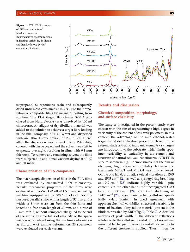
structure of natural cell wall constituents. ATR FT-IR
spectra shown in Fig. 1 demonstrates that the aim of
obtaining high chemical variability between the
treatments MFLC1 and MFLC4 was fully achieved.
On the one hand, aromatic skeletal vibrations at 1595
and 1505 cm-1 [24] as well as syringyl ring breathing
at 1242 cm-1 [25] indicate highly variable lignin
content. On the other hand, the unconjugated C=O
band at 1733 cm-1 [24] and C–O stretching at
1242 cm-1 [25] reveal variable hemicellulose, specif-
ically xylan, content. In good agreement with
apparent chemical variability, structural variability in
terms of fraction of crystalline material present in the
fibrils is revealed by XRD (Fig. 2; Table 2). A detailed
analysis of peak width of the different reflections
attributed to the cellulose I crystal did not reveal any
measurable change in terms of crystallite size due to
the different treatments applied. Thus it may be
Figure 1 ATR FT-IR spectra
of different variants of
fibrillated material.
Representative spectral regions
indicating variability in lignin
and hemicellulose (xylan)
content are indicated.
J Mater Sci (2017) 52:60–72 63
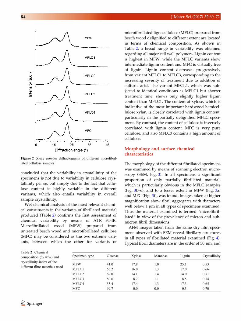
concluded that the variability in crystallinity of the
specimens is not due to variability in cellulose crys-
tallinity per se, but simply due to the fact that cellu-
lose content is highly variable in the different
variants, which also entails variability in overall
sample crystallinity.
Wet-chemical analysis of the most relevant chemi-
cal constituents in the variants of fibrillated material
produced (Table 2) confirms the first assessment of
chemical variability by means of ATR FT-IR.
Microfibrillated wood (MFW) prepared from
untreated beech wood and microfibrillated cellulose
(MFC) may be considered as the two extreme vari-
ants, between which the other for variants of
microfibrillated lignocellulose (MFLC) prepared from
beech wood delignified to different extent are located
in terms of chemical composition. As shown in
Table 2, a broad range in variability was obtained
regarding all major cell wall polymers. Lignin content
is highest in MFW, while the MFLC variants show
intermediate lignin content and MFC is virtually free
of lignin. Lignin content decreases progressively
from variant MFLC1 to MFLC3, corresponding to the
increasing severity of treatment due to addition of
sulfuric acid. The variant MFCL4, which was sub-
jected to identical conditions as MFLC1 but shorter
treatment time, shows only slightly higher lignin
content than MFLC1. The content of xylose, which is
indicative of the most important hardwood hemicel-
lulose xylan, is closely correlated with lignin content,
particularly in the partially delignified MFLC speci-
mens. By contrast, the content of cellulose is inversely
correlated with lignin content. MFC is very pure
cellulose, and also MFLC3 contains a high amount of
cellulose.
Morphology and surface chemicalcharacteristics
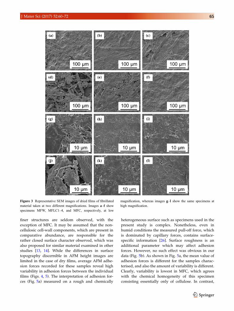
The morphology of the different fibrillated specimens
was examined by means of scanning electron micro-
scopy (SEM, Fig. 3). In all specimens a significant
proportion of only partially fibrillated material,
which is particularly obvious in the MFLC samples
(Fig. 3b–e), and to a lesser extent in MFW (Fig. 3a)
and MFC (Fig. 3f), was found. Images taken at higher
magnification show fibril aggregates with diameters
well below 1 lm in all types of specimens examined.
Thus the material examined is termed ‘‘microfibril-
lated’’ in view of the prevalence of micron and sub-
micron fibril dimensions.
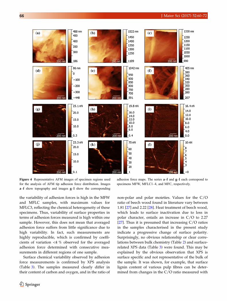
AFM images taken from the same dry film speci-
mens observed with SEM reveal fibrillary structures
in all types of fibrillated material examined (Fig. 4).
Typical fibril diameters are in the order of 50 nm, and
Table 2 Chemical
composition (% w/w) and
crystallinity index of the
different fibre materials used
Specimen type Glucose Xylose Mannose Lignin Crystallinity
MFW 41.0 17.8 1.0 25.1 0.53
MFLC1 56.2 16.0 1.3 17.0 0.66
MFLC2 62.0 14.1 1.4 14.0 0.71
MFLC3 80.6 8.7 1.1 8.5 0.74
MFLC4 53.4 17.4 1.3 17.3 0.65
MFC 99.7 0.0 0.0 0.3 0.70
Figure 2 X-ray powder diffractograms of different microfibril-
lated cellulose samples.
64 J Mater Sci (2017) 52:60–72
finer structures are seldom observed, with the
exception of MFC. It may be assumed that the non-
cellulosic cell-wall components, which are present in
comparative abundance, are responsible for the
rather closed surface character observed, which was
also proposed for similar material examined in other
studies [13, 14]. While the differences in surface
topography discernible in AFM height images are
limited in the case of dry films, average AFM adhe-
sion forces recorded for these samples reveal high
variability in adhesion forces between the individual
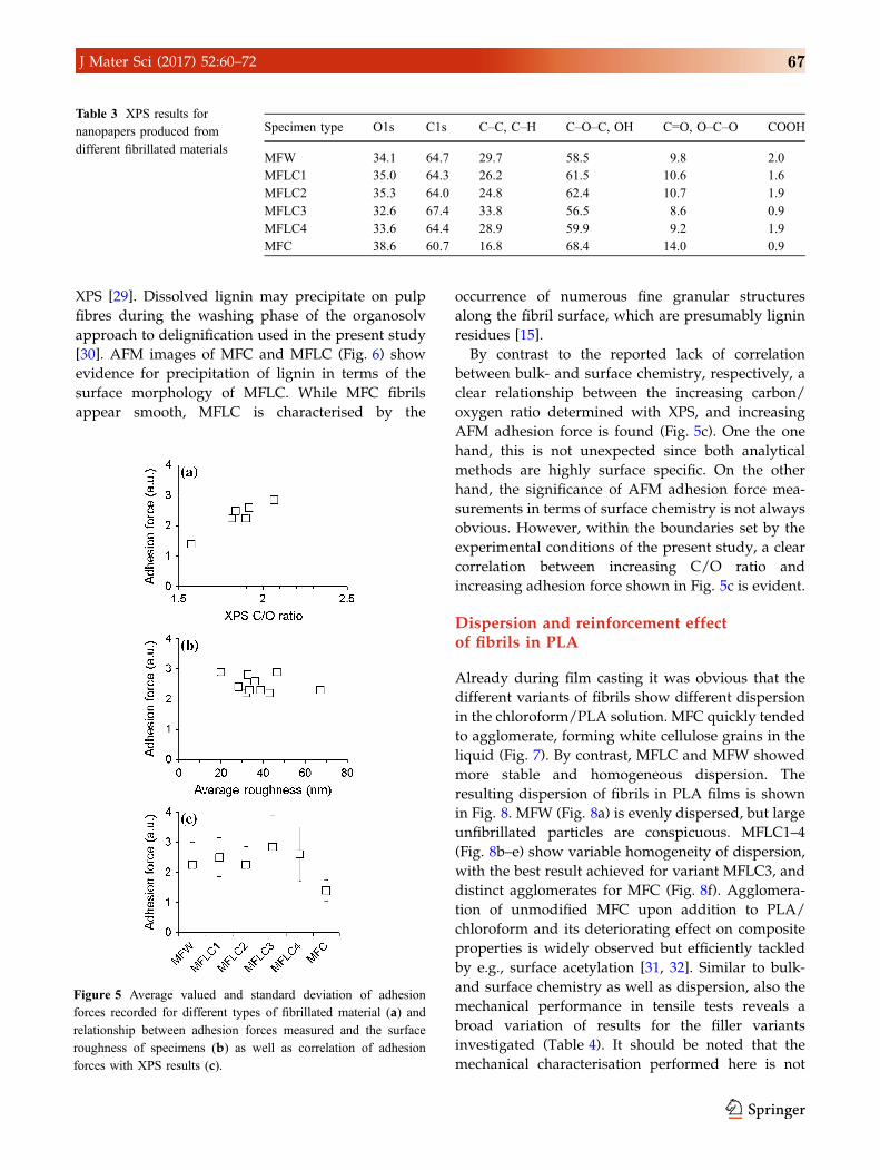
films (Figs. 4, 5). The interpretation of adhesion for-
ces (Fig. 5a) measured on a rough and chemically
heterogeneous surface such as specimens used in the
present study is complex. Nonetheless, even in
humid conditions the measured pull-off force, which
is dominated by capillary forces, contains surface-
specific information [26]. Surface roughness is an
additional parameter which may affect adhesion
forces. However, no such effect was obvious in our
data (Fig. 5b). As shown in Fig. 5a, the mean value of
adhesion forces is different for the samples charac-
terised, and also the amount of variability is different.
Clearly, variability is lowest in MFC, which agrees
with the chemical homogeneity of this specimen
consisting essentially only of cellulose. In contrast,
Figure 3 Representative SEM images of dried films of fibrillated
material taken at two different magnifications. Images a–f show
specimens MFW, MFLC1–4, and MFC, respectively, at low
magnification, whereas images g–l show the same specimens at
high magnification.
J Mater Sci (2017) 52:60–72 65
the variability of adhesion forces is high in the MFW
and MFLC samples, with maximum values for
MFLC3, reflecting the chemical heterogeneity of these
specimens. Thus, variability of surface properties in
terms of adhesion forces measured is high within one
sample. However, this does not mean that averaged
adhesion force suffers from little significance due to
high variability. In fact, such measurements are
highly reproducible, which is confirmed by coeffi-
cients of variation \4 % observed for the averaged
adhesion force determined with consecutive mea-
surements in different regions of one sample.
Surface chemical variability observed by adhesion
force measurements is confirmed by XPS analysis
(Table 3). The samples measured clearly differ in
their content of carbon and oxygen, and in the ratio of
non-polar and polar moieties. Values for the C/O
ratio of beech wood found in literature vary between
1.81 [27] and 2.22 [28]. Heat treatment of beech wood,
which leads to surface inactivation due to loss in
polar character, entails an increase in C/O to 2.27
[27]. Thus it is presumed that increasing C/O ratios
in the samples characterised in the present study
indicate a progressive change of surface polarity.
Surprisingly, no obvious relationship or clear corre-
lations between bulk chemistry (Table 2) and surface-
related XPS data (Table 3) were found. This may be
explained by the obvious observation that XPS is
surface specific and not representative of the bulk of
the sample. It was shown, for example, that surface
lignin content of various pulp fibres can be deter-
mined from changes in the C/O ratio measured with
Figure 4 Representative AFM images of specimen regions used
for the analysis of AFM tip adhesion force distribution. Images
a–f show topography and images g–l show the corresponding
adhesion force maps. The series a–f and g–l each correspond to
specimens MFW, MFLC1–4, and MFC, respectively.
66 J Mater Sci (2017) 52:60–72
XPS [29]. Dissolved lignin may precipitate on pulp
fibres during the washing phase of the organosolv
approach to delignification used in the present study
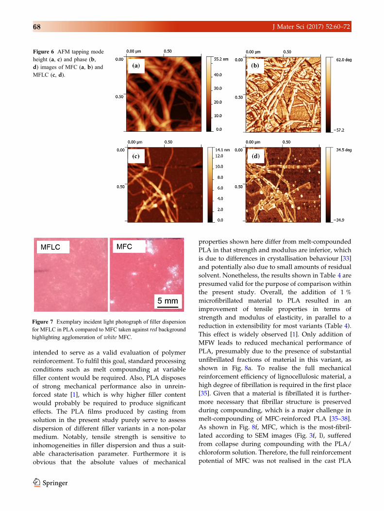
[30]. AFM images of MFC and MFLC (Fig. 6) show
evidence for precipitation of lignin in terms of the
surface morphology of MFLC. While MFC fibrils
appear smooth, MFLC is characterised by the
occurrence of numerous fine granular structures
along the fibril surface, which are presumably lignin
residues [15].
By contrast to the reported lack of correlation
between bulk- and surface chemistry, respectively, a
clear relationship between the increasing carbon/
oxygen ratio determined with XPS, and increasing
AFM adhesion force is found (Fig. 5c). One the one
hand, this is not unexpected since both analytical
methods are highly surface specific. On the other
hand, the significance of AFM adhesion force mea-
surements in terms of surface chemistry is not always
obvious. However, within the boundaries set by the
experimental conditions of the present study, a clear
correlation between increasing C/O ratio and
increasing adhesion force shown in Fig. 5c is evident.
Dispersion and reinforcement effectof fibrils in PLA
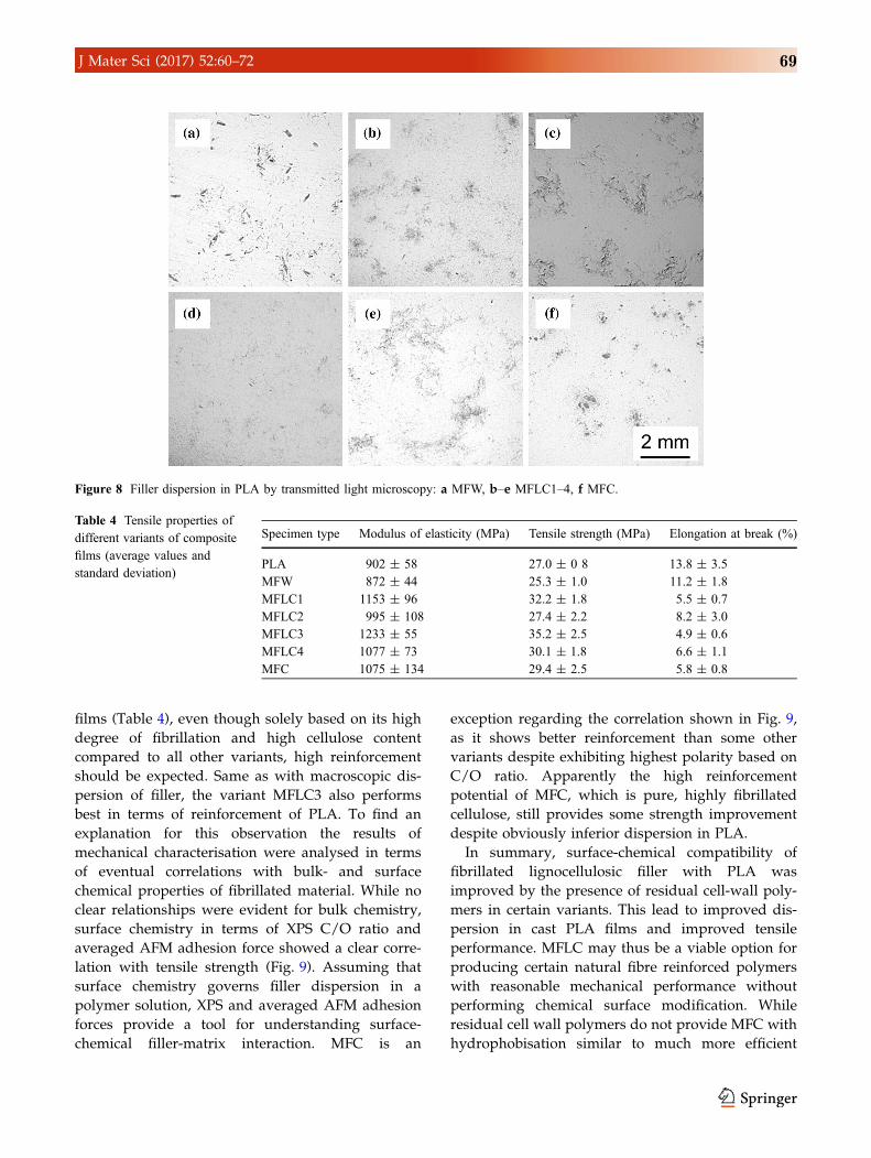
Already during film casting it was obvious that the
different variants of fibrils show different dispersion
in the chloroform/PLA solution. MFC quickly tended
to agglomerate, forming white cellulose grains in the
liquid (Fig. 7). By contrast, MFLC and MFW showed
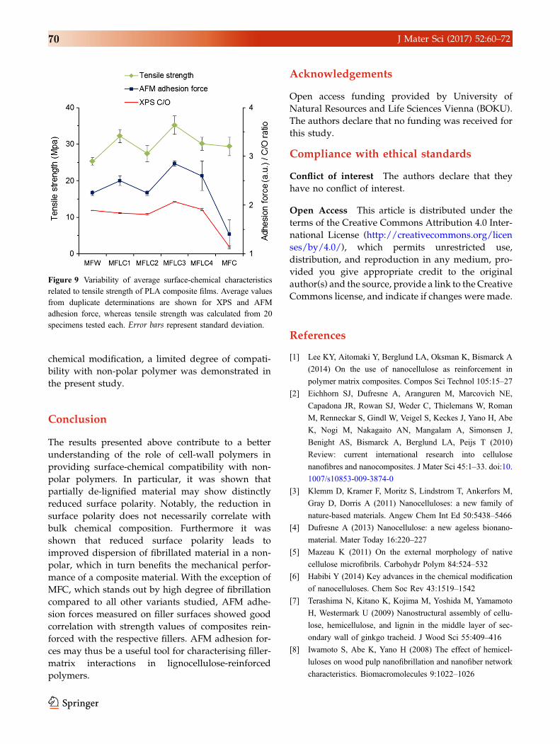
more stable and homogeneous dispersion. The
resulting dispersion of fibrils in PLA films is shown
in Fig. 8. MFW (Fig. 8a) is evenly dispersed, but large
unfibrillated particles are conspicuous. MFLC1–4
(Fig. 8b–e) show variable homogeneity of dispersion,
with the best result achieved for variant MFLC3, and
distinct agglomerates for MFC (Fig. 8f). Agglomera-
tion of unmodified MFC upon addition to PLA/
chloroform and its deteriorating effect on composite
properties is widely observed but efficiently tackled
by e.g., surface acetylation [31, 32]. Similar to bulk-
and surface chemistry as well as dispersion, also the
mechanical performance in tensile tests reveals a
broad variation of results for the filler variants
investigated (Table 4). It should be noted that the
mechanical characterisation performed here is not
Figure 5 Average valued and standard deviation of adhesion
forces recorded for different types of fibrillated material (a) and
relationship between adhesion forces measured and the surface
roughness of specimens (b) as well as correlation of adhesion
forces with XPS results (c).
Table 3 XPS results for
nanopapers produced from
different fibrillated materials
Specimen type O1s C1s C–C, C–H C–O–C, OH C=O, O–C–O COOH
MFW 34.1 64.7 29.7 58.5 9.8 2.0
MFLC1 35.0 64.3 26.2 61.5 10.6 1.6
MFLC2 35.3 64.0 24.8 62.4 10.7 1.9
MFLC3 32.6 67.4 33.8 56.5 8.6 0.9
MFLC4 33.6 64.4 28.9 59.9 9.2 1.9
MFC 38.6 60.7 16.8 68.4 14.0 0.9
J Mater Sci (2017) 52:60–72 67
intended to serve as a valid evaluation of polymer
reinforcement. To fulfil this goal, standard processing
conditions such as melt compounding at variable
filler content would be required. Also, PLA disposes
of strong mechanical performance also in unrein-
forced state [1], which is why higher filler content
would probably be required to produce significant
effects. The PLA films produced by casting from
solution in the present study purely serve to assess
dispersion of different filler variants in a non-polar
medium. Notably, tensile strength is sensitive to
inhomogeneities in filler dispersion and thus a suit-
able characterisation parameter. Furthermore it is
obvious that the absolute values of mechanical
properties shown here differ from melt-compounded
PLA in that strength and modulus are inferior, which
is due to differences in crystallisation behaviour [33]
and potentially also due to small amounts of residual
solvent. Nonetheless, the results shown in Table 4 are
presumed valid for the purpose of comparison within
the present study. Overall, the addition of 1 %
microfibrillated material to PLA resulted in an
improvement of tensile properties in terms of
strength and modulus of elasticity, in parallel to a
reduction in extensibility for most variants (Table 4).
This effect is widely observed [1]. Only addition of
MFW leads to reduced mechanical performance of
PLA, presumably due to the presence of substantial
unfibrillated fractions of material in this variant, as
shown in Fig. 8a. To realise the full mechanical
reinforcement efficiency of lignocellulosic material, a
high degree of fibrillation is required in the first place
[35]. Given that a material is fibrillated it is further-
more necessary that fibrillar structure is preserved
during compounding, which is a major challenge in
melt-compounding of MFC-reinforced PLA [35–38].
As shown in Fig. 8f, MFC, which is the most-fibril-
lated according to SEM images (Fig. 3f, l), suffered
from collapse during compounding with the PLA/
chloroform solution. Therefore, the full reinforcement
potential of MFC was not realised in the cast PLA
Figure 7 Exemplary incident light photograph of filler dispersion
for MFLC in PLA compared to MFC taken against red background
highlighting agglomeration of white MFC.
Figure 6 AFM tapping mode
height (a, c) and phase (b,
d) images of MFC (a, b) and
MFLC (c, d).
68 J Mater Sci (2017) 52:60–72
films (Table 4), even though solely based on its high
degree of fibrillation and high cellulose content
compared to all other variants, high reinforcement
should be expected. Same as with macroscopic dis-
persion of filler, the variant MFLC3 also performs
best in terms of reinforcement of PLA. To find an
explanation for this observation the results of
mechanical characterisation were analysed in terms
of eventual correlations with bulk- and surface
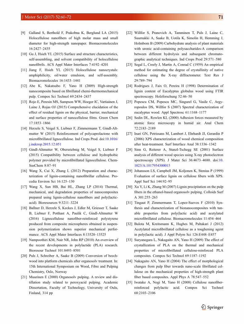
chemical properties of fibrillated material. While no
clear relationships were evident for bulk chemistry,
surface chemistry in terms of XPS C/O ratio and
averaged AFM adhesion force showed a clear corre-
lation with tensile strength (Fig. 9). Assuming that
surface chemistry governs filler dispersion in a
polymer solution, XPS and averaged AFM adhesion
forces provide a tool for understanding surface-
chemical filler-matrix interaction. MFC is an
exception regarding the correlation shown in Fig. 9,
as it shows better reinforcement than some other
variants despite exhibiting highest polarity based on
C/O ratio. Apparently the high reinforcement
potential of MFC, which is pure, highly fibrillated
cellulose, still provides some strength improvement
despite obviously inferior dispersion in PLA.
In summary, surface-chemical compatibility of
fibrillated lignocellulosic filler with PLA was
improved by the presence of residual cell-wall poly-
mers in certain variants. This lead to improved dis-
persion in cast PLA films and improved tensile
performance. MFLC may thus be a viable option for
producing certain natural fibre reinforced polymers
with reasonable mechanical performance without
performing chemical surface modification. While
residual cell wall polymers do not provide MFC with
hydrophobisation similar to much more efficient
Figure 8 Filler dispersion in PLA by transmitted light microscopy: a MFW, b–e MFLC1–4, f MFC.
Table 4 Tensile properties of
different variants of composite
films (average values and
standard deviation)
Specimen type Modulus of elasticity (MPa) Tensile strength (MPa) Elongation at break (%)
PLA 902 ± 58 27.0 ± 0 8 13.8 ± 3.5
MFW 872 ± 44 25.3 ± 1.0 11.2 ± 1.8
MFLC1 1153 ± 96 32.2 ± 1.8 5.5 ± 0.7
MFLC2 995 ± 108 27.4 ± 2.2 8.2 ± 3.0
MFLC3 1233 ± 55 35.2 ± 2.5 4.9 ± 0.6
MFLC4 1077 ± 73 30.1 ± 1.8 6.6 ± 1.1
MFC 1075 ± 134 29.4 ± 2.5 5.8 ± 0.8
J Mater Sci (2017) 52:60–72 69
chemical modification, a limited degree of compati-
bility with non-polar polymer was demonstrated in
the present study.
Conclusion
The results presented above contribute to a better
understanding of the role of cell-wall polymers in
providing surface-chemical compatibility with non-
polar polymers. In particular, it was shown that
partially de-lignified material may show distinctly
reduced surface polarity. Notably, the reduction in
surface polarity does not necessarily correlate with
bulk chemical composition. Furthermore it was
shown that reduced surface polarity leads to
improved dispersion of fibrillated material in a non-
polar, which in turn benefits the mechanical perfor-
mance of a composite material. With the exception of
MFC, which stands out by high degree of fibrillation
compared to all other variants studied, AFM adhe-
sion forces measured on filler surfaces showed good
correlation with strength values of composites rein-
forced with the respective fillers. AFM adhesion for-
ces may thus be a useful tool for characterising filler-
matrix interactions in lignocellulose-reinforced
polymers.
Acknowledgements
Open access funding provided by University of
Natural Resources and Life Sciences Vienna (BOKU).
The authors declare that no funding was received for
this study.
Compliance with ethical standards
Conflict of interest The authors declare that they
have no conflict of interest.
Open Access This article is distributed under the
terms of the Creative Commons Attribution 4.0 Inter-
national License (http://creativecommons.org/licen
ses/by/4.0/), which permits unrestricted use,
distribution, and reproduction in any medium, pro-
vided you give appropriate credit to the original
author(s) and the source, provide a link to the Creative
Commons license, and indicate if changes were made.
References
[1] Lee KY, Aitomaki Y, Berglund LA, Oksman K, Bismarck A
(2014) On the use of nanocellulose as reinforcement in
polymer matrix composites. Compos Sci Technol 105:15–27
[2] Eichhorn SJ, Dufresne A, Aranguren M, Marcovich NE,
Capadona JR, Rowan SJ, Weder C, Thielemans W, Roman
M, Renneckar S, Gindl W, Veigel S, Keckes J, Yano H, Abe
K, Nogi M, Nakagaito AN, Mangalam A, Simonsen J,
Benight AS, Bismarck A, Berglund LA, Peijs T (2010)
Review: current international research into cellulose
nanofibres and nanocomposites. J Mater Sci 45:1–33. doi:10.
1007/s10853-009-3874-0
[3] Klemm D, Kramer F, Moritz S, Lindstrom T, Ankerfors M,
Gray D, Dorris A (2011) Nanocelluloses: a new family of
nature-based materials. Angew Chem Int Ed 50:5438–5466
[4] Dufresne A (2013) Nanocellulose: a new ageless bionano-
material. Mater Today 16:220–227
[5] Mazeau K (2011) On the external morphology of native
cellulose microfibrils. Carbohydr Polym 84:524–532
[6] Habibi Y (2014) Key advances in the chemical modification
of nanocelluloses. Chem Soc Rev 43:1519–1542
[7] Terashima N, Kitano K, Kojima M, Yoshida M, Yamamoto
H, Westermark U (2009) Nanostructural assembly of cellu-
lose, hemicellulose, and lignin in the middle layer of sec-
ondary wall of ginkgo tracheid. J Wood Sci 55:409–416
[8] Iwamoto S, Abe K, Yano H (2008) The effect of hemicel-
luloses on wood pulp nanofibrillation and nanofiber network
characteristics. Biomacromolecules 9:1022–1026
Figure 9 Variability of average surface-chemical characteristics
related to tensile strength of PLA composite films. Average values
from duplicate determinations are shown for XPS and AFM
adhesion force, whereas tensile strength was calculated from 20
specimens tested each. Error bars represent standard deviation.
70 J Mater Sci (2017) 52:60–72
[9] Galland S, Berthold F, Prakobna K, Berglund LA (2015)
Holocellulose nanofibers of high molar mass and small
diameter for high-strength nanopaper. Biomacromolecules
16:2427–2435
[10] Gu J, Hsieh YL (2015) Surface and structure characteristics,
self-assembling, and solvent compatibility of holocellulose
nanofibrils. ACS Appl Mater Interfaces 7:4192–4201
[11] Jiang F, Hsieh YL (2015) Holocellulose nanocrystals:
amphiphilicity, oil/water emulsion, and self-assembly.
Biomacromolecules 16:1433–1441
[12] Abe K, Nakatsubo F, Yano H (2009) High-strength
nanocomposite based on fibrillated chemi-thermomechanical
pulp. Compos Sci Technol 69:2434–2437
[13] Rojo E, Peresin MS, Sampson WW, Hoeger IC, Vartiainen J,
Laine J, Rojas OJ (2015) Comprehensive elucidation of the
effect of residual lignin on the physical, barrier, mechanical
and surface properties of nanocellulose films. Green Chem
17:1853–1866
[14] Herzele S, Veigel S, Liebner F, Zimmermann T, Gindl-Alt-
mutter W (2015) Reinforcement of polycaprolactone with
microfibrillated lignocellulose. Ind Crops Prod. doi:10.1016/
j.indcrop.2015.12.051
[15] Gindl-Altmutter W, Obersriebnig M, Veigel S, Liebner F
(2015) Compatibility between cellulose and hydrophobic
polymer provided by microfibrillated lignocellulose. Chem-
SusChem 8:87–91
[16] Wang X, Cui X, Zhang L (2012) Preparation and charac-
terization of lignin-containing nanofibrillar cellulose. Pro-
cedia Environ Sci 16:125–130
[17] Wang X, Sun HB, Bai HL, Zhang LP (2014) Thermal,
mechanical, and degradation properties of nanocomposites
prepared using lignin-cellulose nanofibers and poly(lactic-
acid). Bioresources 9:3211–3224
[18] Ballner D, Herzele S, Keckes J, Edler M, Griesser T, Saake
B, Liebner F, Potthast A, Paulik C, Gindl-Altmutter W
(2016) Lignocellulose nanofiber-reinforced polystyrene
produced from composite microspheres obtained in suspen-
sion polymerization shows superior mechanical perfor-
mance. ACS Appl Mater Interfaces 8:13520–13525
[19] Nampoothiri KM, Nair NR, John RP (2010) An overview of
the recent developments in polylactide (PLA) research.
Bioresour Technol 101:8493–8501
[20] Puls J, Schreiber A, Saake B (2009) Conversion of beech-
wood into platform chemicals after organosolv treatment. In:
15th International Symposium on Wood, Fibre and Pulping
Chemistry, Oslo, Norway
[21] Muurinen E (2000) Organosolv pulping. A review and dis-
tillation study related to peroxyacid pulping. Academic
Dissertation, Faculty of Technology, University of Oulu,
Finland, 314 pp
[22] Willfor S, Pranovich A, Tamminen T, Puls J, Laine C,
Suurnakki A, Saake B, Uotila K, Simolin H, Hemming J,
Holmbom B (2009) Carbohydrate analysis of plant materials
with uronic acid-containing polysaccharides-A comparison
between different hydrolysis and subsequent chromato-
graphic analytical techniques. Ind Crops Prod 29:571–580
[23] Segal L, Creely J, Martin A, Conrad C (1959) An empirical
method for estimating the degree of crystallinity of native
cellulose using the X-ray diffractometer. Text Res J
29:789–794
[24] Rodrigues J, Faix O, Pereira H (1998) Determination of
lignin content of Eucalyptus globulus wood using FTIR
spectroscopy. Holzforschung 52:46–50
[25] Popescu CM, Popescu MC, Singurel G, Vasile C, Argy-
ropoulos DS, Willfor S (2007) Spectral characterization of
eucalyptus wood. Appl Spectrosc 61:1168–1177
[26] Sedin DL, Rowlen KL (2000) Adhesion forces measured by
atomic force microscopy in humid air. Anal Chem
72:2183–2189
[27] Inari GN, Petrissans M, Lambert J, Ehrhardt JJ, Gerardin P
(2006) XPS characterization of wood chemical composition
after heat-treatment. Surf Interface Anal 38:1336–1342
[28] Sinn G, Reiterer A, Stanzl-Tschegg SE (2001) Surface
analysis of different wood species using X-ray photoelectron
spectroscopy (XPS). J Mater Sci 36:4673–4680. doi:10.
1023/A:1017954300015
[29] Johansson LS, Campbell JM, Koljonen K, Stenius P (1999)
Evaluation of surface lignin on cellulose fibers with XPS.
Appl Surf Sci 144:92–95
[30] Xu Y, Li K, Zhang M (2007) Lignin precipitation on the pulp
fibers in the ethanol-based organosolv pulping. Colloids Surf
A 301:255–263
[31] Tingaut P, Zimmermann T, Lopez-Suevos F (2010) Syn-
thesis and characterization of bionanocomposites with tun-
able properties from poly(lactic acid) and acetylated
microfibrillated cellulose. Biomacromolecules 11:454–464
[32] Bulota M, Kreitsmann K, Hughes M, Paltakari J (2012)
Acetylated microfibrillated cellulose as a toughening agent
in poly(lactic acid). J Appl Polym Sci 126:E448–E457
[33] Suryanegara L, Nakagaito AN, Yano H (2009) The effect of
crystallization of PLA on the thermal and mechanical
properties of microfibrillated cellulose-reinforced PLA
composites. Compos Sci Technol 69:1187–1192
[34] Nakagaito AN, Yano H (2004) The effect of morphological
changes from pulp fiber towards nano-scale fibrillated cel-
lulose on the mechanical properties of high-strength plant
fiber based composites. Appl Phys A 78:547–552
[35] Iwatake A, Nogi M, Yano H (2008) Cellulose nanofiber-
reinforced polylactic acid. Compos Sci Technol
68:2103–2106
J Mater Sci (2017) 52:60–72 71
[36] Jonoobi M, Mathew AP, Abdi MM, Makinejad MD, Oks-
man K (2012) A comparison of modified and unmodified
cellulose nanofiber reinforced polylactic acid (PLA) pre-
pared by twin screw extrusion. J Polym Environ 20:991–997
[37] Jonoobi M, Harun J, Mathew AP, Oksman K (2010)
Mechanical properties of cellulose nanofiber (CNF)
reinforced polylactic acid (PLA) prepared by twin screw
extrusion. Compos Sci Technol 70:1742–1747
[38] Eyholzer C, Tingaut P, Zimmermann T, Oksman K (2012) Dis-
persion and reinforcing potential of carboxymethylated nanofibril-
lated cellulose powders modified with 1-hexanol in extruded
poly(lacticacid) (PLA)composites. JPolymEnviron20:1052–1062
72 J Mater Sci (2017) 52:60–72
Recommended