1
TITLE PAGE
Regulation of adrenergic, serotonin and dopamine receptors to inhibit diabetic retinopathy:
monotherapies versus combination therapies†
Timothy S. Kern#, Yunpeng Du, Jie Tang, Chieh Allen Lee, Haitao Liu, Alyssa Dreffs, Henri
Leinonen, David A. Antonetti, Krzysztof Palczewski
Center for Translational Vision Research, Gavin Herbert Eye Institute (T.S.K., Y.D., H.L., K.P.),
Department of Physiology and Biophysics (K.P.), and Department of Chemistry (K.P.),
University of California Irvine, Irvine, CA, USA. Veterans Administration Medical Center, Long
Beach Healthcare System, Research Service, Long Beach, CA, USA (T.S.K.) Department of
Ophthalmology, Case Western Reserve University, Cleveland, OH, USA (J.T., C.A.L.). Kellogg
Eye Center, Department of Ophthalmology and Visual Sciences, University of Michigan, Ann
Arbor MI, USA (A.D., D.A.A.).
This article has not been copyedited and formatted. The final version may differ from this version.Molecular Pharmacology Fast Forward. Published on August 15, 2021 as DOI: 10.1124/molpharm.121.000278
at ASPE
T Journals on M
arch 19, 2022m
olpharm.aspetjournals.org
Dow
nloaded from
2
RUNNING TITLE PAGE
Running Title: Treatment of diabetic retinopathy with GPCR ligands.
Total number of manuscript pages: 15
Figures: 8
Tables: 2
Words in Abstract: 238
Words in Introduction:633
Words in Discussion: 1380
#CORRESPONDENCE: Timothy S. Kern, Ph.D. Department of Ophthalmology, University of
California, Irvine, CA, and Research Service, VA Long Beach Healthcare System, Long Beach,
CA; [email protected];
ABBREVIATIONS:
bromocriptine (BRM), diabetic retinopathy (DR), dimethyl sulfoxide (DMSO), dose of minimal
inhibition of retinal oxidative stress (DMIox), doxazosin (DOX), endothelial cells (ECs), G-protein
coupled receptors (GPCRs), inducible isoform of nitric oxide synthase (iNOS), intercellular
adhesion molecule1 (ICAM1), metoprolol (MTP), phosphate buffered saline (PBS),
streptozotocin (STZ), tamsulosin (TAM). wildtype (WT)
This article has not been copyedited and formatted. The final version may differ from this version.Molecular Pharmacology Fast Forward. Published on August 15, 2021 as DOI: 10.1124/molpharm.121.000278
at ASPE
T Journals on M
arch 19, 2022m
olpharm.aspetjournals.org
Dow
nloaded from

3
Abstract
We compared monotherapies and combinations of therapies that regulate G-protein coupled
receptors (GPCR) with respect to their abilities to inhibit early stages of diabetic retinopathy
(DR) in streptozotocin-diabetic mice. Metoprolol (MTP; 0.04-1.0 mg/kgbw/day), bromocriptine
(BRM; 0.01-0.1 mg/kgbw/day), doxazosin (DOX; 0.01-1.0 mg/kgbw/day) or tamsulosin (TAM;
0.05-0.25 mg/kgbw/day) were injected individually daily for 2 months in dose-response studies
to assess their effects on the diabetes-induced increases in retinal superoxide and leukocyte-
mediated cytotoxicity against vascular endothelial cells (ECs), both of which abnormalities have
been implicated in the development of DR. Each of the individual drugs inhibited the diabetes-
induced increase in retinal superoxide at the higher concentrations tested, but the inhibition was
lost at lower doses. To determine if combination therapies had superior effects over individual
drugs, we intentionally selected for each drug a low dose that had little or no effect on the
diabetes-induced retinal superoxide for use separately or in combinations in 8-month studies of
retinal function, vascular permeability and capillary degeneration in diabetes. At the low doses
used, combinations of the drugs generally were more effective than individual drugs, but the low
dose MTP alone totally inhibited diabetes-induced reduction in a vision task, BRM or DOX alone
totally inhibited the vascular permeability defect, and DOX alone totally inhibited diabetes-
induced degeneration of retinal capillaries. Although low-dose MTP, BRM, DOX or TAM
individually had beneficial effects on some endpoints, combination of the therapies better
inhibited the spectrum of DR lesions evaluated.
Keywords: Systems pharmacology, diabetic retinopathy, GPCRs, Tamsulosin, Bromocriptine,
Metoprolol, Doxazosin, retina
This article has not been copyedited and formatted. The final version may differ from this version.Molecular Pharmacology Fast Forward. Published on August 15, 2021 as DOI: 10.1124/molpharm.121.000278
at ASPE
T Journals on M
arch 19, 2022m
olpharm.aspetjournals.org
Dow
nloaded from

4
Significance Statement:
The pathogenesis of early stages of diabetic retinopathy remain incompletely understood, but
multiple different cell-types are believed to be involved in the pathogenic process. We have
compared the effects of monotherapies to those of combinations of drugs that regulate G-
protein coupled receptor (GPCR) signaling pathways with respect to their relative abilities to
inhibit the development of early diabetic retinopathy.
This article has not been copyedited and formatted. The final version may differ from this version.Molecular Pharmacology Fast Forward. Published on August 15, 2021 as DOI: 10.1124/molpharm.121.000278
at ASPE
T Journals on M
arch 19, 2022m
olpharm.aspetjournals.org
Dow
nloaded from

5
Introduction
Despite many years of research, the pathogenesis of early stages of diabetic retinopathy (DR)
remains incompletely understood. Numerous postulates have been put forth to explain the early
development of the retinopathy, based on single molecular abnormalities (Kowluru and Mishra,
2015; Liu et al., 2019a; Sahajpal et al., 2019; Tonade et al., 2017) or abnormalities in single
retinal cell-types, including ECs, pericytes, ganglion cells, Müller cells, retinal pigment
epithelium, and retinal photoreceptor cells (Beltramo and Porta, 2013; Kern and Barber, 2008;
Kowluru and Mohammad, 2020; Liu et al., 2019a; Simo et al., 2018; Simo et al., 2010; Tonade
et al., 2017; Wang et al., 2015; Xu et al., 2011). Therapeutic approaches to inhibit the
retinopathy have focused primarily on targeted treatment of only single defects. Since
redundancy of biological pathways allows cells and tissues to compensate when several
aspects of that system are perturbed (Berger and Iyengar, 2009), however, therapy with a single
drug is less likely to normalize a perturbed system than is a combination of drugs that act on
different targets within the same or even unrelated networks.
G-protein coupled receptors (GPCRs) comprise a large diverse superfamily of membrane
proteins encoded by more than 800 genes (Fredriksson et al., 2003) that transduce a wide
variety of extracellular signals, including photons, ions, small organic molecules and proteins,
into intracellular second messengers through activation of one or more G proteins, including the
Gs, Gi and Gq subtypes (Marinissen and Gutkind, 2001). Often, agonism at Gs-coupled GPCRs
increases the activity of adenylyl cyclase, leading to increased generation of cAMP; whereas
agonism at Gi-coupled receptors has an opposite (inhibitory) effect. The Gq-coupled receptor-
signaling pathway differs from Gs- and Gi-coupled receptors in that it signals via the
phospholipase C pathway, eventually leading to Ca2+ release from the endoplasmic reticulum
and activation of NADPH oxidase (Brown, 2007; Inoue et al., 2008; Yamamori et al., 2000).
This article has not been copyedited and formatted. The final version may differ from this version.Molecular Pharmacology Fast Forward. Published on August 15, 2021 as DOI: 10.1124/molpharm.121.000278
at ASPE
T Journals on M
arch 19, 2022m
olpharm.aspetjournals.org
Dow
nloaded from

6
In diabetes, a number of GPCR-regulating drugs, (including doxazosin (DOX), prazosin,
guanabenz, lofexidine, and RO 04-6790) that regulate diverse signaling pathways downstream
of the different G proteins, all had the common effect of inhibiting the diabetes- or glucose-
induced increase in superoxide in retinal cells (Du et al., 2015). This is important because
oxidative stress has been implicated in the pathogenesis of DR (Berkowitz et al., 2009; Kanwar
et al., 2007; Kowluru et al., 2001). Moreover, daily administration of DOX (an α1-adrenergic (Gq)
-receptor antagonist (Hamilton et al., 1985)) for 8 months statistically significantly inhibited
molecular abnormalities and the capillary degeneration that is characteristic of DR (Du et al.,
2015).
Study of transcript abundance has shown that a subset of GPCRs are highly expressed in retina
(Chen et al., 2016b), and that targeting those abundant receptors with agonists or antagonists
resulted in protection from retinal degeneration secondary to bright light or Stargardt disease
(Chen et al., 2016b; Chen et al., 2013; Leinonen et al., 2019; Orban et al., 2017). Moreover,
simultaneous administration of a Gi-receptor agonist, bromocriptine (BRM), along with the Gs-
and Gq-receptor antagonists, metoprolol (MTP) and tamsulosin (TAM), respectively, was found
to exert a better-than-additive (synergistic) protection of photoreceptor cells in those disease
models (Chen et al., 2016b; Chen et al., 2013; Leinonen et al., 2019; Orban et al., 2017).
In the present report, we evaluated the effects of MET, BRM, TAM or DOX monotherapies on
molecular abnormalities that are believed to contribute to the development of DR, and then
investigated whether combinations of these drugs inhibited the development of long-term
pathology of DR (such as retinal permeability, capillary degeneration, and visual dysfunction)
better than did the monotherapies. These drugs were selected, not based on any known
biochemical or functional abnormalities downstream of those receptors in diabetes, but instead
This article has not been copyedited and formatted. The final version may differ from this version.Molecular Pharmacology Fast Forward. Published on August 15, 2021 as DOI: 10.1124/molpharm.121.000278
at ASPE
T Journals on M
arch 19, 2022m
olpharm.aspetjournals.org
Dow
nloaded from

7
based on the abundance of their receptors in the retina, and the previously reported beneficial
effects of these drugs in retinal degenerative diseases.
This article has not been copyedited and formatted. The final version may differ from this version.Molecular Pharmacology Fast Forward. Published on August 15, 2021 as DOI: 10.1124/molpharm.121.000278
at ASPE
T Journals on M
arch 19, 2022m
olpharm.aspetjournals.org
Dow
nloaded from

8
Materials and Methods
Animals and diabetes. All experiments followed the guidelines set forth by the Association for
Research in Vision and Ophthalmology Resolution on Treatment of Animals in Research and by
the institutional Animal Care and Use Committee. Insulin-deficient diabetes was induced in
randomly selected 2-month-old fasted male C57BI/6J mice (Jax #000664, Bar Harbor, MA) by
intraperitoneal injections of streptozotocin (STZ; 60 mg/kg body weight) on 5 consecutive days.
Insulin was given as needed (0-0.2 units every 2-3 days) to maintain body weight while allowing
chronic hyperglycemia, polyuria and hyperphagia. Female rodents are resistant to the
diabetogenic effects of STZ (Leiter, 1982; Rossini et al., 1978), so only males were used in the
present study. Blood glucose and HbA1c were measured as reported previously (Lee et al.,
2013; Tang et al., 2013b). Diabetic mice and age-matched non-diabetic control mice were
studied after 2 months of diabetes ord 8 months of diabetes (i.e., 4 and 10 months of age).
Two experiments were conducted. The first involved daily treatment of diabetic mice for 2
months (a duration of diabetes that results in molecular and some functional changes in the
retina, but retinal vascular abnormalities such as increased permeability and degeneration have
not yet developed); the second study involved drug treatment daily for 8 months of diabetes (a
duration of diabetes that results in a statistically significant increases in retinal vascular
permeability and degeneration compared to nondiabetic controls) (Feit-Leichman et al., 2005).
Drug studies. Dose-response curves were generated by injecting different doses of MET (0.04-
1.0 mg/kgbw/day), BRM (0.01-0.1 mg/kgbw/day), DOX (0.01-1.0 mg/kgbw/day) and TAM (0.05-
0.25 mg/kgbw/day) into diabetic mice daily IP for two months. These drugs were selected for
their ability to modulate signaling systems downstream of GPCRs expressed in high abundance
in the retina (Chen et al., 2013) and for previously reported beneficial effects on the retina in
diabetes and retinal degeneration (Chen et al., 2016b; Chen et al., 2013; Du et al., 2015;
This article has not been copyedited and formatted. The final version may differ from this version.Molecular Pharmacology Fast Forward. Published on August 15, 2021 as DOI: 10.1124/molpharm.121.000278
at ASPE
T Journals on M
arch 19, 2022m
olpharm.aspetjournals.org
Dow
nloaded from

9
Leinonen et al., 2019; Orban et al., 2017). These 2-month dose-response studies were
conducted to evaluate the effects of the individual drugs on diabetes-induced retinal oxidative
stress and other molecular abnormalities that have been implicated in development of the
retinopathy. From these studies, we identified also a low dose of each drug that individually had
little or no effect on the retinal oxidative stress (labelled DMIox), and used that dose of each drug
in 8-month studies to determine if combinations of the drugs more effectively inhibited the early
stages of DR than did monotherapies. We conducted the long-term study using 11 experimental
groups as summarized in Table 1. All therapeutics were administered at identified doses daily
by intraperitoneal injection in DMSO. An initial 2-month study with diabetic mice treated with and
without DMSO showed no statistically significant difference on retinal superoxide, so diabetic
control animals were untreated in subsequent studies. Drugs were obtained from Sigma-Aldrich
(St. Louis, MO).
Retinal superoxide. Freshly isolated retinas were incubated in 200 µL of Krebs-Hepes buffer, pH
7.2, with 5 mM or 25 mM glucose for 5 min at 37°C in 5% CO2. Luminescence indicating the
presence of superoxide was measured 5 min after addition of lucigenin to 0.54 mM (Du et al.,
2003; Du et al., 2010; Gubitosi-Klug et al., 2008; Kern et al., 2010; Kern et al., 2007; Veenstra et
al., 2015). Luminescence intensity is reported in arbitrary units per mg protein.
Immunoblots. Retinal homogenates were separated by SDS-PAGE and incubated with either
anti-intercellular adhesion molecule-1 (α-ICAM1; 1:2000 dilution; #MAB796; R&D Systems,
Minneapolis, MN) or the anti-inducible isoform of nitric oxide synthase (α-iNOS; 1:1000 dilution;
#SC-7271; Santa Cruz Biotechnology, Inc., Santa Cruz, CA). Protein levels were quantified
relative to ß-actin loading controls (1:3000 dilution; #AB8226; Abcam, Inc., Cambridge, MA) in
the same samples.
This article has not been copyedited and formatted. The final version may differ from this version.Molecular Pharmacology Fast Forward. Published on August 15, 2021 as DOI: 10.1124/molpharm.121.000278
at ASPE
T Journals on M
arch 19, 2022m
olpharm.aspetjournals.org
Dow
nloaded from

10
Optokinetic assessment of visual function. The maximum spatial frequency that resulted in head
tracking was determined as the spatial frequency threshold. After 2 months of diabetes, contrast
sensitivity was measured at a single spatial frequency (0.064 c/d) with the Virtual Optomotor
system, as previously described (Lee et al., 2013; Liu et al., 2015b; Prusky et al., 2004). The
grader was masked with respect to the animals’ experimental group. Although non-diabetic mice
could be differentiated from diabetic animals based on body weight, investigators could not
discern drug treatment group identity.
Leukocyte-mediated cytotoxicity of ECs (Li et al., 2012; Talahalli et al., 2013; Tang et al., 2013a;
Tang et al., 2013b; Tian et al., 2013; Veenstra et al., 2013). Immortalized retinal ECs (Su et al.,
2003) were grown in control medium (DMEM with 5 mM glucose) containing 10% serum. The
serum concentration was reduced to 2% just before cells were placed either in normal glucose
(5 mM) or high glucose (30 mM). Media was changed every other day for 3 days. When cells
reached 80% confluence (~300,000 cells), freshly isolated leukocytes from blood (100,000 cells)
were added and incubated for additional 6 h, after which cells were collected and washed with
PBS. Cells were stained with an antibody against CD144 (1:50 dilution, BD Biosciences
Pharmingen, San Diego, CA) to identify ECs; and the viability of the ECs was identified by flow
cytometry, based on 7-AAD (7-aminoactinomycin D; BD, San Diego, CA) staining. EC death
was expressed as the percentage of ECs that stained positively with dye. Approximately 10,000
cells were counted in each sample. Experiments were repeated twice with similar results each
time.
Capillary degeneration. Diabetes-induced vascular pathology was measured in retinas from
animals diabetic for 8 months and their age-matched controls, as described previously (Li et al.,
2011; Li et al., 2012; Veenstra et al., 2013). Briefly, formalin-fixed retinas were digested with 40
U/mL elastase (Calbiochem, San Diego, CA) (Veenstra et al., 2015). When totally freed of
This article has not been copyedited and formatted. The final version may differ from this version.Molecular Pharmacology Fast Forward. Published on August 15, 2021 as DOI: 10.1124/molpharm.121.000278
at ASPE
T Journals on M
arch 19, 2022m
olpharm.aspetjournals.org
Dow
nloaded from

11
neural cells, the isolated retinal vasculature was laid out on a glass microscope slide, dried, and
stained with hematoxylin and PAS (periodic acid-Schiff). Degenerated (acellular) capillaries
were quantified in a masked manner in 6-7 field areas corresponding to the mid-retina.
Permeability. Retinal permeability was measured in the other eye from animals in the long-term
studies, using a fluorescently labeled tracer as described previously (Antonetti et al., 1998; Du
et al., 2015; Liu et al., 2015a; Liu et al., 2019b). Briefly, sterile fluorescein isothiocyanate (FITC)-
BSA (bovine serum albumin; 50 µg/µl; Sigma-Aldrich A9771) in phosphate buffered saline
(NaCl, 0.138 M; KCl, 0.0027 M; pH 7.4) was injected into the tail vein of mice at 100 µg/g bw.
The dye circulated for 20 min before blood samples were collected and eyes were enucleated.
One eye was fixed in ice cold 4% paraformaldehyde, infused with sucrose, and then frozen in
O.C.T. in isopentane on dry ice. Retinal cryosections were imaged by fluorescence microscopy.
Two sections per eye were imaged equi-distant on either side of the optic disc in the inner and
outer plexiform layers to generate an average image pixel density in the neural retina, exclusive
of any vessels, from a total of 4 images. Relative average fluorescence values were normalized
to the relative plasma fluorescence for final determinations of retinal dye accumulation.
Statistical analyses. Data are expressed as means ± SD. All statistical analyses were performed
by ANOVA, followed by Fischer's LSD (least significant difference) post-hoc test (StatView, SAS
Institute, Inc.). Inter-group comparisons in the ANOVA post-hoc tests were made between all
groups with appropriate correction for multiple comparisons; but results are reported here only
for the comparisons of D control versus N control and D control versus drug-treated D, except
as noted in the text. Values of P<0.05 (two-tailed) are considered statistically significant.
Individuals performing assays were masked as to which group the animals were in, with the
exception that nondiabetic animals could be differentiated from diabetics based on body weight
during visual function tests (but it was impossible to visibly differentiate among the various
This article has not been copyedited and formatted. The final version may differ from this version.Molecular Pharmacology Fast Forward. Published on August 15, 2021 as DOI: 10.1124/molpharm.121.000278
at ASPE
T Journals on M
arch 19, 2022m
olpharm.aspetjournals.org
Dow
nloaded from

12
diabetic groups). The 2-month studies served both a hypothesis-testing role (GPCR-modifying
drugs inhibit diabetes-induced increase in retinal superoxide) stated prior to the onset of the
study, and an exploratory role (determine the DMIox for each drug). The 8-month study was
hypothesis-testing (combinations of therapies that modify GPCR signaling better inhibit DR than
do monotherapies). All comparisons tested a pre-specified statistical null hypothesis that there
was no statistically significant difference between the diabetic control group and diabetics
treated with GPCR-modifying drugs. Use of the term “significant” in this manuscript always
refers to statistical significance. Group sizes needed at the end of the experiment (n=6 per
group) were estimated by sample size analysis based on previously reported differences
between diabetic and nondiabetic controls with respect to retinal capillary degeneration, and
initial group sizes were then increased in anticipation of some mortality or reversal of diabetes.
This article has not been copyedited and formatted. The final version may differ from this version.Molecular Pharmacology Fast Forward. Published on August 15, 2021 as DOI: 10.1124/molpharm.121.000278
at ASPE
T Journals on M
arch 19, 2022m
olpharm.aspetjournals.org
Dow
nloaded from

13
Results
Diabetes. As intended, all diabetic groups had HbA1c and blood glucose concentrations that
were greater than those found in age-matched, non-diabetic controls. None of the drug
treatments had an effect on glycemia. Average body weights, non-fasting glucose levels, and
HbA1c concentrations for the animal groups in the 8-month experiment are summarized in
Table 1. Mortality during the 8-month experiment also is summarized in the Table. Clinical data
from a representative 2-month study is shown in Supplemental data; Table S1, and was similar
to that in the 8-month study.
Two-month diabetes. Diabetes of 2-month duration significantly increased retinal superoxide
(P<0.001; Fig 1a). When MTP, BRM, DOX or TAM were administered individually to diabetic
animals for 2 months, the higher dosages tested of each drug significantly (P<0.01) inhibited the
diabetes-induced increase in retinal superoxide (Fig1b), providing evidence that
pharmacologically targeting GPCRs protects the retina against oxidative stress in diabetes. In
order to minimize the likelihood of undesirable side effects of these drugs and to determine if
combinations of the drugs resulted in additive or greater-than-additive inhibition of lesions of the
retinopathy, we sought to identify a lower dose of each monotherapy at which there was little or
no inhibition of the diabetes-induced increase in retinal superoxide (defined as the Dose of
Minimal Inhibition of retinal oxidative stress; DMIox). Based on the reported equation to convert
the dose of each drug used in humans to a comparable dose for mice (Food and Drug
Administration, 2005), the dosages used in the present study are below clinically used daily
dosages, with the exception of TAM (see Supplement; Table S2). Using the data in Fig 1b, an
estimated ED50 for inhibition of the diabetes-induced increase in retinal superoxide was
calculated for each of the individual drugs, and is summarized in Table 2.
This article has not been copyedited and formatted. The final version may differ from this version.Molecular Pharmacology Fast Forward. Published on August 15, 2021 as DOI: 10.1124/molpharm.121.000278
at ASPE
T Journals on M
arch 19, 2022m
olpharm.aspetjournals.org
Dow
nloaded from

14
Whereas administration of any of the individual drugs for 2 months at their DMIox led to little or
no inhibition of the diabetes-induced retinal oxidative stress, co-administration of MTP, BRM,
DOX, or TAM with any other of the drugs (also at their DMIox) statistically significantly inhibited
the retinal oxidative stress (Fig 2). These data demonstrate what appears to be a greater than
additive effect of the drugs against retinal oxidative stress in diabetes, and provide evidence that
signaling pathways regulated by adrenergic and serotonin and/or dopamine receptors contribute
to diabetes-induced oxidative stress. Strikingly, however, the benefit of therapy combinations
was less evident when BRM was substituted with a selective D2-like receptor agonist,
pramipexole (Fig. 3).
Diabetes of 2-month duration also significantly increased the expression of pro-inflammatory
proteins, iNOS and ICAM1 (both P<0.05; Fig 4 and Supplement; Fig S1). Both of these pro-
inflammatory proteins have been implicated in the pathogenesis of vascular lesions in early DR
(Joussen et al., 2004; Zheng et al., 2007). Individual drugs administered daily for 2 months at
their DMIox did not inhibit diabetes-induced increases in iNOS or ICAM1, but many of the drug
combinations did so, despite being at their DMIox (Fig. 4). In response to the drug combinations,
the diabetes-induced increase in retinal iNOS was inhibited almost totally, and the increase in
ICAM1 was inhibited by about half.
We previously implicated leukocytes as contributing to the development of vascular lesions in
DR (Li et al., 2012; Talahalli et al., 2013; Tian et al., 2013; Veenstra et al., 2013). and the killing
of retinal ECs by leukocytes from either diabetic patients (Tian et al., 2013) or mice (Li et al.,
2012) has been demonstrated ex vivo. Because systemically delivered drugs do not affect only
retinal cells, we assessed the effects of MTP, BRM, DOX, and TAM individually and in
combinations on the leukocyte-mediated cytotoxicity against retinal ECs using leukocytes
isolated from the blood of animals treated with drugs for 2 months. Daily administration of either
This article has not been copyedited and formatted. The final version may differ from this version.Molecular Pharmacology Fast Forward. Published on August 15, 2021 as DOI: 10.1124/molpharm.121.000278
at ASPE
T Journals on M
arch 19, 2022m
olpharm.aspetjournals.org
Dow
nloaded from

15
MTP or BRM as monotherapies at their DMIox to diabetic animals did not statistically significantly
inhibit leukocyte-mediated cytotoxicity against retinal ECs, but their combinations with other
drugs did (Fig. 5). In contrast, DOX statistically significantly inhibited the diabetes-induced
leukocyte-mediated EC death as a monotherapy and in any of the combinations tested (Fig 5).
Contrast sensitivity and spatial frequency threshold are psychophysical measures that assess
the function of retina-to-mid-brain (superior colliculus) visual pathways. Diabetes for a 2-3-
month duration in wild-type C57Bl/6J mice (4-5 months age) caused a significant decrease in
both parameters of visual function compared to nondiabetic controls (Fig 6; both P<0.05). Daily
administration of MTP alone or BRM+DOX or MTP+BRM+DOX all statistically significantly
inhibited the diabetes-induced decrease in spatial frequency threshold. None of the therapies or
combinations improved the diabetes-induced reduction in contrast sensitivity measurement at
the single spatial frequency evaluated.
8-month diabetes. Based on the drug studies at 2 months of diabetes (described above), we
initiated long-term studies using MTP, BRM, DOX, and TAM at their DMIox drug dosages. The
drugs were administered individually or in combinations daily for 8 months to determine the
effects of the drugs on clinically meaningful endpoints of DR.
As expected, diabetes of 8-month duration caused a statistically significant increase in the
number of degenerated retinal capillaries (Fig 7). Neither MTP nor BRM at their DMIox inhibited
degeneration of retinal capillaries in diabetic mice over the 8-month period, but the capillary
degeneration was statistically significantly inhibited by either DOX or TAM monotherapies at
their DMIox. The triple combination therapies of either MTP+BRM+DOX or MTP+BRM+TAM also
statistically significantly inhibited the diabetes-induced degeneration of retinal capillaries.
This article has not been copyedited and formatted. The final version may differ from this version.Molecular Pharmacology Fast Forward. Published on August 15, 2021 as DOI: 10.1124/molpharm.121.000278
at ASPE
T Journals on M
arch 19, 2022m
olpharm.aspetjournals.org
Dow
nloaded from

16
Diabetes of 8-month duration significantly increased also leakage of FITC-BSA into regions of
the neural retina occupied by the vascular beds (inner plexiform layer (IPL) and outer plexiform
layer (OPL)) (both P<0.001; Fig 8), and this permeability defect responded somewhat differently
to therapies than did the capillary degeneration. Neither MTP nor TAM had any effect on
leakage of FITC-albumin into the IPL or OPL, whereas BRM and DOX monotherapies at their
DMIox very significantly inhibited the leakage defects (both P<0.001). Because of the strong
therapeutic effect of BRM and DOX monotherapies in this assay, none of the drug combinations
showed a better inhibition of the permeability defect than those monotherapies. Nonetheless the
permeability defects were almost totally inhibited in diabetic mice treated with MET+BRM+DOX,
and permeability in those animals was indistinguishable from N controls. BRM monotherapy
was significantly more effective at inhibiting the permeability defect in both retinal layers than
MET+BRM+TAM (P<0.0002). Comparing the two triple therapies, MET+BRM+DOX was
significantly more effective at inhibiting the permeability defect than was MET+BRM+TAM
(p<0.01 in both retinal layers). In contrast to the beneficial effect of DOX on permeability, TAM
(the other Gq-linked inhibitor tested) had no such beneficial effect on retinal permeability in vivo
unless combined with others of the tested drugs (Fig. 8).
This article has not been copyedited and formatted. The final version may differ from this version.Molecular Pharmacology Fast Forward. Published on August 15, 2021 as DOI: 10.1124/molpharm.121.000278
at ASPE
T Journals on M
arch 19, 2022m
olpharm.aspetjournals.org
Dow
nloaded from

17
Discussion
In the present study, we sought to investigate whether the use of multiple drugs that act on
complimentary GPCR signaling systems might result in better inhibition of DR than can be
achieved using a single drug therapy. When used alone, MET, BRM, DOX, or TAM significantly
inhibited several diabetes-induced molecular abnormalities in the retina at the high doses
tested, and were less effective at lower doses.
Using a kinetic assay to study the effect of these drugs on G protein activation by GPCRs, Chen
et al (Chen et al., 2016b) reported that BRM activates dopamine receptors D4R and D2R, as
well as inhibiting the α1A-adrenergic receptor, ADRA1A. DOX was found to be a full antagonist
for ADRA1A. TAM demonstrated complex activity on the four receptors, and MTP showed no
agonistic or antagonistic effects on any of the four GPCRs tested. Knowledge about which
receptors are being influenced by the drugs used, and the biological effects of those drugs, is
expected to provide insight into the molecular mechanism(s) by which pathology of DR develops
and might be inhibited. The most likely explanation for the powerful effect of BRM is as a
dopamine R2 agonist. Dopamine is known to counter VEGF-induced permeability (Bhattacharya
et al., 2008). The beneficial actions of DOX on diabetes-induced degeneration of retinal
capillaries were reproduced also by pharmacologic inhibition of NADPH oxidase (Du et al.,
2015), thus suggesting that superoxide generation or calcium release following hyperglycemia-
mediated activation of ADRA1A are involved in the capillary degeneration.
Since oxidative stress has been implicated in the pathogenesis of at least the vascular lesions
of DR, it was surprising that monotherapies of DOX or BRM at doses that did not inhibit retinal
superoxide (DMIox) nevertheless caused a significant inhibition of retinal vascular permeability in
diabetes. Likewise, degeneration of retinal capillaries was strongly inhibited by DOX
monotherapy at a dose that did not inhibit retinal superoxide. Thus, the effect of a drug on
This article has not been copyedited and formatted. The final version may differ from this version.Molecular Pharmacology Fast Forward. Published on August 15, 2021 as DOI: 10.1124/molpharm.121.000278
at ASPE
T Journals on M
arch 19, 2022m
olpharm.aspetjournals.org
Dow
nloaded from
18
retinal oxidative stress does not necessarily predict its effect on vascular or neural lesions of the
retinopathy.
Individual drugs inhibited some lesions of the retinopathy, but did not correct all of the retinal
abnormalities in diabetes. BRM inhibited the retinal permeability defect in diabetes at the low
dose tested, but did not significantly inhibit the retinal capillary degeneration or visual
dysfunction. MTP alone inhibited the loss of visual acuity as estimated from spatial frequency
threshold, but had no significant effect on either retinal capillary permeability or degeneration in
diabetes. DOX inhibited the defects in capillary permeability and degeneration, but showed no
similar beneficial effect on visual function.
Previous studies showed that simultaneous administration of low doses of MTP, TAM, and BRM
resulted in a synergistic (super-additive) inhibition of retinal photoreceptor cell degeneration
(Chen et al., 2016b; Chen et al., 2013; Leinonen et al., 2019; Orban et al., 2017). Data in the
present study indicates that this combinatorial approach has beneficial effects in the retina also
in diabetes. Combinations of MTP, BRM, and DOX or TAM at doses that individually had little or
no beneficial effect on the diabetes-induced increase in retinal superoxide levels resulted in a
significant inhibition of the retinal oxidative stress, and showed a similar, although less strong,
beneficial effect on expression of pro-inflammatory proteins in the retina. Moreover,
combinations of MTP+BRM+DOX or MTP+BRM+TAM at their DMIox mitigated a greater number
of the long-term complications of DR than did individual drugs, illustrating the superiority of the
combinatorial approach when drugs are applied at low dosages. We interpret that combinations
of the drugs tested apparently led to stimulation of Gi/o signaling by activating the dopamine
receptors D2R and D4R, as well as inhibition of Gs and Gq signaling by antagonizing D1R and
the α1A-adrenergic receptor ADRA1A, respectively. Cross-talk is known to occur among
signaling pathways, so manipulation of a particular GPCR not only affects the signaling
This article has not been copyedited and formatted. The final version may differ from this version.Molecular Pharmacology Fast Forward. Published on August 15, 2021 as DOI: 10.1124/molpharm.121.000278
at ASPE
T Journals on M
arch 19, 2022m
olpharm.aspetjournals.org
Dow
nloaded from

19
pathways regulated by that GPCR, but also can bring about changes in other signaling
pathways. Manipulation of these receptors using the same drugs also had broad effects on the
retinal transcriptome in retinal degeneration (Chen et al., 2016b), showing that the combined
lower-dose GPCR-targeted treatments better preserved patterns of retinal gene expression that
were similar to those of the normal retina than did higher-dose monotherapies.
Receptors affected by these drugs are found in the retina (Chen et al., 2013; Ruan et al., 2020),
but it cannot be assumed that the beneficial effects of the drugs were mediated via drug effects
in those retinal cells. The drugs used by us act also on other cell-types not residing in the eye
(including leukocytes), resulting in anti-inflammatory and anti-oxidant effects (Chen et al.,
2016b; Dunzendorfer and Wiedermann, 2000; García-Prieto et al., 2017; Grisanti et al., 2011;
Heijnen et al., 1996; King, 2017; Kintscher et al., 2001; Takahashi et al., 2005). Leukocytes
have been implicated in the capillary degeneration and altered permeability in diabetes (Li et al.,
2012; Liu et al., 2019a; Talahalli et al., 2013; Tian et al., 2013; Veenstra et al., 2013) perhaps
via leukostasis, leukocyte-mediated cytotoxicity against the ECs, and/or release of bioactive
compounds from leukocytes circulating through the retinal vasculature. The present results
show that DOX monotherapy and the triple combinations of drugs statistically significantly
inhibited both the leukocyte-mediated cytotoxicity against retinal ECs as well as the retinal
capillary degeneration in diabetes.
The term “systems pharmacology” describes a therapeutic approach whereby multiple drugs
modify different signaling pathways that act additively to achieve a positive therapeutic effect
(Berger and Iyengar, 2009; Chen et al., 2016a; Chen et al., 2016b; Chen et al., 2013; Hansen et
al., 2011; Orban et al., 2017). Importantly, this unbiased approach can lead to beneficial effects
even when specific knowledge is lacking about the molecular mechanisms of the abnormalities
in cells or tissues. Another potential advantage of the systems pharmacology approach is that
This article has not been copyedited and formatted. The final version may differ from this version.Molecular Pharmacology Fast Forward. Published on August 15, 2021 as DOI: 10.1124/molpharm.121.000278
at ASPE
T Journals on M
arch 19, 2022m
olpharm.aspetjournals.org
Dow
nloaded from

20
combinations of certain drugs have been reported to have synergistic beneficial therapeutic
effects at low drug doses (Chen et al., 2016b; Chen et al., 2013; Leinonen et al., 2019; Orban et
al., 2018), thus minimizing potential dose-dependent side effects of the drugs.
Differences between DOX and TAM with respect to the diabetes-induced increase in
permeability, and between pramipexole and BRM with respect to retinal oxidative stress in the
present results indicate that we cannot assume that all β- and α-adrenergic antagonists (Gs- and
Gq-coupled, respectively) or D2-like dopamine-receptor agonists (Gi-coupled) individually or in
combination would be effective therapeutic approaches to inhibit DR. It seems likely that BRM´s
diverse receptor actions contribute to the beneficial effects of the drug on DR. BRM shows high
potency also for Gi-coupled 5-HT1A and 5-HT1D receptors, and targeting these receptors with
agonists have been shown to decrease oxidative stress and damage in retinal pigment
epithelium and retina injury models (Collier et al., 2011; Coyner et al., 2016; Thampi et al.,
2012).
A weakness of the current study is that we did not demonstrate that the lower concentrations of
drugs actually reduced the risk of undesirable side-effects (such as tachycardia and
hypertension). Nevertheless, administration of drugs at the lowest doses that show desirable
effects is an accepted tenet of pharmacology.
Early DR is not a single abnormality, but is comprised of a spectrum of lesions that can occur
individually in other conditions. For example, retinal vascular permeability is increased also in
ischemia-reperfusion injury and age-related macular degeneration (Abcouwer et al., 2013;
Erickson et al., 2007), capillary degeneration occurs also in silicosis and aging (Kuwabara et al.,
1961; Soliman et al., 2015), retinal microaneurysms develop also in macroglobulinemia
(Vasileiou et al., 2020; Xu et al., 2015), and dysfunction of retinal neurons develops in a variety
This article has not been copyedited and formatted. The final version may differ from this version.Molecular Pharmacology Fast Forward. Published on August 15, 2021 as DOI: 10.1124/molpharm.121.000278
at ASPE
T Journals on M
arch 19, 2022m
olpharm.aspetjournals.org
Dow
nloaded from
21
of conditions, suggesting that these lesions do not necessarily develop by identical mechanisms
and thus might not respond equally to the same therapies. The monotherapies tested had
beneficial effects on some, but not all, components of the retinopathy, so a combination of the
therapies seems best suited for having beneficial effects on the broadest spectrum of lesions.
Whether or not higher concentrations of the individual drugs tested here might inhibit the full
spectrum of lesions that characterize DR is not known, but higher drug doses are expected to
increase the likelihood of side-effects. We conclude that there is value in therapy development
that focuses on ameliorating dysfunctions of multiple pathways and networks in DR rather than
targeting single abnormalities.
This article has not been copyedited and formatted. The final version may differ from this version.Molecular Pharmacology Fast Forward. Published on August 15, 2021 as DOI: 10.1124/molpharm.121.000278
at ASPE
T Journals on M
arch 19, 2022m
olpharm.aspetjournals.org
Dow
nloaded from

22
Acknowledgements
We thank Dr. N. Sheibani, PhD (University of Wisconsin-Madison) for the immortalized mouse
retinal endothelial cells (Su et al., 2003), Kathryn Zongolowicz and Heather Butler from the
Case Western Reserve University NEI Core grant for the evaluation of visual function, and
Aicha Saadane, PhD, Emma M. Lessieur, PhD, and Jianying Kiser, PhD for experimental and
animal support.
Funding Footnote
†This work was supported by the National Institutes of Health National Eye Institute and
[EY022938, R24 EY024864, EY012021, EY011373 and EY007003]; the National Institutes of
Health National Institute of Diabetes and Digestive and Kidney Diseases [DK020572]; and the
Department of Veterans Affairs [BX003604].
Dr. Kern is the recipient of a Research Career Scientist award from the Department of Veterans
Affairs. K. Palczewski is the Irving H. Leopold Chair of Ophthalmology at the Gavin Herbert Eye
Institute, Department of Ophthalmology, University of California, Irvine. The authors
acknowledge unrestricted grants to the Department of Ophthalmology at Case Western Reserve
University and at the University of California-Irvine from Research to Prevent Blindness (New
York, NY).
Financial Disclosure
T.S.K and K.P. are inventors of U.S. patent no. 10272106B2 (“Compositions and methods of
treating diabetic retinopathy”) issued to Case Western Reserve University. K.P. also is an
inventor of related U.S. patent nos. 8722669 (“Compounds and methods of treating ocular
disorders”) and 20080275134 (“Methods for treatment of retinal degenerative disease”) issued
This article has not been copyedited and formatted. The final version may differ from this version.Molecular Pharmacology Fast Forward. Published on August 15, 2021 as DOI: 10.1124/molpharm.121.000278
at ASPE
T Journals on M
arch 19, 2022m
olpharm.aspetjournals.org
Dow
nloaded from

23
to Case Western Reserve University. The values of these patents may be affected by this
publication.
Authorship Contributions.
Participated in research design: Kern, Palczewski
Conducted experiments: Du, Tang, Lee, Liu, Dreffs.
Performed data analysis: Kern, Du, Antonetti.
Wrote or contributed to the writing of the manuscript: Kern, Leinonen, Antonetti, Palczewski
This article has not been copyedited and formatted. The final version may differ from this version.Molecular Pharmacology Fast Forward. Published on August 15, 2021 as DOI: 10.1124/molpharm.121.000278
at ASPE
T Journals on M
arch 19, 2022m
olpharm.aspetjournals.org
Dow
nloaded from
24
References
Abcouwer SF, Lin CM, Shanmugam S, Muthusamy A, Barber AJ and Antonetti DA (2013)
Minocycline prevents retinal inflammation and vascular permeability following ischemia-
reperfusion injury. J Neuroinflammation 10: 149.
Antonetti DA, Barber AJ, Khin S, Lieth E, Tarbell JM and Gardner TW (1998) Vascular
permeability in experimental diabetes is associated with reduced endothelial occludin
content: vascular endothelial growth factor decreases occludin in retinal endothelial
cells. Penn State Retina Research Group. Diabetes 47(12): 1953-1959.
Beltramo E and Porta M (2013) Pericyte loss in diabetic retinopathy: mechanisms and
consequences. Curr Med Chem 20(26): 3218-3225.
Berger SI and Iyengar R (2009) Network analyses in systems pharmacology. Bioinformatics
25(19): 2466-2472.
Berkowitz BA, Gradianu M, Bissig D, Kern TS and Roberts R (2009) Retinal ion regulation in a
mouse model of diabetic retinopathy: natural history and the effect of Cu/Zn superoxide
dismutase overexpression. Invest Ophthalmol Vis Sci 50: 2351-2358.
Bhattacharya R, Sinha S, Yang S-P, Patra C, Dutta S, Wang E and Mukhopadhyay D (2008)
The neurotransmitter dopamine modulates vascular permeability in the endothelium. J
Mol Signal 3: 14-14.
Brown GC (2007) Mechanisms of inflammatory neurodegeneration: iNOS and NADPH oxidase.
Biochem Soc Trans 35: 1119-1121.
Chen Y, Kern TS, Kiser PD and Palczewski K (2016a) Eyes on systems pharmacology.
Pharmacol Res 114: 39-41.
Chen Y, Palczewska G, Masuho I, Gao S, Jin H, Dong Z, Gieser L, Brooks MJ, Kiser PD, Kern
TS, Martemyanov KA, Swaroop A and Palczewski K (2016b) Synergistically acting
agonists and antagonists of G protein-coupled receptors prevent photoreceptor cell
degeneration. Sci Signal 9(438): ra74.
This article has not been copyedited and formatted. The final version may differ from this version.Molecular Pharmacology Fast Forward. Published on August 15, 2021 as DOI: 10.1124/molpharm.121.000278
at ASPE
T Journals on M
arch 19, 2022m
olpharm.aspetjournals.org
Dow
nloaded from

25
Chen Y, Palczewska G, Mustafi D, Golczak M, Dong Z, Sawada O, Maeda T, Maeda A and
Palczewski K (2013) Systems pharmacology identifies drug targets for Stargardt
disease-associated retinal degeneration. J Clin Invest 123: 5119-5134.
Collier RJ, Patel Y, Martin EA, Dembinska O, Hellberg M, Krueger DS, Kapin MA and Romano
C (2011) Agonists at the serotonin receptor (5-HT(1A)) protect the retina from severe
photo-oxidative stress. Invest Ophthalmol Vis Sci 52(5): 2118-2126.
Coyner AS, Ryals RC, Ku CA, Fischer CM, Patel RC, Datta S, Yang P, Wen Y, Hen R and
Pennesi ME (2016) Retinal Neuroprotective Effects of Flibanserin, an FDA-Approved
Dual Serotonin Receptor Agonist-Antagonist. PLoS One 11(7): e0159776.
Du Y, Cramer M, Lee CA, Tang J, Muthusamy A, Antonetti DA, Jin H, Palczewski K and Kern
TS (2015) Adrenergic and serotonin receptors affect retinal superoxide generation in
diabetic mice: relationship to capillary degeneration and permeability. FASEB J 29(5):
2194-2204.
Du Y, Miller CM and Kern TS (2003) Hyperglycemia increases mitochondrial superoxide in
retina and retinal cells. Free Radic Biol Med 35: 1491-1499.
Du Y, Tang J, Li G, Berti-Mattera L, Lee CA, Bartkowski D, Gale D, Monahan J, Niesman MR,
Alton G and Kern TS (2010) Effects of p38 MAPK inhibition on early stages of diabetic
retinopathy and sensory nerve function. Invest Ophthalmol Vis Sci 51: 2158-2164.
Dunzendorfer S and Wiedermann CJ (2000) Modulation of neutrophil migration and superoxide
anion release by metoprolol. J Mol Cell Cardiol 32(6): 915-924.
Erickson KK, Sundstrom JM and Antonetti DA (2007) Vascular permeability in ocular disease
and the role of tight junctions. Angiogenesis 10: 103-117.
Feit-Leichman RA, Kinouchi R, Takeda M, Fan Z, Mohr S, Kern TS and Chen DF (2005)
Vascular Damage in a Mouse Model of Diabetic Retinopathy: Relation to Neuronal and
Glial Changes. Invest Ophthalmol Vis Sci 46: 4281-4287.
This article has not been copyedited and formatted. The final version may differ from this version.Molecular Pharmacology Fast Forward. Published on August 15, 2021 as DOI: 10.1124/molpharm.121.000278
at ASPE
T Journals on M
arch 19, 2022m
olpharm.aspetjournals.org
Dow
nloaded from
26
Food and Drug Administration (2005) Guidance for Industry. Estimating the Maximum Safe
Starting Dose in Initial Clinical Trials for Therapeutics in Adult Healthy Volunteers.
Fredriksson R, Lagerstrom MC, Lundin LG and Schioth HB (2003) The G-protein-coupled
receptors in the human genome form five main families. Phylogenetic analysis,
paralogon groups, and fingerprints. Mol Pharmacol 63(6): 1256-1272.
García-Prieto J, Villena-Gutiérrez R, Gómez M, Bernardo E, Pun-García A, García-Lunar I,
Crainiciuc G, Fernández-Jiménez R, Sreeramkumar V, Bourio-Martínez R, García-Ruiz
JM, del Valle AS, Sanz-Rosa D, Pizarro G, Fernández-Ortiz A, Hidalgo A, Fuster V and
Ibanez B (2017) Neutrophil stunning by metoprolol reduces infarct size. Nature
Communications 8(1): 14780.
Grisanti LA, Perez DM and Porter JE (2011) Modulation of immune cell function by α(1)-
adrenergic receptor activation. Current topics in membranes 67: 113-138.
Gubitosi-Klug RA, Talahalli R, Du Y, Nadler JL and Kern TS (2008) 5-Lipoxygenase, but not
12/15-Lipoxygenase, contributes to degeneration of retinal capillaries in a mouse model
of diabetic retinopathy. Diabetes 57: 1387-1393.
Hamilton CA, Reid JL and Vincent J (1985) Pharmacokinetic and pharmacodynamic studies
with two alpha-adrenoceptor antagonists, doxazosin and prazosin in the rabbit. Br J
Pharmacol 86(1): 79-87.
Hansen J, Zhao S and Iyengar R (2011) Systems pharmacology of complex diseases. Ann N Y
Acad Sci 1245: E1-5.
Heijnen CJ, Rouppe van der Voort C, Wulffraat N, van der Net J, Kuis W and Kavelaars A
(1996) Functional alpha 1-adrenergic receptors on leukocytes of patients with
polyarticular juvenile rheumatoid arthritis. J Neuroimmunol 71(1-2): 223-226.
Inoue T, Suzuki Y, Yoshimaru T and Ra C (2008) Reactive oxygen species produced up- or
downstream of calcium influx regulate proinflammatory mediator release from mast cells:
role of NADPH oxidase and mitochondria. Biochim Biophys Acta 1783: 789-802.
This article has not been copyedited and formatted. The final version may differ from this version.Molecular Pharmacology Fast Forward. Published on August 15, 2021 as DOI: 10.1124/molpharm.121.000278
at ASPE
T Journals on M
arch 19, 2022m
olpharm.aspetjournals.org
Dow
nloaded from

27
Joussen AM, Poulaki V, Le ML, Koizumi K, Esser C, Janicki H, Schraermeyer U, Kociok N,
Fauser S, Kirchhof B, Kern TS and Adamis AP (2004) A central role for inflammation in
the pathogenesis of diabetic retinopathy. FASEB J 18: 1450-1452.
Kanwar M, Chan PS, Kern TS and Kowluru RA (2007) Oxidative damage in the retinal
mitochondria of diabetic mice: possible protection by superoxide dismutase. Invest
Ophthalmol Vis Sci 48(8): 3805-3811.
Kern TS and Barber AJ (2008) Retinal ganglion cells in diabetes. J Physiol 586: 4401-4408.
Kern TS, Du Y, Miller CM, Hatala DA and Levin LA (2010) Overexpression of Bcl-2 in vascular
endothelium inhibits the microvascular lesions of diabetic retinopathy. Am J Pathol 176:
2550-2558.
Kern TS, Miller CM, Du Y, Zheng L, Mohr S, Ball SL, Kim M, Jamison JA and Bingaman DP
(2007) Topical administration of nepafenac inhibits diabetes-induced retinal
microvascular disease and underlying abnormalities of retinal metabolism and
physiology. Diabetes 56(2): 373-379.
King KR (2017) Stunning neutrophils changes the forecast: No more showers. Sci Transl Med
9(388): eaan3775.
Kintscher U, Kon D, Wakino S, Goetze S, Graf K, Fleck E, Hsueh WA and Law RE (2001)
Doxazosin inhibits monocyte chemotactic protein 1-directed migration of human
monocytes. J Cardiovasc Pharmacol 37(5): 532-539.
Kowluru RA and Mishra M (2015) Oxidative stress, mitochondrial damage and diabetic
retinopathy. Biochim Biophys Acta 1852: 2474-2483.
Kowluru RA and Mohammad G (2020) Epigenetics and Mitochondrial Stability in the Metabolic
Memory Phenomenon Associated with Continued Progression of Diabetic Retinopathy.
Sci Rep 10(1): 6655.
This article has not been copyedited and formatted. The final version may differ from this version.Molecular Pharmacology Fast Forward. Published on August 15, 2021 as DOI: 10.1124/molpharm.121.000278
at ASPE
T Journals on M
arch 19, 2022m
olpharm.aspetjournals.org
Dow
nloaded from
28
Kowluru RA, Tang J and Kern TS (2001) Abnormalities of retinal metabolism in diabetes and
experimental galactosemia. VII. Effect of long-term administration of antioxidants on the
development of retinopathy. Diabetes 50(8): 1938-1942.
Kuwabara T, Carroll JM and Cogan DG (1961) Retinal vascular patterns. III. Age, hypertension,
absolute glaucoma, injury. Arch Ophthalmol 65: 708-716.
Lee CA, Li G, Patel MD, Petrash JM, Benetz BA, Veenstra A, Amengual J, Von Lintig J, Burant
C, Tang J and Kern TS (2013) Diabetes-induced impairment in visual function in mice:
contributions of p38 MAPK, RAGE, leukocytes, and aldose reductase. Invest Ophthalmol
Vis Sci 93: 135-143.
Leinonen H, Choi EH, Gardella A, Kefalov VJ and Palczewski K (2019) A Mixture of U.S. Food
and Drug Administration-Approved Monoaminergic Drugs Protects the Retina From Light
Damage in Diverse Models of Night Blindness. Invest Ophthalmol Vis Sci 60(5): 1442-
1453.
Leiter EH (1982) Multiple low-dose streptozotocin-induced hyperglycemia and insulitis in C57BL
mice: influence of inbred background, sex, and thymus. Proc Natl Acad Sci U S A 79(2):
630-634.
Li G, Tang J, Du Y, Lee CA and Kern TS (2011) Beneficial effects of RAGE-Ig fusion protein on
early diabetic retinopathy and tactile allodynia. Mol Vis 17: 3156-3165.
Li G, Veenstra AA, Talahalli RR, Wang X, Gubitosi-Klug RA, Sheibani N and Kern TS (2012)
Marrow-derived cells regulate the development of early diabetic retinopathy and tactile
allodynia in mice. Diabetes 61: 3294-3303.
Liu H, Lessieur EM, Saadane A, Lindstrom SI, Taylor PR and Kern TS (2019a) Neutrophil
elastase contributes to the pathological vascular permeability characteristic of diabetic
retinopathy. Diabetologia 62: 2365-2374.
This article has not been copyedited and formatted. The final version may differ from this version.Molecular Pharmacology Fast Forward. Published on August 15, 2021 as DOI: 10.1124/molpharm.121.000278
at ASPE
T Journals on M
arch 19, 2022m
olpharm.aspetjournals.org
Dow
nloaded from

29
Liu H, Tang J, Du Y, Lee CA, Golczak M, Muthusamy A, Antonetti DA, Veenstra AA, Amengual
J, von Lintig J, Palczewski K and Kern TS (2015a) Retinylamine Benefits Early Diabetic
Retinopathy in Mice. J Biol Chem 290(35): 21568-21579.
Liu H, Tang J, Du Y, Saadane A, Samuels I, Veenstra A, Kiser JZ, Palczewski K and Kern TS
(2019b) Transducin1, Phototransduction and the Development of Early Diabetic
Retinopathy. Invest Ophthalmol Vis Sci 60(5): 1538-1546.
Liu H, Tang J, Lee CA and Kern TS (2015b) Metanx and early stages of diabetic retinopathy.
Invest Ophthalmol Vis Sci 56: 647-653.
Marinissen MJ and Gutkind JS (2001) G-protein-coupled receptors and signaling networks:
emerging paradigms. Trends Pharmacol Sci 22(7): 368-376.
Orban T, Leinonen H, Getter T, Dong Z, Sun W, Gao S, Veenstra A, Heidari-Torkabadi H, Kern
TS, Kiser PD and Palczewski K (2017) A therapeutic combination of GPCR modulators
that protects photoreceptors from degeneration. J Pharmacol Exp Ther 364: 207-220.
Orban T, Leinonen H, Getter T, Dong Z, Sun W, Gao S, Veenstra A, Heidari-Torkabadi H, Kern
TS, Kiser PD and Palczewski K (2018) A Combination of G Protein-Coupled Receptor
Modulators Protects Photoreceptors from Degeneration. J Pharmacol Exp Ther 364(2):
207-220.
Prusky GT, Alam NM, Beekman S and Douglas RM (2004) Rapid quantification of adult and
developing mouse spatial vision using a virtual optomotor system. Invest Ophthalmol Vis
Sci 45: 4611-4616.
Rossini AA, Williams RM, Appel MC and Like AA (1978) Sex differences in the multiple-dose
streptozotocin model of diabetes. Endocrinology 103(4): 1518-1520.
Ruan Y, Böhmer T, Jiang S and Gericke A (2020) The Role of Adrenoceptors in the Retina.
Cells 9(12): 2594.
Sahajpal N, Kowluru A and Kowluru RA (2019) The Regulatory Role of Rac1, a Small Molecular
Weight GTPase, in the Development of Diabetic Retinopathy. J Clin Med 8(7).
This article has not been copyedited and formatted. The final version may differ from this version.Molecular Pharmacology Fast Forward. Published on August 15, 2021 as DOI: 10.1124/molpharm.121.000278
at ASPE
T Journals on M
arch 19, 2022m
olpharm.aspetjournals.org
Dow
nloaded from

30
Simo R, Stitt AW and Gardner TW (2018) Neurodegeneration in diabetic retinopathy: does it
really matter? Diabetologia 61(9): 1902-1912.
Simo R, Villarroel M, Corraliza L, Hernandez C and Garcia-Ramirez M (2010) The retinal
pigment epithelium: something more than a constituent of the blood-retinal barrier--
implications for the pathogenesis of diabetic retinopathy. J Biomed Biotechnol 2010:
190724.
Soliman MK, Sarwar S, Hanout M, Sadiq MA, Agarwal A, Gulati V, Nguyen QD and Sepah YJ
(2015) High-resolution adaptive optics findings in talc retinopathy. Int J Retina Vitreous
1: 10.
Su X, Sorenson CM and Sheibani N (2003) Isolation and characterization of murine retinal
endothelial cells. Mol Vis 9: 171-178.
Takahashi HK, Iwagaki H, Tamura R, Katsuno G, Xue D, Sugita S, Mori S, Yoshino T, Tanaka
N and Nishibori M (2005) alpha1-Adrenergic receptor antagonists induce production of
IL-18 and expression of ICAM-1 and CD40 in human monocytes. J Immunother 28(1):
40-43.
Talahalli R, Zarini S, Tang J, Li G, Murphy R, Kern TS and Gubitosi-Klug RA (2013) Leukocytes
regulate retinal capillary degeneration in the diabetic mouse via generation of
leukotrienes. J Leukoc Biol 93: 135-143.
Tang J, Du Y, Petrash JM, Sheibani N and Kern TS (2013a) Deletion of aldose reductase from
mice inhibits diabetes-induced retinal capillary degeneration and superoxide generation.
PLoS One 8(4): e62081.
Tang J, Lee CA, Du Y, Sun Y, Pearlman E, Sheibani N and Kern T (2013b) MyD88-dependent
pathways in leukocytes affect the retina in diabetes. PLoS One 8: e68871.
Thampi P, Rao HV, Mitter SK, Cai J, Mao H, Li H, Seo S, Qi X, Lewin AS, Romano C and
Boulton ME (2012) The 5HT1a receptor agonist 8-Oh DPAT induces protection from
This article has not been copyedited and formatted. The final version may differ from this version.Molecular Pharmacology Fast Forward. Published on August 15, 2021 as DOI: 10.1124/molpharm.121.000278
at ASPE
T Journals on M
arch 19, 2022m
olpharm.aspetjournals.org
Dow
nloaded from
31
lipofuscin accumulation and oxidative stress in the retinal pigment epithelium. PLoS One
7(4): e34468.
Tian P, Ge H, Liu H, Kern T, Du L, Guan L, Su S and Liu P (2013) Leukocytes from diabetic
patients kill retinal endothelial cells: effects of berberine. Mol Vision 19: 2092-2105.
Tonade D, Liu H, Palczewski K and Kern TS (2017) Photoreceptor cells produce inflammatory
products that contribute to retinal vascular permeability in a mouse model of diabetes.
Diabetologia 60(10): 2111-2120.
Vasileiou V, Kotoula M, Tsironi E, Androudi S and Papageorgiou E (2020) Bilateral vision loss in
Waldenstrom's macroglobulinemia. Ann Hematol 99(1): 193-194.
Veenstra A, Liu H, Lee CA, Du Y, Tang J and Kern TS (2015) Diabetic Retinopathy: Retina-
Specific Methods for Maintenance of Diabetic Rodents and Evaluation of Vascular
Histopathology and Molecular Abnormalities. Curr Protoc Mouse Biol 5(3): 247-270.
Veenstra AA, Tang J and Kern TS (2013) Antagonism of CD11b with Neutrophil Inhibitory
Factor (NIF) inhibits vascular lesions in diabetic retinopathy. PLoS One 8(10): e78405.
Wang JJ, Zhu M and Le YZ (2015) Functions of Muller cell-derived vascular endothelial growth
factor in diabetic retinopathy. World J Diabetes 6(5): 726-733.
Xu HZ, Song Z, Fu S, Zhu M and Le YZ (2011) RPE barrier breakdown in diabetic retinopathy:
seeing is believing. J Ocul Biol Dis Infor 4(1-2): 83-92.
Xu LT, Courtney RJ and Ehlers JP (2015) Bevacizumab therapy and multimodal ultrawide-field
imaging in immunogammopathy maculopathy secondary to Waldenstrom's
macroglobulinemia. Ophthalmic Surg Lasers Imaging Retina 46(2): 262-265.
Yamamori T, Inanami O, Nagahata H, Cui Y and Kuwabara M (2000) Roles of p38 MAPK, PKC
and PI3-K in the signaling pathways of NADPH oxidase activation and phagocytosis in
bovine polymorphonuclear leukocytes. FEBS Lett 467: 253-258.
This article has not been copyedited and formatted. The final version may differ from this version.Molecular Pharmacology Fast Forward. Published on August 15, 2021 as DOI: 10.1124/molpharm.121.000278
at ASPE
T Journals on M
arch 19, 2022m
olpharm.aspetjournals.org
Dow
nloaded from
32
Zheng L, Du Y, Miller C, Gubitosi-Klug RA, Ball S, Berkowitz BA and Kern TS (2007) Critical
role of inducible nitric oxide synthase in degeneration of retinal capillaries in mice with
streptozotocin-induced diabetes. Diabetologia 50: 1987-1996.
This article has not been copyedited and formatted. The final version may differ from this version.Molecular Pharmacology Fast Forward. Published on August 15, 2021 as DOI: 10.1124/molpharm.121.000278
at ASPE
T Journals on M
arch 19, 2022m
olpharm.aspetjournals.org
Dow
nloaded from

33
Figure legends
In the panels, each hollow circle corresponds to data from an individual mouse, and the vertical
bars depict group means. The analysis steps had been decided before seeing the data. Sample
sizes were unequal at the time of sample collection because of tissue availability or mortality).
Fig 1. Individually administered drugs that modulate GPCR-pathways inhibited the diabetes-
induced increase in retinal oxidative stress. (a) Diabetes (Diab) of 2-month duration caused a
two-fold increase in the superoxide level in the retina compared to that in age-matched
nondiabetic (N) controls. (B) Daily administration of MTP, BRM, DOX, or TAM individually to
diabetic animals for 2 months totally inhibited the diabetes-induced increase in retinal
superoxide at the highest concentrations administered. The lowest doses tested (also
administered daily for 2 months) were less effective, and led to the identification of a dose of
each drug that was regarded as the DMIox reported in Table 2. The magnitude of the diabetes-
induced increase in retinal superoxide shown in (a) applies also to all graphs in (b).
Sample size is indicated below each column. *P<0.05; **P<0.001 compared to diabetic control.
Fig 2.Super-additive inhibition of retinal superoxide by combination therapies. By definition, daily
administration of drugs individually at their DMIox for 2 months had little or no effect on the
diabetes-induced retinal oxidative stress. Combinations with other test drugs (also at their
DMIox), however, caused a marked (essentially total) inhibition of the oxidative stress.
Nondiabetic (N) control = 100%. Sample size is indicated below each column. *P<0.05;
**P<0.001 compared to diabetic control.
Fig 3. Replacement of BRM by a different dopaminergic Gi agonist (Pramipexole (Pram);
selective to D2-, D3-, D4-receptors) resulted in less inhibition of retinal superoxide, even when
combined with MTP and DOX. Pram was administered daily at two sub-efficacious doses (0.1
This article has not been copyedited and formatted. The final version may differ from this version.Molecular Pharmacology Fast Forward. Published on August 15, 2021 as DOI: 10.1124/molpharm.121.000278
at ASPE
T Journals on M
arch 19, 2022m
olpharm.aspetjournals.org
Dow
nloaded from

34
and 0.01 mg/kg/day) as a monotherapy and in combination with MTP and DOX (which were
administered at their DMIox) for 2 months. As a monotherapy, Pram at the higher dose only
modestly, albeit statistically significantly, inhibited the superoxide increase. Sample size is
indicated below each column. *P<0.05; **P<0.001 compared to diabetic control.
Fig 4. Effect of drugs on the diabetes-induced increase in expression of pro-inflammatory
proteins in retina in diabetes. The diabetes-induced increases in expression of iNOS (A) and
ICAM1 (B) in the retina were only partially inhibited by daily administration of MTP, BRM, DOX,
or TAM individually at their DMIox; but these proteins in the retina were strongly inhibited by
most combinations of the drugs. Representative western blots are included in the Supplement,
Fig S1. Sample size is indicated below each column. *P<0.05 compared to diabetic control.
Fig 5. Leukocytes isolated from mice diabetic for 2 months caused more cytotoxicity to retinal
ECs than did leukocytes from age-matched non-diabetic (N) mice. This effect was only partially
inhibited (not statistically significantly) by MTP or BRM individually at their DMIox doses, but the
inhibition was essentially total by DOX monotherapy or by the drug combinations. Leukocytes
were collected from bone marrow of animals in the different experimental groups, and were co-
cultured ex vivo with retinal ECs for 6 hours. EC death was quantified by flow cytometry.
Sample size is indicated below each column. *P<0.05; **P<0.001 compared to diabetic control.
Fig 6. Impairment of visual function in diabetic C57Bl/6J mice compared to nondiabetic
C57Bl/6J mice at 4-5 months of age (2-3 months of diabetes in appropriate animals), and
effects of drugs at their DMIox. Spatial frequency threshold (A) and contrast sensitivity (B) were
reduced slightly (but statistically significantly) in diabetic C57Bl/6J mice compared to
nondiabetic (N) controls. Contrast sensitivity was measured only at a single fixed spatial
frequency (at 0.064 c/d). Daily administration of MTP monotherapy or BRM+DOX or
This article has not been copyedited and formatted. The final version may differ from this version.Molecular Pharmacology Fast Forward. Published on August 15, 2021 as DOI: 10.1124/molpharm.121.000278
at ASPE
T Journals on M
arch 19, 2022m
olpharm.aspetjournals.org
Dow
nloaded from

35
MTP+BRM+DOX almost totally preserved spatial frequency threshold, but none of the
monotherapies or combinations had a similar beneficial effect on the contrast sensitivity
measurement. Sample size is indicated below each column. *P<0.05 compared to diabetic
control.
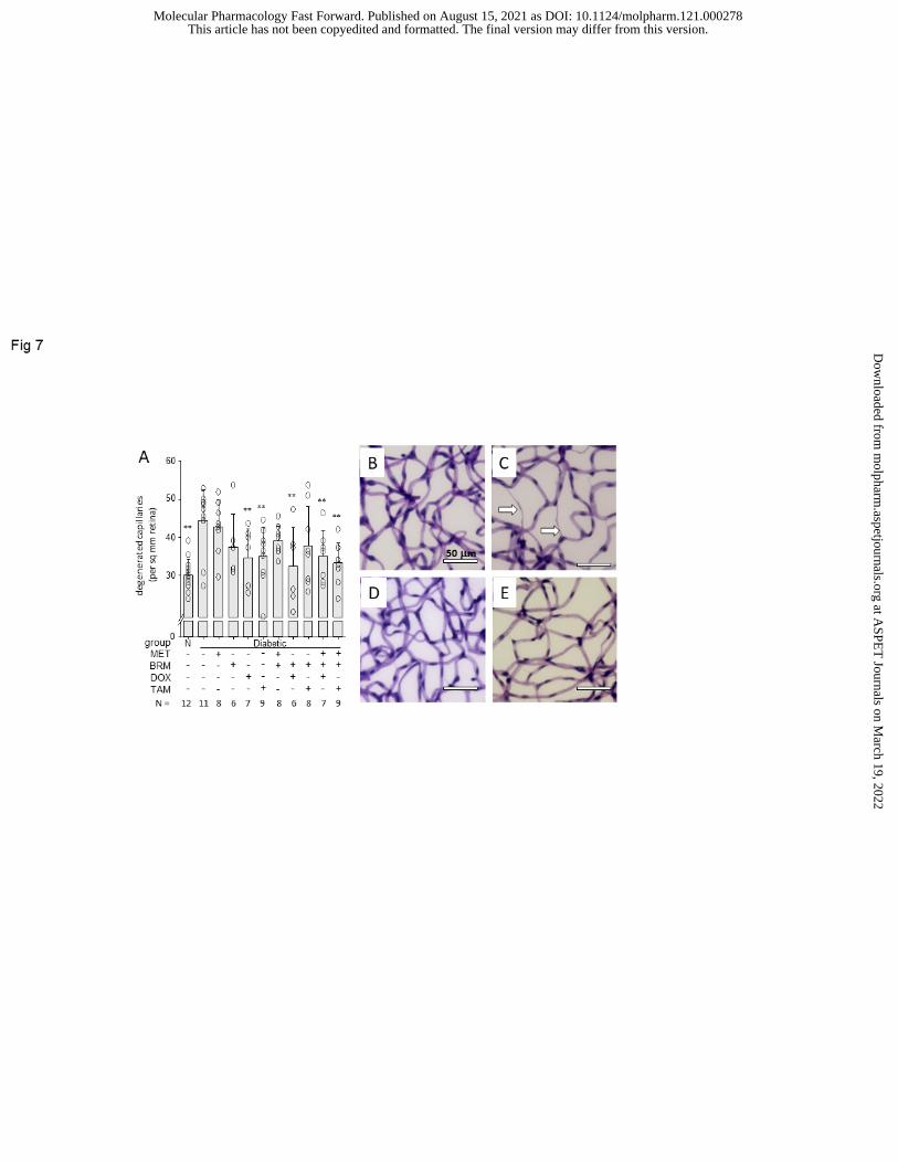
Fig 7. Diabetes-induced degeneration of retinal capillaries at 8 months of study. MTP, BRM,
DOX, and TAM were administered daily at their DMIox doses individually or in combinations. (A)
Graphical summary of the number of degenerated capillaries in mid-retina of nondiabetic and
diabetic mice at 8 months of diabetes (10 months of age; sample size is indicated below each
column). Individually at their DMIox doses, neither MTP nor BRM appreciably inhibited the
capillary degeneration, but DOX or TAM monotherapies did statistically significantly inhibit the
vaso-obliteration. Most combinations inhibited the capillary degeneration. (B-E) show
representative photomicrographs demonstrating the retinal vasculature at the same age. (B) WT
nondiabetic, (C) WT diabetic control, (D) DOX as a monotherapy, (E) MTP+BRM+DOX as a
combination therapy. Degenerated retinal capillaries (acellular capillaries; illustrated by white
arrows) are about 50% more numerous in WT diabetic mice than in WT nondiabetic mice. The
effect of each of the other drugs or drug combinations to inhibit the capillary degeneration is
assessed compared to the WT diabetic group. Calibration bar indicates 50 µm actual
dimensions. *P<0.05; **P<0.001 compared to diabetic control. Mean ±SD.
Fig 8. Leakage of albumin into the neural retina in diabetes. Diabetes-induced degeneration of
retinal capillaries at 8 months of study. Diabetes of 8-month duration more than doubled the
accumulation of FITC-BSA in the (A) inner plexiform layer (IPL) and (B) outer plexiform layer
(OPL) of the retina, as evidenced by diffuse green color in IPL and OPL. Bright green spots
indicate FITC-BSA within retinal capillaries, and are not included in the quantification for
permeability determination. All drugs were administered daily at their DMIox. Neither MTP nor
This article has not been copyedited and formatted. The final version may differ from this version.Molecular Pharmacology Fast Forward. Published on August 15, 2021 as DOI: 10.1124/molpharm.121.000278
at ASPE
T Journals on M
arch 19, 2022m
olpharm.aspetjournals.org
Dow
nloaded from

36
TAM inhibited the diabetes-induced defect in permeability, but both BRM and DOX totally and
significantly inhibited the defect in both layers. (C-E) show representative photomicrographs
illustrating fluorescence in the neural retina. (C) WT nondiabetic, (D) WT diabetic control, (E)
MTP+BRM+DOX as a combination therapy administered daily to diabetic mice. FITC-albumin
was injected intravenously, allowed to circulate for 20 minutes, and then fluorescence in areas
of the IPL or OPL was quantified from retinal cross-sections. Calibration bar indicates actual
dimensions. Sample size is indicated below each column. *P<0.05; **P<0.001 compared to
diabetic control. Mean ±SD
This article has not been copyedited and formatted. The final version may differ from this version.Molecular Pharmacology Fast Forward. Published on August 15, 2021 as DOI: 10.1124/molpharm.121.000278
at ASPE
T Journals on M
arch 19, 2022m
olpharm.aspetjournals.org
Dow
nloaded from

37
Table 1. Clinical data of experimental groups over 8 months of diabetes
Group n* BW
(g)
Blood glucose
(nonfasting; mg/dl)
HbA1c
(%)
Nondiabetic control 12 48 ±4 145 ±15 2.9 ±0.1
Diabetes control 11 29 ±4 467 ±46 9.5 ±1.3
Diabetes +
MTP 8 27 ±3 482 ±39 9.9 ±0.4
BRM 6 27 ±3 468 ±46 9.7 ±0.8
DOX 7 28 ±3 491 ±17 9.7 ±0.8
BRM+MTP 8 30 ±5 443 ±41 9.0 ±1.7
BRM+DOX 6 27 ±3 450 ±44 9.9 ±1.1
MTP+BRM+DOX 7 29 ±2 463 ±49 9.6 ±0.3
TAM 9 28 ±2 460 ±45 10.1 ±0.9
BRM+TAM 8 27 ±2 497 ±44 10.1 ±0.4
MTP+BRM+TAM 7 29 ±3 445 ±45 9.9 ±0.4
* All experimental groups were n=10 at start of experiment, except n=12 for Nondiabetic
and Diabetes controls
This article has not been copyedited and formatted. The final version may differ from this version.Molecular Pharmacology Fast Forward. Published on August 15, 2021 as DOI: 10.1124/molpharm.121.000278
at ASPE
T Journals on M
arch 19, 2022m
olpharm.aspetjournals.org
Dow
nloaded from

38
Table 2. Drug dosages administered daily in 8-month study, and estimated ED50
Drug DMIox
(mg/kgbw/day)
Daily dose
administered
(mg/kgbw/day)
Estimated ED50 for inhibition of the
diabetes-induced increase in retinal
superoxide (mg/kgbw/day)
MET 0.04 0.04 0.08
BRM 0.01 0.01 0.02
DOX 0.01 0.01 0.02
TAM 0.15 0.15 0.15
This article has not been copyedited and formatted. The final version may differ from this version.Molecular Pharmacology Fast Forward. Published on August 15, 2021 as DOI: 10.1124/molpharm.121.000278
at ASPE
T Journals on M
arch 19, 2022m
olpharm.aspetjournals.org
Dow
nloaded from

This article has not been copyedited and formatted. The final version may differ from this version.Molecular Pharmacology Fast Forward. Published on August 15, 2021 as DOI: 10.1124/molpharm.121.000278
at ASPE
T Journals on M
arch 19, 2022m
olpharm.aspetjournals.org
Dow
nloaded from

This article has not been copyedited and formatted. The final version may differ from this version.Molecular Pharmacology Fast Forward. Published on August 15, 2021 as DOI: 10.1124/molpharm.121.000278
at ASPE
T Journals on M
arch 19, 2022m
olpharm.aspetjournals.org
Dow
nloaded from

This article has not been copyedited and formatted. The final version may differ from this version.Molecular Pharmacology Fast Forward. Published on August 15, 2021 as DOI: 10.1124/molpharm.121.000278
at ASPE
T Journals on M
arch 19, 2022m
olpharm.aspetjournals.org
Dow
nloaded from

This article has not been copyedited and formatted. The final version may differ from this version.Molecular Pharmacology Fast Forward. Published on August 15, 2021 as DOI: 10.1124/molpharm.121.000278
at ASPE
T Journals on M
arch 19, 2022m
olpharm.aspetjournals.org
Dow
nloaded from

This article has not been copyedited and formatted. The final version may differ from this version.Molecular Pharmacology Fast Forward. Published on August 15, 2021 as DOI: 10.1124/molpharm.121.000278
at ASPE
T Journals on M
arch 19, 2022m
olpharm.aspetjournals.org
Dow
nloaded from

This article has not been copyedited and formatted. The final version may differ from this version.Molecular Pharmacology Fast Forward. Published on August 15, 2021 as DOI: 10.1124/molpharm.121.000278
at ASPE
T Journals on M
arch 19, 2022m
olpharm.aspetjournals.org
Dow
nloaded from
This article has not been copyedited and formatted. The final version may differ from this version.Molecular Pharmacology Fast Forward. Published on August 15, 2021 as DOI: 10.1124/molpharm.121.000278
at ASPE
T Journals on M
arch 19, 2022m
olpharm.aspetjournals.org
Dow
nloaded from

This article has not been copyedited and formatted. The final version may differ from this version.Molecular Pharmacology Fast Forward. Published on August 15, 2021 as DOI: 10.1124/molpharm.121.000278
at ASPE
T Journals on M
arch 19, 2022m
olpharm.aspetjournals.org
Dow
nloaded from
Recommended