Richard HynesHHMI/MIT
MGH Tumor Microcirculation Course
Cambridge, MA
June 4, 2003
”Cell Adhesion in Tumor Growth, Progression and Angiogenesis"
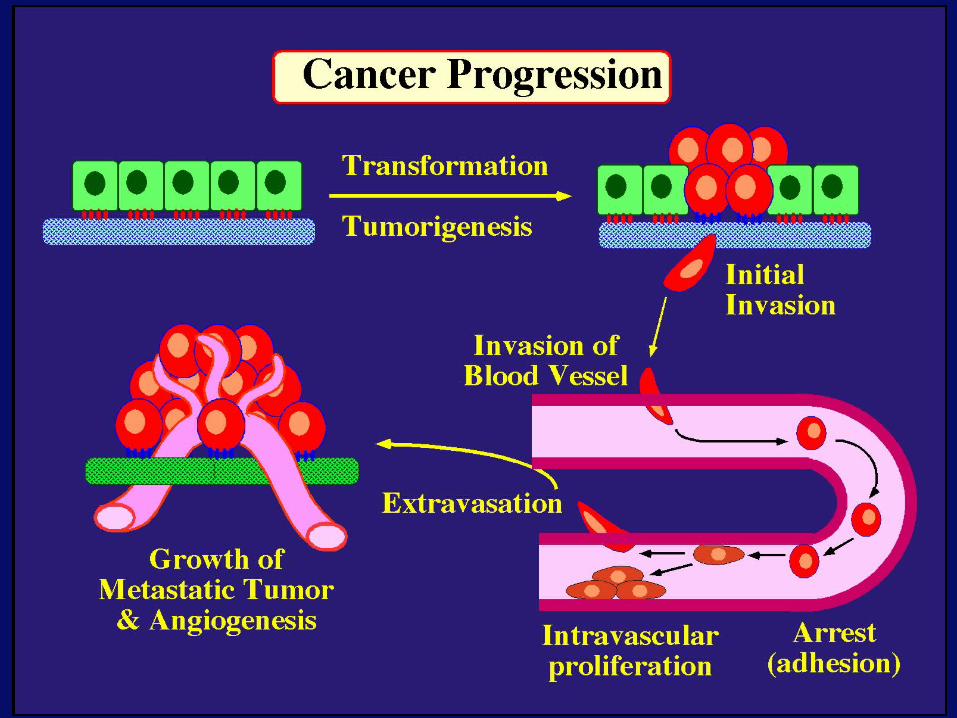
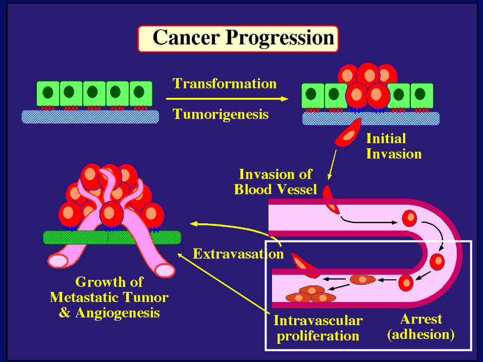
GROWTH of PRIMARY TUMORand INITIAL INVASION
Angiogenesis-also Lymphangiogenesis (not shown)
Loss of cell adhesion
Further loss of cell adhesion
Gain of cell migration
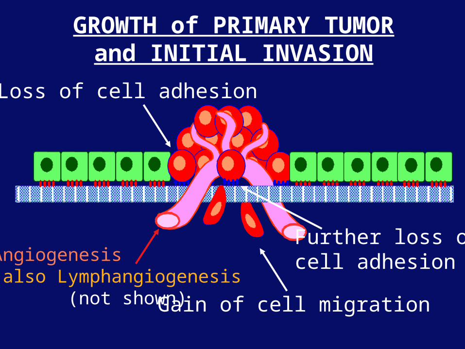
Angiogenesis andLymphangiogenesis
• Essential for growth of primary tumor(and later of metastases)
• Involves extensive migration and adhesionof endothelial cells and pericytes
• Involves organization of basement membranes
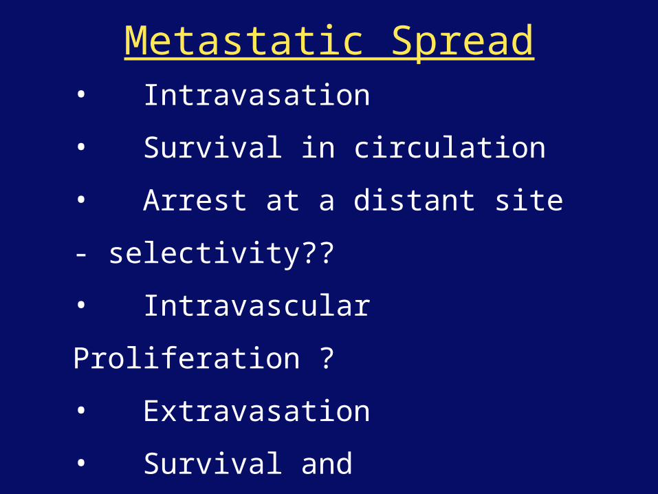
Metastatic Spread• Intravasation
• Survival in circulation
• Arrest at a distant site - selectivity??
• Intravascular Proliferation ?
• Extravasation
• Survival and proliferation at the new site
• Angiogenesis again
• All of these involve cell adhesion
U
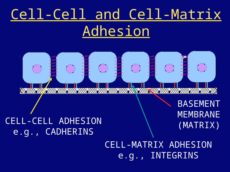
CELL-CELL ADHESIONe.g., CADHERINS
CELL-MATRIX ADHESIONe.g., INTEGRINS
BASEMENTMEMBRANE(MATRIX)
Cell-Cell and Cell-Matrix Adhesion
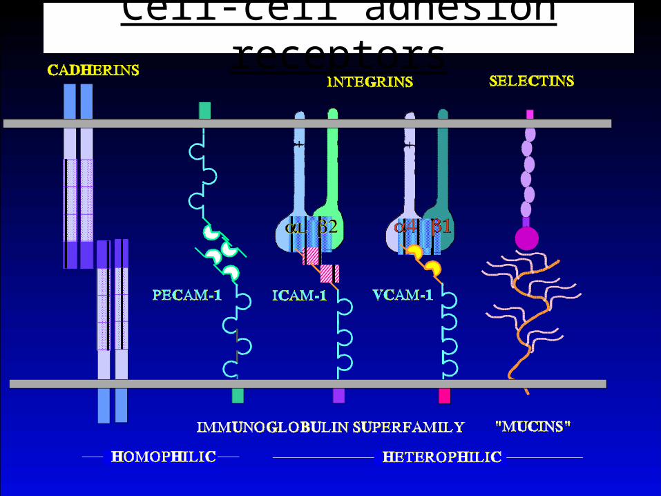
Cell-cell adhesion receptors
S
S
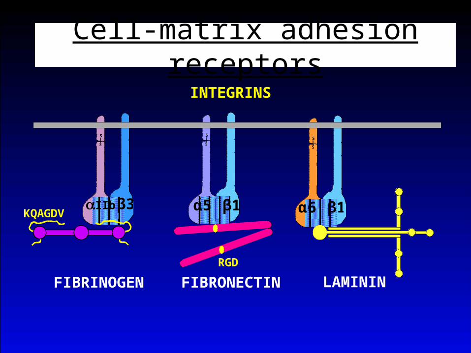
INTEGRINS
FIBRINOGEN
S
SS
S
FIBRONECTIN LAMININ
αIIb β3 α5 β1 α6 β1
RGD
KQAGDV
CELL-MATRIX ADHESIONCell-matrix adhesion receptors
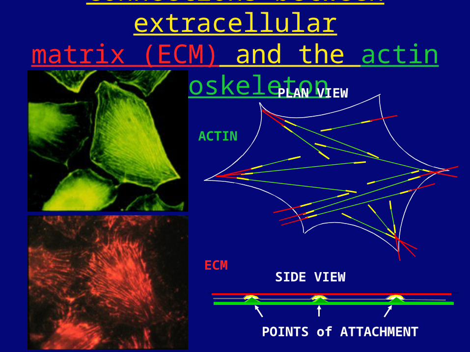
Connections between extracellularmatrix (ECM) and the actin cytoskeleton
ACTIN
ECM
PLAN VIEW
SIDE VIEW
POINTS of ATTACHMENT
The Molecular Linkage Between Actin and ECM via Integrins
β1α5
FAK TALIN
INTEGRIN
ACTINFILAMENTS
MEMBRANE
MATRIX
CDK
SIGNALLINGEVENTS
RGD
PROTEOGLYCAN
src
PKC
TENSIN
VIN
αAαA
PAXILLIN
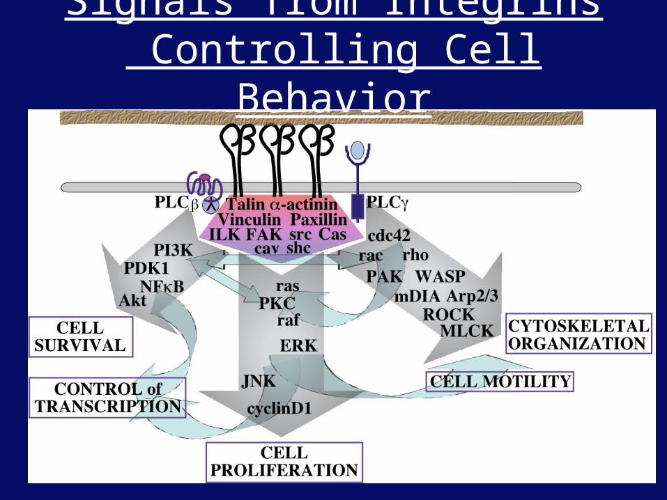
Signals from Integrins Controlling Cell Behavior
β
Functions of Cell Adhesion Receptors
• Mediate adhesion to adjacent cells and to ECM
• Control cell shape, polarity and migration
• Control cell proliferation, survival, gene expression and differentiation
How do these functions impact tumor progression?
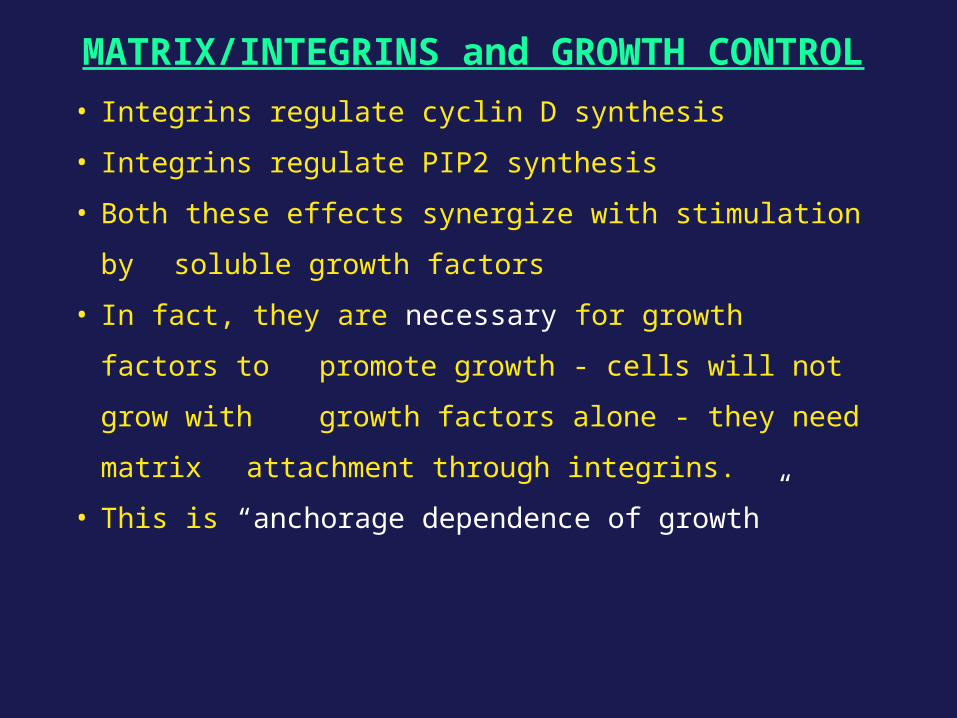
MATRIX/INTEGRINS and GROWTH CONTROL
• Integrins regulate cyclin D synthesis
• Integrins regulate PIP2 synthesis
• Both these effects synergize with stimulation by
soluble growth factors
• In fact, they are necessary for growth factors to
promote growth - cells will not grow with growth
factors alone - they need matrix attachment
through integrins.
• This is “anchorage dependence of growth”
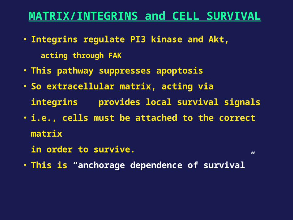
MATRIX/INTEGRINS and CELL SURVIVAL
• Integrins regulate PI3 kinase and Akt,
acting through FAK
• This pathway suppresses apoptosis
• So extracellular matrix, acting via integrins
provides local survival signals
• i.e., cells must be attached to the correct matrix
in order to survive.
• This is “anchorage dependence of survival”
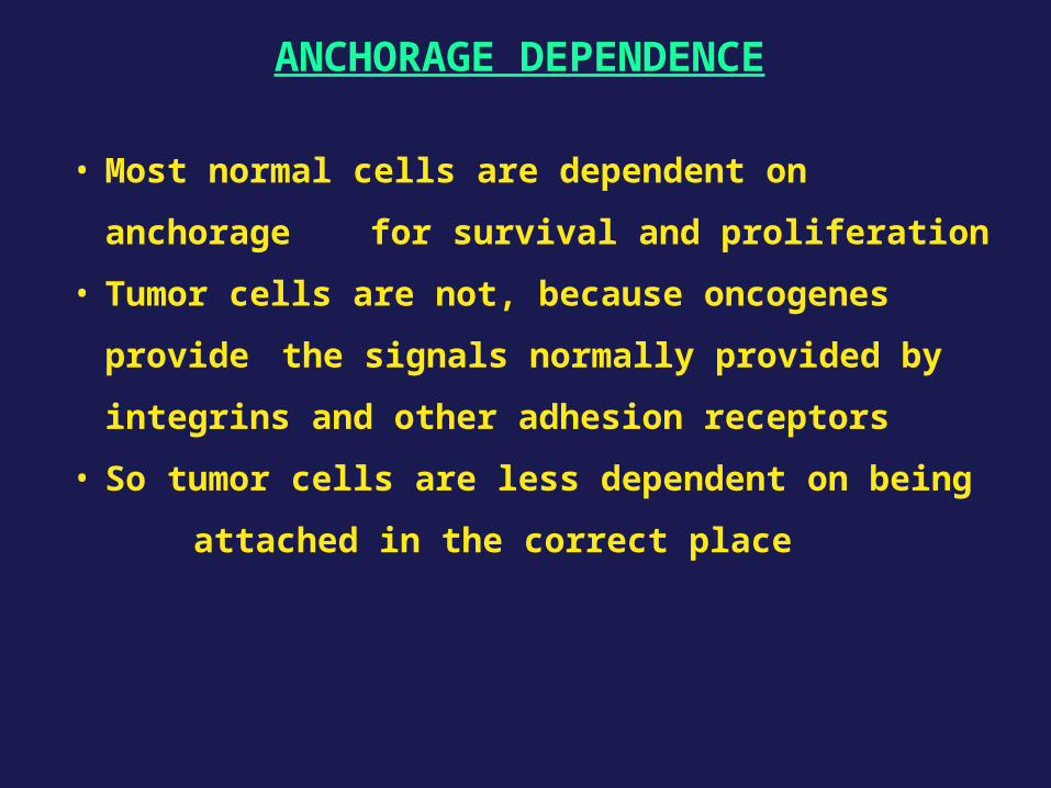
ANCHORAGE DEPENDENCE
• Most normal cells are dependent on anchorage
for survival and proliferation
• Tumor cells are not, because oncogenes provide
the signals normally provided by
integrins and other adhesion receptors
• So tumor cells are less dependent on being
attached in the correct place
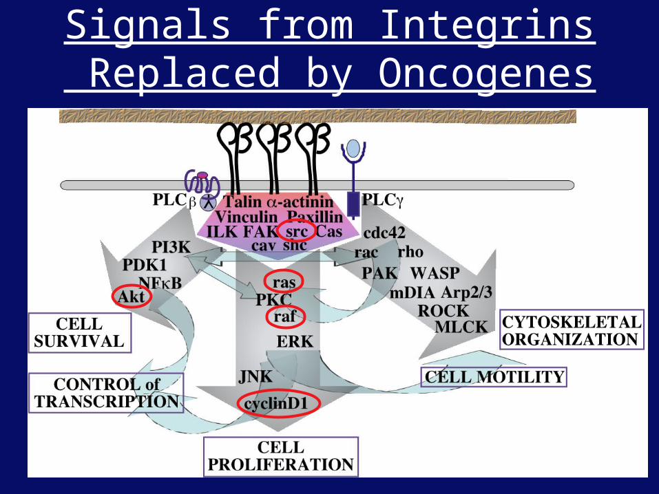
Signals from Integrins Replaced by Oncogenes
β
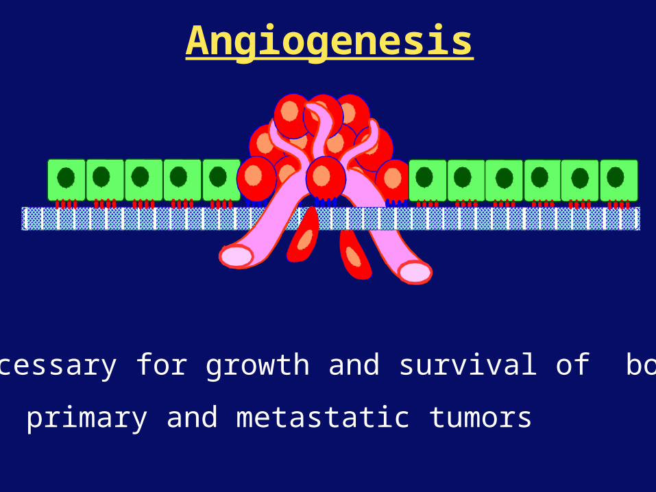
Angiogenesis
Necessary for growth and survival of both
primary and metastatic tumors
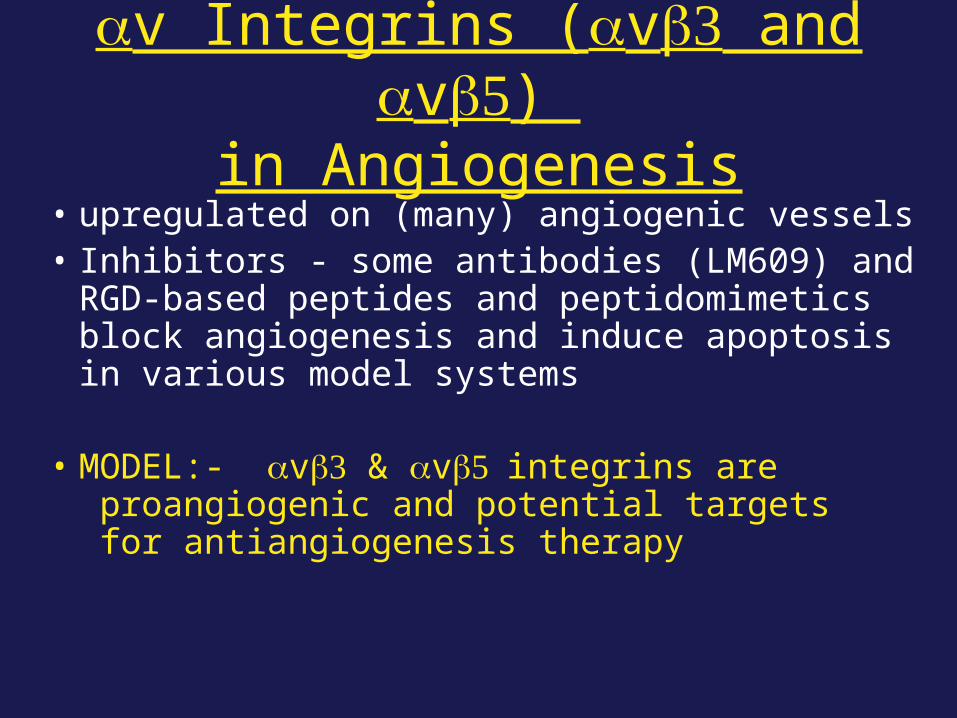
αv Integrins (αvβ and αvβ5) in Angiogenesis
• upregulated on (many) angiogenic vessels• Inhibitors - some antibodies (LM609) and RGD-
based peptides and peptidomimetics block angiogenesis and induce apoptosis in various model systems
• MODEL:- αvβ & αvβ5integrins are proangiogenic and potential targets for antiangiogenesis therapy
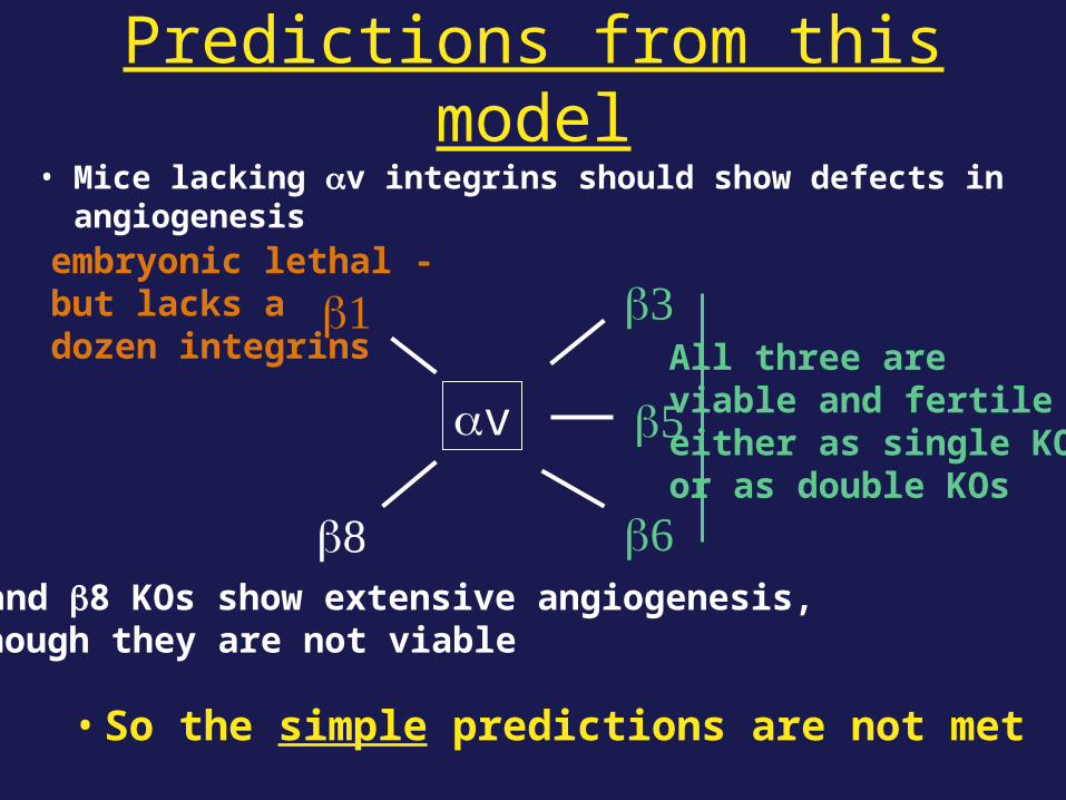
Predictions from this model• Mice lacking αv integrins should show defects in angiogenesis
• So the simple predictions are not met
αv
β β
β5
βAll three areviable and fertileeither as single KOsor as double KOs
αv and β8 KOs show extensive angiogenesis, although they are not viable
β1embryonic lethal -but lacks a dozen integrins
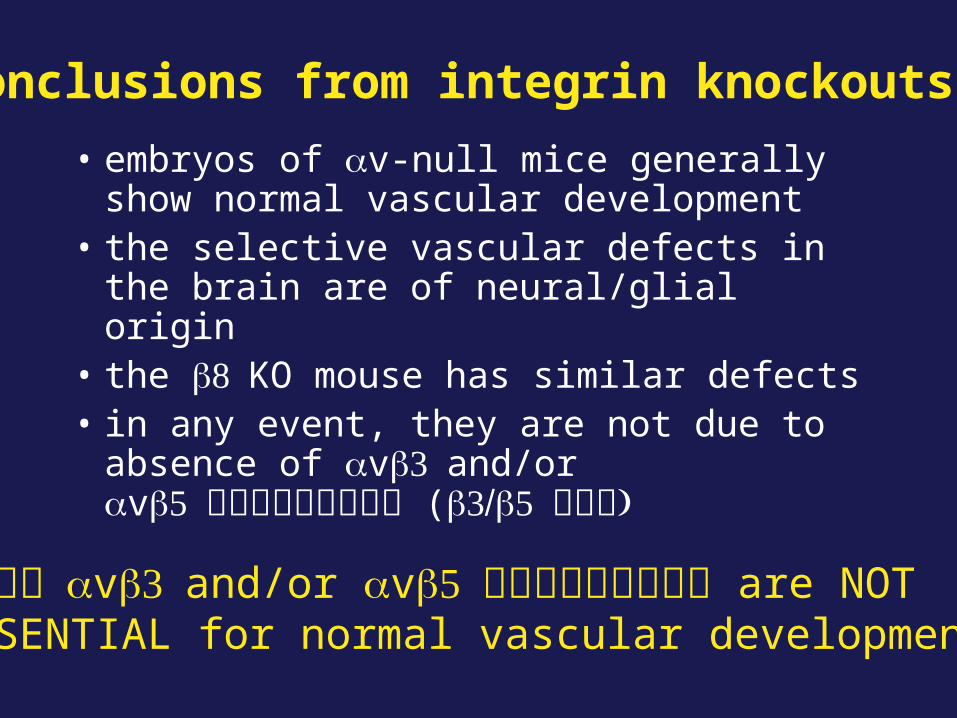
• embryos of αv-null mice generally show normal vascular development
• the selective vascular defects in the brain are of neural/glial origin
• the βKO mouse has similar defects • in any event, they are not due to absence of
αvβand/or αvβ5 (ββ5
Conclusions from integrin knockouts
αvβand/or αvβ5 are NOT ESSENTIAL for normal vascular development
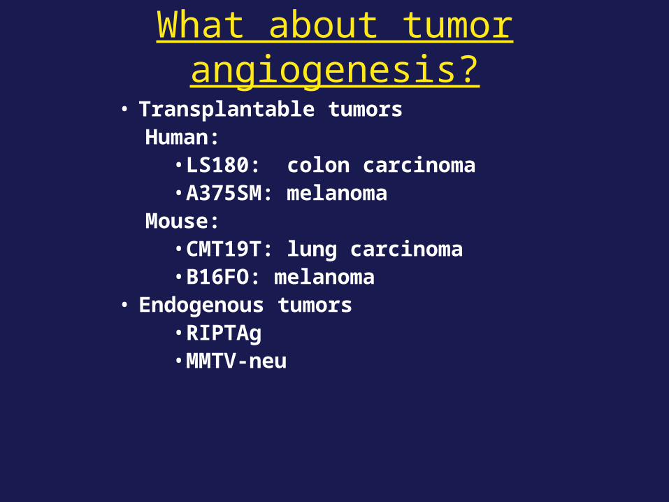
What about tumor angiogenesis?
• Transplantable tumorsHuman:
• LS180: colon carcinoma • A375SM: melanoma
Mouse:• CMT19T: lung carcinoma• B16FO: melanoma
• Endogenous tumors• RIPTAg• MMTV-neu
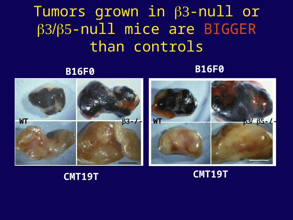
Tumors grown in β-null or ββ5-null mice are BIGGER than controls
B16F0
CMT19T
WT
B16F0
CMT19T
WTβ-/- ββ5-/-
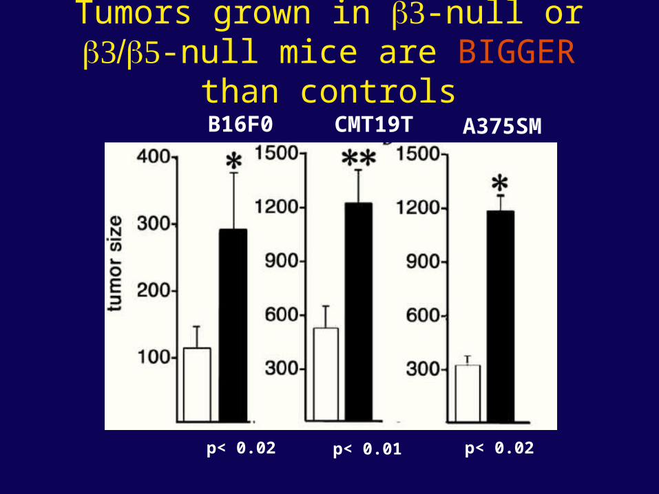
Tumors grown in β-null or ββ5-null mice are BIGGER than controls
B16F0 CMT19T A375SM
p< 0.01p< 0.02 p< 0.02
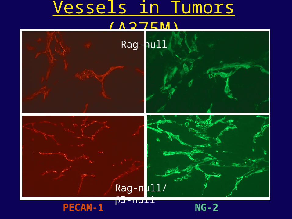
Vessels in Tumors (A375M)Rag-null
Rag-null/β3-null
PECAM-1 NG-2
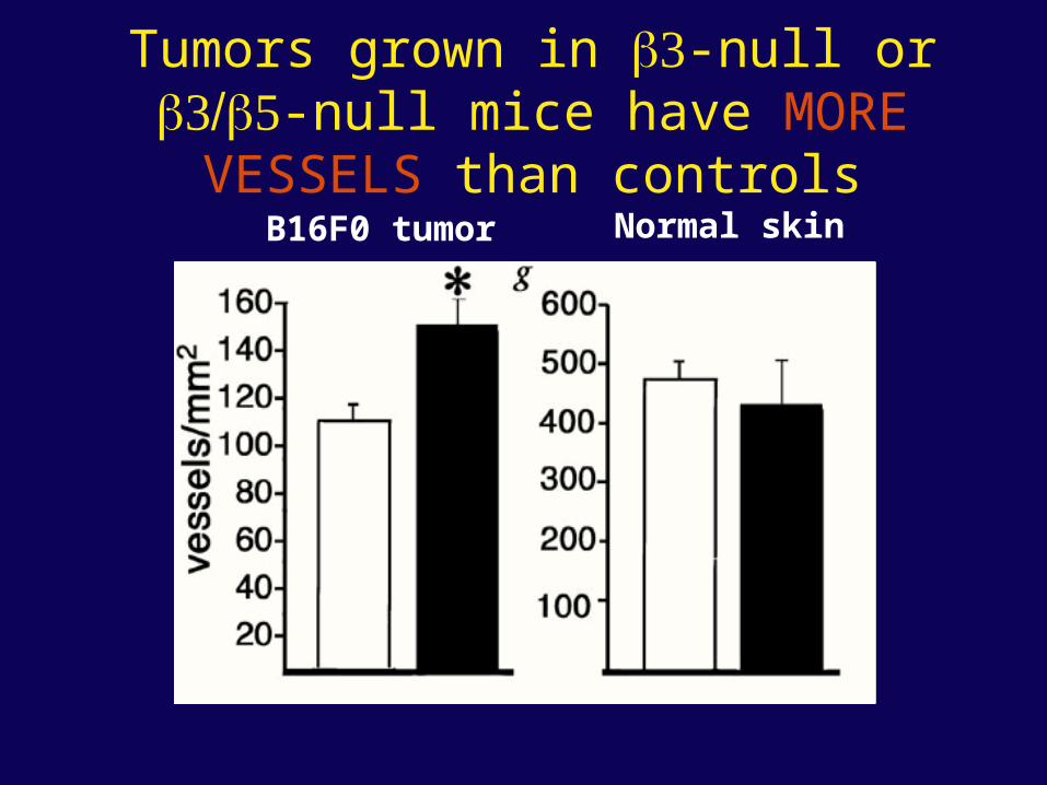
Tumors grown in β-null or ββ5-null mice have MORE VESSELS than controls
B16F0 tumor Normal skin
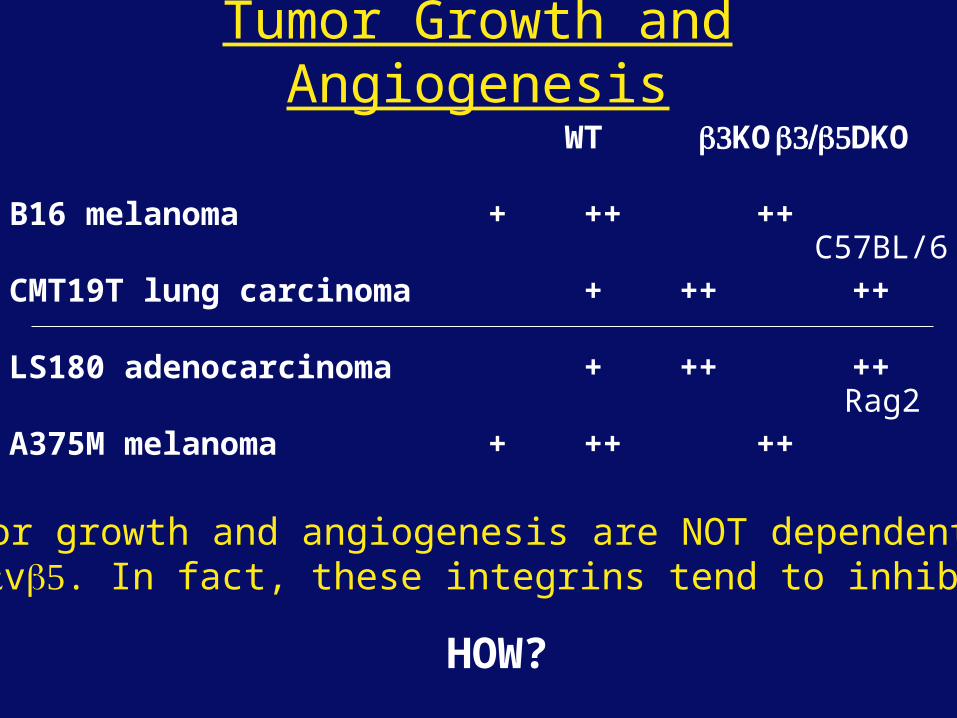
Tumor Growth and Angiogenesis
So:- tumor growth and angiogenesis are NOT dependent onαvβ orαvβ5. In fact, these integrins tend to inhibit them.
HOW?
WT βKO ββ5DKO
B16 melanoma + ++ ++
CMT19T lung carcinoma + ++ ++
LS180 adenocarcinoma + ++ ++
A375M melanoma + ++ ++
C57BL/6
Rag2
α5β1 Integrin and Fibronectin in Angiogenesis
• both are upregulated on angiogenic vessels• mice lacking α5β1 die with vascular defects• mice lacking die with vascular defects• antibodies to either inhibit angiogenesis• peptides blocking their interaction inhibit
angiogenesis• that is - genetics and inhibitor studies conform here• Fibronectin and α5β1 integrin are proangiogenic• They appear good targets for antiangiogenesis
A new way of thinking about αv integrins in angiogenesis
• The original model of their being proangiogenic does not explain all the data
• Perhaps they are actually antiangiogenic or negative regulators some or all the time
• The negative regulation model does a better, although not a perfect job of explaining the data
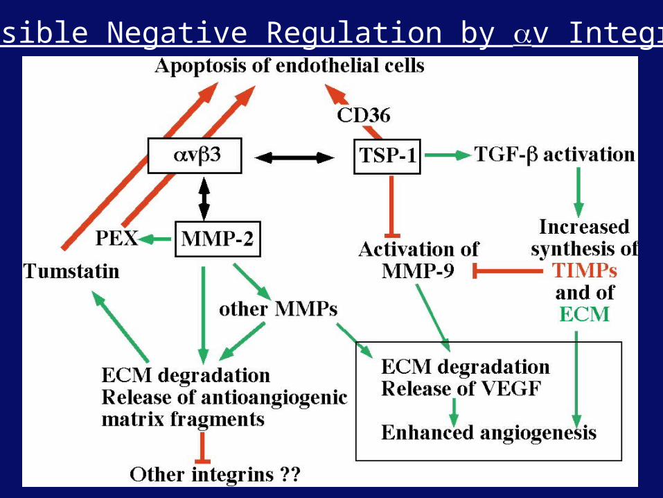
Possible Negative Regulation by αv Integrins
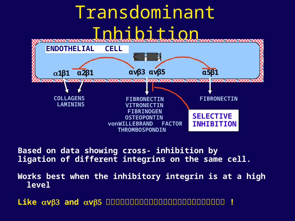
Transdominant Inhibition
α1β1 α2β1 α5β1
FIBRONECTIN
αvβ3 αvβ5
FIBRONECTINVITRONECTINFIBRINOGEN
OSTEOPONTINvon WILLEBRAND FACTOR
THROMBOSPONDIN
COLLAGENSLAMININS
ENDOTHELIAL CELL
SELECTIVEINHIBITION
Based on data showing cross- inhibition byligation of different integrins on the same cell.
Works best when the inhibitory integrin is at a high level
Like αvβ and αvβ5 !
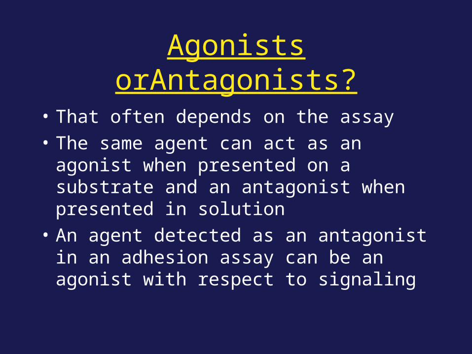
Agonists orAntagonists?
• That often depends on the assay
• The same agent can act as an agonist when presented on a substrate and an antagonist when presented in solution
• An agent detected as an antagonist in an adhesion assay can be an agonist with respect to signaling
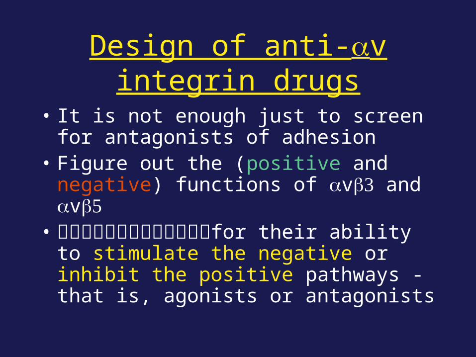
Design of anti-αv integrin drugs
• It is not enough just to screen for antagonists of adhesion
• Figure out the (positive and negative) functions of αvβ and αvβ5
• for their ability to stimulate the negative or inhibit the positive pathways - that is, agonists or antagonists
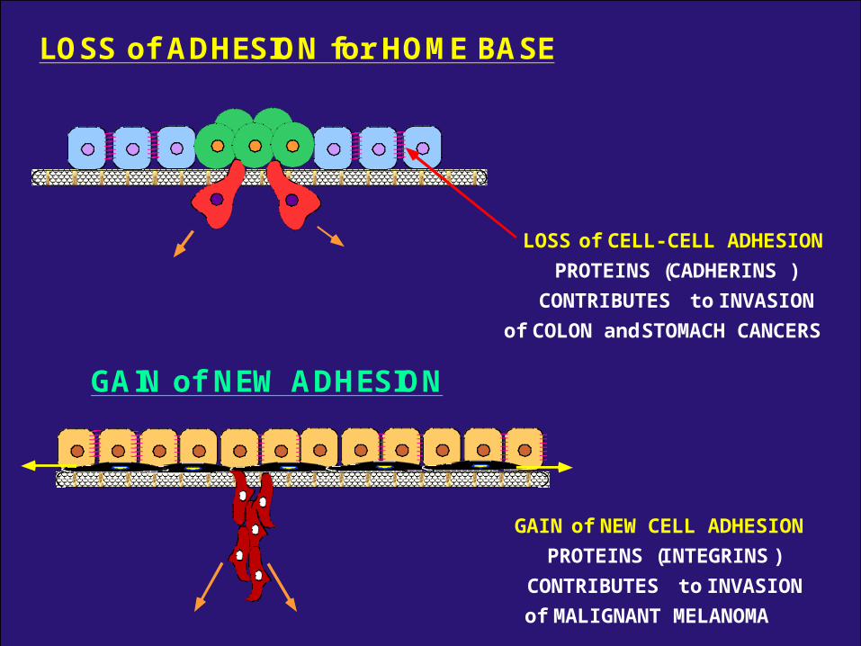
LOSS of ADHESION for HOME BASE
LOSS of CELL-CELL ADHESION
PROTEINS (CADHERINS)
CONTRIBUTES to INVASION
of COLON and STOMACH CANCERS
GAIN of NEW ADHESION
GAIN of NEW CELL ADHESION
PROTEINS (INTEGRINS)
CONTRIBUTES to INVASION
of MALIGNANT MELANOMA
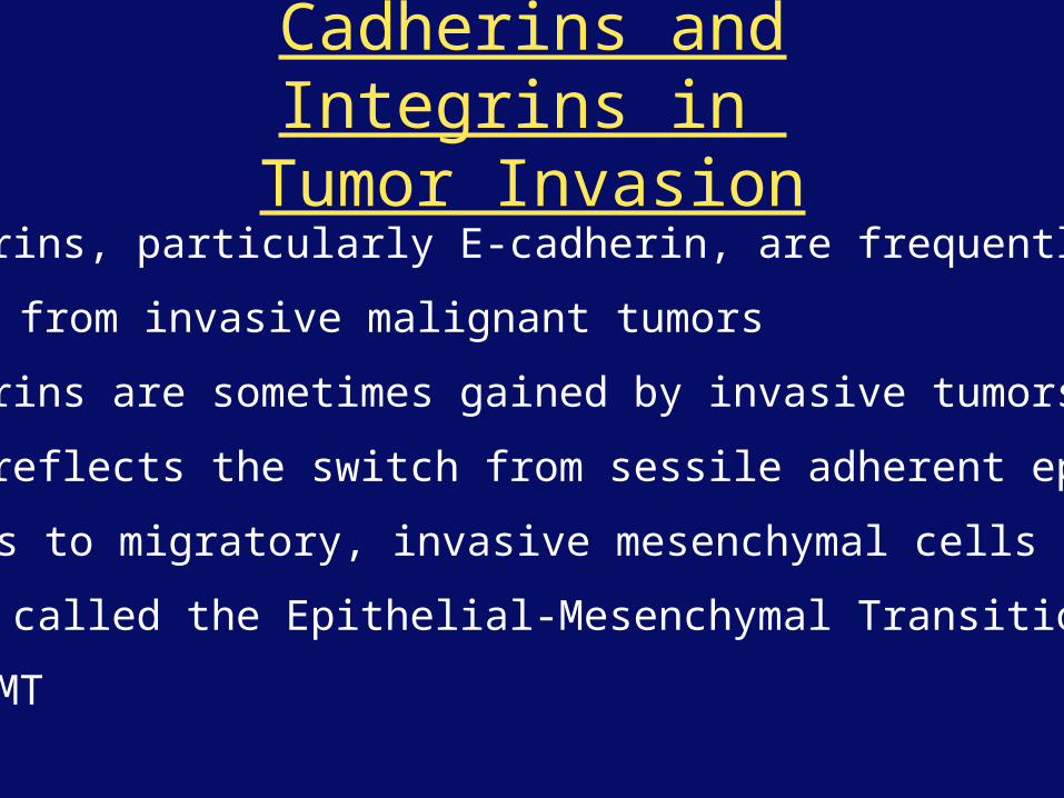
Cadherins and Integrins in Tumor Invasion
• Cadherins, particularly E-cadherin, are frequently
lost from invasive malignant tumors
• Integrins are sometimes gained by invasive tumors
• This reflects the switch from sessile adherent epithelial
cells to migratory, invasive mesenchymal cells
• Often called the Epithelial-Mesenchymal Transition
or EMT
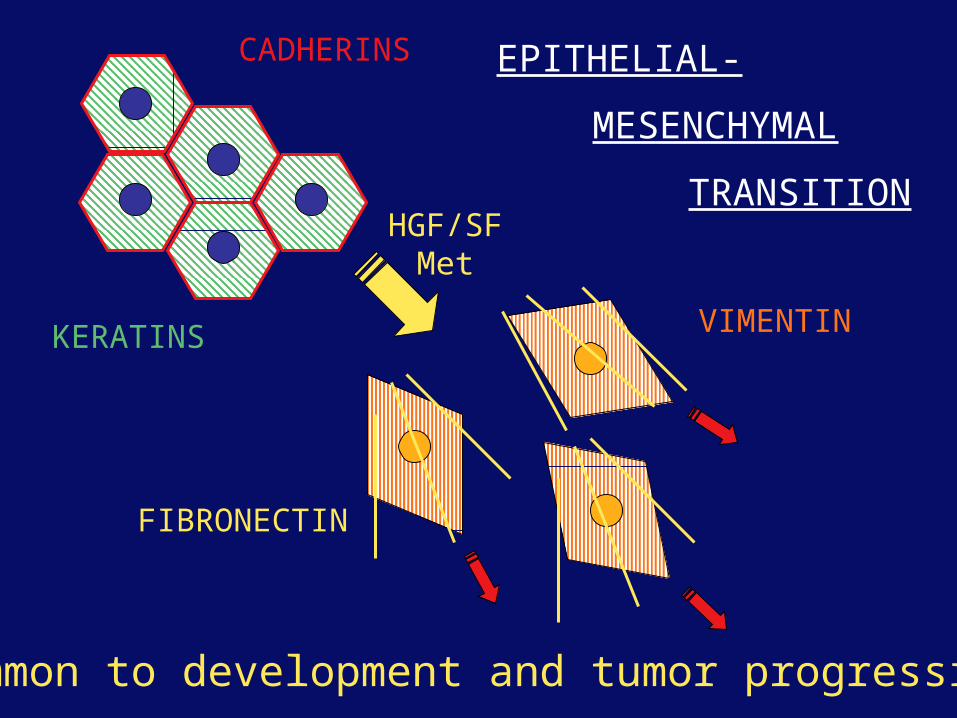
CADHERINS
KERATINS VIMENTIN
FIBRONECTIN
EPITHELIAL-
MESENCHYMAL
TRANSITIONHGF/SF
Met
Common to development and tumor progression
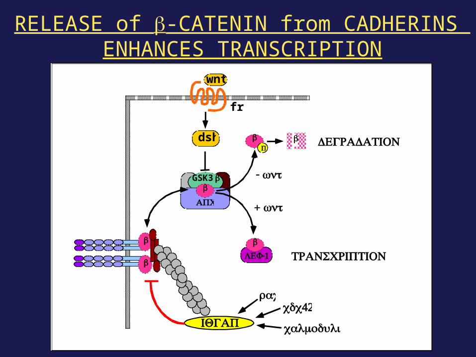
wnt
frz
dsh
GSK3ββ
β
β
β
LEF-1
β β
-wnt
+wntAPC
TRANSCRIPTION
PDEGRADATION
IQGAP
rac42cdc
calmodulin
RELEASE of β-CATENIN from CADHERINS ENHANCES TRANSCRIPTION
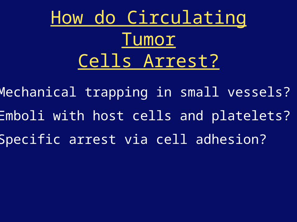
How do Circulating TumorCells Arrest?
Mechanical trapping in small vessels?
Emboli with host cells and platelets?
Specific arrest via cell adhesion?
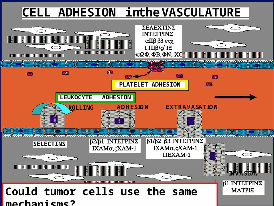
LEUKOCYTE ADHESION
PLATELET ADHESION
ROLLING ADHESION EXTRAVASATION
INVASION
CELL ADHESION in the VASCULATURE
SELECTINS β2/β1INTEGRINS,ICAMs -1VCAM
β1INTEGRINSMATRIX
β1/β2/βINTEGRINS,ICAMs -1VCAM
-1PECAM
SELECTINSINTEGRINSαIIbβetcGPIb/ /V IX,vWF ,FB ,FN CO
Could tumor cells use the same mechanisms?
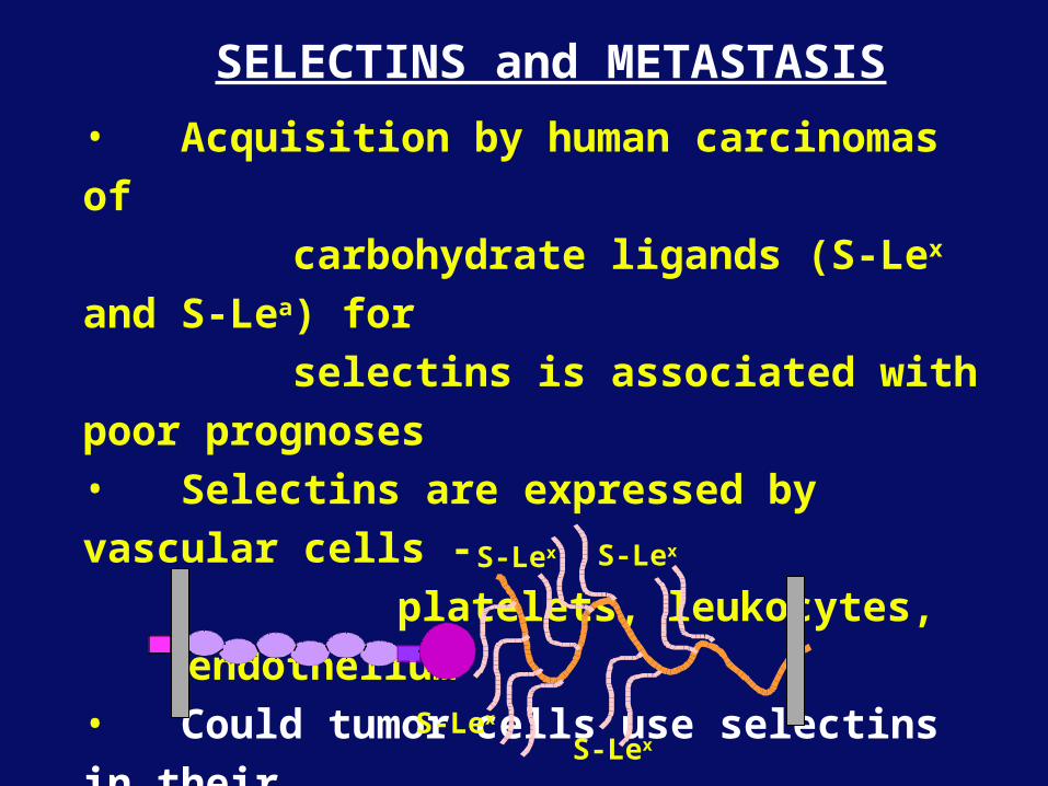
SELECTINS and METASTASIS
• Acquisition by human carcinomas of
carbohydrate ligands (S-Lex and S-Lea)
for
selectins is associated with poor
prognoses• Selectins are expressed by vascular cells -
platelets, leukocytes, endothelium• Could tumor cells use selectins in their
metastatic spread?
S-LexS-Lex
S-LexS-Lex
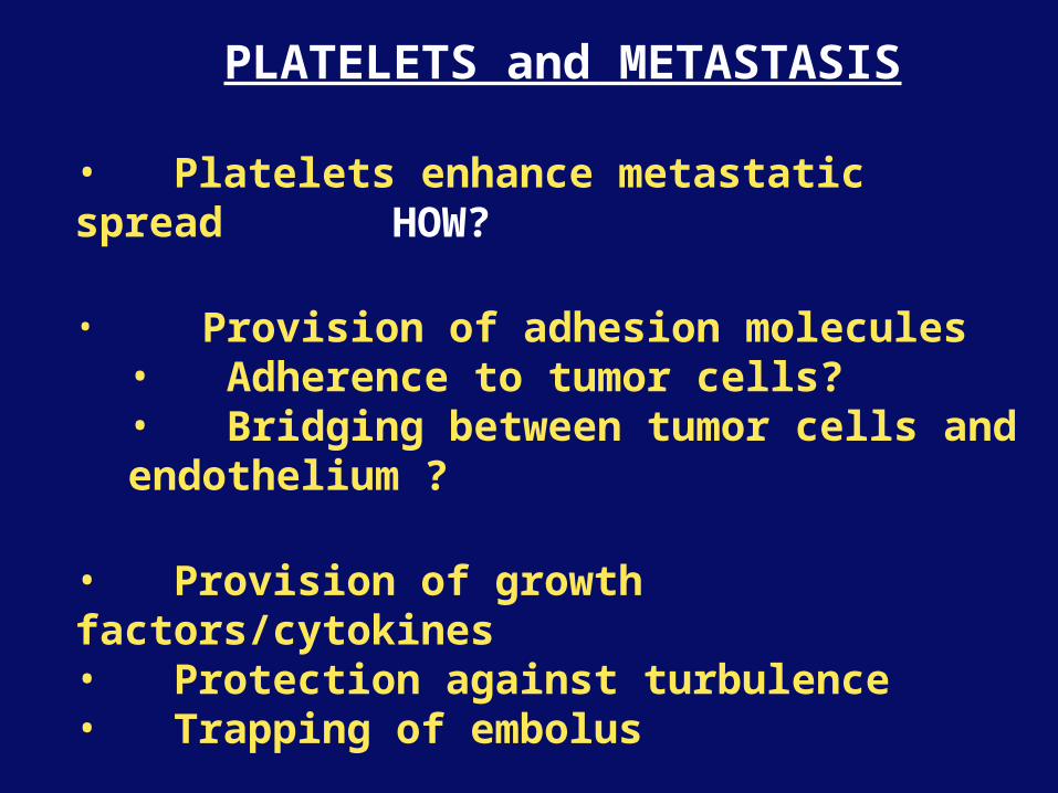
PLATELETS and METASTASIS
• Platelets enhance metastatic spreadHOW?
• Provision of adhesion molecules• Adherence to tumor cells?• Bridging between tumor cells and endothelium ?
• Provision of growth factors/cytokines• Protection against turbulence• Trapping of embolus
• Could selectins or integrins play a role?
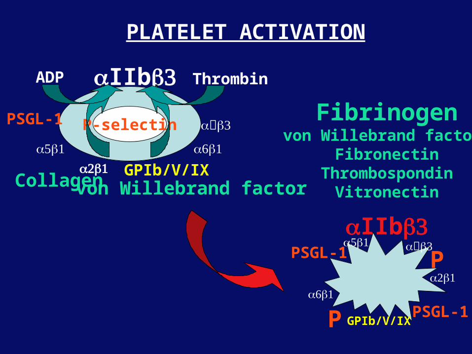
GPIb/V/IX
α5β1α2β1
αβ
αβ1
αIIbβ
Collagen von Willebrand factor
Fibrinogenvon Willebrand factor
FibronectinThrombospondin
Vitronectin
P-selectin
PLATELET ACTIVATION
αIIbβ
ThrombinADP
P
P
GPIb/V/IX
α5β1
α2β1
αβ
αβ1
PSGL-1
PSGL-1
PSGL-1
S-Lex
S-Lex
S-Lex
S-Lex
S-Lex
S-Lex
S-Lex
S-Lex
S-Lex
S-Lex
S-Lex
S-Lex
S-Lex
S-Lex
S-Lex
S-Lex S-Lex
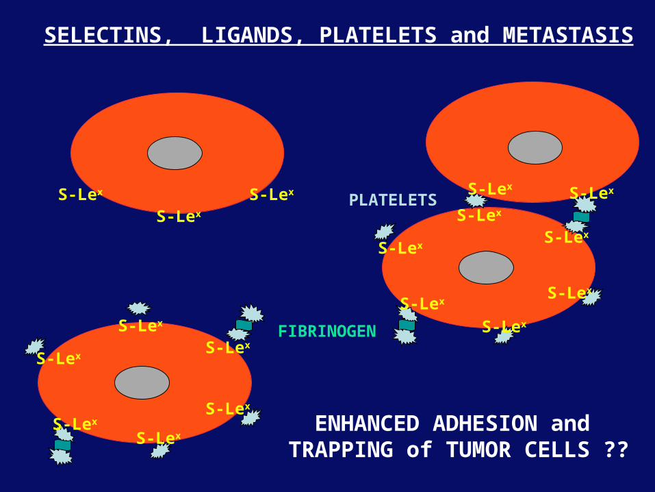
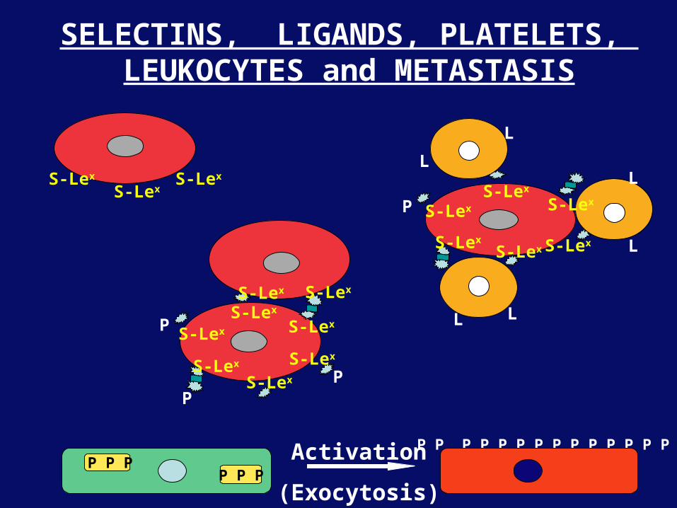
SELECTINS, LIGANDS, PLATELETS and METASTASIS
PLATELETS
FIBRINOGEN
ENHANCED ADHESION and TRAPPING of TUMOR CELLS ??
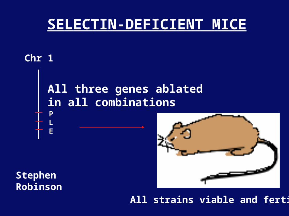
Chr 1
PLE
SELECTIN-DEFICIENT MICE
All strains viable and fertile
All three genes ablatedin all combinations
StephenRobinson
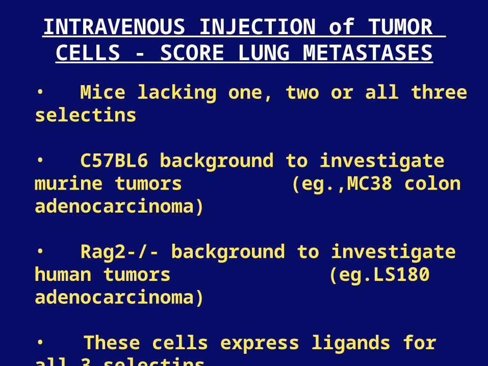
INTRAVENOUS INJECTION of TUMOR CELLS - SCORE LUNG METASTASES
• Mice lacking one, two or all three selectins
• C57BL6 background to investigate murine tumors (eg.,MC38 colon adenocarcinoma)
• Rag2-/- background to investigate human tumors (eg.LS180 adenocarcinoma)
• These cells express ligands for all 3 selectins
Daniela Taverna - and collaboration with Ajit Varki/Lubor Borsig
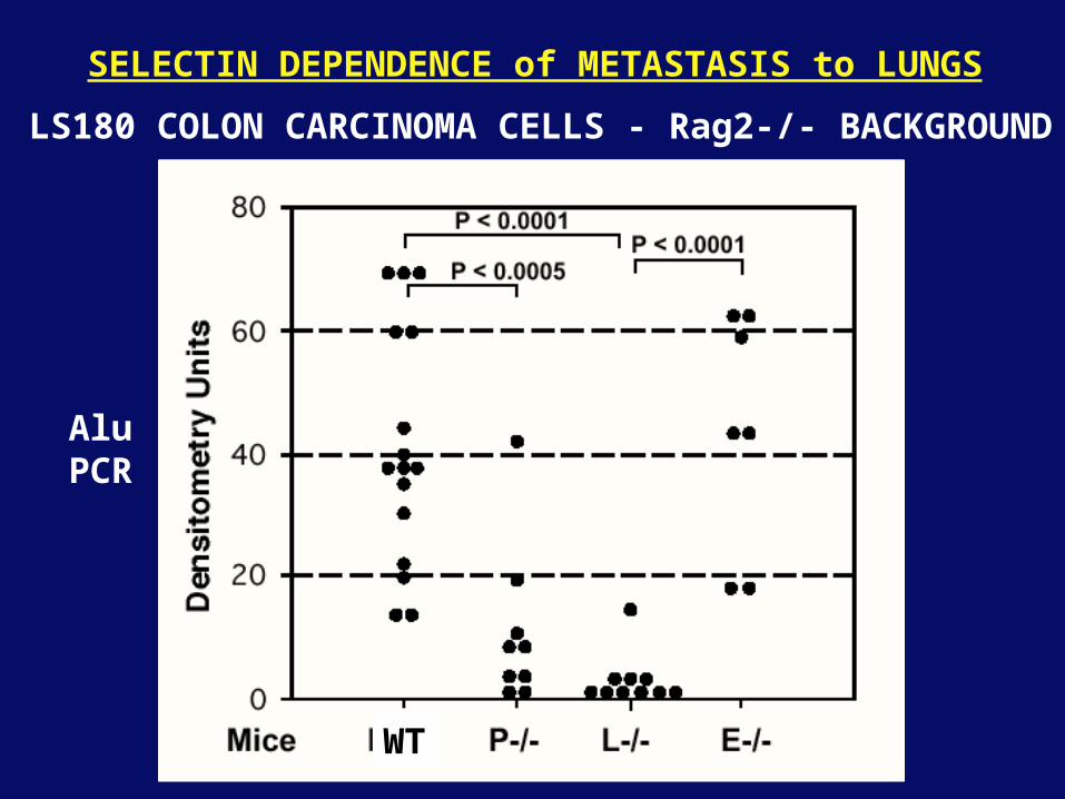
LS180 COLON CARCINOMA CELLS - Rag2-/- BACKGROUND
WT
AluPCR
SELECTIN DEPENDENCE of METASTASIS to LUNGS
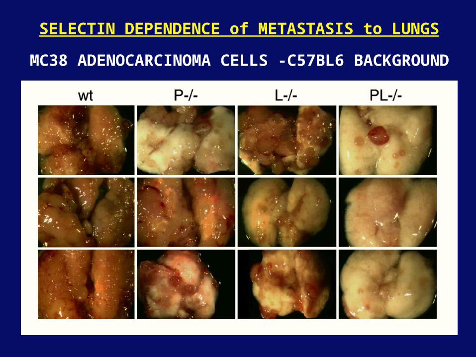
SELECTIN DEPENDENCE of METASTASIS to LUNGS
MC38 ADENOCARCINOMA CELLS -C57BL6 BACKGROUND
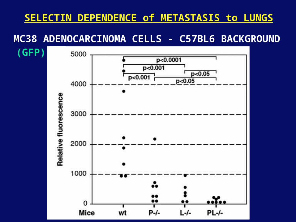
SELECTIN DEPENDENCE of METASTASIS to LUNGS
MC38 ADENOCARCINOMA CELLS - C57BL6 BACKGROUND(GFP)
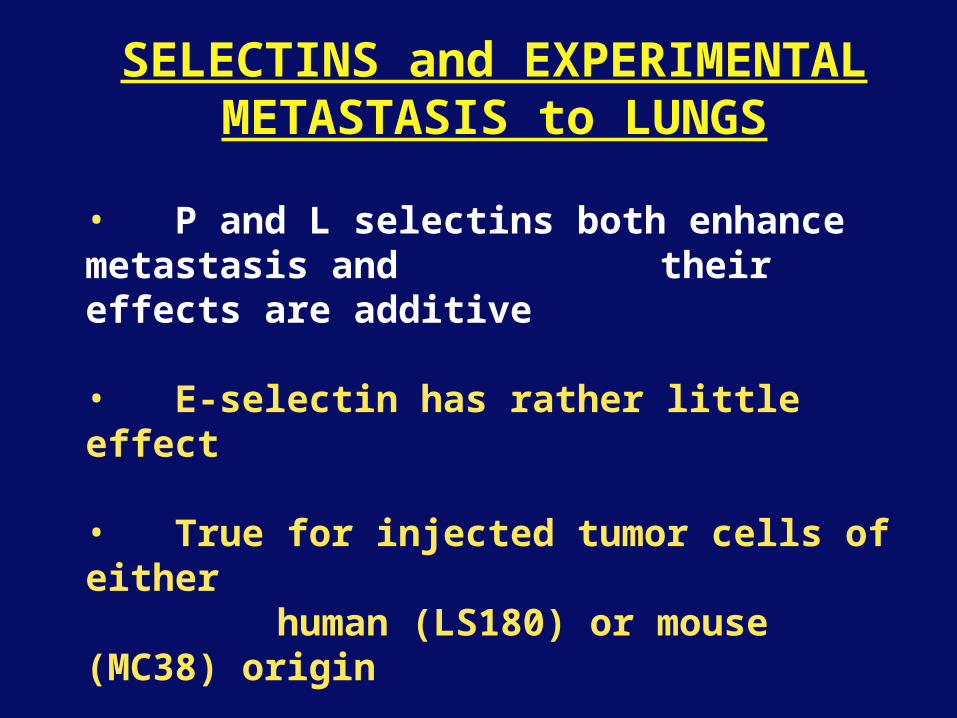
SELECTINS and EXPERIMENTALMETASTASIS to LUNGS
• P and L selectins both enhance metastasis and their effects are additive
• E-selectin has rather little effect
• True for injected tumor cells of eitherhuman (LS180) or mouse (MC38)
origin
• Selectin ligands on the tumor cells may be contributing to metastasis
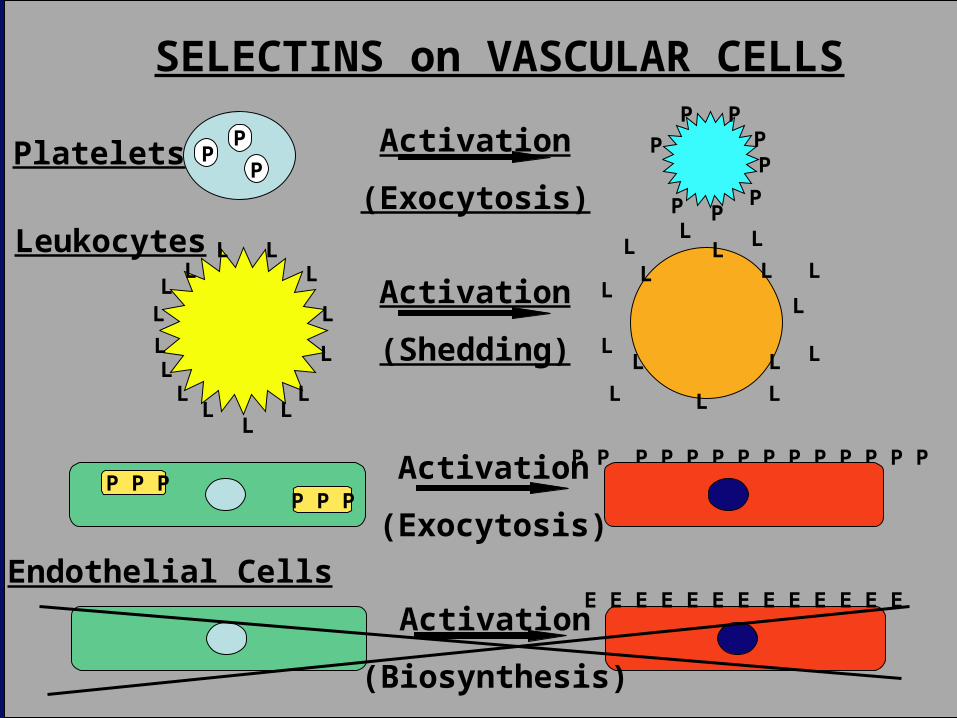
Endothelial CellsE E E E E E E E E E E E E
Activation
(Biosynthesis)
P P P P P P P P P P P P P PP P P
P P P
Activation
(Exocytosis)
SELECTINS on VASCULAR CELLS
L
Platelets
Leukocytes
PP
PP P
PP
PPP
PActivation
(Exocytosis)
Activation
(Shedding) L
L L
L
L
L
L
P
L
L
L
LLL
LL
L
LL
LL
L
LL L
L
L
L
L
LL
S-Lex
S-LexS-Lex
S-Lex
S-LexS-Lex
S-Lex
S-Lex
S-Lex
S-Lex S-Lex
SELECTINS, LIGANDS, PLATELETS, LEUKOCYTES and METASTASIS
S-Lex
S-Lex
S-Lex
S-Lex
S-Lex
S-Lex
P P P P P P P P P P P P P PP P P
P P P
Activation
(Exocytosis)
L L
L
L
L
L
P
P
PP
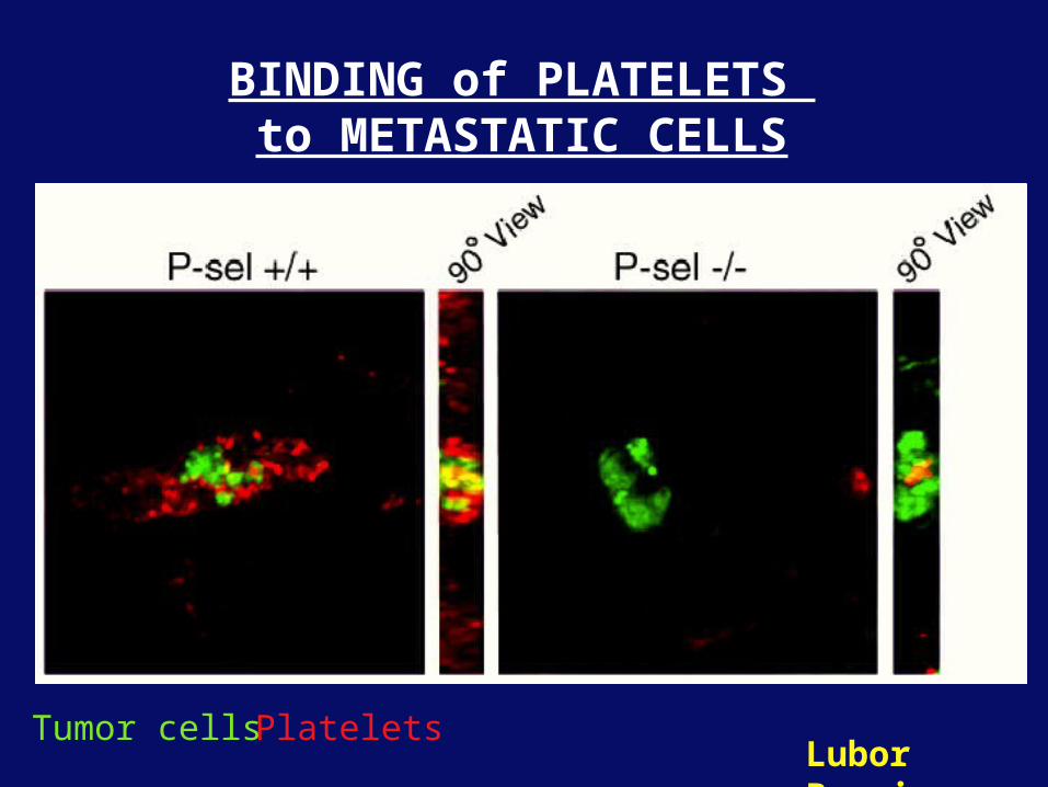
BINDING of PLATELETS to METASTATIC CELLS
Lubor BorsigTumor cells Platelets
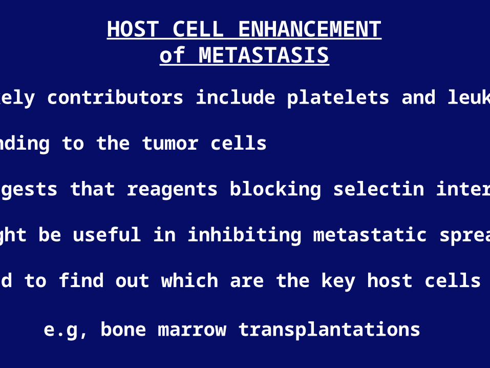
• Likely contributors include platelets and leukocytes
binding to the tumor cells
• Suggests that reagents blocking selectin interactions
might be useful in inhibiting metastatic spread
• Need to find out which are the key host cells
e.g, bone marrow transplantations
HOST CELL ENHANCEMENTof METASTASIS
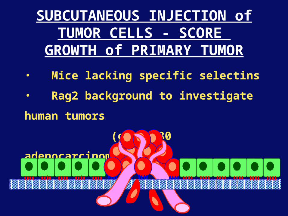
SUBCUTANEOUS INJECTION ofTUMOR CELLS - SCORE
GROWTH of PRIMARY TUMOR
• Mice lacking specific selectins
• Rag2 background to investigate human tumors
(eg.LS180 adenocarcinoma)
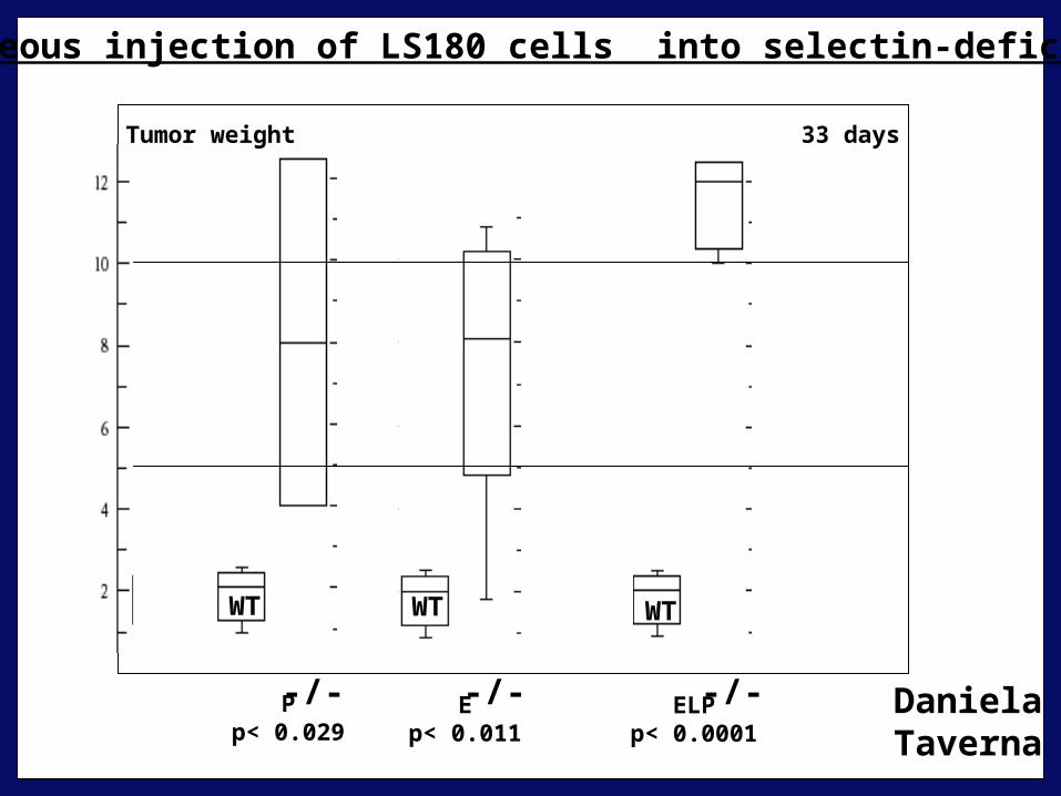
Subcutaneous injection of LS180 cells into selectin-deficient mice
Pp< 0.029
Ep< 0.011
ELPp< 0.0001
33 daysTumor weight
DanielaTaverna
-/- -/- -/-
WT WT WT
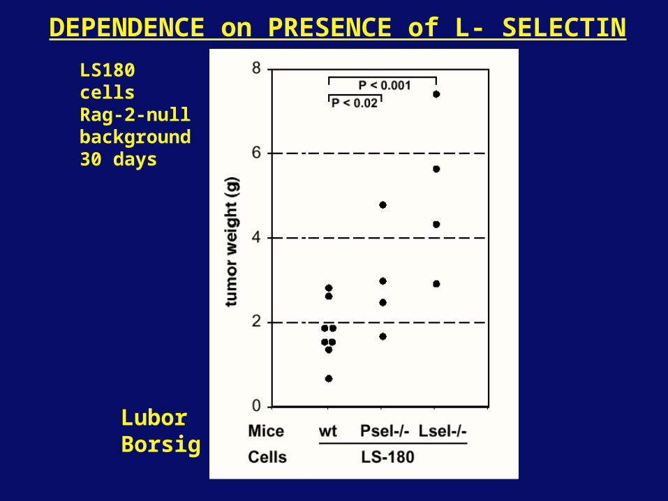
DEPENDENCE on PRESENCE of L- SELECTIN
LS180 cellsRag-2-null background30 days
LuborBorsig
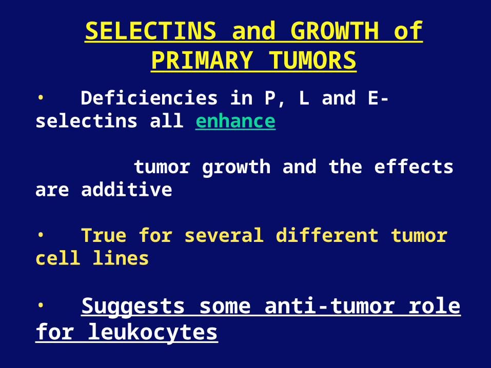
SELECTINS and GROWTH ofPRIMARY TUMORS
• Deficiencies in P, L and E-selectins all enhance
tumor growth and the effects are additive
• True for several different tumor cell lines
• Suggests some anti-tumor role for leukocytes
• Rag-2 -/- mice lack B, T and NK-T cells
• Macrophages, NK cells, platelets, endothelium ???
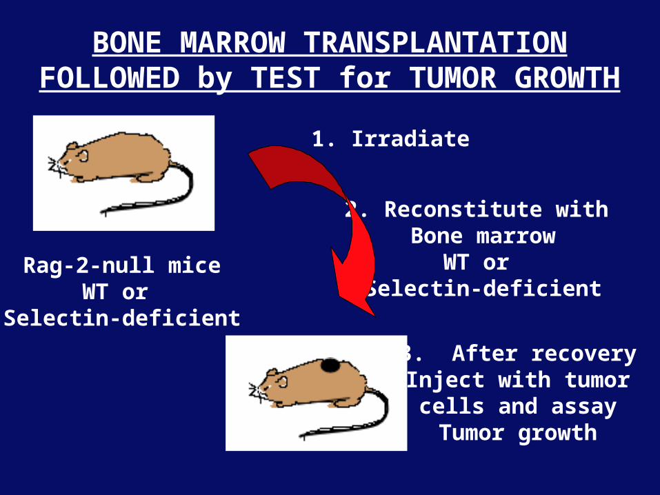
BONE MARROW TRANSPLANTATIONFOLLOWED by TEST for TUMOR GROWTH
Rag-2-null miceWT or
Selectin-deficient
2. Reconstitute with Bone marrow
WT or Selectin-deficient
1. Irradiate
3. After recoveryInject with tumor
cells and assayTumor growth
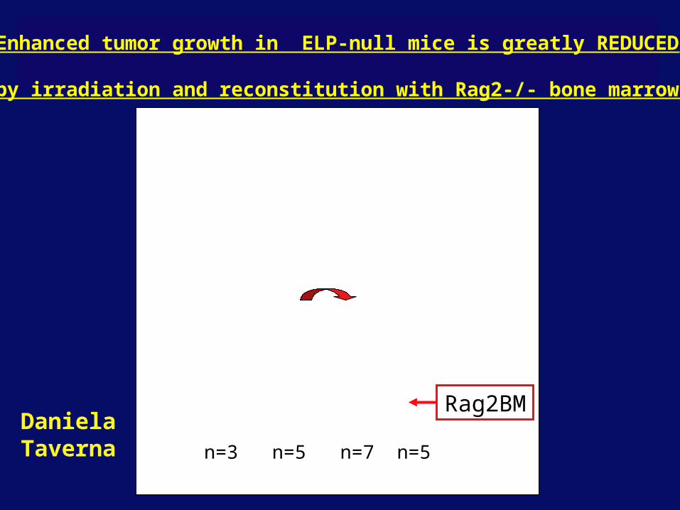
Enhanced tumor growth in ELP-null mice is greatly REDUCED
by irradiation and reconstitution with Rag2-/- bone marrow
Rag2BM
n=3 n=5 n=7 n=5
DanielaTaverna
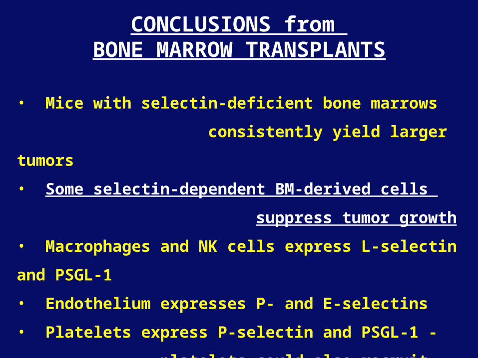
CONCLUSIONS from BONE MARROW TRANSPLANTS
• Mice with selectin-deficient bone marrows
consistently yield larger tumors
• Some selectin-dependent BM-derived cells
suppress tumor growth
• Macrophages and NK cells express L-selectin and PSGL-1
• Endothelium expresses P- and E-selectins
• Platelets express P-selectin and PSGL-1 -
platelets could also recruit other cell types

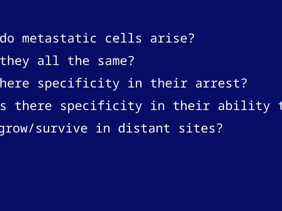
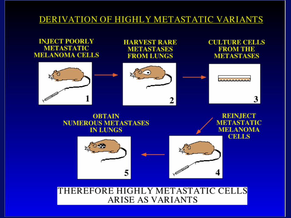
How do metastatic cells arise?
Are they all the same?
Is there specificity in their arrest?
Or is there specificity in their ability to
grow/survive in distant sites?
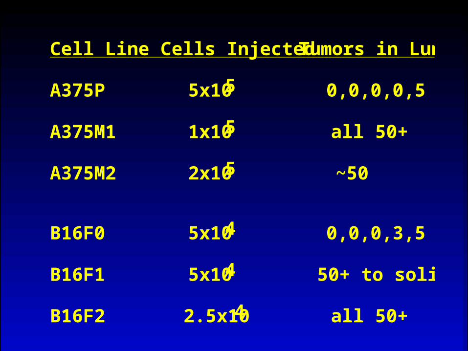
Cell Line Cells Injected Tumors in Lungs
A375P 5x10⁵ 0,0,0,0,5
A375M1 1x10⁵ all 50+
A375M2 2x10⁵ ~50
B16F0 5x10⁴ 0,0,0,3,5
B16F1 5x10⁴ 50+ to solid
B16F2 2.5x10⁴ all 50+
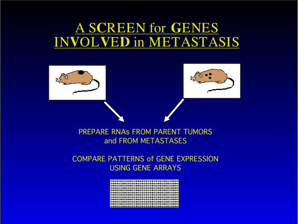
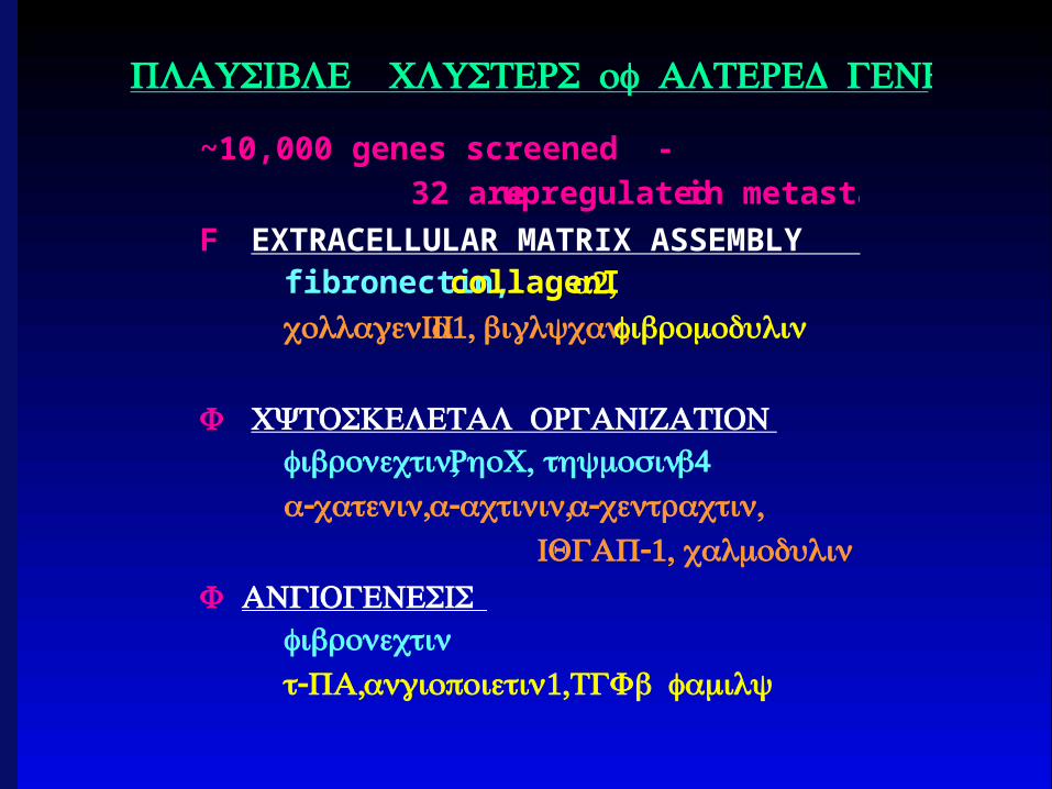
~10,000 genes screened -
32 are upregulated in metastases
F EXTRACELLULAR MATRIX ASSEMBLYfibronectin, collagenIα2,collagenIIIα1, ,biglycanfibromodulin
F CYTOSKELETAL ORGANIZATION,fibronectin ,RhoCthymosinβ4
α- ,cateninα- ,actininα- ,centractin -1,IQGAP calmodulin
F ANGIOGENESISfibronectin- ,t PA angiopoietin1,TGFβfamily
PLAUSIBLE CLUSTERS of ALTERED GENES
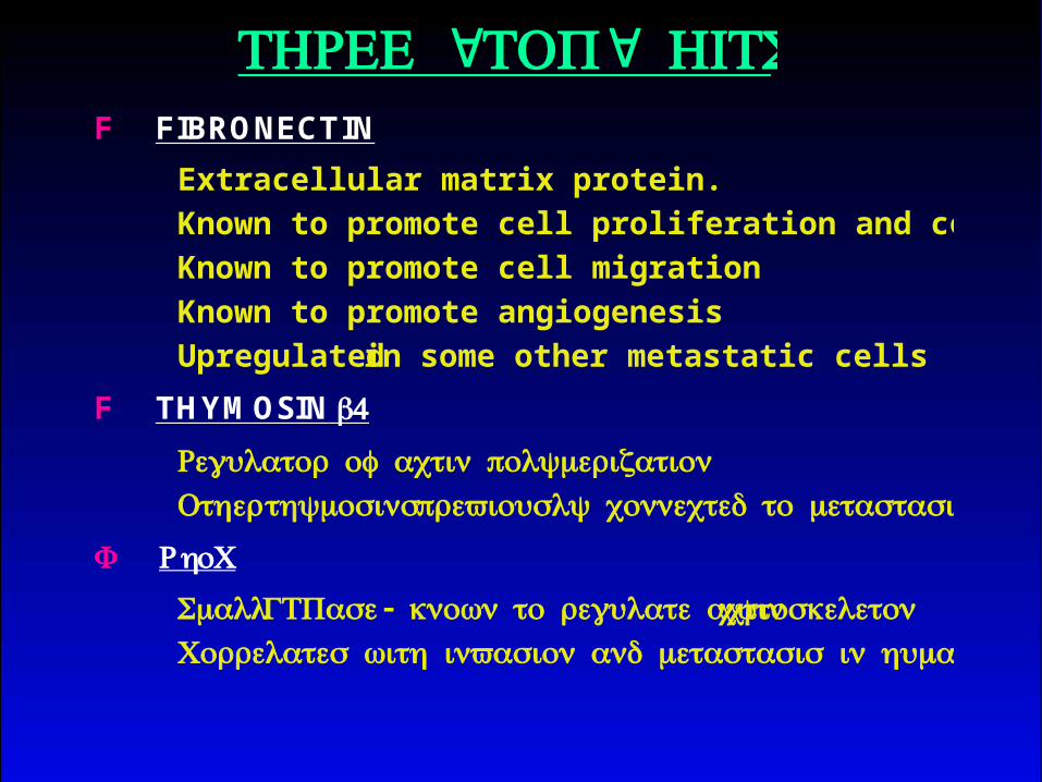
F FIBRONECTIN
Extracellular matrix protein.
Known to promote cell proliferation and cell survival
Known to promote cell migration
Known to promote angiogenesis
Upregulated in some other metastatic cells
F THYMOSIN β4
Regulator of actin polymerizationOtherthymosins previously connected to metastasis
F RhoC
SmallGTPase- known to regulate actincytoskeleton Correlates with invasion and metastasis in human cancers
" "THREE TOP HITS
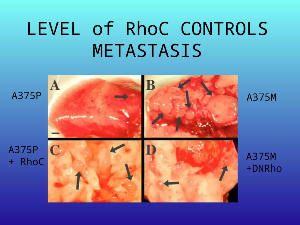
LEVEL of RhoC CONTROLS METASTASIS
A375P
A375P+ RhoC
A375M
A375M+DNRho
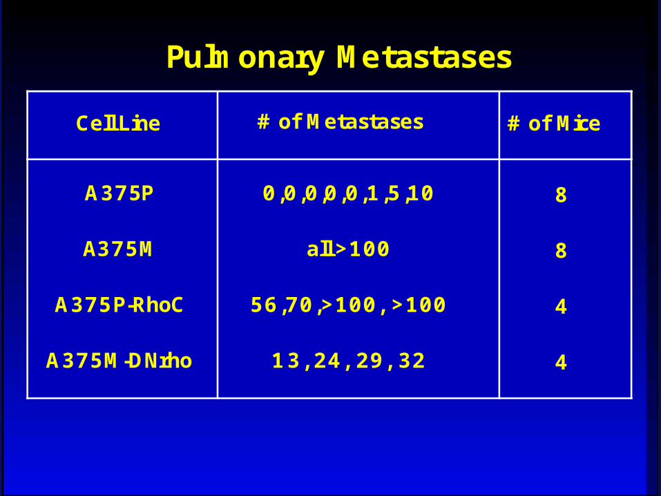
Cell Line # of Metastases # of Mice
A375P
A375M
A375P-RhoC
A375M-DNrho
0,0,0,0,0,1,5,10
all >100
56,70,>100, >100
13, 24, 29, 32
8
8
4
4
Pulmonary Metastases
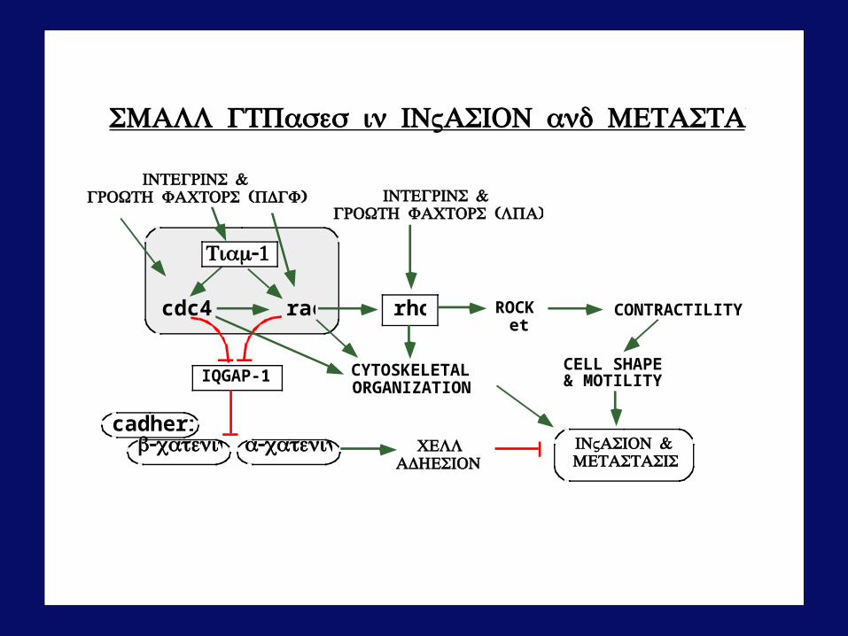
cdc42 rac rho ROCKetc
CONTRACTILITY
IQGAP-1 CYTOSKELETALORGANIZATION
CELL SHAPE& MOTILITY
cadherinα-cateninβ-catenin CELL
ADHESION&INVASION
METASTASIS
&INTEGRINS ( )GROWTH FACTORS PDGF &INTEGRINS
( )GROWTH FACTORS LPA
-1Tiam
SMALL GTPases in INVASION and METASTASIS
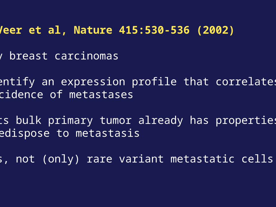
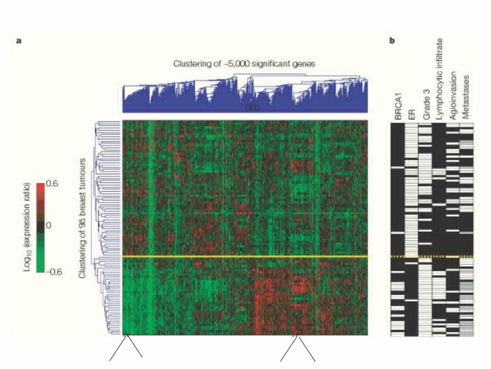
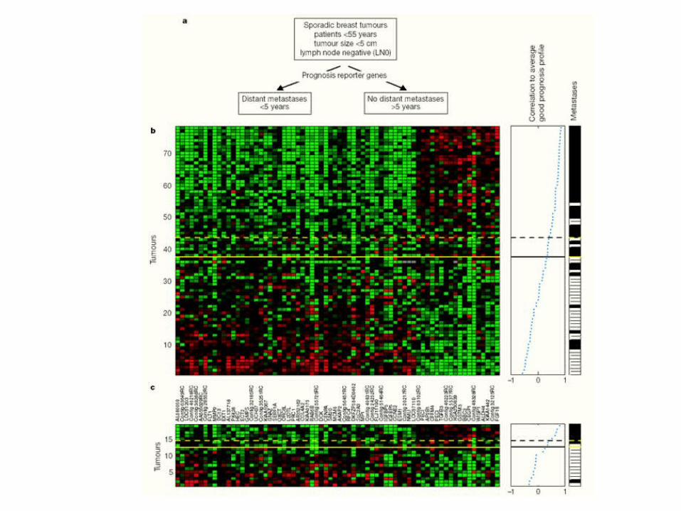
Van’t Veer et al, Nature 415:530-536 (2002)
Primary breast carcinomas
Can identify an expression profile that correlates withincidence of metastases
Suggests bulk primary tumor already has properties that predispose to metastasis
That is, not (only) rare variant metastatic cells
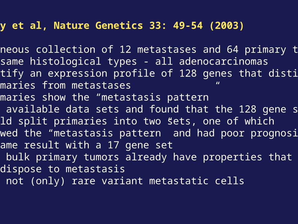
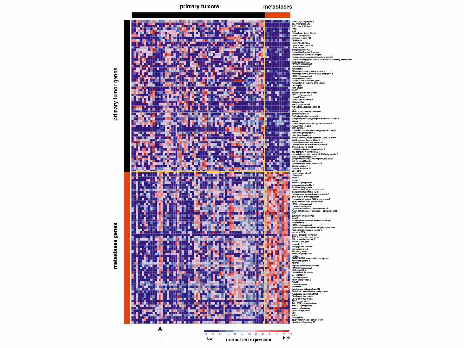
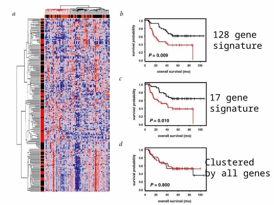
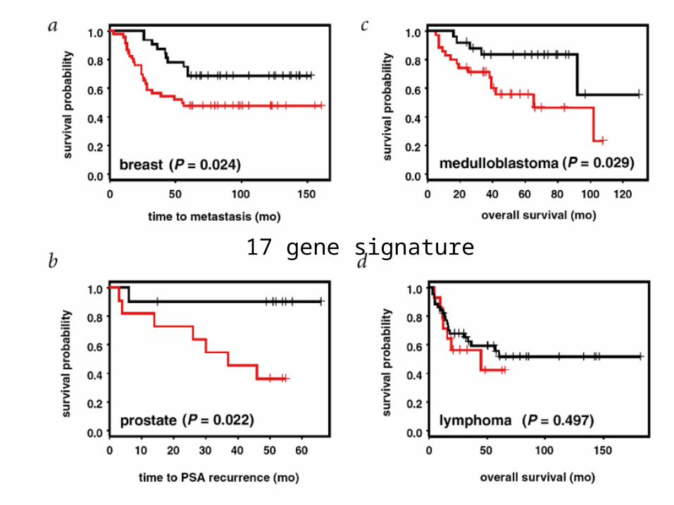
Ramaswamy et al, Nature Genetics 33: 49-54 (2003)
Miscellaneous collection of 12 metastases and 64 primary tumors of same histological types - all adenocarcinomas
Can identify an expression profile of 128 genes that distinguishesprimaries from metastases
Some primaries show the “metastasis pattern”Analyzed available data sets and found that the 128 gene set
could split primaries into two sets, one of whichshowed the “metastasis pattern” and had poor prognosis- same result with a 17 gene set
Suggests bulk primary tumors already have properties that predispose to metastasis
That is, not (only) rare variant metastatic cells
17 gene signature
128 gene signature
Clusteredby all genes
17 gene signature
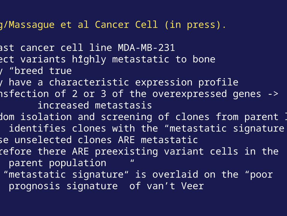
Kang/Massague et al Cancer Cell (in press).
Breast cancer cell line MDA-MB-231Select variants highly metastatic to boneThey “breed true”They have a characteristic expression profileTransfection of 2 or 3 of the overexpressed genes ->
increased metastasisRandom isolation and screening of clones from parent line
identifies clones with the “metastatic signature”These unselected clones ARE metastaticTherefore there ARE preexisting variant cells in the
parent populationThe “metastatic signature” is overlaid on the “poor
prognosis signature” of van’t Veer
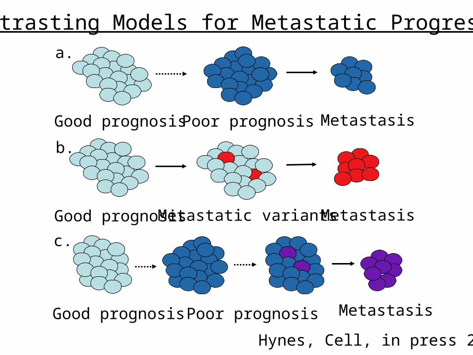
Good prognosis Poor prognosis Metastasis
a.
Good prognosis Metastatic variants Metastasis
b.
Good prognosis Poor prognosis
c.
Metastasis
Contrasting Models for Metastatic Progression
Hynes, Cell, in press 2003
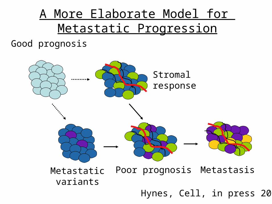
Good prognosis
Poor prognosis Metastasis
Stromal response
Metastaticvariants
A More Elaborate Model for Metastatic Progression
Hynes, Cell, in press 2003
SS
SS
SS
SS
SS
ANTIBODIES
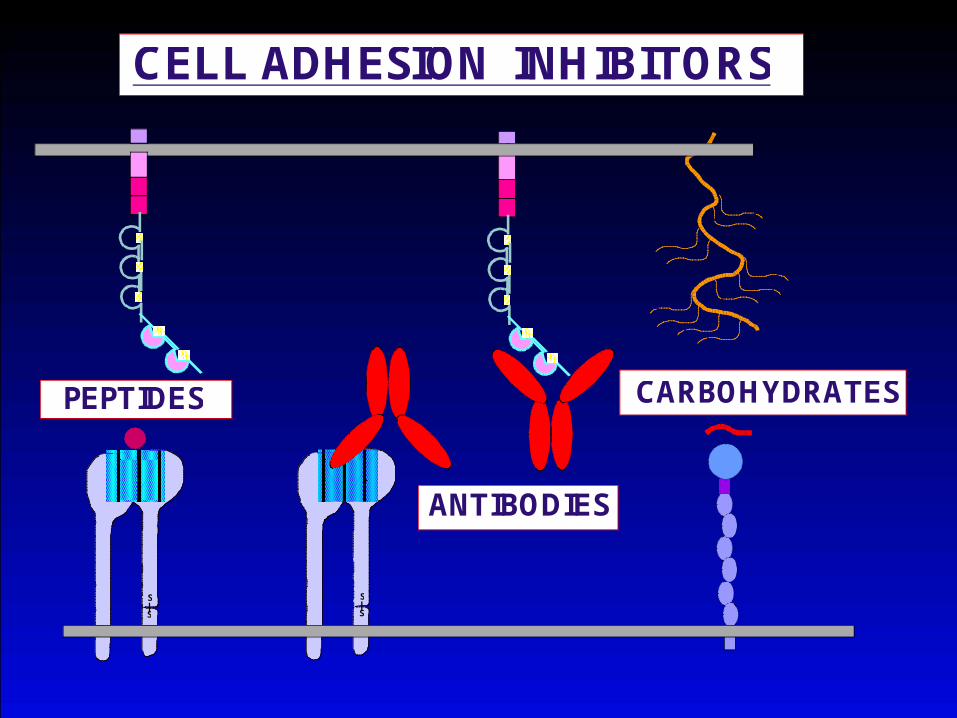
CELL ADHESION INHIBITORS
S
S
S
S
SS
SS
SS
SS
SS
CARBOHYDRATESPEPTIDES
Recommended