Safety and Clinical Performance of the Safety and Clinical Performance of the Drug Eluting Absorbable Metal Scaffold Drug Eluting Absorbable Metal Scaffold
in the Treatment of Subjects with de in the Treatment of Subjects with de Novo Lesions in Native Coronary Novo Lesions in Native Coronary
Arteries-BIOSOLVE-II Arteries-BIOSOLVE-II
Michael Haude, MDMichael Haude, MDOn behalf of the BIOSOLVE-II InvestigatorsOn behalf of the BIOSOLVE-II Investigators
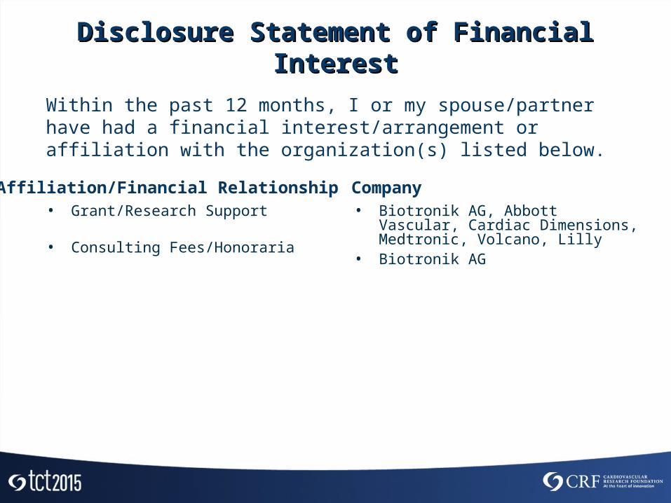
Disclosure Statement of Financial InterestDisclosure Statement of Financial Interest
• Grant/Research Support
• Consulting Fees/Honoraria
• Biotronik AG, Abbott Vascular, Cardiac Dimensions, Medtronic, Volcano, Lilly
• Biotronik AG
Within the past 12 months, I or my spouse/partner have had a financial interest/arrangement or affiliation with the organization(s) listed below.
Affiliation/Financial Relationship Company

BackgroundBackgroundEvolution of the BIOTRONIK Magnesium ScaffoldEvolution of the BIOTRONIK Magnesium Scaffold
*Composite of cardiac death, target vessel myocardial infarction, clinically driven target lesion revascularization and CABG

Study DesignStudy Design121 patients with de novo coronary artery
stenosis and successful DREAMS 2G implantation (2.5x20, 3.0x20, 3.5x25 mm)
1 month, Clinical FUP
6 month •Clinical FUP (mandatory)•Angiographic FUP (mandatory) •IVUS / OCT (Subgroup only) •Vasomotion (Subgroup only)
12 month •Clinical FUP (mandatory)•Angiographic FUP (voluntary)•IVUS / OCT (voluntary)•Vasomotion (voluntary)
3 year, Clinical FUP
2 year, Clinical FUP
DESIGN Prospective, multi-center, FIM.
PRIMARY ENDPOINT In-segment late lumen loss @ 6-month
COORDINATING CLINICAL INVESTIGATOR Prof. M.Haude, Lukaskrankenhaus GmbH,
Neuss, Germany
CORELAB Cardialysis, Rotterdam, The Netherlands
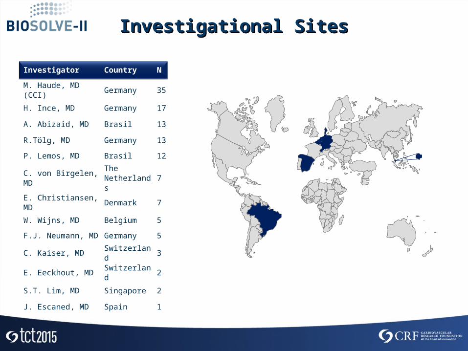
Investigator Country N
M. Haude, MD (CCI) Germany 35
H. Ince, MD Germany 17
A. Abizaid, MD Brasil 13
R.Tölg, MD Germany 13
P. Lemos, MD Brasil 12
C. von Birgelen, MDThe Netherlands
7
E. Christiansen, MD Denmark 7
W. Wijns, MD Belgium 5
F.J. Neumann, MD Germany 5
C. Kaiser, MD Switzerland 3
E. Eeckhout, MD Switzerland 2
S.T. Lim, MD Singapore 2
J. Escaned, MD Spain 1
Investigational SitesInvestigational Sites
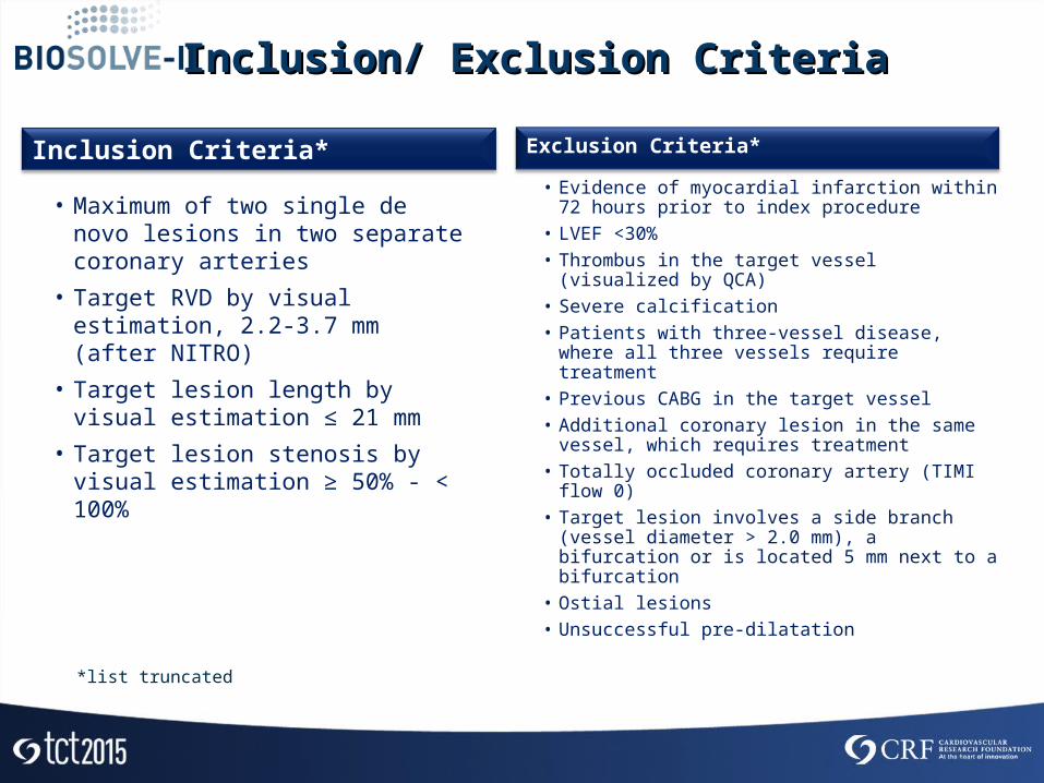
Inclusion Criteria*
• Maximum of two single de novo lesions in two separate coronary arteries
• Target RVD by visual estimation, 2.2-3.7 mm (after NITRO)
• Target lesion length by visual estimation ≤ 21 mm
• Target lesion stenosis by visual estimation ≥ 50% - < 100%
Inclusion/ Exclusion CriteriaInclusion/ Exclusion Criteria
Exclusion Criteria*
• Evidence of myocardial infarction within 72 hours prior to index procedure
• LVEF <30%• Thrombus in the target vessel (visualized by
QCA)• Severe calcification• Patients with three-vessel disease, where all
three vessels require treatment• Previous CABG in the target vessel• Additional coronary lesion in the same vessel,
which requires treatment• Totally occluded coronary artery (TIMI flow 0)• Target lesion involves a side branch (vessel
diameter > 2.0 mm), a bifurcation or is located 5 mm next to a bifurcation
• Ostial lesions• Unsuccessful pre-dilatation
*list truncated
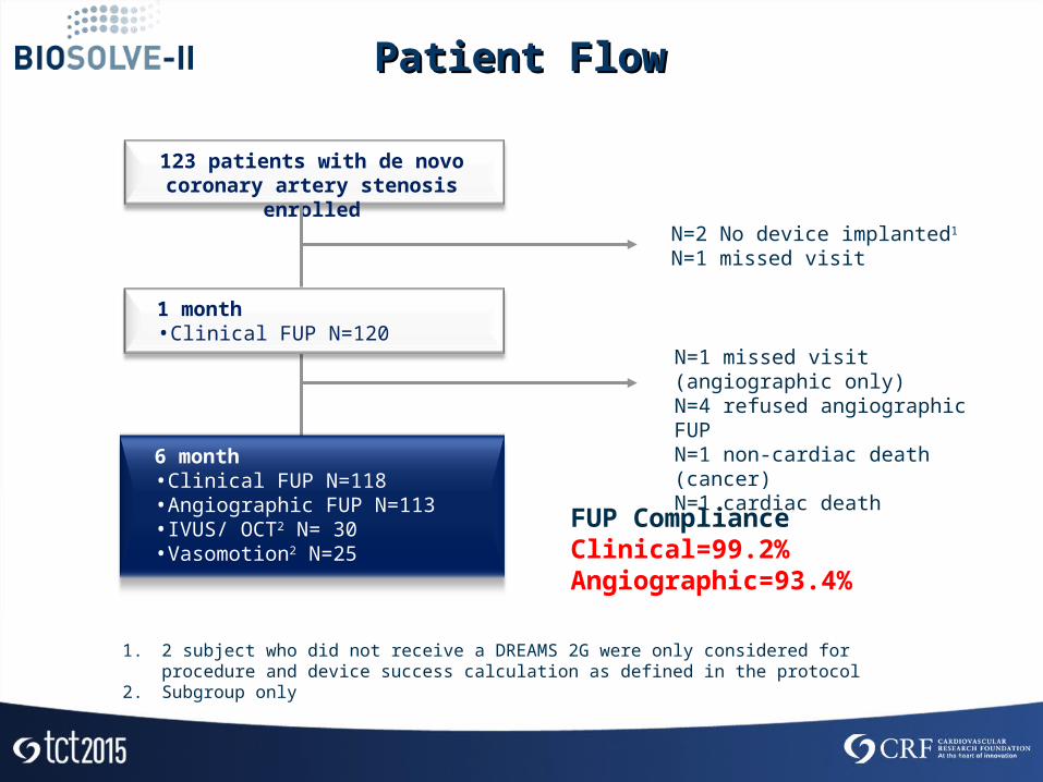
Patient FlowPatient Flow
123 patients with de novo coronary artery stenosis enrolled
1 month •Clinical FUP N=120
6 month •Clinical FUP N=118 •Angiographic FUP N=113 •IVUS/ OCT2 N= 30•Vasomotion2 N=25
N=2 No device implanted1
N=1 missed visit
N=1 missed visit (angiographic only)N=4 refused angiographic FUPN=1 non-cardiac death (cancer)N=1 cardiac death
FUP Compliance Clinical=99.2%Angiographic=93.4%
1. 2 subject who did not receive a DREAMS 2G were only considered for procedure and device success calculation as defined in the protocol
2. Subgroup only

Lesion Characteristics N (%)
Lesion Length (mm ± SD) 12.61 ± 4.53
RVD (mm ± SD) 2.68 ± 0.40
AHA/ ACC Lesion Class B2/C 53 (43.8)
CalcificationModerate/Severe
13 (10.6)
Baseline & Lesion Characteristics Baseline & Lesion Characteristics N=123N=123
Baseline Characteristics N (%)
Age (mean ± SD) 65.2±10.3
Male 78 (63.4)
Hypertension 101 (82.1)
Hyperlipidemia 74 (60.2)
Smoking 67 (54.5)
Diabetes mellitus 36 (29.3)
Insulin dependent 11 (30.6)
Non-Insulin dependent 25 (69.4)
History of MI 29 (23.6)
Previous percutaneous Intervention
44 (35.8)
Lesion Location N (%)
LAD 47 (38.2)
LCx 29 (23.6)
RCA 45 (36.6)
Intermediate Branch 2 (1.6)

BaselineN=123
Post-ProcedureN=121
6-monthN=113
Lesion Length (mm) 12.61±4.53 NA NA
In-segment RVD (mm) 2.68±0.40 2.69±0.39 2.55±0.41
In-scaffold RVD (mm) NA 2.78±0.36 2.59±0.40
In-segment MLD (mm) 1.19±0.32 2.16±0.40 1.89±0.43
In-scaffold MLD (mm) NA 2.45±0.32 2.00±0.44
In-segment acute gain (mm) NA 0.96±0.40 NA
In-scaffold acute gain (mm) NA 1.25±0.35 NA
In-segment DS (%) 55.2±10.3 19.7±8.3 25.9±12.3
In-scaffold DS (%) NA 11.7±5.2 22.6±12.9
In-segment LLL (mm) NA NA 0.27±0.37
In-scaffold LLL (mm) NA NA 0.44±0.36
In-segment Binary Restenosis (%) NA NA 5.4
In-Scaffold Binary Restenosis (%) NA NA 5.4
QCA ResultsQCA Results
Data are presented as mean ±SD unless aotherwise stated; NA = not applicable

Vasomotion Results at 6-month (N=25)Vasomotion Results at 6-month (N=25)Mean Lumen Diameter
Proximal (mm±SD)
Mean Lumen DiameterScaffold (mm±SD)
Mean Lumen DiameterDistal
(mm±SD)2.68±0.45 2.57±0.56 2.76±0.46 2.60±0.29 2.49±0.34 2.66±0.33 2.39±0.35 2.09±0.50 2.39±0.40
80% (20/25) demonstrate ≥ 3% vasomotion after Ach or Nitro
Ach = Acetylcholine
Nitro = Nitroglycerine

Post-procedure 6-month ∆6-month vs post[95% CI]
p-value
Vessel area (mm2) 14.06±3.17 14.21±3.14 0.15[-0.13-0.42] 0.289
Scaffold area (mm2) 6.24±1.15 6.21±1.22 -0.03[-0.29-023] 0.803
Plaque area (mm2) 7.76±2.41 8.06±2.23 0.29[0.11-0.47] 0.002
NIH area (mm2) NA 0.08±0.09 NA NA
IVUS AnalysisIVUS AnalysisSubgroup N=30Subgroup N=30
NIH area
Scaffold Area
Lumen area Vessel area
Plaque area
NA = Not Applicable
NIH= Neointimal Hyperplasia
Compared to BIOSOLVE-I NIH Area was reduced by 73% from 0.30±0.41mm2 to 0.08±0.09mm2
Post-procedure 6-month
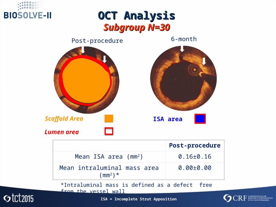
OCT AnalysisOCT AnalysisSubgroup N=30Subgroup N=30
ISA areaScaffold Area
Lumen area
Post-procedure
Mean ISA area (mm2) 0.16±0.16
Mean intraluminal mass area (mm2)* 0.00±0.00
Post-procedure 6-month
*Intraluminal mass is defined as a defect free from the vessel wall
ISA = Incomplete Strut Apposition
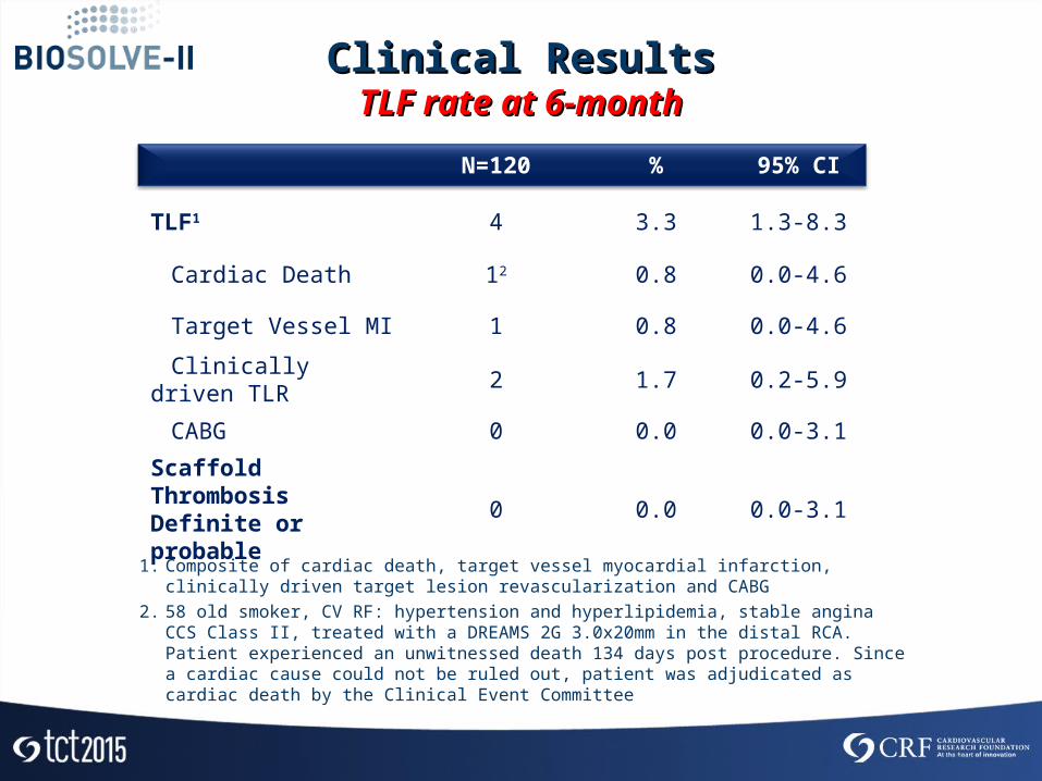
N=120 % 95% CI
TLF1 4 3.3 1.3-8.3
Cardiac Death 12 0.8 0.0-4.6
Target Vessel MI 1 0.8 0.0-4.6
Clinically driven TLR 2 1.7 0.2-5.9
CABG 0 0.0 0.0-3.1
Scaffold Thrombosis Definite or probable 0 0.0 0.0-3.1
Clinical ResultsClinical ResultsTLF rate at 6-monthTLF rate at 6-month
1. Composite of cardiac death, target vessel myocardial infarction, clinically driven target lesion revascularization and CABG
2. 58 old smoker, CV RF: hypertension and hyperlipidemia, stable angina CCS Class II, treated with a DREAMS 2G 3.0x20mm in the distal RCA. Patient experienced an unwitnessed death 134 days post procedure. Since a cardiac cause could not be ruled out, patient was adjudicated as cardiac death by the Clinical Event Committee
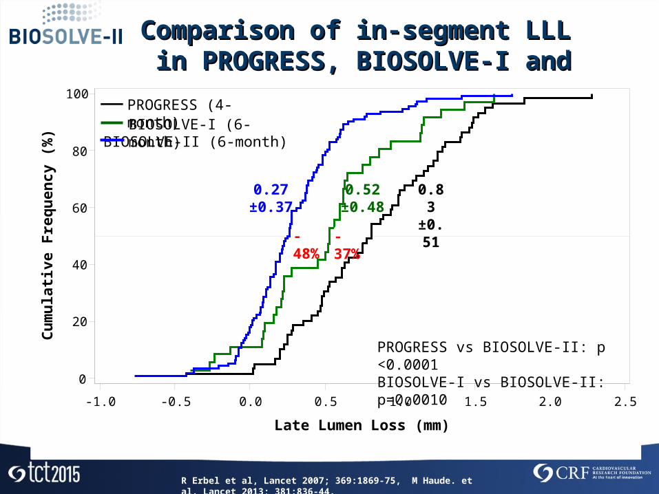
Comparison of in-segment LLL Comparison of in-segment LLL in PROGRESS, BIOSOLVE-I and BIOSOLVE-IIin PROGRESS, BIOSOLVE-I and BIOSOLVE-II
-1.0 -0.5 0.0 0.5 1.0 1.5 2.0 2.5
Late Lumen Loss (mm)
0
20
40
60
80
100
Cum
ulati
ve F
requ
ency
(%)
0.83±0.51
0.52±0.48
0.27±0.37
BIOSOLVE-I (6-month)PROGRESS (4-month)
BIOSOLVE-II (6-month)
PROGRESS vs BIOSOLVE-II: p <0.0001BIOSOLVE-I vs BIOSOLVE-II: p=0.0010
R Erbel et al, Lancet 2007; 369:1869-75, M Haude. et al. Lancet 2013; 381:836-44.
-48% -37%
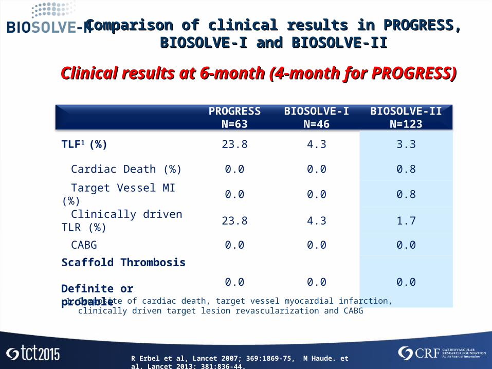
PROGRESS
N=63BIOSOLVE-I
N=46BIOSOLVE-II
N=123
TLF1 (%) 23.8 4.3 3.3
Cardiac Death (%) 0.0 0.0 0.8
Target Vessel MI (%) 0.0 0.0 0.8
Clinically driven TLR (%) 23.8 4.3 1.7
CABG 0.0 0.0 0.0
Scaffold Thrombosis Definite or probable 0.0 0.0 0.0
Clinical results at 6-month (4-month for PROGRESS)Clinical results at 6-month (4-month for PROGRESS)
1. Composite of cardiac death, target vessel myocardial infarction, clinically driven target lesion revascularization and CABG
Comparison of clinical results in PROGRESS, BIOSOLVE-I and Comparison of clinical results in PROGRESS, BIOSOLVE-I and BIOSOLVE-IIBIOSOLVE-II
R Erbel et al, Lancet 2007; 369:1869-75, M Haude. et al. Lancet 2013; 381:836-44.

ConclusionConclusion
• DREAMS 2G in BIOSOLVE-II demonstrates significantly improved in-segment LLL (0.27±0.37mm) compared to it`s precursor devices tested in the PROGRESS (0.83±0.37mm) and the BIOSOLVE-I study (0.52±0.48mm)
• Vasomotion of the scaffolded vessel segment was demonstrated at 6 months
• IVUS results in a subgroup of 30 subjects demonstrate a preservation of the scaffold area with a low neo-intimal area at 6-month
• No intra-luminal masses were observed by OCT at any time in a subgroup of 25 subjects
• DREAMS 2G in BIOSOLVE-II demonstrates a low TLF (3.3%) and TLR (1.7%) rate at 6-month, which is comparable to other absorbable scaffolds and permanent drug eluting stents
• No definite or probable scaffold thrombosis was observed with DREAMS 2G tested in BIOSOLVE-II or any of it`s precursor devices tested in PROGRESS and BIOSOLVE-I in a total of 232 subjects

Published Online in THE LANCET on October 12, 2015Published Online in THE LANCET on October 12, 2015http://dx.doi.org/10.1016/S0140-6736(15)00447-Xhttp://dx.doi.org/10.1016/S0140-6736(15)00447-X

acute 3-12 months 12 months1 month
Mgabsorption
Mgabsorption
Mgabsorption
Biotronik Magnesium ScaffoldBiotronik Magnesium ScaffoldMagnesium Absorption ProcessMagnesium Absorption Process
Recommended