University of Massachusetts Amherst
Up-regulating Apoptosis in Cancerous Cells using the Bcl-2 Family of Proteins and Apoptosis
Biology 499T/P Honors Thesis
Natalie L. Saloio5/3/2016
Saloio 1
ABSTRACT
The Bcl-2 family of proteins plays a crucial role in negatively and positively regulating
apoptosis. Cancerous phenotypes create a cellular environment where apoptosis is negatively
regulated, which makes manipulations of the Bcl-2 family of proteins an appropriate method for
cancer therapy. This paper addresses potential manipulations which can be done to the pro-
apoptotic and anti-apoptotic Bcl-2 family of proteins which will increase the level of intrinsic
apoptosis in tumor cells, and act as cancer therapies. Scientists, through their literature, have
reviewed drugs which mimic the actions of the Bcl-2 family of proteins to either increase or
decrease levels of intrinsic apoptosis in cells and many therapies have been produced which
trigger the extrinsic pathway of apoptosis, but none of the processes involved manipulating the
actual Bcl-2 family of proteins and very few involve triggering the intrinsic apoptotic pathway.
This thesis proposes increasing expression of the Bcl-2 BH3-only protein, and inhibiting the BH4
domain of anti-apoptotic Bcl-2 proteins using recombinant protein expression, small molecule
inhibitors, and gene therapy. These therapeutic methods adhere to targeted cancer therapy
which allows consumption of higher doses without damaging the rest of the patient’s cells by
directing the drug to target only tumor cells, making it a more effective drug with a higher
efficacy and less side effects

Saloio 2
INTRODUCTION
Cellular Mechanisms of Apoptosis
Cell death is an existential component of living organisms. When our cells absorb too
many ultraviolet rays, radiation, toxins, and free radicals, DNA damage occurs, and our cells use
DNA repair mechanisms to mend the damage. If the damage is not repaired, the cell will be
signaled to self-destruct through pathways of programmed cell death, such as apoptosis. If the
cell’s pathways of programmed cell death are inhibited, damaged cells may accrue mutations that
lead to transformation of the cell, which will lead to tumor formation, possibly cancer, and
eventually, death of the organism.
Cell death can be regulated and accidental (Kroemer et al. 2009). Oncosis is an accidental
type of cell death, characterized by swelling of the nucleus and cytoplasm after a lethal injury
(Kroemer et al. 2009). Programmed cell death is a form of regulated cell death that is mediated
by an intracellular process (Kroemer et al. 2009). There are many forms of programmed cell
death, such as apoptosis, autophagic cell death, necroptosis, and pryoptosis, classified by their
morphological appearance, enzymological criteria, functional aspects, or immunological
characteristics (Kroemer et al. 2009). Apoptosis is a very common process of programmed cell
death that occurs in multicellular organisms (Kroemer et al. 2009).
Apoptosis is characterized by its specific morphological features, responsible for
rounding-up of the cell, retraction of pseudopodes, reduction of cellular and nuclear volume,
nuclear fragmentation, minor modification of cytoplasmic organelles, plasma membrane
blebbing, and engulfment by resident phagocytes (Kroemer et al. 2009). The three important
biochemical features of apoptosis include protein cleavage, DNA breakdown, and phagocytic
recognition. Apoptosis is a necessary function of cells throughout development and aging of

Saloio 3
organisms as a balancing mechanism to maintain cell populations in tissues (Ouyang et al. 2012).
For example, apoptosis is the process responsible for creating the space between our fingers and
toes as we are developing as human embryos. It is also required for proper functioning of the
immune system, such as eliminating activated or auto-aggressive immune cells during
maturation in the central lymphoid organs, or in peripheral tissues (Ouyang et al. 2012). To
accommodate for the different functions of apoptosis, there are two pathways it encounters.
The two pathways of apoptosis are intrinsic and extrinsic. These pathways are
differentiated by the apoptotic signals received from inside and outside the cell. The intrinsic
pathway of apoptosis is a response to signaling molecules originating from inside the cell, i.e.,
the cell kills itself because it senses stress. The signaling molecules originating inside the cell can
be triggered by severe cell stress, such as, DNA damage, chromosome rearrangement, cytotoxic
stimuli, etc. (Ouyang et al. 2012). In contrast, the extrinsic pathway of apoptosis, is a response to
signaling molecules originating from outside the cell (Ouyang et al. 2012). In extrinsic apoptosis,
receptor-mediated signals stimulate cell death, i.e., receptors receive extracellular signaling
molecules which direct the cell to kill itself. Receptors which receive the extracellular signals
and initiate extrinsic apoptosis are the tumor necrosis factor (TNF) receptor and the first
apoptosis signal (FAS) receptor. A process which would trigger receptor-mediated signals to
initiate the external pathway of apoptosis is modulation of immune function, e.g., extrinsic
apoptosis is used by white blood cells to kill viral infections (Ouyang et al. 2012). Another
process the extrinsic apoptotic pathway is used for is the separation of human toes and fingers
during development (Ouyang et al. 2012). Both intrinsic and extrinsic pathways are required for
proper organism function, especially when there are malfunctions in cellular pathways.
The Relationship Between Apoptosis and Cancer
Saloio 4
Cells with malfunctioning apoptotic pathways lack the ability to activate programmed
cell death processes, such as apoptosis: one defining characteristic of a cancerous cell phenotype
(Ouyang et al. 2012). In signal transduction pathways which promote cell survival and growth,
such as the Protein Kinase B (AKT) pathway, pro-apoptotic proteins are always active unless an
extracellular survival/growth signal binds to the survival/growth receptor (Zheng et al. 2015).
When the survival/growth signal binds to the survival/growth receptor, an anti-apoptotic protein
will inhibit apoptosis (Zheng et al. 2015). An example of a mutation which could cause cancer in
the AKT signaling pathway is if the pro-apoptotic protein, in charge of initiating apoptosis, had a
loss-of-function mutation, where it no longer initiated apoptosis: this would lead to non-stop cell
reproduction (an over proliferation of cells). Another example of a mutation in the AKT
signaling pathway which could cause cancer is a gain-of-function mutation in the anti-apoptotic
protein, where it is constitutively active, inhibiting apoptosis: this would also lead to an over
1proliferation of cells. Although the two mutations affect two different proteins in the pathway,
the end result for both was the cell’s inability to initiate apoptosis. The pro-apoptotic and anti-
apoptotic proteins in the AKT pathway are both a part of the Bcl2-family of proteins. After
gaining an understanding of how a lack of apoptosis can lead to cancer, one can propose
potential cancer therapy methods by finding mechanisms to ensure that apoptosis is activated in
cancerous cells using the Bcl-2 family of proteins.
Bcl-2 Family of Proteins
The B-cell lymphoma 2 (Bcl-2) family of intracellular proteins located in the cytoplasm
and mitochondria are one of the many proteins which regulate intrinsic apoptosis (Shamas-Din et
al. 2011). There are two sub-families of Bcl-2 proteins, categorized by whether they positively
(pro-apoptotic Bcl-2) or negatively (anti-apoptotic Bcl-2) regulate apoptosis (Shamas-Din et al.

Saloio 5
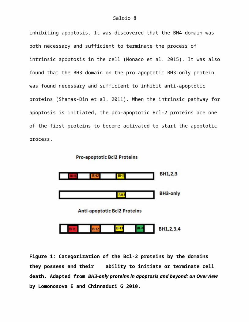
2011). The anti-apoptotic Bcl2 proteins which negatively regulate apoptosis have domains 1,2, 3,
and 4 (BH1,2,3,4). The pro-apoptotic Bcl-2 proteins that positively regulate apoptosis have two
sub-families of proteins, differentiated by their domains: BH3-only and BH1,2,3 (Shamas-Din et
al. 2011). Bcl-2 anti-apoptotic proteins have one domain which the Bcl-2 pro-apoptotic proteins
lack; the BH4 domain (Shamas-Din et al. 2011). Since the BH4 domain is the only domain
differentiating pro-apoptotic from anti-apoptotic activity in the Bcl-2 family of proteins,
scientists have inquired whether it was a key player in inhibiting apoptosis. It was discovered
that the BH4 domain was both necessary and sufficient to terminate the process of intrinsic
apoptosis in the cell (Monaco et al. 2015). It was also found that the BH3 domain on the pro-
apoptotic BH3-only protein was found necessary and sufficient to inhibit anti-apoptotic proteins
(Shamas-Din et al. 2011). When the intrinsic pathway for apoptosis is initiated, the pro-apoptotic
Bcl-2 proteins are one of the first proteins to become activated to start the apoptotic process.
Figure 1: Categorization of the Bcl-2 proteins by the domains they possess and their
ability to initiate or terminate cell death. Adapted from BH3-only proteins in apoptosis and
beyond: an Overview by Lomonosova E and Chinnaduri G 2010.

Saloio 6
The Process of Intrinsic Apoptosis
Bcl-2 Pro-apoptotic Protein Dimerization and Mitochondrial Outer Membrane Permeabilization
When internal cellular stress triggers the initiation of the intrinsic pathway of apoptosis,
the BH3-only pro-apoptotic proteins inhibit the anti-apoptotic proteins. BH3-only proteins
essentially inhibit the inhibitors, by binding the anti-apoptotic Bcl-2 proteins and forming a
sequestration complex. After the BH3-only proteins inhibit the inhibitors, the pro-apoptotic Bcl-
2 proteins are set free in the cytoplasm and their activity is encouraged (Tait and Green 2010).
Two of the same pro-apoptotic Bcl-2 proteins, Bcl-2 antagonist killer (BAK), dimerize through
their BH3 domains, producing a homodimer. One BAK is located in the cytoplasm and the other
on the outer membrane of the mitochondria. The dimerization of the two BAK Bcl-2 pro-
apoptotic proteins, initiates mitochondrial outer membrane permeabilization (Tait and Green
2010). The BAK protein localized in the cytoplasm is controlled by anti-apoptotic Bcl-2
proteins when apoptosis is not taking place. When one of the BAK proteins is activated its BH3
domain is exposed, and is inserted into the hydrophobic groove of another BAK molecule (Tait
and Green 2010). When BAK in the cytoplasm and BAK on the outer membrane of the
mitochondria are not dimerizing, and apoptosis is not taking place, an anti-apoptotic Bcl-2
protein is inhibiting BAK in the cytoplasm. When the cell is signaled to apoptosis, a Bcl-2 BH-3-
only protein localized in the cytoplasm inhibits the anti-apoptotic Bcl-2 protein, allowing BAK
in the cytoplasm to dimerize with BAK on the outer mitochondrial membrane.
Mitochondria contain an outer and inner phospholipid bilayer membrane separated by the
intermembrane space differentiated by their protein and lipid compositions (Tait and Green
2010). The inner membrane is composed of about twenty percent lipids and eighty percent
proteins, making it only permeable to oxygen, carbon dioxide, and water (Tait and Green 2010).
In contrast, the outer membrane is composed of fifty percent lipids and fifty percent proteins

Saloio 7
allowing the membrane to be highly permeable to small proteins like cytochrome c (Tait and
Green 2010). The cells are committed to apoptosis when intermembrane space proteins, such as
cytochrome c, are released into the cytoplasm, during mitochondrial outer membrane
permeabilization (MOMP).
The Release of Cytochrome C
Cytochrome c is an apoptosis-dependent protein released from the mitochondrial
membrane, which initiates a cascade of events that leads to the activation of the apoptosome,
caspase proteases, and other proteins required for the completion of cell demolition. Cytochrome
c does this by binding to an apoptotic protease activating factor (APAF), and releasing the
caspase recruitment domain (CARD)(Martinou and Youle 2011). Many APAF proteins
aggregate together through their CARD domains. This leads to the formation of an apoptosome,
a large quaternary structure which recruits and activates an initiator caspase, leading to a positive
feedback event of caspases required to complete apoptosis (Tait and Green 2010).
The Activation of Caspases
Most apoptosis pathways are caspase-dependent, and therefore require the activation of
these enzymes to perform proteolysis, the breakdown of proteins (Czabotar et al. 2014).
Caspases initiate cell shrinkage, nuclear fragmentation, chromatin condensation and membrane
blebbing, completing the process of apoptosis (Tait and Green 2010). The caspase family of
proteins is a family of twelve enzymes (Ouyang et al. 2012). They are known as zygomens,
meaning they are always inactive until a biochemical change in the cell, such as internal damage
or death factors, causes their activation (Brentnall et al. 2013). This family of enzymes has
cysteines and histidine residues in their active sites, which act by stabilizing the cleavage of
apoptosis-targeted proteins at specific aspartic acids, a characteristic which makes them very
Saloio 8
selective (Ouyang et al. 2012). The initiator caspase 9 plays an important role in initiating the
proteolytic cascade of the other 12 caspases required to completely demolish the cell (Tait and
Green 2010). This is because caspase 9 is the only one which has a CARD domain which allows
it to bind to the CARD domain on the apoptosome (Tait and Green 2010).
Therapy Proposals
For this project, I seek to uncover methods which could act as cancer therapies by
enabling or disabling the functions of proteins involved in the intrinsic pathway of apoptosis. I
will be analyzing and reviewing scientific literature to research the Bcl-2 family of proteins as
well as other proteins which regulate the intrinsic pathway of apoptosis. It is my assertion that
the proteins and pathways involved in intrinsic apoptosis can be exploited to create an increase in
apoptotic activity in cancerous cells.
There are many ways in which the intrinsic apoptotic pathway and the proteins involved
can be manipulated and exploited to increase the frequency of programmed cell death in cells
containing malfunctioning pathways which have lost the ability to activate apoptosis. After
analyzing this pathway, I hypothesize that increasing the expression of the pro-apoptotic Bcl-2
BH3-only proteins and inhibiting the BH4 domain on the anti-apoptotic Bcl-2 proteins are both
manipulations, that individually, could increase the levels of apoptosis and prevent tumor
growth. Since the goal of this therapy is to induce apoptosis, it will need to be a targeted event,
which kills only cancer cells. Correctly targeting cancer cells is required for the efficacy of the
treatment.
STRATEGIES FOR THERAPY
Inhibiting the BH4 Domain of Anti-apoptotic Bcl-2 Proteins
Saloio 9
The BH4 domain on the anti-apoptotic Bcl-2 proteins binds to BAK in the cytoplasm,
and inhibits it from dimerizing with BAK on the outer mitochondrial membrane. The anti-
apoptotic Bcl-2 protein binds to the VDAC1 channel and inhibits its activity, inhibiting the
influx of calcium ions which prevents MOMP (Monaco et al. 2015). The dimerization of BAK
proteins are required for MOMP, which is required for the release of cytochrome c and the
completion of apoptosis. Inhibiting the BH4 domain on the anti-apoptotic Bcl-2 proteins will
increase the level of apoptosis in the cell, and eventually kill the tumor. The inhibition of the
anti-apoptotic Bcl-2 protein BH4 domain can be completed through small molecule inhibitors.
To use a small molecule inhibitor against the anti-apoptotic Bcl-2 BH4 domain, a target
residue must be selected and surface binding pockets must be detected in the BH4 domain,
favorable for small molecule binding (Johnson and Karanicolas 2015). Random ligand selectivity
must be studied through the Bcl-2 BH4 domain, and the ligand protein interaction can be
detected through screening (Ishima 2015). The small molecule inhibitor will be transported into
the cancer cells using lipid nanoparticles (Rostami et al. 2014).
Small molecule inhibitors force the inhibitor-bound protein to adopt a confirmation that is
opposite or distinct from its unbound conformation. Therefore, inhibiting the BH4 domain of the
anti-apoptotic Bcl-2 protein is expected to force the anti-apoptotic Bcl-2 proteins to gain pro-
apoptotic activity. The BH4 domain of Bcl-2 (a specific anti-apoptotic Bcl-2 protein) is required
for the protein’s anti-apoptotic function (Chen and Deng 2015). Knowing this, the BH4 domain
could be responsible for the anti-apoptotic characteristics of all the anti-apoptotic Bcl-2 proteins.
Chen and Xingming found that when the BH4 domain is cleaved or removed from the anti-
apoptotic Bcl-2 proteins, they act as pro-apoptotic Bcl-2 proteins, promoting apoptosis (Chen
and Deng 2015). The BH4 domain on the anti-apoptotic Bcl-2 proteins inhibits the BH3-only
Saloio 10
Bcl2 pro-apoptotic proteins from dimerizing on the outer mitochondrial membrane, which
prevents MOMP, the release of cytochrome c, the activation of caspases, and the completion of
apoptosis. The BH4 domain also inhibits the influx of calcium ions required for the release of
cytochrome c, and therefore the completion of apoptosis.
Increasing Expression of Bcl-2 BH3-only Protein
Increasing the expression of the pro-apoptotic Bcl-2 BH3-only proteins will allow the
anti-apoptotic Bcl-2 proteins to be inhibited, releasing BAK located in the cytoplasm, allowing it
to dimerize with BAK on the outer mitochondrial membrane, leading to a constantly
permeabilized mitochondria outer membrane, allowing the continuous release of cytochrome c.
The continuous release of cytochrome c will ensure the activation of the apoptosome and the
initiation of the cascade of caspase activation completing the process of apoptosis. Increasing the
expression of the pro-apoptotic Bcl-2 BH3-only protein can be accomplished through
recombinant protein expression and gene therapy.
To obtain recombinant protein expression, the gene of interest which codes for the
protein of interest will be cloned in vector, transformed into the host, induced and purified
(Rosano and Ceccarelli 2014). For this therapy, multiple copies of the pro-apoptotic BH3-only
Bcl-2 protein are desired. The first step in making a recombinant protein is isolating the gene of
interest which codes for the protein of interest (Rosano and Ceccarelli 2014). Therefore,
restriction endonuclease (a restriction enzyme) will be used to cleave and isolate the BH3-only
gene from the genome. This gene will then be added to a vector, making a construct (Rosano and
Ceccarelli 2014). The construct will then be inserted into cancer cells. To successfully deliver the
construct to cancer cells only, gene therapy will be used (Rosano and Ceccarelli 2014).

Saloio 11
Vectors used for recombinant proteins can be viruses or bacteria (Duffy et al. 2013). A
popular bacterial vector is plasmid from E. coli (Duffy et al. 2013). There are many popular viral
vectors used for recombinant proteins such as adenovirus, baculovirus, herpes virus, retrovirus,
lentivirus, etc. (Rosano and Ceccarelli 2014). Viral vectors can both integrate their genome into
the cell’s chromosome, so it can self-replicate, or they can be transient and replicate once
(requiring readministration) (Duffy et al. 2013). Viral vectors must include an origin of
replication, promoter, selection marker, and a cloning site (Rosano and Ceccarelli 2014). The
promoter on the viral vector must be specific to cancer cells (Duffy et al. 2013). The viral vector
will act as the gene delivery vehicle in the host (Westphal et al. 2013).
A common virus used as a viral vector in cancer gene therapy is the adenovirus
(Westphal et al. 2013). Adenovirus is a vehicle which could transport therapeutic genes into
hosts in gene therapy (Westphal et al. 2013). Since human contact is very common with
adenovirus it is very likely that the patient of this gene therapy has developed antibodies against
it, which means the patient could reject the therapy more easily than if the expression vector was
a virus which doesn’t come into human contact very much (Wold and Toth 2013). Adenovirus
serves as a good virus vector because its genome is very well known and can be easily modified,
it efficiently delivers the viral genome to target cells, and it can be produced in large quantities
(Westphal et al. 2013). Adenoviruses will need to be readministered because the extra genes are
not replicated when the cell undergoes cell replication, i.e., it is a transient vector (Westphal et
al. 2013). Using a viral vector, such as adenovirus, which needs to be readministered, is a safe
approach to the therapy because if something went wrong and the adenovirus vector was
targeting the wrong cells, the problem could be fixed before it did too much damage. This is in
contrast to using a viral vector which inserts its genome into the targeted cell’s genome and
Saloio 12
essentially self-replicates, such as a lentivirus. Self-replicating vectors which inflict cell death
could do a lot of damage in a short amount of time if the vector was targeting the wrong cells.
Multiple conditions will be tested to obtain the desired protein. A western blot analysis
will be performed to detect the presence of the protein in the cancerous cell. To direct this
protein into cancerous cells it will be required to use receptor-mediated gene transfer with
receptor-ligand interactions where the ligand is coupled to the Bcl-2 BH3-only DNA complex
(Giacca and Zacchigna 2012). This will ensure proper cellular targeting and delivery (Giacca and
Zacchigna 2012). Receptor-mediated gene transfer will convey the recombinant Bcl-2 BH3-only
protein from the site of administration to the surface of cancerous cells, and facilitate endocytosis
(Giacca and Zacchigna 2012).
DISCUSSION
Both therapeutic mechanisms are dependent on a normal functioning intrinsic
mitochondrial apoptotic pathway. If there is a mutation in this apoptotic pathway, such as a loss-
of-function mutation, both of these mechanisms will not work. Some examples of mutations in
this apoptotic pathway which could negate both therapeutic mechanisms is; a mutation which
prevents BAK dimerization even after a small molecule inhibited the BH4 domain and BAK in
the cytoplasm was released; a mutation which prevents MOMP even after BAK dimerization; a
mutation which prevents the release of cytochrome c into the cytoplasm even after MOMP, etc.
Small Molecule Inhibitors
A difficult aspect of producing small molecule inhibitors is finding the appropriate ligand
that will inhibit the specific domain (ligand selectivity) and for the purpose of this therapy;
finding a ligand that will inhibit the BH4 domain of anti-apoptotic Bcl-2 proteins. Scientists have
been combating this struggle by starting the process with an unbound protein structure and

Saloio 13
observing low-energy conformations that include deep-surface pockets, which enables inhibitors
to recognize low-lying excited states of the protein that are naturally occurring (Johnson and
Karanicolas 2015). Another solution could be viewing the interaction from the perspective of the
potential ligand, where an inhibitor is expected to act against a given protein, only if the protein
surface has a suitable pocket for the ligand to bind to under normal physiological conditions
(Johnson and Karanicolas 2015). The process of finding ligand selectivity can be completed with
the aid of computer software such as Rosetta (Johnson and Karanicolas 2015). Ligand selectivity
that is already known for certain domains in proteins that are very similar to the structure of the
proteins being used for therapy is very helpful in determining an effective ligand.
The Bcl2-family of proteins have posed great targets for creating small molecule
inhibitors to work against their anti-apoptotic activity (Johnson and Karanicolas 2015).
Therefore, selectivity of ligands has already been achieved towards some of the anti-apoptotic
Bcl-2 proteins (Johnson and Karanicolas 2015). This makes easier the search for an appropriate
ligand to bind to the BH4 domain of the anti-apoptotic Bcl-2 proteins.
Inhibiting the BH4 domain on the anti-apoptotic Bcl-2 proteins, through small molecule
inhibitors, and increasing the expression of the pro-apoptotic BH3-only Bcl-2 protein will
increase the level of apoptosis in the cell, and eventually kill the tumor, as long as the small
molecule inhibitor inhibits the BH4 domain of the anti-apoptotic Bcl-2 proteins. If the small
molecule inhibitor interacts with domains of proteins other than the BH4 domain of anti-
apoptotic Bcl-2 proteins, and inhibits the growth of non-cancerous, this could fatally harm the
organism.
Recombinant Protein Production
Saloio 14
Increasing the expression of the Bcl-2 BH3-only proteins through recombinant proteins
pose difficulties in some areas, including the potential for low protein activity and detection,
protein toxicity, and codon bias (Rosano and Ceccarelli 2014). If the protein imposes harmful
effects on the cell, its detection through western blot analysis could be very low or zero (Rosano
and Ceccarelli 2014). When introducing the recombinant proteins into the host cell it is
important to monitor the growth of the protein before it is induced due to the fact that it may be
toxic to the cell and perform unnecessary damaging functions which interfere with proliferation
and homeostasis (Rosano and Ceccarelli 2014). Codon biases can occur when the frequency of
synonymous codons in the foreign coding DNA is significantly different than the hosts which
could result in low levels of tRNAs and indirectly lead to low levels of expression of the
recombinant protein (Rosano and Ceccarelli 2014).
Recombinant protein production can be limiting when using retroviral vectors. Based on
the packaging constraints imposed by the viral proteins, vector genomes themselves have limited
space for insertion of foreign sequences (Rosano and Ceccarelli 2014). Along with the issue of
limited space comes the question of the stability of the vectors (Rosano and Ceccarelli 2014).
The natural processes which are required to proceed in a retrovirus vector, such as reverse
transcriptase, require a high level of rearrangement and instability which could pose problems in
the transduction and delivery of the vector into the cell (Rosano and Ceccarelli 2014). Due to
immune responses, sustaining long term gene expression of the therapeutic gene (in this case the
Bcl-2 BH3-only) using viral vectors is a challenge, especially amongst the other signals or drugs
administered to the host (Rosano and Ceccarelli 2014). To help avoid this, the use of a gene’s
own promoter, other than the viral promoter, can help stabilize long-term gene expression
(Rosano and Ceccarelli 2014). Another vector which could be used is a plasmid found in bacteria

Saloio 15
cells, such as E. coli. To conclude, integration of authentic genomic elements into a viral vector
can help avoid common problems by sustaining gene expression and preventing an immune
response (Rosano and Ceccarelli 2014).
Gene Therapy
When executing gene therapy, there are many variables which must be considered
carefully in order for success, such as conquering intracellular and extracellular barriers (Giacca
and Zacchigna 2012). Common intracellular trafficking barriers occur when the transported
protein is unable to escape intracellular vesicles (Giacca and Zacchigna 2012). To help the
transported protein successfully escape the vesicle, vesicular releasing agents can be applied
when introducing the new gene to the host cell (Giacca and Zacchigna 2012). Vesicular releasing
agents include lysosomotropic agents, glycerol, virus particles, membrane disruptive peptides,
and photosynthesizing compounds (Giacca and Zacchigna 2012).
After the protein is released from the vesicle, it must avoid being degraded by the
cytoplasm and it must be successfully localized to the nucleus (Giacca and Zacchigna 2012). A
nuclear localization signal along with DNA carrier molecules will help ensure that the protein
enters the nucleus (Giacca and Zacchigna 2012).
Once the protein is located in the nucleus, it is at risk for DNA damage due to factors
such as immune responses, transcriptional shut off, inefficient nuclear trafficking, etc. (Giacca
and Zacchigna 2012). When the protein is in the nucleus, DNA damage can be prevented by
including specific sequences, derived from viruses or chromosomes, to the transferred DNA
which assure either integration of the DNA into the host chromosome, or extrachromosomal
replication of the transferred DNA with equal segregation to daughter cells (Wirth et al. 2013).
Saloio 16
Overcoming extracellular barriers include avoiding undesired interactions with plasma,
degradative enzymes, nontarget tissues, immune responses and allowing endocytosis to
successfully occur (Wirth et al. 2013). Some of the solutions to overcome these barriers include
studying the physicochemical structure of the protein and creating the best transfection
environment based on its features (Wirth et al. 2013). To help enable endocytosis, active
endogenous transport mechanisms can be used (Wirth et al. 2013). To avoid an immunological
response, polyplexes can be constructed in a specific manner (Wirth et al. 2013). Since the
positive charge DNA complexes possess attract the complement system, a solution could pose
coating the DNA positive charges with macromolecules, which could inhibit interactions with
the complement system of the host (Wirth et al. 2013). Viral delivery of gene therapy and
targeting cancer cells can be problematic with a viral vector. Using an adenovirus for gene
therapy can be risky because if the efficiency of infection is not %100, the therapy would not
work. Another option would be to use a targeted lipid nanoparticle delivery system.
Extrinsic Pathway of Apoptosis
The extrinsic pathway of apoptosis is a commonly manipulated pathway for cancer
therapies because it is initiated through the activation of death receptors which are always
present on the surface of the cells. Activating receptors which initiate apoptosis makes for a good
therapy because the receptor is always present. An example of a ligand which is a valuable target
for potential cancer therapies because it activates more than one death receptor, is the TNF-
related apoptosis-inducing ligand (TRAIL) (Sayers 2011). This ligand is studied heavily and
commonly manipulated for potential cancer therapies (Sayers 2011). Therapies include
administering TRIAL monoclonal antibodies (McGrath 2011). Another mechanism that could
work is using an adenoviral vectors to deliver TRIAL directly into tumor location (McGrath

Saloio 17
2011). Since the death receptor which TRIAL binds to is always present on the surface of the cell
this therapy would be more efficient than therapies involving intrinsic apoptosis.
CONCLUSION
Both proposed therapies in this paper share the commonality of targeting programmed
cell death pathways. Using small molecule inhibitors to interfere with the anti-apoptotic
properties that the BH4 domain usually expresses and changing its conformation to expresses
pro-apoptotic properties will interfere with its ability to inhibit apoptosis, and therefore
encourage apoptosis. Producing recombinant proteins and using gene therapy to increase the
expression of the BH3-only Bcl-2 protein, a protein which inhibits anti-apoptotic activity, will
also encourage a higher level of apoptosis. The two strategies proposed ensures that tumor cells
have the ability to initiate apoptosis.
Enabling cancer cells the ability to induce apoptosis is crucial since a common
occurrence in the development of tumor cells is the cell’s inability to activate it’s apoptotic
pathways. Enabling healthy cells which are normally signaled to proliferate, the ability to induce
apoptosis will kill the patient. It is important to remember that for both therapies it is required
that they only target cancer cells. Producing a cancer therapy which only targets the cancer cells
is an enormous improvement from chemotherapy, radiation, and other non-targeted therapies,
because it enables patients to receive higher doses of a higher potency, with significantly less
side effects.

Saloio 18
REFERENCES CITED
Brentnall M, Rodriguez-Menocal L, De Guevara RL, Cepero E, Boise LH. 2013. Caspase-9,caspase-3 and caspase-7 have distinct roles during intrinsic apoptosis. BMC Cell Biol. 14:32.
Chen G, Deng X. 2015. Targeting Bcl2 in cancer. Oncoscience 2:813–814.Czabotar PE, Lessene G, Strasser A, Adams JM. 2014. Control of apoptosis by the BCL-2 protein family: implications for physiology and therapy. Nat. Rev. Mol. Cell Biol. 15:49–63.
Duffy MR, Parker AL, Kalkman ER, White K, Kovalskyy D, Kelly SM, Baker AH. 2013. Identification of novel small molecule inhibitors of adenovirus gene transfer using a high throughput screening approach. J. Controlled Release 170:132–140.
Giacca M, Zacchigna S. 2012. Virus-mediated gene delivery for human gene therapy. J.Controlled Release 161:377–388.Ishima R. 2015. Protein-Inhibitor Interaction Studies Using NMR. Appl. NMR Spectrosc. 1:143–181.
Johnson DK, Karanicolas J. 2015. Selectivity by Small-Molecule Inhibitors of ProteinInteractions Can Be Driven by Protein Surface Fluctuations. PLoS Comput. Biol. 11. [accessed 2016 Apr 28]. http://www.ncbi.nlm.nih.gov/pmc/articles/PMC4338137/
Kroemer G, Galluzzi L, Vandenabeele P, Abrams J, Alnemri E, Baehrecke E, Blagosklonny M,El-Deiry W, Golstein P, Green D, et al. 2009. Classification of cell death. Cell Death Differ. 16:3–11.
McGrath EE. 2011. The Tumor Necrosis Factor-Related Apoptosis-Inducing Ligand and Lung Cancer: Still Following the Right TRAIL? J. Thorac. Oncol. 6:983–987.
Monaco G, Decrock E, Arbel N, van Vliet AR, La Rovere RM, De Smedt H, Parys JB, AgostinisP, Leybaert L, Shoshan-Barmatz V, et al. 2015. The BH4 domain of anti-apoptotic Bcl-XL, but not that of the related Bcl-2, limits the voltage-dependent anion channel 1 (VDAC1)-mediated transfer of pro-apoptotic Ca2+ signals to mitochondria. J. Biol. Chem. 290:9150–9161.Ouyang L, Shi Z, Zhao S, Wang F-T, Zhou T-T, Liu B, Bao J-K. 2012. Programmed cell death pathways in cancer: a review of apoptosis, autophagy and programmed necrosis. Cell Prolif. 45:487–498.
Rosano GL, Ceccarelli EA. 2014. Recombinant protein expression in escherichia coli: advances
Saloio 19
and challenges. Front. Microbiol. 5. [accessed 2016 Feb 2]. http://www.ncbi.nlm.nih.gov/pmc/articles/PMC4029002/
Rostami E, Kashanian S, Azandaryani AH, Faramarzi H, Dolatabadi JEN, Omidfar K. 2014.Drug targeting using solid lipid nanoparticles. Chem. Phys. Lipids 181:56–61.Sayers TJ. 2011. Targeting the extrinsic apoptosis signaling pathway for cancer therapy. Cancer Immunol. Immunother. 60:1173–1180.
Shamas-Din A, Brahmbhatt H, Leber B, Andrews DW. 2011. BH3-only proteins: Orchestrators of apoptosis. Biochim. Biophys. Acta BBA - Mol. Cell Res. 1813:508–520.
Tait SWG, Green DR. 2010. Mitochondria and cell death: outer membrane permeabilization andbeyond. Nat. Rev. Mol. Cell Biol. 11:621–632.
Westphal M, Ylä-Herttuala S, Martin J, Warnke P, Menei P, Eckland D, Kinley J, Kay R, RamZ. 2013. Adenovirus-mediated gene therapy with sitimagene ceradenovec followed by intravenous ganciclovir for patients with operable high-grade glioma (ASPECT): a randomised, open-label, phase 3 trial. Lancet Oncol. 14:823–833.
Wirth T, Parker N, Ylä-Herttuala S. 2013. History of gene therapy. Gene 525:162–169.Wold WSM, Toth K. 2013. Adenovirus Vectors for Gene Therapy, Vaccination and Cancer Gene Therapy. Curr. Gene Ther. 13:421–433.
Zheng D, Zhu G, Liao S, Yi W, Luo G, He J, Pei Z, Li G, Zhou Y. 2015. Dysregulation of thePI3K/Akt signaling pathway affects cell cycle and apoptosis of side population cells in nasopharyngeal carcinoma. Oncol. Lett. 10:182–188.
Recommended