6
Sarcoidosis and Kidney Disease
Tulsi Mehta, Anirban Ganguli and Mehrnaz Haji-Momenian Department of Medicine, Washington Hospital Center,
Washington DC, USA
1. Introduction
Sarcoidosis is an illness of granulomatous inflammation with multi-organ association. While
most individuals exhibit pulmonary pathology, renal involvement is not without prevalence
or significance. This chapter will review the current epidemiology of the disease and explore
the two major pathways in the pathogenesis of renal sarcoidosis, mainly granulomatous
deposition and deranged calcium management. With these concepts addressed, further
inquiries into intrinsic renal disease will be provided along with explanations of
renovascular complications, obstructive nephropathy, and transplant pathology. Each
ailment will be accompanied by common presentation, more detailed pathophysiology,
appropriate diagnostics, and current treatment recommendations. This chapter will seek to
purvey a comprehensive but concise exploration of renal sarcoidosis.
2. Epidemiology & susceptibility
Sarcoidosis can affect a wide range of racial and ethnic groups but it has high prevalence in
northern European countries, Japan, and the United States1. Certain countries have skewed
incidences, for example: black Americans are three times more likely than white Americans
to develop the disease (Iannuzzi et al. 2007). However, across the racial and ethnic groups,
females are more prone to the illness than males (Iannuzzi et al. 2007). The disease manifests
itself typically in patients less than 50 years of age and mainly in the third of fourth decade
of life (Iannuzzi et al. 2011). A patient with a first degree relative with the disease has a five-
fold increase of developing sarcoidosis. Nevertheless, this risk still does not exceed 1%
(Iannuzzi et al. 2011). Patient susceptibility also increases with certain associations of
genetics and environmental factors. Discoveries into HLA gene products and the
butyrophilin-like2 (BTNL2) gene are the latest areas of genetic interests (Iannuzzi et al.
2007). A variety of environmental triggers including wood-burning stoves, tree pollen,
inorganic particles, insecticides, and mold have also been scrutinized in addition to
mycobacteria and propionibacteria antigens (Iannuzzi et al. 2007, 2011). In fact, combinations
of genetic and environmental activators have also been examined, for example: HLA-DQB1
and water damage or high humanity in the workplace (Iannuzzi et al. 2007). However, it
seems that a ubiquitous number of agents may initiate a similar immunologic pathway that
is pathognomonic for sarcoidosis.
www.intechopen.com

Chronic Kidney Disease
88
3. Manifestations & pathogenesis
Sarcoidosis mainly affects the pulmonary system, with an over 90% occurrence rate in the afflicted, presenting as mostly hilar lymphadenopathy but also including pulmonary hyperternsion and obstructive and restrictive airway disease (Iannuzzi et al. 2011). Other major organ systems disturbed include the skin, the eye, the heart, and the nervous system with approximately 25 to 30% involvement (Iannuzzi et al 2011). Renal sarcoidosis is in fact rare with exact number relating prevalence difficult to come by. Unfortunately, the etiology for nephron-related disease is quite vast and it has been hard to delineate pure renal manifestations from simple metabolic disturbances (Berliner et al 2006). In order to understand the extent and pathogenesis of renal involvement, two central pathways for nephron insult has been validated including granulomatous deposition and deranged calcium management. While these pathways are by no means the only two routes of renal involvement, they are the most significant and the overriding themes for renal insult.
3.1 Granuloma formation
Many aspects of this process still require elucidation yet strong evidence reveals that granuloma formation centers on T cells reacting with unclear triggers and certain gene products to illicit cascades that either lead to complete resolution of inflammation or to irreversible fibrosis (Iannuzzi et al. 2007). Specifically, antigen presenting cells including macrophages with susceptible HLA or BTNL2 gene products present triggers including organic, inorganic, and infectious agents to the CD4 T cell. Once initiated, numerous peripheral cytokines, interleukins, and immune modulators steer T cells into a T Helper 1 or T Helper 2 response; where with the former, resolution of inflammation is more probable but with the later, fibrosis and irreversible damage is more probable (Iannuzzi et al. 2007, 2011). This deposition of macrophages, giant cells, and T helper cells form the pathognomonic, non-caseating granulomas that defines sarcoidosis (Casella and Allon 1983) See Figure 1. In renal disease, these granulomas are primarily in the cortex but may also be found in the medulla or capsule (Casella and Allon 1983). This process is the basis for granulomatous interstitial nephritis, which will be further discussed subsequently.
3.2 Deranged calcium management
Despite the granulomatous inflammation that marks sarcoidosis, deranged calcium homeostasis has a greater effect on the kidneys than the invasive granulomas themselves. Activated pulmonary macrophages express 1-α hydroxylase, which has important implications in maintaining appropriate levels of calcium in the body. In normal physiology, calcium balance is attained through the intricate interactions of parathyroid hormone (PTH), calcium, phosphorus, and Vitamin D. PTH upregulates renal 1-α hydroxylase, a cytochrome P450 enzyme located in the proximal tubule, to metabolize 25-hydroxy vitamin D to 1, 25-dihydroxy vitamin D, the bioactive form of Vitamin D, also known as calcitriol. Calcitriol, in turn, promotes calcium absorption in the intestines, kidneys, and bones. When calcium levels are adequate, normal physiological negative feedback mechanisms halt the PTH and calcitriol cycle. However, in sarcoidosis, extra-renal production of 1-α hydroxylase inappropriately increases calcitriol levels thereby increasing serum calcium and decreasing PTH. Unlike its renal equivalent, the granulomatous 1-α hydroxylase is immuned from the normal negative feedback mechanisms of hypercalcemia and is therefore unregulated,
www.intechopen.com

Sarcoidosis and Kidney Disease
89
(Iannuzzi MC et al. N Engl J Med 2007;357:2153-2165.)
Fig. 1. Hypothesized Immunopathogenesis of Granuloma Formation.
causing disturbed calcium homeostasis. This not only causes hypercalcemia, hypercalciuria and possibly subsequent nephrolithiasis and nephrocalcinosis, which itself is the most common cause of progressive renal failure. The clinical consequences of each imbalance range from trivial presentation to overt pathology including dehydration, renal colic, and end-stage renal disease. Diagnosis may be established by laboratory findings, ultrasonography, and computed tomography. General treatments incorporate adequate oral hydration, minimization of dietary calcium and vitamin D, avoidance of UV light exposure, and possibly corticosteroid therapy (Sharma 1996).
www.intechopen.com
Chronic Kidney Disease
90
Hypercalcemia may cause decrease glomerular filtration rate by vasoconstricting the afferent arterioles and thereby decreasing renal blood flow (Berliner et al 2006). Additionally, it may cause tubular necrosis, tubulointerstitial non-granulomatous inflammation with calcium deposits ultimately causing nephrocalcinosis and chronic kidney disease (Berliner et al 2006). Hypercalciuria, which is three times as more common as hypercalcemia, predisposes patients to calcium oxalate nephrolithiasis, which may ultimately lead to obstruction or chronic pyelonephritis (Berliner et al 2006 and Sharma 1996). Renovascular complications as well as obstructive nephropathy will also be further discussed subsequently.
4. Obstructive nephropathy
Abnormal calcium metabolism is a well known feature of sarcoidosis. Hypercalcemia and hypercalcuria is related to endogenous vitamin D. It is suggested that excess vitamin D may result in increased intestinal calcium absorption and consequent hypercalcemia, hypercalcuria and renal calculi. Hypercalcuria is defined as using excretion of 300 mg/day in men or 250 mg/day in women, about 2-5% healthy adults exhibit hypercalcuria. Hypercalcuria is the most common renal manifestation. It is caused by glomerular filtration of excess blood calcium and suppression by high calcitriol levels on PTH activity. It affects 50% of patients with sarcoidosis, often with an insidious onset because most patients remain normocalcemic. Sharma suggests that 10% of patients with sarcoidosis are diagnosed with hypercalcemia whereas 30% of patients with sarcoidosis show an increase in serum calcium. (Sharma, 1996)
In 1988, Foster described eight patients where he described extra uveitis may be the presenting sign of sarcoidosis. It was the first study that suggested that there may be unexpected presenting signs of sarcoidosis. (Foster, 1988) One of these symptoms may be nephrolithiasis. In a study from Italy, the charts 618 patients with histologically proven sarcoidosis was reviewed in 1978-92 in order to identify nephrolithiasis as a presenting feature of sarcoidosis. (Rizzato et al 1995) The authors concluded that calculi were the presenting feature of sarcoidosis in 6 out of 618 patients (1%) and was the first manifestation of disease in 14 (2. 2%) of the patients. In another 9 patients who presented with pulmonary involvement, persistent hematuria or pyuria led to discovery of stones via ultrasound or intravenous pyelography. Given that this is an uncommon disease, there is a very small chance that a physician seeing a patient for the first time with a new kidney stone will later prove to be is sarcoidosis. In the literature, the overall prevalence of nephrolithiasis is 10% in patients with sarcoidosis. (Muther et al 1981 and Rizzato 1995) The incidence of 2.2% exceeds more than 20 times the expected yearly rate of renal calculi in the general population (36 per 100, 000 in women and 123 in men in Rochester (Johnson et al 1979), 122 in California (Hiatt et al 1982) and 68 in Kyoto –Osaka. (Yoshida and Okada, 1990) In course of chronic sarcoidosis, approximately 10-13.8% of patients have at least 1 asymptomatic stone. (Lebacq, 1970)
Treatment of hypercalcuria involves minimization of dietary calcium and Vitamin D, avoidance of UV exposure, and dietary oxalate restriction. This is because an increase in intestinal calcium absorption caused by excess in 1, 25 dihydroxyvitamin D may result in an increase in urinary oxalate excretion especially if diet is low in calcium. Overabsorption of calcium leaves less of this divalent cation to complex with oxalate in the proximal intestine so more oxalate is delivered to the colon in which anion is hyperabsorbed. Corticosteriods
www.intechopen.com
Sarcoidosis and Kidney Disease
91
are usually necessary to normalize these parameters as they can decrease inflammatory activity and reduce calcitriol syntheses.
Retroperitoneal lymph nodes can enlarge sufficiently to cause urethral obstruction.
(Frailly et al 1990). Sarcoidosis has also been shown to be responsible for bilateral hydronephrosis on the basis of retroperitoneal lymph node enlargement, with resolution after corticosteroid treatment. (Miyazaki 1995).
5. Glomerular diseases associated with sarcoidosis
Glomerular involvement in sarcoidosis is not very common. The spectrum of commonly reported glomerular diseases include focal segmental sclerosis, membranous glomerulonephritis (GN), mesangioproliferative glomerulonephritis, mesangiocapillary glomerulonephritis, IgA nephropathy and crescentric glomerulonephritis. (Sheffield 1997)
The exact mechanisms of glomerular disease in sarcoidosis are not known. Due to the absence of a consistent glomerular pathology and a well described etiological pathway, most cases are believed to be coincidental associations. Broadly speaking, abnormalities in both the humoral and cellular immune system in sarcoidosis contribute to the development of immune complex –type glomerulonephritis which also explains why immunoglobulin and complement deposition are commonly observed in renal biopsies in sarcoidosis. (Gobel et al 2001).
5.1 Membranous glomerulonephritis
Overall, membranous glomerulonephritis (MGN) is the most commonly reported glomerular pathology. Amongst 39 cases of glomerular diseases reported in sarcoidosis, Vanhille et al found that 13 were MGN, largely occurring late in the course of overt disease. (Vanhille et al 1986) Khan et al. described a 56-yr-old woman with pulmonary sarcoidosis who developed heavy proteinuria. A renal biopsy revealed both interstitial granulomas and membranous glomerulonephritis. (Khan et al 1999) Rarely patients may be diagnosed to have sarcoidosis during the work up for secondary causes of nephrotic syndrome. Dimitriades et al. described a 13-yr-old girl who presented with the nephrotic syndrome and renal biopsy showed membranous nephropathy. (Dimitriades 1999) Typical subepithelial deposits were found with electron microscopy. Bilateral hilar adenopathy was present, which suggested sarcoidosis. The diagnosis was confirmed by a bone marrow biopsy, which disclosed noncaseating granulomas. The patient was treated with corticosteroids and cyclophosphamide, and her condition stabilized. In an experimental study, Maruyama et al, induced subepithelial deposits in pigs injected with heterologous antibodies to angiotensin converting enzyme (ACE). Confocal microscopy showed co localization of the granular deposits of ACE and anti ACE goat IgG on the outer aspect of glomerular basement. The authors conjectured that a similar autoimmune process may cause membranous GN in sarcoidosis. While traditionally idiopathic MGN is steroid resistant, most cases of MGN associated with sarcoidosis seem to respond to high dose steroid therapy especially if there is coexistent granulomatous interstitial nephritis (GIN) (Khan et al 1999). Others used pulse methylprednisolone plus oral cyclophosphamide to show remission of the nephrotic state. (Dimitriades et al 1999) See Figure 2. for histology of membranous nephropathy in sarcoidosis.
www.intechopen.com

Chronic Kidney Disease
92
Fig. 2. (A) Immunofluorescence shows granular IgG deposits along the glomerular basement membrane consistent with membranous glomerulonephritis. (B) Left forearm biopsy with epithelioid granulomas. A star-shaped asteroid body is visible within a giant cell. Magnifications: x800 in A (IgG); x500 in B (hematoxylin and eosin). Gobel U et al. JASN 2001;12:616-623
5.2 Minimal change disease
Nephrotic syndrome due to minimal change disease (MCD) also has been described in patients with sarcoidosis. Mundlein et al, described a patient with Grave’s disease with steroid dependent MCD who achieved complete remission with cyclophosphamide. (Mundlein et al 1996) Patient was subsequently diagnosed to have typical chest findings of pulmonary sarcoidosis. In contrast, Parry and Falk described a case of longstanding pulmonary sarcoidosis that later went on to develop steroid resistant MCD not responding
www.intechopen.com

Sarcoidosis and Kidney Disease
93
to high dose steroids or cyclophosphamide. (Parry et al 1997) The patient had to be started on cyclosporine which was given for a year and a sustained remission was attained. Spontaneous occurrence and remission of heavy proteinuria coinciding with the relapse of the disease is also well described. (Mery 2005) The authors postulated that there is a functional and transient increase of glomerular permeability to proteins secondary to release of vascular permeability factor like lymphokines by activated T cells.
5.3 Crescentic glomerulonephritis
Crescent Glomerulonephrits (GN) has also been frequently reported in patients with
sarcoidosis and co-existing ANCA associated vasculitis. ANCA are autoantibodies found in
some autoimmune diseases, recognized by their reactivity with cytoplasmic antigens in
neutrophils; two groups are recognized: c-ANCA, reacting with proteinase 3, is found in
polyangiitis and Churg-Strauss syndrome; p-ANCA, reacting with myeloperoxidase is
found in Wegener granulomatosis. Auinger et al described a patient with rapidly
progressive glomerulonephritis and hepatosplenomegaly with no prior diagnosis of
sarcoidosis whose renal biopsy showed crescentic GN. (Auinger et al 1997) Diagnosis of
sarcoidosis was made with raised angiotensin converting enzyme (ACE) levels and both
liver and kidney biopsies showing interstitial noncaseating granulomas. Patient was started
on high dose steroids with which renal function improved. Subsequently, the patient
developed anti- myeloperoxidase (MPO) antibodies. In contrast, Ahuja et al reported a
patient with crescentic GN in the setting of Wegener's granulomatosis (WG). (Ahuja et al
1996). Patient responded well to long term oral cyclophosphamide treatment. Subsequently,
the patient developed biopsy-confirmed pulmonary sarcoidosis months later. Given such
close associations, it is believed that these sarcoidosis and granulomatous vasculitis like WG
may have some common mechanisms. See Figure 3.
5.4 Other glomerular diseases
Rare associations of sarcoidosis with post-infectious GN have also been noted. Michaels et
al. described two patients with sarcoidosis : one with recent history of pneumonia and
other with elevated antistreptolysin O titres who developed acute renal failure with active
urinary sediments and nephrotic range proteinuria (Michaels et al 2000). Biopsies
disclosed diffuse endocapillary proliferative GN with hump-like epithelial deposits. Both
patients responded well to corticosteroids with resolution of proteinuria and azotemia.
Similarly IgA nephropathy (IgAN), coexisting with sarcoidosis is not unusual given the
wide prevalence of IgAN. Taylor and Nishiki described a case of IgAN in sarcoidosis
typically presenting as nephritic syndrome that responded well to steroids. (Taylor at el
1996 and Nishiki et al 2010) Renal amyloidosis (AA type) has also noted in patients with
long standing sarcoidosis with the classical presentation of steroid resistant nephrotic
syndrome with slow progression to end stage renal disease. (Tchenio et al. 1996 and
Rainfray et al 1988).
6. Tubulointerstitial diseases
After excluding abnormalities affecting calcium homeostasis, tubulointerstitial diseases are the most commonly encountered renal abnormalities in sarcoidosis. They are
www.intechopen.com
Chronic Kidney Disease
94
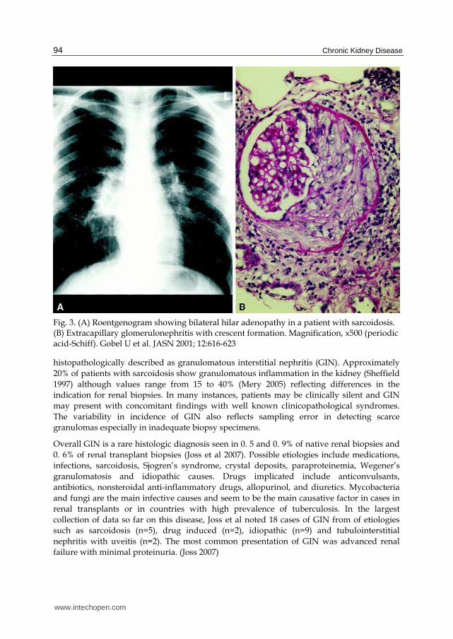
Fig. 3. (A) Roentgenogram showing bilateral hilar adenopathy in a patient with sarcoidosis. (B) Extracapillary glomerulonephritis with crescent formation. Magnification, x500 (periodic acid-Schiff). Gobel U et al. JASN 2001; 12:616-623
histopathologically described as granulomatous interstitial nephritis (GIN). Approximately
20% of patients with sarcoidosis show granulomatous inflammation in the kidney (Sheffield
1997) although values range from 15 to 40% (Mery 2005) reflecting differences in the
indication for renal biopsies. In many instances, patients may be clinically silent and GIN
may present with concomitant findings with well known clinicopathological syndromes.
The variability in incidence of GIN also reflects sampling error in detecting scarce
granulomas especially in inadequate biopsy specimens.
Overall GIN is a rare histologic diagnosis seen in 0. 5 and 0. 9% of native renal biopsies and
0. 6% of renal transplant biopsies (Joss et al 2007). Possible etiologies include medications,
infections, sarcoidosis, Sjogren’s syndrome, crystal deposits, paraproteinemia, Wegener’s
granulomatosis and idiopathic causes. Drugs implicated include anticonvulsants,
antibiotics, nonsteroidal anti-inflammatory drugs, allopurinol, and diuretics. Mycobacteria
and fungi are the main infective causes and seem to be the main causative factor in cases in
renal transplants or in countries with high prevalence of tuberculosis. In the largest
collection of data so far on this disease, Joss et al noted 18 cases of GIN from of etiologies
such as sarcoidosis (n=5), drug induced (n=2), idiopathic (n=9) and tubulointerstitial
nephritis with uveitis (n=2). The most common presentation of GIN was advanced renal
failure with minimal proteinuria. (Joss 2007)
www.intechopen.com
Sarcoidosis and Kidney Disease
95
Despite great clinical variability, the most common clinical syndrome associated with sarcoidosis and GIN is chronic kidney disease with decline in renal function usually over weeks to months (Jean-Philllipe 2005). Acute renal failure as an initial presentation is also well known (O’Riordan et al 2001). Renal dysfunction may progress at variable rates but can irreversibly progress to end stage renal disease despite high dose glucocorticoid treatment. (Tsiouris et al 1999) Consistent with a pattern of tubulointerstitial disease, proteinuria is either absent or mild. Urine analysis shows leucocytes and granular casts. Rarely, patient may present as frank hematuria lasting several weeks. (Mills et al 1994) Functional tubular abnormalities can occur in as much as 50% of cases of sarcoidosis when aggressively investigated which include renal glycosuria, urinary sodium and potassium wasting, Fanconi’s Syndrome, decreased urinary concentration ability, proximal or distal tubular acidosis. (Muther et al 1981) It is uncertain whether the presence of interstitial lesions solely contributes to these abnormalities but hypercalcemia and hypergammaglobulinemia also play a pathogenic role. (Mery, 2005)
GIN is usually associated with enlarged kidneys mimicking polycystic kidney disease or renal carcinoma. (Mery, 2005) Renal sonogram shows bilateral renal masses which are either hyper- or hypoechoic in comparison to adjacent renal parenchyma. Computer tomogram shows the renal masses to be low intensity. A Gallium-76 citrate scan commonly reveals increased uptake suggesting active granulomatous inflammation. (Mery and Kenouch, 1988) Serum ACE concentration is a poor marker of active renal lesion and may even be normal in active GIN with severe renal failure. (Hannedouche et al 1990)
In most cases, a diagnosis of GIN is made in the context of typical extra-renal manifestations of sarcoidosis and/or hyperkalemia. Rarely renal involvement may be isolated and preceding other sites of the disease for months to years. Some have even considered isolated GIN as a localized form of sarcoidosis. In such isolated cases, it is important to rule out drug induced interstitial nephritis which is far more easily treatable cause of GIN that sarcoidosis itself. (Muther et al 1981) Another syndrome commonly associated with Sjogren’s Syndrome but also reported with sarcoidosis is the “TINU syndrome or the Dobrin Syndrome (Sinnamon et al 2008) which is characterized by acute interstitial nephritis, anterior uveitis and epitheliod granulomas in bone marrow and lymph nodes. The renal lesion consists of interstitial infiltrates mainly composed of mononuclear cells and few eosinophils. Although no interstitial granulomas are seen in TINU, the interstitial cell infiltrate is the same as a sarcoid granuloma. Therefore it is possible that some cases described as Dobrin syndrome may be atypical forms of sarcoidosis.
Analyzing all cases of GIN, Joss et al, noted that the background diagnosis of sarcoidosis was known in only 1 of 5 patients of GIN who eventually were categorized as sarcoid GIN. Mean age of presentation was 56. 8 years. ACE levels were elevated in a minority of patients (1 out of 5) and hypercalcemia was seen in only 2 patients. Pulmonary findings of hilar lymphadenopathy was seen in only 1 patient and one had the TINU syndrome.
(Joss et al 2007)
Renal pathology of GIN consists of the typical non-caseating granuloma widely distributed throughout the cortex and the medulla, although the density of these lesions may differ from patient to patient. (Mery 2005) The sarcoid granuloma consists of lymphocytes, mononuclear, cells and plasma cells. The center of the granuloma consists of epitheliod and
www.intechopen.com

Chronic Kidney Disease
96
multinucleate giant cells both of which are derived from activated macrophages. Multinucleate giant cells are formed by the coalescence of epitheliod cells. Lymphocytes largely consist of T-helper cells (CD 4+) in the center and CD 8+ lymphocytes in the periphery. Some granulomas have small arteries in their center. Although granulomas may also form in drug induced interstitial nephritis it is less well formed than in sarcoidosis. Varying degrees of fibrosis may also be present. The severity of fibrosis correlates with tubular atrophy and degeneration. In the absence of any predominant glomerular pathology, the glomeruli are either normal or show mesangial hypertrophy and thickening of the basement membrane. Electron microscopy may show fusion of epithelial foot processes (Farge et al 1986). However, there are no significant immune deposits in either the glomeruli or tubules as seen by immunoflourescent microscopy. In a significant number of cases, immunoflourescence with anti-ACE serum showed localization in the sarcoid granuloma in addition to normal staining of the brush border of the proximal tubules. (Mery et al 1988) See Figure 4.
Fig. 4. Renal biopsy showed a granulomatous interstitial nephritis with a broadened interstitial, cellular infiltrates and granuloma with typical multinucleated giant cells (arrowheads). Kettritz R et al. Nephrol. Dial. Transplant. 2006; 21:2690-2694
www.intechopen.com
Sarcoidosis and Kidney Disease
97
In contrast to conventional pathological dogma, Joss et al showed that asteroid bodies and calcification were not common in sarcoid GIN. (Joss et al 2007) Interestingly, asteroid bodies were seen in 1 case of drug induced AIN. However, lymphocyte cuffing and giant cell infiltration were prominent in sarcoid granulomas in the kidney. Necrosis and eosinophil infiltration of the interstitum was more common in drug induced GIN as compared to sarcoidosis. It is now believed that idiopathic GIN, TINU and sarcoidosis represents a clinicopathological spectrum and that idiopathic GIN or TINU may subsequently develop typical extra-renal manifestations of sarcoidosis.
6.1 Treatment
The mainstay of treatment of sarcoid GIN is glucocorticoids. Initial treatment requires a daily dose of prednisone or prednisolone preferably 1-1. 5 mg/kg. Response to treatment can often be dramatic in terms of improvement of renal insufficiency. The best response to glucocorticoids was noted in a study by Mahevas et al. in which 47 patients with renal sarcoid received prednisolone while 10 also received pulse methylprednisolone. (Mahevas et al 2009) The authors concluded that at 24 months, a complete and partial remission occurred in 30 and 5 patients respectively. But no response was noted in patients with severe interstitial fibrosis of greater than 50%. Underlying functional tubular dysfunction improves with progressive drop in serum creatinine. An important point to realize here is that steroid treatment has to be prolonged and must exceed at least 6 months as nephropathy relapses very frequently with short term therapy (Gene and Cheviot 1988). A commonly followed strategy is to give the initial dose for 2 months followed by progressive taper and switching to an alternate –day therapy. A maintenance therapy period for 1 year at least is recommended. Serial renal biopsies have shown a regression of granuloma in conjunction with improvement of renal function (Farge et al 1986) although given the variability in results (Gene and Cheviot 1988) routine biopsies after starting steroids is not recommended. Treatment in advanced disease is often associated with interstitial fibrosis along with focal segmental glomerulosclerosis and vascular lesions. However, vascular lesions are more common with long term corticosteroid therapy and are associated with delayed development of hypertension which is a major contributor to progression of renal failure. (Mery and Kenouch 1988)
While analyzing outcomes of steroid treatment in a heterogeneous population of GIN, Joss et al, presented data of 16 patients of which 5 were labeled as sarcoidosis. Patients were treated with prednisolone (starting dose of 0. 55mg/kg) (Joss et al 2007) for a mean period of 25 months and then followed up for a period of 45 months. Overall, renal function stabilized or improved at the end of the study with mean GFR improving from 21 to 56 ml/min. One patient who was on dialysis at the beginning of therapy was able to discontinue dialysis within 3 months. Six patients relapsed on dose reduction of which 4 were sarcoid GIN who required azathioprine to break steroid dependence. Sarcoid patients required longer treatment (36 months) as compared to idiopathic or TINU patients. The greatest renal recovery occurred in the first year of treatment. There was no difference in renal outcome when analyzing the degree of interstitial fibrosis. Age less than 60 years was associated with a better outcome. Table 1 summarizes data on treatment of GIN in some important studies so far.
Long term results with steroid therapy in sarcoid GIN have not been rigorously tested in randomized controlled trials. In a large case series of 39 patients with sarcoid renal disease,
www.intechopen.com

Chronic Kidney Disease
98
Parameter Joss et al Robson et al. O’Riordan et al.
Hannedouche et al.
Brause et al.
n 18 161 7 5 6 5
Cause Mixed Idiopathic/ TINU/sarcoid
Idiopathic Isolated sarcoid
Sarcoid Sarcoid
Age (yr) 55 56 69 48 to 71 62 61
Male gender (%) 61 56 71 60 50 60
Renal function at baseline
BaselineCC (ml/min)
21 24 14 6 NA NA
Baseline creatinine (μmol/L)
373 357 420 NA 566 396
Hypercalcemia 3/18 3/16 2/7 0/5 2/6 1/5
Raised serum ACE
4/17 4/15 3/7 1/5 4/4 1/5
Improved renal function
17/18 15/16 5/7 4/5 6/6 5/5
Long-term RRT 0/18 0/16 2/7 0/5 0/6 0/5
Prednisolone (%) 89 88 100 100 100 100
Mean follow-up (mo)
45 48 25 35 75 NA
Renal function at last visit
ECC (ml/min) at end of therapy
56 53 22 20 NA NA
Creatinine (μmol/L) at end of study
159 159 296 NA 192 225
1 Data excluding the two cases of drug-induced GIN.
Table 1. Comparison on treatment of GIN in literature.
17 patients with biopsy-proven tubulo-interstitial nephritis with significant renal
impairment were analyzed over a one year period of corticosteroid therapy. (Robson et al
2003). All patients were initially started on prednisolone at 0. 5 mg/kg body weight at a
daily dose of 30–60 mg which was tapered by 5 mg each week once the renal function has
improved and/or stabilized. Thereafter, patients were maintained on 5–7. 5 mg daily
indefinitely. Mean duration of study was 84 months. Estimated glomerular filtration rate
(eGFR) at baseline was 26. 814 ml/min which improved to 49. 65. 2 ml/min (P<0. 01) at 1
year, and 47. 96. 8 ml/min (P<0. 05) at last review. Interestingly, the response to treatment
was similar regardless of the degree of renal impairment at baseline, race and the degree of
tubulo-interstitial scarring on renal biopsy. Three patients developed side effects that could
www.intechopen.com

Sarcoidosis and Kidney Disease
99
be attributed to steroids which included acute psychosis and type 2 diabetes. Long term use
of corticosteroids, especially in adolescents, can cause substantial side effects including
diabetes, growth retardation and cataract. Alternative agents that have been attempted in
treating sarcoid GIN include mycophenolate (Moudgil 2002) and mizoribine (Rajakariar et al
2006 and Ito et al 2009) which are limited to case reports and have been primarily used in
pediatric patients to break steroid dependence or ameliorate significant side effects. Other
agents which have been tried in systemic extra-renal sarcoidosis include mycophenolate
mofetil, methotrexate, azathioprine, antimalarials, and phosphodiesterase inhibitors such as
pentoxifylline and thalidomide although no data on treating renal sarcoidosis exists.
(Baughman 2003) There has been great interest in the use of TNF-antagonists as another
modality to treat sarcoid GIN in order to avoid use of steroids. TNF-alpha, which is
expressed by monocytes, is critical in the development of these noncaseating granulomas.
TNF-alpha receptor antagonists have also been shown to prevent the initiation and
perpetuation of inflammation and subsequent interstitial fibrosis. Etanercept is a soluble
TNF-alpha receptor fusion protein that binds TNF-alpha. Infliximab and adalimumab are
monoclonal antibodies that bind specifically to and neutralize TNF-alpha. While etanercept
is an ineffective agent in the treatment of systemic sarcoidosis, (Ulz et al 2003) infliximab has
been shown to be effective in a case of renal sarcoid. Thumfart et al, described the case of a
boy presenting with severe arterial hypertension and acute renal failure caused by an
isolated sarcoid granulomatous interstitial nephritis. Renal function improved initially with
prednisone treatment but later, the patient showed signs of severe steroid toxicity and
progressive renal failure. Monthly treatment with infliximab was initiated resulting in a
steady improvement in renal function and resolution of renal granulomata, as well as
reduction in antihypertensive medication. (Thumfart 2005) Ahmed et al presented a patient
with acute renal failure due to isolated granulomatous infiltration of the renal parenchyma.
(Ahmed et al 2007) Renal biopsy showed granulomatous interstitial nephritis with
noncaseating granulomas. There was no evidence of extrarenal sarcoid involvement.
Prednisone 60mg daily resulted in significant improvement in renal function. Due to
recurrent flares while tapering the prednisone and steroid toxicity, treatment with
infliximab was instituted and resulted in stabilization of renal function. This case
demonstrated that steroid-dependant or refractory renal sarcoidosis cases may respond to
infliximab. We recently reported the case of a 46-year-old woman with multi-organ
sarcoidosis, type 2 diabetes, subnephrotic-range proteinuria, hypertension and recurrent
episodes of hypercalcemia-induced acute kidney injury who was referred for evaluation of
worsening renal function and nephrotic range proteinuria. (Gupta et al 2008) A kidney
biopsy showed sarcoid GIN with moderate-to-severe chronic tubulointerstitial disease,
hypertensive vasculopathy, and diabetic glomerulosclerosis. Because steroids had caused
multiple side effects including diabetes, hypertension and obesity and attempts to wean
steroids had caused hypercalcemia and acute renal failure, Adalimumab (HumiraTM) 40
mg/0. 8 cc weekly for 6 months was initiated. After 6 months of treatment with
adalimumab, serum creatinine improved from 345 μmol/L (3. 9 mg/dL) to 1. 8 mg/dl (her
baseline for years) and proteinuria improved from 10 g/day to 3. 5 g in 24 hours
respectively. A repeat biopsy showed persistent diabetic glomerulosclerosis, moderate
chronic tubulointerstitial inflammation with complete resolution of interstitial epitheliod
granulomas. Although adalimumab and infliximab are generally safe, some side effects
www.intechopen.com
Chronic Kidney Disease
100
include risk of lymphoma and reactivation of latent tuberculosis (Denys et al 2007). These
agents may hold promise for the future once large scale randomized studies are available to
show consistent benefits with minimal side effects.
7. Renovascular diseases associated with sarcoidosis
Renovascular diseases secondary to sarcoidosis are distinctly rare and attributed to a form
of secondary vasculitides. Systemic vasculitis associated with sarcoidosis has been
reported as an isolated entity in the literature after excluding other common causes of
vasculitis. It is predominantly large vessel vasculitis although few instances of small
vessel vasculitis have been reported. In a large case series and review of literature on
sarcoid vasculitis, Fernandes et al, noted that most cases were children and clinical
presentation resembled hypersensitivity vasculitis, Takayasu’s arteritis, polyarteritis
nodosa or microscopic polyangitis. (Fernandes 2000) Clinical features included fever,
peripheral adenopathy, hilar adenopathy, rash, pulmonary parenchymal disease,
musculoskeletal symptoms, and scleritis or iridocyclitis with biopsy showing necrotizing
sarcoid granulomata. Interestingly, no renal involvement was noted. Notably the authors
found large vessel vasculitis largely in the African American population while small
vessel vasculitis predominantly affected white races. Godin et al described a known case
of pulmonary sarcoidosis with persistent hypertension. (Godin et al 1980) Diagnostic
evaluation for renovascular hypertension included aortography which showed severe
stenosis of right renal artery. Surgical exploration showed extensive periaortic and
perirenal fibrosis with extrinsic compression of renal artery. Pathological examination of
the kidney revealed epitheloid infiltration of the adventia of renal artery suggestive of
sarcoid angitis. Surgical biopsy was performed on both kidneys. The right kidney,
protected by arterial stenosis, was slightly altered, while the left kidney showed extensive
interstitial, tubular, and glomerular lesions which included focal and segmental
hyalinosis. Marcussen et al, reported an autopsy case of a middle aged man who died of
myocardial infarction secondary to fulminent vasculitis. (Marcussen and Lund 1989)
Pathology showed widespread giant cell vasculitis with simultaneous involvement of the
renal arteries, veins, and arterioles along with typical interstitial sarcoid granuloma.
Shintaku et al, showed granulomatous inflammation of small renal vessels and crescentric
GN on the autopsy of a patient with pulmonary hemorrhage and rapidly progressive
renal failure. (Shintaku et al 1989) Thus, sarcoid angitis, especially causing small vessel
vasculitis in the kidney may represent a very severe form of sarcoidosis. In their review,
Fernandes et al, noted that four out of six patients responded well to steroid treatment
alone but had relapses when attempts were made to taper or withdraw steroids.
(Fernandes 2000) Frequently, there is an overlap between sarcoidosis and well known
causes of granulomatous vasculitis. For instance, Watson et al described a case of
longstanding pulmonary sarcoidosis presenting with rapidly progressive renal failure
with p-ANCA positivity. (Watson 1996) Renal biopsy demonstrated focal and segmental
fibrinoid necrosis with crescentric GN and focal fibrinoid necrosis in arterial wall, but no
granulomata and pauci-immune deposits on immunofluorescence. Unlike patients with
ANCA positive vasculitis, the index case responded poorly to pulse steroids and
cyclophosphamide and progressed rapidly to end stage renal disease.
www.intechopen.com

Sarcoidosis and Kidney Disease
101
8. Kidney transplantation in patients with sarcoidosis
The usual cause of end stage renal disease in sarcoidosis requiring renal replacement
therapy is usually due to hypercalcemic nephropathy rather than granulomatous interstitial
nephritis or a glomerular disease. The outcome in renal transplantation in patients with
sarcoidosis has been described in the literature. The first recurrence of sarcoid GIN in renal
allograft was diagnosed 6 years after deceased donor kidney transplantation in a patient
that was diagnosed with GIN before transplantation (Shen et al 1986). A recent French study
aimed to describe a multicenter experience with kidney transplantation in patients with
sarcoidosis. (Aouizerate et al 2010) In this study, the authors retrospectively identified 18
patients who underwent renal transplantation. Patient medical charts, demographics were
reviewed. The median time between the last sarcoidosis episode and renal transplantation
was 78 (8 to 900) months. Only 3 out of 18 patients had been on immunosuppression prior to
transplantation. Vast majority of the patients had in the past received steroids and other
immunosuppression for their sarcoid before transplantation. Renal disease was attributable
to biopsy proven renal sarcoid in 10 out of the 18 patients and was attributed to other causes
in 8 patients. Mean age of transplantation was 43. 5 +/- 11 years. 17 out of 18 patients had a
deceased donor transplant. Mean donor age was 36. 5 +/ 15 years. Mean cold ischemia time
was 16. 6 +/- 8 hours. 11 patients received induction therapy with anti-thymocyte globulin
or Il-2 receptor antagonists. Maintenance immunosuppression included calcineurin inhibitor
(CNI) for all patients, mycophenolate mofetil or azathiporine, sirolimus and corticosteroids
for 16 out of the 18 patients. At the end of the 42 month follow up period, patient and death
censored graft survival was 94. 4% and mean GFR was 60 cc/min per 1. 73 m2. Recurrence
of sarcoidosis after renal transplantation was observed in 5 (27%) of patients. The median
period between renal transplantation and recurrence was 13 months and four of five
patients exhibited recurrence in the first 18 months after renal transplantation. Recurrences
involved in the same organ in four of five patients and included renal involvement in three
patients and lung and liver involvement in one patient. Mean GFR at end of follow-up was
significantly lower in the three patients with recurrence than that for the entire cohort. (31
versus 60 cc/min per 1. 73 m2). Analysis of the recurrences showed that they occur in the
first 18 months after transplantation. Primary disease related to sarcoidosis was strongly
associated with recurrence (40% in the group with renal sarcoidosis versus 12. 5% in a group
with a primary nephropathy, and median period between last episode of sarcoidosis and
renal transplantation was shorter in the case of sarcoidosis recurrence (42 versus 78 months
respectively). This study showed that patients with initial renal involvement display
sensitivity to disease recurrence in allograft. The incidence of recurrence was significant as
all patients were maintained on triple immunosuppressive therapy including steroids and
mycophenolate mofetil. This study showed that renal transplant can be conducted safely in
transplant patients with sarcoidosis, but recurrences do occur and affect overall graft
outcome.
Kukura reported a case of recurrence of sarcoidosis in the renal allograft during pregnancy.
(Kukura et al 2004) This was a 27 yr old female diagnosed with sarcoidosis at age 14 by
lacrimal and parotid gland biopsy. 4 years after presentation, she developed hypertension
and renal insufficiency. Kidney biopsy showed interstitial nephritis and nephrosclerosis, but
no granulomas. Patient was eventually started in hemodialysis and underwent kidney
www.intechopen.com

Chronic Kidney Disease
102
transplantation with excellent graft function with a creatinine of 1. 32 mg/dl and a negative
urinalysis. Patient was maintained on cyclosporine, azathioprine and prednisone 25 mg by
mouth daily. 2 years after transplantation once the steroids were withdrawn, patient
continued to have good kidney function with an allograft biopsy showing mild chronic
allograft nephropathy only. Immunosuppression consisted of azathioprine and
cyclosporine. At 3 years after kidney transplantation, patient became pregnant. 29 weeks
into pregnancy, renal function worsened. Biopsy showed numerous noncaseating
granulomas bound to the arteries, initial arteritis in one artery, mild interstitial mononuclear
inflammation and tubulitis. Graft function improved with pulse methylprednisolone and
tapered steroids were used. After delivery, renal allograft biopsy was performed 6 months
which showed baseline disease of mild chronic allograft nephropathy and sporadic
granulomas. This case demonstrates that steroid withdrawal after kidney transplantation
may lead to sarcoidosis recurrence.
The implication that sarcoid reflects a disease phenomena related to the immunologic
stimulus makes sarcoidosis an unlikely diagnosis to be made in an immunosuppressed
patient such as an organ transplant recipient. However, Schmidt et al showed that after
kidney transplantation, sarcoidosis can occur in the lung and pleura. (Schmidt et al 1999) In
this case, a 41 yr old with history of IgA nephropathy and no past medical history received a
living related kidney transplant and had been receiving tacrolimus therapy. He was found
to have a large pleural effusion 17 months after kidney transplant. Diagnosis of sarcoidosis
was established by identifying noncaseating granulomas, some with multinucleated giant
cells in the pleural and lung tissue. All viral and bacterial workup was negative. The
effusion resolved after initiating corticosteroid therapy. One month into therapy, the
effusion resolved and patient continued to be asymptomatic twenty months after therapy.
The authors did not speculate on the pathogenesis of granuloma formation since both
tacrolimus and corticosteroids interfere with T lymphocyte function and granuloma
formation. They speculated that activation of tissue chemokines of the IP-10 type during the
posttransplant period, along with subsequent recruitment of lymphocytes and macrophages
may have resulted in the sarcoidosis.
9. Conclusion
Sarcoidosis is a disease that primarily affects the reticuloendothelial system but can affect all
tissues and organ systems. In this chapter, we described the effects of sarcoidosis on the
kidneys. This disease affects patients worldwide and is defined pathologically by the
presence of noncaseating granulomas in the involved tissue. The etiology of sarcoidosis has
yet to be determined but some have proposed a possible infectious etiology. Commonly
sarcoid patients present with hypercalcemia, hypercalcuria, and nephrolithiasis due to the
overproduction of calcitriol from the epitheliod granulomas. We also described the rare
glomerular and renovascular manifestations of sarcoidosis. Granulomatous interstitial
nephritis is most commonly associated with sarcoidosis. It is a histological diagnosis and
can be treated with both steroids and TNF-alpha antagonists. Kidney transplantation is safe
in patients with sarcoidosis but we must keep in mind the disease can recur in the allograft.
In conclusion, sarcoidosis is a complex disease and presents both a diagnostic and
management challenge to the physician.
www.intechopen.com
Sarcoidosis and Kidney Disease
103
10. References
Ahmed MM, Mubashir E, Dossabhoy NR. Isolated renal sarcoidosis: a rare presentation of a
rare disease treated with infliximab. Clin Rheumatol. 2007; 26(8):1346-9.
Ahuja TS, Mattana J, Valderrama E, Sankaran R, Singhal PC, Wagner JD: Wegener's
granulomatosis followed by development of sarcoidosis. Am J Kidney Dis. 1996;
28:893 -898. 47.
Aouizerate, Jessie, Matignon, Marie et al: Renal Transplantation in Patients with Sarcoidosis:
A French Multicenter Study. Clinical Journal of American Society of Nephrology
2010 5: 2101-2108.
Auinger M, Irsigler K, Breiteneder S, Ulrich W: Normocalcemic hepatorenal sarcoidosis with
crescentic glomerulonephritis. Nephrol Dial Transplant. 1997; 12:1474 -1477.
Baughman, R. P, Lynch, J. Difficult treatment issues in sarcoidosis. J Intern Med. 2003 Jan;
253(1):41-5.
Berliner AR, Haas M, Choi MJ. Sarcoidosis: The Nephrologist’s Perspective. Am J Kid Dis
2006; 48(5):856-870.
Brause M, Magnusson K, Degenhardt S, Helmchen U, Grabensee B: Renal involvement in
sarcoidosis: A report of 6 cases. Clin Nephrol. 2002; 57 :142– 148.
Casella FJ, Allon M. The Kidney in Sarcoidosis. J Am Soc Nephrol 1993; 3: 1555-1562.
Denys, B. Bogaerts, Y. Coenegrachts, K. Et al. Steroid-resistant sarcoidosis: is antagonism of
TNF-α the answer? Clinical Science. 2007; 11: 281–289.
Dimitriades C, Shetty AK, Vehaskari M, Craver RD, Gedalia A: Membranous nephropathy
associated with childhood sarcoidosis. Pediatr Nephrol. 1999;13:444 -447.
Farge D, Loite F, Turner M. Granulomatous nephritis and chronic renal failure in
sarcoidosis. Long term follow up studies in two patients. American J Nephrol. 1986;
6:22-27.
Fernandes SR, Singsen BH, Hoffman GS. Sarcoidosis and systemic vasculitis. Semin
Arthritis Rheum. 2000 Aug; 30(1):33-46.
Foster S. Ocular manifestations of sarcoidosis preceding systemic manifestations. In: Grassi
C. Rizzato G, Pzzi E, eds. Sarcoidosis and other granuloamtous disorders. Amsterdam:
Elsevier; 1988: 1977-81.
Fraioli P, Montemurro L, Castrignano L, Rizzato G. Retroperitoneal involvement in
sarcoidosis. Sarcoidosis. 1990;7(2):101.
Gobel U, Kettritz R, Schneider W, Luft F. The protean face of renal sarcoidosis. J Am Soc
Nephrol. 2001; 12:616–623.
Godin M et al. Sarcoidosis, retroperitoneal fibrosis, renal artery involvement and unilateral
focal glomerulosclerosis. Archives of Internal Medicine. 1980;140:1240-1242
Guenel J and Chevet D. Interstitial nephropathies in sarcoidosis. Effect of corticosteroid
therapy and long-term evolution. Retrospective study of 22 cases. Nephrologie.
1988;9(6):253-7.
Gupta, R. Beaudet, Lisa and Mehta, Tulsi. Treatment of sarcoid granulomatous interstitial
nephritis with adalimumab. NDT Plus. 2008;2(2): 139-142.
Hannedouche, T., Grateau, G., Noël, et al. Renal Granulomatous Sarcoidosis: Report of Six
Cases. Nephrolo Dial Transplant. 1990;5(1):18-24.
Hiatt R, Dales L. Friedman G. Frequency of urolithiasis in a prepaid medical care program.
Americal Journal Epidemiol 1982: 115: 255-65.
www.intechopen.com
Chronic Kidney Disease
104
Iannuzzi MC, Rybicki BA, Teirstein AS. Medical Progress: Sarcoidosis. N Engl J Med 2007:
357(21). 2153-2165.
Iannuzzi MC, Fontana JR. Sarcoidosis: Clinical Presentation, Immunopathogenesis, and
Therapeutics. JAMA 2011; 305(4):391-399.
Ito, Shuichi, Harada, Tomonori, Nakamura, Tomoko et al. Mizoribine for renal sarcoidosis:
effective steroid tapering and prevention of recurrence. Pediatr Nephrol 2009;
24:411-414.
Johnson C, Wilson D, O’Fallon W et al. Renal stone epidemiology: a 25 year study in
Rochester, Minnesota. Kidney International 1979: 16: 624-31.
Joss, Nicola, Morris, Scott and Young, B et al. Granulomatous Interstitial Nephritis. CJASN
2007; 2(2); 222-230.
Kettritz, R, Goebel U, Fiebeler, A, et al. The protean face of sarcoidosis revisited. Nephrol
Dial Transplant 2006; 21:2690-2694.
Khan IH, Simpson JG, Catto GR, MacLeod AM: Membranous nephropathy and
granulomatous interstitial nephritis in sarcoidosis. Nephron. 1999 66: 459 -461.
Kukura S, Viklicky o, Lacha J, et al: Recurrence of sarcoidosis in renal allograft during
pregnancy. Nephrology Dialysis Transplant 2004; 19: 1640-1642.
Lebacq E, Desmet V, Verhaegen H. Renal involvement in sarcoidosis. Postgrad Med J 1970;
46: 526.
Mahévas M, Lescure FX, Boffa J et al. Renal sarcoidosis: clinical, laboratory, and histologic
presentation and outcome in 47 patients. Medicine (Baltimore). 2009;88(2):98.
Marcussen N and Lund C. Combined sarcoidosis and disseminated visceral giant cell
vasculitis. Pathology, Research and Practice. 1989;184:325-330.
Maruyama S, Cantu E 3rd, Demartino C, Vladutiu A, Caldwell PR, Wang CY, D'Agati V,
Godman G, Stern DM, Andres G. Membranous glomerulonephritis induced in the
pig by antibody to angiotensin-converting enzyme: considerations on its relevance
to the pathogenesis of human idiopathic membranous glomerulonephritis. J Am
Soc Nephrol. 1999 Oct;10(10):2102-8.
Mery, Jean-Phillippe. The patient with sarcoidosis. Oxford textbook of Clinical Nephrology,
3rd Edition (2005), Volume 1, Oxford University Press:733-740.
Mery J. P., Kenouch S. Les atteintes de l'interstitium rénal au cours des maladies
systémiques. Seminares d’uro-nephrologie Pitie-Salpetriere, 1988; 57-89
Moudgil, A., Przygodzki, R. and Kher. K. Successful steroid-sparing treatment of renal
limited sarcoidosis with mycophenolate mofetil. Pediatric Nephrol 2006;21(2):281-
285.
Michaels S, Sabnis SG, Oliver JD, Guccion JG: Renal sarcoidosis with superimposed
glomerulonephritis presenting as acute renal failure. Am J Kidney Dis. 2000; 36:1 -6.
Mills, PR, Burns AP, Dorman AM, Sweny PJ, Moorhead JF. Granulomatous sarcoid
nephritis presenting as frank haematuria. Nephrol Dial Transplant. 1994;9(11):1649-
51.
Miyazaki E, Tsuda T, et al. : Sarcoidosis presenting as bilateral hydronephrosis. Intern Med
35: 579-582, 1996
Mundlein E, Greten T, Ritz E: Graves' disease and sarcoidosis in a patient with minimal-
change glomerulonephritis. Nephrol Dial Transplant. 1996; 11:860 -862.
www.intechopen.com

Sarcoidosis and Kidney Disease
105
Muther, R., Mc Carron D et al. Renal manifestations of sarcoidosis. Arch Intern Medicine
1981;141 :643-645.
Nishiki M, Murakami Y, Yamane Y, Kato Y: Steroid-sensitive nephrotic syndrome,
sarcoidosis, and thyroiditis: A new syndrome? Nephrol Dial Transplant. 1999;
14:2008 -2010.
O'Riordan E, Willert RP, Reeve R et al. Isolated sarcoid granulomatous interstitial nephritis:
review of five cases at one center. Clin Nephrol. 2001 Apr;55(4):297-302.
Parry RG, Falk C: Minimal change disease in association with sarcoidosis. Nephrol Dial
Transplant. 1997;12 : 2159-2160.
Rainfray M. Renal amyloidosis complicating sarcoidosis. Thorax. 1988;43:422-423.
Rajakariar, E., Sharples, J et al. Sarcoid tubulo-interstitial nephritis: Long-term outcome and
response to corticosteroid therapy. Kid Int. 2006; 70: 165–169.
Rizzato G, Fraioli P, Montemurro L. Nephrolithiasis as a presenting feature of chronic
sarcoidosis. Thorax 1995; 50:555.
Rizzato G: Sarcoidosis in Italy. Sarcoidosis 9(supl): 145-147. 1995
Robson MG, Banerjee D, Hopster D, Cairns HS: Seven cases of granulomatous interstitial
nephritis in the absence of extrarenal sarcoid. Nephrol Dial Transplant. 2003 ;18
:280– 284.
Schmidt, R., Bender, f. Change, W: Sarcoidosis After renal Transplantation. Transplantation
68(9) 1420-1423, 1999.
Sharma OP. Vitamin D, Calcium, and Sarcoidosis. Chest 1996; 109(2): 535-539.
Sheffield EA: Pathology of sarcoidosis. Clin Chest Med. 1997;18: 741-753.
Shen S, Hall-Craggs, M. et al : Recurrent sarcoid granulomatous nephritis and reactive
tuberculin skin test in a renal transplant recipient. American Journal of Medicine
80:699-702, 1986.
Shintaku M, Mase K, Ohtsuki H, Yasumizu R, Yasunaga K, Ikehara S: Generalized
sarcoidlike granulomas with systemic angiitis, crescentic glomerulonephritis, and
pulmonary hemorrhage. Report of an autopsy case. Arch Pathol Lab Med. 1989;
113:1295 -129.
Sinnamon, T. Courtney, Harron, C et al. Tubulointerstitial nephritis and uveitis (TINU)
syndrome: epidemiology, diagnosis and management. Nephrol Dial Transpl Plus.
2008; 2(1): 112-116.
Taylor JE, Ansell ID: Steroid sensitive nephrotic syndrome and renal impairment in a
patient with sarcoidosis. Nephrol Dial Transplant. 1996; 11:355 -356.
Tchenio X et al. Amylose renale AA au cours d’une sarcoidose. Revue des Maladies
Respiratoires. 1996; 13. 601-602.
Thumfart J. Isolated sarcoid granulomatous interstitial nephritis responding to infliximab
therapy. Am J Kidney Dis 2005;45:411-414.
Tsiouris N, Kovacs B, Daskal I I, Brent LH, Samuels A. End stage renal disease in sarcoidosis
of the kidney. 1999. Am J Kidney Disease. 1999;34: E21
Ulz JP, Limper AH, Kalra S, et al. Etanercept for the treatment of stage II and III progressive
pulmonary sarcoidosis. Chest 2003;124:177.
Vanhille, Ph et al. Glomerulonephrite rapidement progressive a depots mesangiuax d’IgA
au cours d’une sarcoidose. Nephrologie. 1986;5: 207-209.
www.intechopen.com

Chronic Kidney Disease
106
Watson G, Hill C M, Biggart J D et al. Sarcoidosis and primary systemic vasculitis. Nephrol
Dial Transplant. 1996;11: 1631-1633. 54.
Yoshida O, Okada Y. Epidemiology of urolithiasis in Japan: a chronological and
geographical study. Urol Int 1990: 45: 104-11.
www.intechopen.com
Chronic Kidney DiseaseEdited by Prof. Monika Göőz
ISBN 978-953-51-0171-0Hard cover, 444 pagesPublisher InTechPublished online 16, March, 2012Published in print edition March, 2012
InTech EuropeUniversity Campus STeP Ri Slavka Krautzeka 83/A 51000 Rijeka, Croatia Phone: +385 (51) 770 447 Fax: +385 (51) 686 166www.intechopen.com
InTech ChinaUnit 405, Office Block, Hotel Equatorial Shanghai No.65, Yan An Road (West), Shanghai, 200040, China
Phone: +86-21-62489820 Fax: +86-21-62489821
Chronic kidney disease is an increasing health and economical problem in our world. Obesity and diabetesmellitus, the two most common cause of CKD, are becoming epidemic in our societies. Education on healthylifestyle and diet is becoming more and more important for reducing the number of type 2 diabetics andpatients with hypertension. Education of our patients is also crucial for successful maintenance therapy. Thereare, however, certain other factors leading to CKD, for instance the genetic predisposition in the case ofpolycystic kidney disease or type 1 diabetes, where education alone is not enough.
How to referenceIn order to correctly reference this scholarly work, feel free to copy and paste the following:
Tulsi Mehta, Anirban Ganguli and Mehrnaz Haji-Momenian (2012). Sarcoidosis and Kidney Disease, ChronicKidney Disease, Prof. Monika Göőz (Ed.), ISBN: 978-953-51-0171-0, InTech, Available from:http://www.intechopen.com/books/chronic-kidney-disease/sarcoidosis-and-kidney-disease
Recommended