Screening of soil fungi in order to biosynthesize AgNPsand evaluation of antibacterial and antibiofilm activities
SEYEDE SOHEILA MOUSAVI1, PARINAZ GHADAM1,*
and PARISA MOHAMMADI21 Department of Biotechnology, Faculty of Biological Sciences, Alzahra University, Tehran 1993893973, Iran2 Department of Microbiology, Faculty of Biological Sciences, Alzahra University, Tehran 1993893973, Iran
*Author for correspondence ([email protected])
MS received 25 August 2019; accepted 10 March 2020
Abstract. The biosynthesis of nanoparticles (NPs) has recently attracted a lot of research attention due to its being an
eco-friendly and economical method. NPs are formed under normal temperatures and pressures. The shape and size of
NPs can be controlled by choosing a suitable pH and temperature. In this study, 24 strains of fungi isolated from desert
soils were screened for AgNP synthesis. The MS17 isolated was chosen as the superior strain capable of rapidly
synthesizing monodisperse AgNPs. The optimum conditions for AgNP synthesis were investigated. AgNPs were char-
acterized by UV–visible spectrophotometry, dynamic light scattering, X-ray diffraction, transmission electron microscopy
and Fourier-transform infrared. The NPs produced were found to be in the form of Ag/AgCl with a size range of 5–15 nm.
Then, the NPs were capped by proteins and carbohydrates, which play an important role in NP stability. The NPs were
capable of antimicrobial activities against the standard bacterial pathogens, Pseudomonas aeruginosa ATCC 27853,
Escherichia coli ATCC 25922, Bacillus subtilis ATCC 6633, Staphylococcus aureus ATCC 1431 and the multidrug-
resistant P. aeruginosa B52 and P. aeruginosa 48.
Keywords. Soil fungi; AgNPs; antimicrobial activity.
1. Introduction
Historically, silver has been widely used for several purposes,
such as fabricating jewellery and food containers and also in
medicine [1,2]. Silver nanoparticles (AgNPs) can be applied
in antibacterial agents, biosensors, composite fibres, refrig-
erator superconductors, cosmetics and electronics [3]. There
are three general ways for the synthesis of NPs, including
physical, chemical and biological methods. While chemical
methods use several toxic solvents and produce a degree of
hazardous materials, physical methods are energy-consum-
ing [4]. Biological approaches have been developed in
response to the increasing request for the high-efficiency,
low-cost, non-toxic and biocompatible construction of metal
NPs. Biological resources, including plants, plant products,
algae, fungi, yeasts, bacteria, actinomycetes and viruses, are
capable of producing different types of NPs [5]. Today, fungi
are regarded as a nano-factory for the bio-synthesis of NPs.
Biosynthesizing NPs by fungi has some advantages over
other biological resources because fungi produce high levels
of reducing agents, such as proteins, to reduce metallic ions to
a less toxic form [6]. Various fungi have recently been used
for biotechnological processes, and using the residual
mycelium of these fungi has been proposed as a potential
cost-effective solution [7].
The present study was conducted to screen some
desert-soil fungi in terms of AgNP synthesis. The biosyn-
thesis of AgNPs from the superior strain was optimized and
then characterized. The antibacterial and antibiofilm activ-
ities of the produced NPs were then assayed.
2. Materials and method
2.1 Screening of fungi for the synthesis of AgNPs
The synthesis of NPs of 24 fungal isolates was investigated.
Twenty-three out of 24 belong to Aspergillus and the other
one belongs to Fusarium achieved from the microbial bank
of Alzahra University which was collected from the desert
soil of Khabr National Park (Kerman, Iran). Fungi were
cultured on potato dextrose agar and kept at room temper-
ature for 8 days to produce sufficient conidiospores. Then,
1 ml of the conidiospore suspension with a concentration of
104 conidiospores per ml was inoculated into the 250 ml
flask containing 100 ml liquid culture medium of malt
glucose yeast peptone agar and comprising 0.3% malt
extract, 0.3% yeast extract, 0.5% peptone and 1% glucose
and then incubated at 27–30�C in 120 rpm, for 4 days. After
4 days, fungi were filtered by the sterilized Whatman paper
Bull Mater Sci (2020) 43:214 � Indian Academy of Scienceshttps://doi.org/10.1007/s12034-020-02182-8Sadhana(0123456789().,-volV)FT3](0123456789().,-volV)
no. 1. Thereafter, fungi mycelium was washed by distilled
water to remove the trace of medium components. After-
wards, 10 g of the wet mycelia was suspended into the
250 ml Erlenmeyer flask containing 100 ml of distilled–
sterilized water and incubated for 2 days in the similar
conditions of incubation. After 2 days, the suspensions were
gain filtered through the sterilized Whatman paper no. 1,
and the mycelium-free extract was used for further inves-
tigations. The filtered fungus extract was added to a silver
nitrate solution so that the final concentrations of silver
nitrate became 1 mM. Then, the synthesis of AgNPs kept at
room temperature in a dark place away from air [8].
2.2 Optimization of AgNP production
After selecting the superior strain, the synthesis conditions,
such as the effect of pH (6, 8, 10 and 12), temperature (80,
50�C and room temperature (25–27�C)) and silver nitrate
concentrations (1–5 mM), were optimized and characterized.
2.3 Characterization of AgNPs
The preliminary characterization of AgNP synthesis was
visually done through the observation of colour changes. The
UV–vis spectroscopy measurements were performed on a
SPEKOL 2000 spectrophotometer in the range of 350–700
nm. The hydrodynamic diameter, polydispersity index (PDI)
and distribution of AgNPs were examined by a NanoPhox
90-246V instrument (Sympatec GmbH, Clausthal-Zellerfeld,
Germany). The biosynthesized AgNP solution was dropped
in a cast on a carbon-coated copper grid for transmission
electron microscopy (TEM) analysis (Philips CM30). The
distribution of AgNP size was calculated using Digimizer
software version 4.1.1.0. The X-ray diffraction (XRD) pat-
terns were obtained on a Rigaku instrument, operating at 40
kV and at a current of 30 mA. The Fourier-transform infrared
(FTIR) spectra of AgNPs were recorded by TENSOR 27
spectrometer in the range of 400–4000 cm-1.
2.4 Mechanisms involved in AgNP biosynthesis
2.4a Determination of reducing biomolecules: To
determine the effective agents in biosynthesis of AgNPs,
the biomolecules in the fungal extract were investigated.
The phenol sulphuric acid method was used to determine
the carbohydrate content of fungal extract [9]. For this
reason, 200 ll of phenol solution (5% w/v) with different
concentrations was added to the 400 ll of fungal extract.
Immediately, 1 ml of sulphuric acid (97%) was added. After
10 min, each sample was vortexed for 30 s. After 20 min,
the absorption was obtained at 490 nm. Glucose was applied
as a standard sugar to illustrate the standard curve.
The Bradford method was used to measure the total
protein concentration of samples [10]. The standard protein
was bovine serum albumin. Then, 5 ml of Bradford reagent
was added to 100 ll of different concentrations of standard
protein solution (1.0–1.7 mg ml-1) as well as the fungal
extracts. After 2 min, the absorption was read at 595 nm.
The ninhydrin method was used to determine the amount
of amino acid in the fungal extract [11]. Thereafter, 2 ml of
extract and 2 ml of glycine solution in different concen-
trations (0–100 lg ml-1), as a standard solution, were
separately mixed with 1 ml of ninhydrin (5% ninhydrin in
acetone and potassium cyanide) and incubated in the boiling
water bath for 20 min. Finally, the volume of each sample
was diluted using distilled water up to 10 ml and the
absorbance of the samples was obtained at 570 nm.
2.4b Nitrate reductase assay: The quantitative assay of
nitrate reductase of the fungi extract was performed by the
Harley method [12]. In this method, nitrate as a substrate was
used, and the activity of enzyme is determined after 1 h through
measuring the produced nitrite. Afterwards, 5 ml of extract of
superior strain and 5 ml of assay medium (potassium nitrate
30 mM and propanol 5% (v/v) in phosphate buffer (0.1 M,
pH 7.5)) were mixed and incubated in dark at room temperature
for 1 h and then placed in a hot water bath. After cooling the
tubes at room temperature, 2.5 ml of sulphanilamide solution
(sulphanilamide 1% (w/v) in HCl 25% (v/v)) 2.5 ml of 0.1%
N-(1-naphthyl)ethylenediamine dihydrochloride solution were
added into each tube and mixed. After 20 min, the absorption of
each sample was surveyed in 540 nm. The activity of enzyme
was calculated considering the produced nitrite per hour for the
initial sample (5 ml) and was expressed as nmol nitrite h-1 ml-1.
2.4c Determination of free radical scavenging of fungal
extract using the DPPH method: The antioxidant activity of
fungal extract was evaluated through the free radical scavenging
effect on 1,1-diphenyl-2-picrylhydrazyl (DPPH) radical [13].
Different volumes of fungal extract (25–55 ml) were diluted
with methanol up to 1 ml and the mixture was added to 2 ml of
0.1 mM DPPH methanolic solution. This mixture was
thoroughly mixed and kept in the dark for 1 h. The control
tube contained DPPH and methanol. In this assay, 50lg ml-1 of
ascorbic acid was used as standard. After 1 h, the absorbance of
the samples was obtained at 517 nm. The antioxidant activity of
the sample was calculated according to equation (1):
%Scavenging ¼ AControl � ASample
� �=AControl
� �� 100:
ð1Þ
2.4d SDS-PAGE gel electrophoresis: To evaluate the
protein content of the fungal extract, SDS-PAGE gel
electrophoresis was performed by the Laemmli method [14].
The fresh fungal extract was frozen at -70�C over night and
lyophilized. The produced dried powder was solved in distilled
water and diluted in a 59 buffer (containing 0.125 M Tris-HCl,
pH 6.8, glycerol, beta-mercaptoethanol and bromophenol blue
214 Page 2 of 8 Bull Mater Sci (2020) 43:214

as the tracking dye) and boiled for 10 min. The colloidal AgNPs,
which were centrifuged and collected, were boiled in the
presence of SDS 1% for 10 min. Then, the sample was
centrifuged for 10 min at 10000 rpm, and its supernatant used for
electrophoresis [6]. All molecular weight markers, fungal
extract and AgNP samples were run at room temperature with
the constant voltage of 100 V for 3 h. Finally, the gel was stained
by Coomassie Brilliant Blue dye.
2.5 Antimicrobial activity of mycosynthesized AgNPs
2.5a Minimum inhibitory concentration (MIC)
of AgNPs: In this study, the MIC of AgNPs was determined
using the broth microdilution method [15]. Four standard
strains, Escherichia coli ATCC 25922, Pseudomonas
aeruginosa ATCC 27853, Staphylococcus aureus ATCC
1431 and Bacillus subtilis ATCC 6633 and antibiotic-resistant
strains collected from burns and sputum, P. aeruginosa 48 and
P. aeruginosa B52, were applied to study the antimicrobial
activity of AgNPs. Two dilution series of AgNPs and fungal
extract were prepared using Mueller-Hinton broth and 100 ll of
inoculum with diluted concentration of (1:150) the first tube of
McFarland added into each well of microtiter plate and
incubated at 37�C for 20 h. The lowest concentration, which
completely inhibited the growth of microorganisms, was
selected as MIC.
2.5b Minimum bactericidal concentration (MBC)
of AgNPs: To determine the MBC of AgNPs, 100 ll
from samples that did not show any bacterial growth from
the microtiter plate wells was collected and spread them on
the surface of the nutrient agar plate. Then, the plates were
incubated at 37�C for 24 h [15].
2.5c Minimum biofilm inhibitory concentration (MBIC)
of AgNPs: To determine the MBIC of AgNPs, the P.
aeruginosa 48 and P. aeruginosa B52 strains were used and
the test was carried out according to the CLSI protocol, 2012
[15]. Serial dilution of AgNPs and the fungal extract were
prepared using trypticase soy broth containing 0.2% glucose.
Then, 100 ll of bacterial suspensions containing the
concentration of bacteria equivalent to the first McFarland
tube was added into the wells. Microtiter plates were incubated
at 37�C for 24 and 40 h for P. aeruginosa 48 and P. aeruginosa
B52, respectively. The crystal violet colorimetric method was
used to detect the MBIC.
3. Results and discussion
3.1 Screening of soil fungi for the synthesizing of AgNPs
Soil fungi are one of the diverse sources of extracellular
metallic NP production [16]. Studies have also shown that
microorganisms have various defensive mechanisms to
help them adapt to adverse environmental conditions, such
as unendurable and stressful conditions. One of the well-
known mechanisms for overcoming environmental stresses
in desert fungi is to produce high amounts of melanin and
various carotenoid pigments. Fungal antioxidants play a
protective role against the long-range part of UV radiation
[17]. The present study, therefore, examines microorgan-
isms from a stressful environment. These fungi were iso-
lated from the soil in Khabr National Park and kept at
Alzahra University’s microbial bank and screened for the
production of AgNPs. All of the 24 fungi isolates (23
isolates of Aspergillus and one isolate of Fusarium)
showed an ability to produce AgNPs. Three factors,
including synthesis rate, dispersion and size of AgNPs,
were taken into consideration when selecting the superior
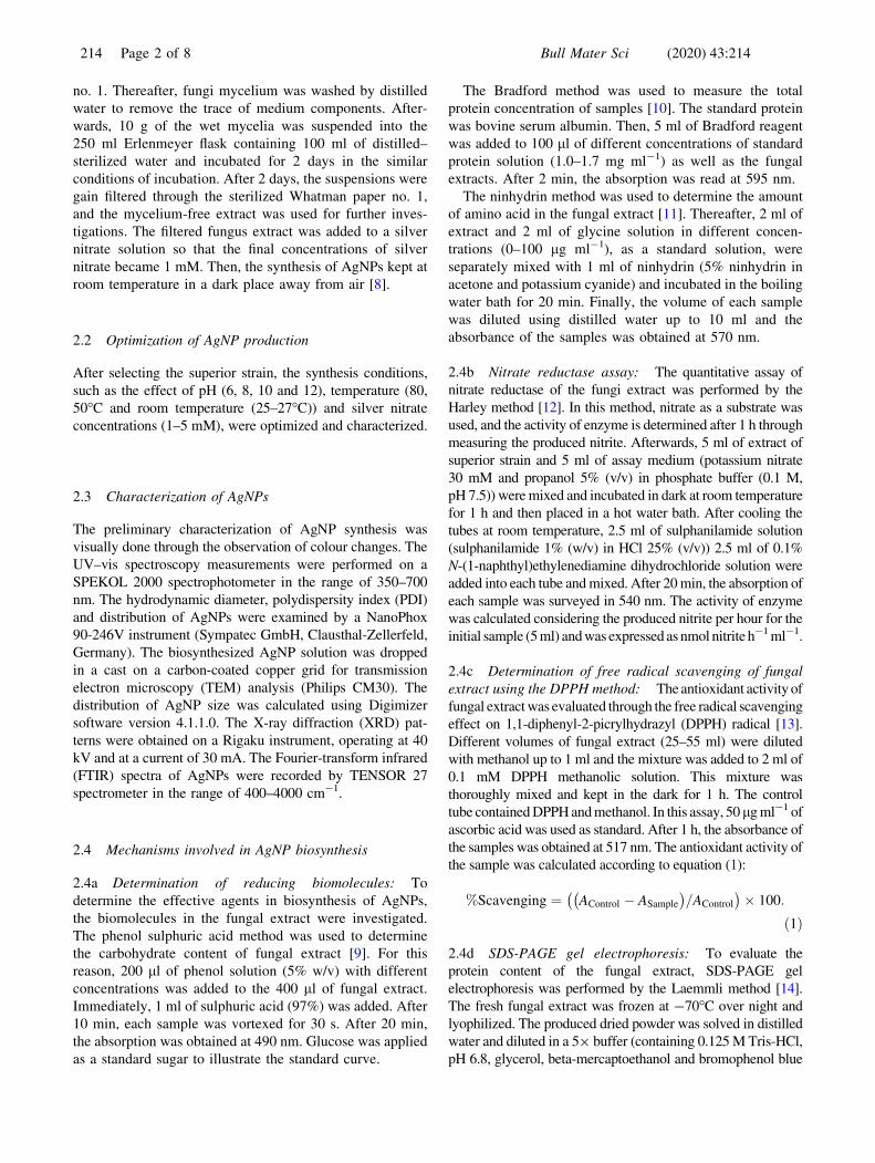
isolate. In nine out of the 24 isolates, the synthesis of
AgNPs was completed in less than 2 h due to the results of
the UV–vis spectra and the surface plasmon resonance
(SPR) peak of the synthesized AgNPs (figure 1). The SPR
peak of AgNPs offered useful information about their size,
shape and dispersion. When the size of AgNPs is
increased, the band gap energy between the capacity band
and the conduction band decreases, and the SPR spectrum
is thus transmitted to higher wavelengths. The peak width
of SPR is directly related to the dispersion size of AgNPs
[18,19]. Among the 24 strains of fungi screened, the MS17
strain (i.e., Aspergillus fumigatus) was selected. The cri-
teria for this selection included producing the highest
amount of AgNPs in less than 1 h, producing small-sized
AgNPs compared to other fungi (kmax = 420 nm) and
yielding low-sized dispersity (i.e., a narrow absorption
peak).
3.2 Optimization of AgNP synthesis
Environmental conditions, such as silver nitrate concentra-
tion, pH and temperature, affect the size, shape and stability
of NPs. AgNPs were synthesized in this study using the
superior strain with various concentrations of silver nitrate,
pHs and temperatures. The UV–vis absorption spectra of
the AgNPs and their stability were then monitored for
2 weeks. Based on the results of the UV–vis spectra of the
synthesized AgNPs in various concentrations of silver
nitrate, the AgNPs that were produced in 1 mM of silver
nitrate had a lower size dispersion, a smaller size and a
higher stability. In acidic pH (= 6), the AgNPs synthesized
by the superior strain showed a very low stability, and an
aggregation of AgNPs was observed immediately after
synthesis. From all the pH values (= 6, 8, 10 and 12), the pH
of 8 was selected since it produced AgNPs that possessed a
higher stability and mono dispersion. The results were
consistent with the findings of previous studies [16,20].
Temperature also plays an important role in the production
and stabilization of AgNPs. The results of a study by Sahoo
et al [21] showed that, at temperatures below 30�C, the size
of AgNPs is 30 nm, and at higher temperatures, the size
Bull Mater Sci (2020) 43:214 Page 3 of 8 214
increases [21]. In the present study, room temperature was
considered the optimal temperature for synthesizing more
stable AgNPs with a lower dispersion. Room temperature,
pH of 8 and 1-mM concentration of AgNO3 were thus
considered the optimal conditions for synthesizing AgNPs
and further analyses were conducted for these conditions.
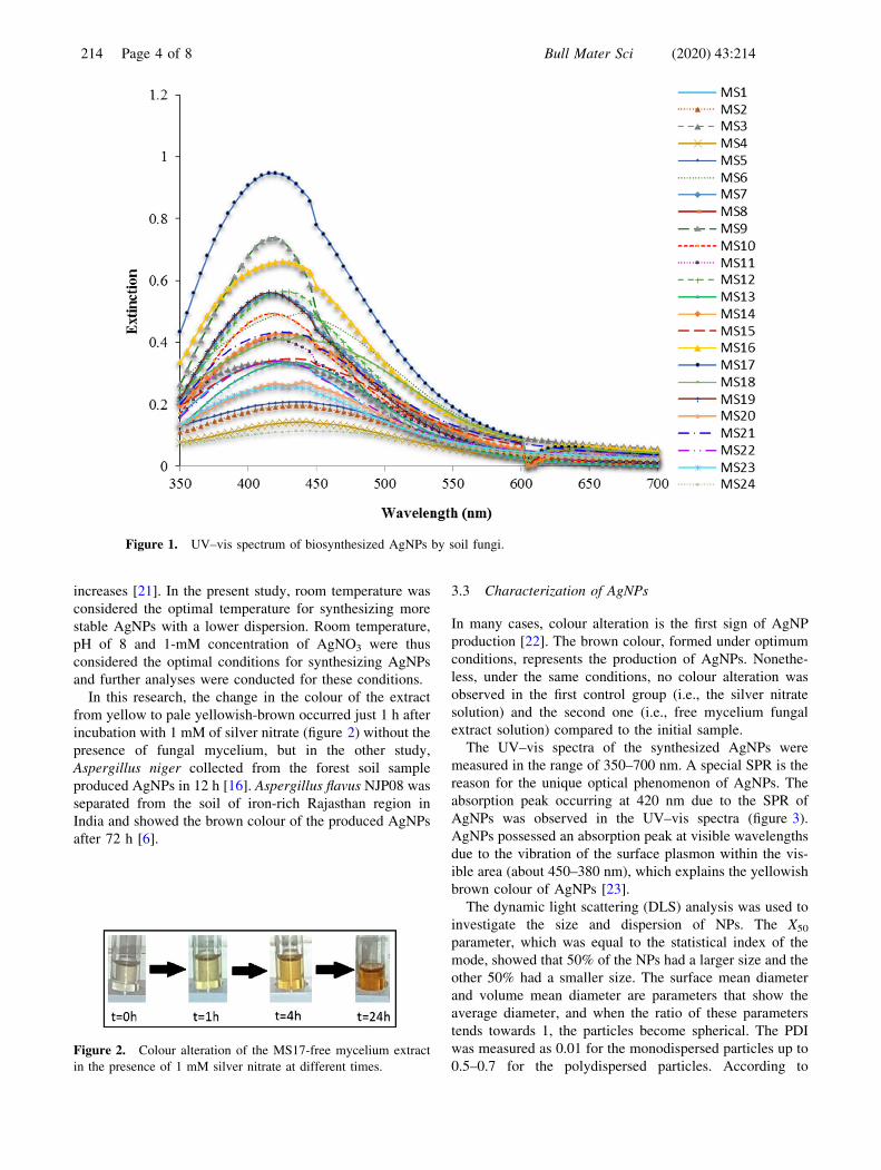
In this research, the change in the colour of the extract
from yellow to pale yellowish-brown occurred just 1 h after
incubation with 1 mM of silver nitrate (figure 2) without the
presence of fungal mycelium, but in the other study,
Aspergillus niger collected from the forest soil sample
produced AgNPs in 12 h [16]. Aspergillus flavus NJP08 was
separated from the soil of iron-rich Rajasthan region in
India and showed the brown colour of the produced AgNPs
after 72 h [6].
3.3 Characterization of AgNPs
In many cases, colour alteration is the first sign of AgNP
production [22]. The brown colour, formed under optimum
conditions, represents the production of AgNPs. Nonethe-
less, under the same conditions, no colour alteration was
observed in the first control group (i.e., the silver nitrate
solution) and the second one (i.e., free mycelium fungal
extract solution) compared to the initial sample.
The UV–vis spectra of the synthesized AgNPs were
measured in the range of 350–700 nm. A special SPR is the
reason for the unique optical phenomenon of AgNPs. The
absorption peak occurring at 420 nm due to the SPR of
AgNPs was observed in the UV–vis spectra (figure 3).
AgNPs possessed an absorption peak at visible wavelengths
due to the vibration of the surface plasmon within the vis-
ible area (about 450–380 nm), which explains the yellowish
brown colour of AgNPs [23].
The dynamic light scattering (DLS) analysis was used to
investigate the size and dispersion of NPs. The X50
parameter, which was equal to the statistical index of the
mode, showed that 50% of the NPs had a larger size and the
other 50% had a smaller size. The surface mean diameter
and volume mean diameter are parameters that show the
average diameter, and when the ratio of these parameters
tends towards 1, the particles become spherical. The PDI
was measured as 0.01 for the monodispersed particles up to
0.5–0.7 for the polydispersed particles. According to
Figure 1. UV–vis spectrum of biosynthesized AgNPs by soil fungi.
Figure 2. Colour alteration of the MS17-free mycelium extract
in the presence of 1 mM silver nitrate at different times.
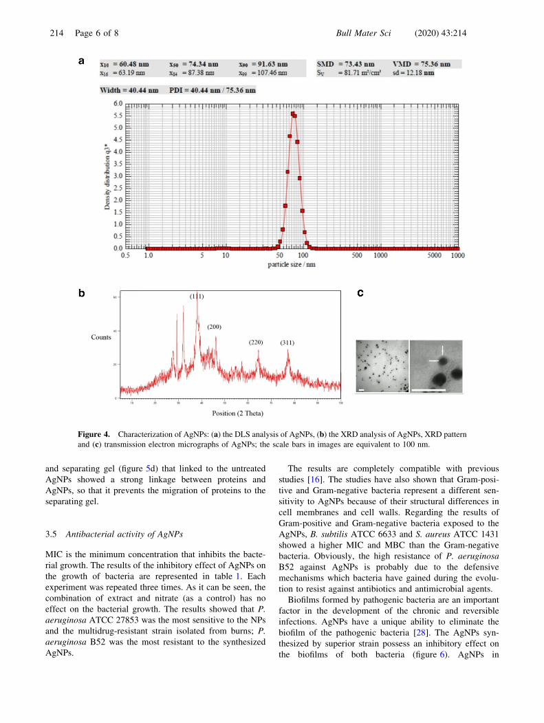
214 Page 4 of 8 Bull Mater Sci (2020) 43:214

figure 4a, the AgNPs synthesized by the superior strain
under optimal conditions showed a spherical shape with the
average hydrodynamic diameter of 75.36 nm, and the PDI
of the obtained particles was 0.27, indicating a mediocre
dispersion.
The XRD analysis revealed the crystalline nature of the
produced AgNPs. The appearance of peaks at 2h = 38� and
64� indicated the presence of Ag0 NPs, and the appearance
of two peaks at 2h = 32� and 46� indicated AgCl NPs in the
samples (figure 4b).
Based on the Scherrer equation and the data obtained
from the XRD analysis, the average crystal size of Ag0 and
AgCl NPs was 20 and 10 nm, respectively. The difference
in the average size that was obtained from both the DLS and
XRD analyses can be attributed to the fact that the hydro-
dynamic diameter of the NPs (AgNPs with surface-bound
agents) is measured in the DLS analysis, whereas only the
size of AgNP crystals, regardless of the capping agents, is
measured in the XRD analysis.
The TEM analysis was also used to determine the size
and shape of the AgNPs. As for the images obtained from
the TEM, the size of the AgNPs was determined using
Digimizer software. The TEM images showed that the
produced AgNPs possessed a spherical shape with a med-
ium-sized dispersion and an average size of 5–20 nm. With
high resolutions, the pale beads can be observed around
AgNPs’ TEM micrographs (figure 4c). This halo is probably
related to the capping agent on the surface of AgNPs that
affects their stability and the colloidal solution of synthe-
sized AgNPs under optimum conditions and can be pre-
served for a month, and the sedimentation was not
observed.
The biomolecules that capped the AgNPs were investi-
gated by FTIR spectroscopy. AgNPs and dried fungal
extract were analysed by FTIR and the results were reported
in the range of 400–4000 cm-1. The 670.95 and 1648 peaks
represent alkene and carboxylic acid, respectively. The
1045.65 peak indicates aliphatic amine. The 1362.45,
1396.41 and 2923.78 peaks are related to the methyl group
and the 3445.58 peak is for primary amine and 3675.88 for
alcoholic and phenolic O–H. The present findings are con-
sistent with the results of the study by Gajbhiye et al [24],
who synthesized AgNPs by the extracellular extract of Al-
ternaria alternata and reported that proteins can link to NPs
via free amines or cysteine residues or by the creation of an
electrostatic interaction through the carbocyclic groups in
enzymes, such that NPs are stabilized by proteins. The
presence of methyl, O–H and alkenyl groups indicates the
presence of carbohydrates in the fungal extract. The pres-
ence of primary amines and carboxylic acid groups is also
the result of amino acids and proteins in the samples.
3.4 The mechanisms involved in the synthesis of AgNPs
Biological systems are able to synthesize AgNPs due to their
reducing and stabilizing agents. Studies show that enzymes,
proteins, amino acids and carbohydrates play an important
role in the AgNP biosynthesis [25–27]. The concentration of
carbohydrate, protein and amino acids in the fungal extract
was 35.6, 37.7 and 2.6 lg ml-1, respectively.
3.4a Nitrate reductase assay: The presence of nitrate
reductase enzyme in the fungal extract of the superior strain
was confirmed only by the qualitative measurement method.
The results of quantitative measurements of nitrate
reductase showed that all of the nitrate reductase enzyme
substrate was consumed by the enzyme which exists in the
fungal extract. Then, the nitrite was reduced to N2 by the
other enzymes that exist in the extract.
3.4b DPPH scavenging of the fungal extract: The
antioxidant capacity of the fungal extract was assessed by
the evaluation of the scavenging of free radical of DPPH.
According to the results, the fungal extract possesses 41%
antioxidant activity.
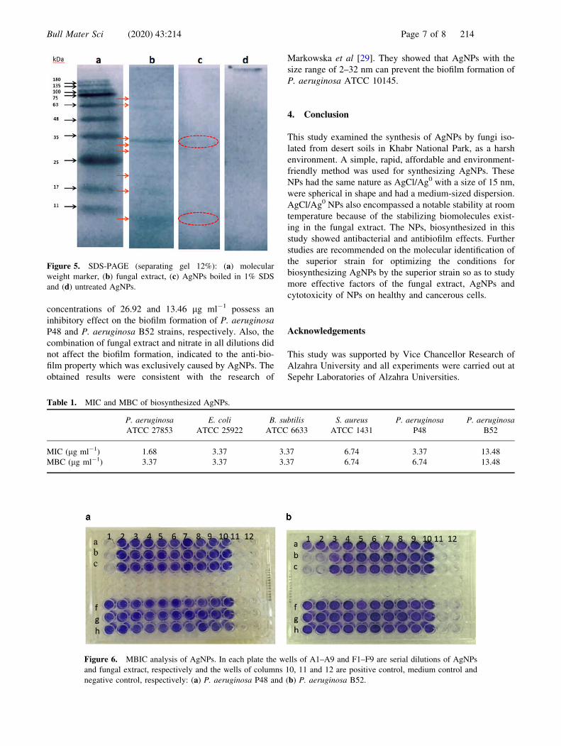
3.4c The SDS-PAGE analysis: The obtained results by
the Bradford and FTIR analysis exhibited the presence of
proteins not only in the fungal extract, but also in the cap of
the NP surface. In order to determine the proteins involved in
the synthesis and stability of the NPs, the SDS-PAGE pattern
obtained from the fungal extract and AgNPs was investigated.
The pattern of fungal proteins exhibited almost 8 bands in the
range of 10–75 kD (figure 5b). These proteins can also affect
the synthesis and the stability of NPs. To identify the proteins
linked to NPs, the synthesized NPs were treated by 1% SDS
and boiling water. SDS leads to denature the proteins and
distinct them from AgNP surface. AgNPs treated by SDS
showed two bands (figure 5c) that could be indicated to both
of these proteins. They functionalized as a capping agent on
the protein surface and affect the stability of NPs. These
findings are also compatible with the FTIR results which had
exhibited the relationship of the fungal extract proteins on
NPs. Furthermore, the broad band between the stacking gel
Figure 3. UV–vis spectrum of biosynthesized AgNPs of the
superior strain after 24 h.
Bull Mater Sci (2020) 43:214 Page 5 of 8 214
and separating gel (figure 5d) that linked to the untreated
AgNPs showed a strong linkage between proteins and
AgNPs, so that it prevents the migration of proteins to the
separating gel.
3.5 Antibacterial activity of AgNPs
MIC is the minimum concentration that inhibits the bacte-
rial growth. The results of the inhibitory effect of AgNPs on
the growth of bacteria are represented in table 1. Each
experiment was repeated three times. As it can be seen, the
combination of extract and nitrate (as a control) has no
effect on the bacterial growth. The results showed that P.
aeruginosa ATCC 27853 was the most sensitive to the NPs
and the multidrug-resistant strain isolated from burns; P.
aeruginosa B52 was the most resistant to the synthesized
AgNPs.
The results are completely compatible with previous
studies [16]. The studies have also shown that Gram-posi-
tive and Gram-negative bacteria represent a different sen-
sitivity to AgNPs because of their structural differences in
cell membranes and cell walls. Regarding the results of
Gram-positive and Gram-negative bacteria exposed to the
AgNPs, B. subtilis ATCC 6633 and S. aureus ATCC 1431
showed a higher MIC and MBC than the Gram-negative
bacteria. Obviously, the high resistance of P. aeruginosa
B52 against AgNPs is probably due to the defensive
mechanisms which bacteria have gained during the evolu-
tion to resist against antibiotics and antimicrobial agents.
Biofilms formed by pathogenic bacteria are an important
factor in the development of the chronic and reversible
infections. AgNPs have a unique ability to eliminate the
biofilm of the pathogenic bacteria [28]. The AgNPs syn-
thesized by superior strain possess an inhibitory effect on
the biofilms of both bacteria (figure 6). AgNPs in
Figure 4. Characterization of AgNPs: (a) the DLS analysis of AgNPs, (b) the XRD analysis of AgNPs, XRD pattern
and (c) transmission electron micrographs of AgNPs; the scale bars in images are equivalent to 100 nm.
214 Page 6 of 8 Bull Mater Sci (2020) 43:214
concentrations of 26.92 and 13.46 lg ml-1 possess an
inhibitory effect on the biofilm formation of P. aeruginosa
P48 and P. aeruginosa B52 strains, respectively. Also, the
combination of fungal extract and nitrate in all dilutions did
not affect the biofilm formation, indicated to the anti-bio-
film property which was exclusively caused by AgNPs. The
obtained results were consistent with the research of
Markowska et al [29]. They showed that AgNPs with the
size range of 2–32 nm can prevent the biofilm formation of
P. aeruginosa ATCC 10145.
4. Conclusion
This study examined the synthesis of AgNPs by fungi iso-
lated from desert soils in Khabr National Park, as a harsh
environment. A simple, rapid, affordable and environment-
friendly method was used for synthesizing AgNPs. These
NPs had the same nature as AgCl/Ag0 with a size of 15 nm,
were spherical in shape and had a medium-sized dispersion.
AgCl/Ag0 NPs also encompassed a notable stability at room
temperature because of the stabilizing biomolecules exist-
ing in the fungal extract. The NPs, biosynthesized in this
study showed antibacterial and antibiofilm effects. Further
studies are recommended on the molecular identification of
the superior strain for optimizing the conditions for
biosynthesizing AgNPs by the superior strain so as to study
more effective factors of the fungal extract, AgNPs and
cytotoxicity of NPs on healthy and cancerous cells.
Acknowledgements
This study was supported by Vice Chancellor Research of
Alzahra University and all experiments were carried out at
Sepehr Laboratories of Alzahra Universities.
Figure 5. SDS-PAGE (separating gel 12%): (a) molecular
weight marker, (b) fungal extract, (c) AgNPs boiled in 1% SDS
and (d) untreated AgNPs.
Figure 6. MBIC analysis of AgNPs. In each plate the wells of A1–A9 and F1–F9 are serial dilutions of AgNPs
and fungal extract, respectively and the wells of columns 10, 11 and 12 are positive control, medium control and
negative control, respectively: (a) P. aeruginosa P48 and (b) P. aeruginosa B52.
Table 1. MIC and MBC of biosynthesized AgNPs.
P. aeruginosa
ATCC 27853
E. coli
ATCC 25922
B. subtilis
ATCC 6633
S. aureus
ATCC 1431
P. aeruginosa
P48
P. aeruginosa
B52
MIC (lg ml-1) 1.68 3.37 3.37 6.74 3.37 13.48
MBC (lg ml-1) 3.37 3.37 3.37 6.74 6.74 13.48
Bull Mater Sci (2020) 43:214 Page 7 of 8 214
References
[1] Klasen H J 2000 Burns 26 131
[2] Castellano J J, Shafii S M, Ko F, Donate G, Wright T E,
Mannari R J et al 2007 Int. Wound J. 4 114
[3] Klaus T, Joerger R, Olsson E and Granqvist C G 1999 Proc.
Natl. Acad. Sci. 96 13614
[4] Mallick K, Witcomb M J and Scurrell M S 2004 J. Mater.
Sci. 39 4459
[5] Thakkar K N, Mhatre S S and Parikh R Y 2010Biol. Med. 6 257
[6] Jain N, Bhargava A, Majomdar S, Tarafdar J C and Panwar J
2011 Nanoscale 3 635
[7] Sastry M, Ahmad A, Khan M I and Kumar R 2003 Curr. Sci.
85 162
[8] Birla S S, Gaikwad S C, Gade A K and Rai M K 2013 Sci.
World J. 2013 1
[9] Albalasmeh A A, Berhe A A and Ghezzehei T A 2013
Carbohydr. Polym. 97 253
[10] Bradford M M 1974 Anal. Biochem. 72 248
[11] Starcher B 2001 Anal. Biochem. 129 125
[12] Jaidev L R and Narasimha G 2010 Colloids Surf. B 81 430
[13] Maizura M, Aminah A and Wan Aida W M 2011 Int. Food
Res. J. 18 529
[14] Laemmli U K 1970 Nature 227 680
[15] CLSI 2012 Methods for dilution antimicrobial susceptibility
tests for bacteria that grow aerobically; approved stan-
dard—CLSI document M07-A9 (Wayne, PA: Clinical and
Laboratory Standards Institute, 9th edn.)
[16] Balakumaran M D 2016 Microbiol. Res. 182 8
[17] Sterflinger K, Tesei D and Zakharova K 2012 Fungal Ecol. 5453
[18] Slistan-Grijalvaa A, Herrera-Urbinab R, Rivas-Silvac J F,
Avalos-Borjad M, Castillon-Barrazad F F and Posada-
Amarillase A 2005 Physica E 27 104
[19] Maccuspie R I, Allen A J and Hackley V A 2011 Nanono-
toxicology 5 140
[20] Mishra A, Tripathy S K, Wahab R, Jeong S H, Hwang I,
Yang Y B et al 2011 Appl. Microbiol. Biotechnol. 92 617
[21] Sahoo P, Kamal S, Kumar T, Sreedhar B, Singh A and
Srivastava S 2009 Def. Sci. J. 59 447
[22] Song J Y, Jang H and Kim B S 2009 Process Biochem. 441133
[23] Mukherjee P, Ahmad A, Mandal D, Satyajyoti S, Sudhakar R S,
Mohammad I K et al 2001 Nano Lett. 1 5174
[24] Gajbhiye M, Kesharwani J, Ingle A, Gade A and Ria M 2009
Nanomed. Nanotechnol. Biol. Med. 5 382
[25] Kumar S A, Abyaneh M K, Gosavi S W, Kulkarni S K,
Pasricha R, Ahmad A et al 2007 Biotechnol. Lett. 29 439
[26] Singh R, Shedbalkar U U, Wadhwani S A and Chopade B A
2015 Appl. Microbiol. Biotechnol. 99 4579
[27] Duran N, Silveria M P, Duran M and Martinez D S T 2005
J. Nanobiotechnol. 7 1
[28] Barapatre A, Aadil K R and Jha H 2016 Bioresour. Bio-
process. 3 8
[29] Markowska K, Grudniak M, Krawczyk K, Wrobel I and
Wolska K I 2014 J. Med. Microbiol. 63 849
214 Page 8 of 8 Bull Mater Sci (2020) 43:214
Recommended
![Results from AgNP method optimization · 2017. 9. 26. · Hg [ng] Ave. Hg/Ag Non-washed AgNPs 67 0.5354 0.00085 Washed AgNPs 45 6.09396 0.01379 Table 1: Results relating absolute](https://img.pdfslide.net/doc/110x75/614862b02918e2056c22a76f/results-from-agnp-method-optimization-2017-9-26-hg-ng-ave-hgag-non-washed.jpg)