1
Sevoflurane Anaesthesia in Dogs :
Clinical Implications and Applications
Ingeborgh Polis
Proefschrift ter verkrijging van de graad van Doctor in de Diergeneeskundige
Wetenschappen (PhD) aan de Faculteit Diergeneeskunde, Universiteit Gent
Promotor: Prof. Dr. F. Gasthuys
Co-promotors: Prof. Dr. L. Van Ham Prof. Dr. Y. Moens
Department of Small Animal Medicine and Clinical Biology
Faculty of Veterinary Medicine
Ghent University
ISBN 90-5864-019-1

2

3
DEDICATION
To my parents, for their lifelong love and encouragement.
To Geert, my support in bad days.
To Bram, the twinkle in my eyes.
Ingeborgh

4

5
CONTENTS
List of abbreviations
GENERAL INTRODUCTION 1
SCIENTIFIC AIMS 5
CHAPTER 1 SEVOFLURANE: PHYSICO-CHEMICAL PROPERTIES 9
AND MAC
Introduction 11 History of inhalant anaesthetic agents 11
Molecular structure and physical properties of sevoflurane 15 Anaesthetic properties of sevoflurane 18
Minimum alveolar concentration (MAC) 24 References 27
CHAPTER 2 SEVOFLURANE: INFLUENCES ON BODY 35
SYSTEMS AND ECONOMIC CONSIDERATIONS
Summary 37
Introduction 37 Effects on central nervous system 38 Effects on cardiovascular system 42
Effects on respiratory system 45 Hepatic effects 47
Renal effects 50 Economic considerations 55
Conclusion 58 References 59
INTRODUCTION TO CHAPTERS 3 AND 4 75
CHAPTER 3 RECOVERY TIMES AND EVALUATION OF CLINICAL 79
HEMODYNAMIC PARAMETERS OF SEVOFLURANE, ISOFLURANE AND HALOTHANE ANAESTHESIA IN
MONGREL DOGS.
Summary 81
Introduction 82
Materials and Methods 83
6
Results 86
Discussion 91
References 99
CHAPTER 4 THE INFLUENCE OF VENTILATION MODE 103
(SPONTANEOUS VENTILATION, IPPV AND PEEP) ON CARDIOPULMONARY PARAMETERS
IN SEVOFLURANE ANAESTHETIZED DOGS.
Summary 105
Introduction 106 Materials and Methods 107
Results 113 Discussion 119 References 126
INTRODUCTION TO CHAPTER 5 131
CHAPTER 5 THE EFFECTS OF INTRATHORACIC PRESSURE DURING 135
CONTINUOUS TWO-LUNG VENTILATION FOR THORACOSCOPY ON THE CARDIORESPIRATORY
PARAMETERS IN SEVOFLURANE ANAESTHETIZED DOGS.
Summary 137 Introduction 138 Materials and Methods 140
Results 146 Discussion 154
References 161
INTRODUCTION TO CHAPTERS 6, 7 AND 8 165
CHAPTER 6 ARTERIAL CATHETERISATION AND VASCULAR 169
ACCESS PORT IMPLANTATION FOR BLOOD SAMPLING AND CONTINUOUS BLOOD PRESSURE
MEASUREMENT IN DOGS.
Summary 171 Introduction 171
Materials and Methods 173 Results 180
Discussion 181 References 185

7
CHAPTER 7 PERIANAESTHETIC CARDIOPULMONARY, SEDATIVE 187
AND ANTINOCICEPTIVE EFFECTS OF A LONG ACTING FORMULATION OF SUFENTANIL ADMINISTERED
BEFORE SEVOFLURANE ANAESTHESIA IN DOGS.
PART I. CARDIOPULMONARY EFFECTS 187
Summary 189
Introduction 190 Materials and Methods 193
Results 199
Discussion 210 References 215
CHAPTER 8 PERIANAESTHETIC CARDIOPULMONARY, SEDATIVE 221
AND ANTINOCICEPTIVE EFFECTS OF A LONG ACTING FORMULATION OF SUFENTANIL ADMINISTERED
BEFORE SEVOFLURANE ANAESTHESIA IN DOGS.
PART II. SEDATIVE AND ANTINOCICEPTIVE EFFECTS 221
Summary 223 Introduction 224 Materials and Methods 225
Results 231 Discussion 237
References 242
GENERAL DISCUSSION 247
References 260
SUMMARY 265
SAMENVATTING 271
DANKWOORD 277
CURRICULUM VITAE 281
PUBLICATIONS 283

8

9
LIST OF ABBREVIATIONS
AA % end tidal anaesthetic agent percentage AR artificial respiration ASA american society of anaesthesiologists BSA body surface area BWT body weight CBF cerebral blood flow CI cardiac index CO cardiac output CO2ET end tidal carbon dioxide percentage CPAP continuous positive airway pressure CV controlled ventilation DAP diastolic arterial blood pressure DPAP diastolic pulmonary artery pressure FA alveolar anaesthetic concentration Fi inspired concentration FiAA % inspiratory anaesthetic agent concentration FiO2 inspiratory oxygen fraction Halo halothane HCO3
- plasma bicarbonate concentration HPV hypoxic pulmonary vasoconstriction HR heart rate ID internal diameter IM intramuscular IPPV intermittent positive pressure ventilation Iso isoflurane ITP intrathoracic pressure IV intravenous LA long acting LVSWI left ventricular stroke work index MAC minimum alveolar concentration MAP mean arterial blood pressure Min-max minimum and maximum MPAP mean pulmonary artery pressure MVV minute ventilation volume OLV one lung ventilation PA alveolar partial pressure PAP pulmonary artery pressure PaCO2 arterial carbon dioxide tension PCV packed cell volume PCWP pulmonary capillary wedge pressure

LIST OF ABBREVIATIONS
10
PEEP positive end expiratory pressure Pinsp inspiratory pressure PO per os PO2 arterial oxygen tension PVR pulmonary vascular resistance RAP right atrial pressure RR respiratory rate RVSWI right ventricular stroke work index SAP systolic arterial blood pressure SBC standard bicarbonate concentration SBE standard base excess SC subcutaneous Sevo sevoflurane SI stroke index SPAP systolic pulmonary artery pressure SpO2 % peripheral haemoglobin saturation SpV spontaneous ventilation SV stroke volume SVR systemic vascular resistance TLV two lung ventilation TV tidal volume VAP vascular access port

1
GENERAL INTRODUCTION

2

GENERAL INTRODUCTION
3
The last decades a very impressive progress has been made
in diagnostic as well as surgical techniques. As a consequence the
need for a safe and stable long-standing anaesthesia during these
procedures increases. Inhalation anaesthetics are very useful for this
purpose. Halothane has being used for several decades in veterinary
medicine and is still a valuable compound in many clinical settings.
However, halothane is not an ideal anaesthetic drug. This is
not surprising as an ideal volatile anaesthetic compound has to fulfil
many criteria: minimal or no depressing effects on vital functions such
as respiration and circulation, rapid onset of action, beneficial
interaction with premedication and anaesthesia-inducing drugs, not to
mention low health hazard for the anaesthetists, low flammability and
the lack of need for expensive vaporizers.
The last decades several new volatile anaesthetics have been
developed such as isoflurane, desflurane and sevoflurane in order to
obtain drugs with more beneficial and less side effects than the
previous ones. Some of these drugs like isoflurane have been studied
extensively in dogs, in experimental as well as clinical settings. The
results of such studies indicate that new drugs may be superior for
some but not all aspects leading to nuanced conclusions.
Sevoflurane has been studied widely in humans. The findings
are interesting in order to have an idea of the profile of this almost
unknown drug in veterinary medicine. In humans its lack of airway
pungency and mainly its low blood-gas solubility induce a fast
induction and recovery from anaesthesia. Sevoflurane has little
influence on cerebral perfusion and intracranial pressure. Depression
of cardiac output is only reported at higher concentrations. Both
characteristics make it the agent of choice in neurological and cardiac

GENERAL INTRODUCTION
4
patients. However one should be careful by extrapolating these
results to canine medicine. Therefore several studies were undertaken
in dogs in order to investigate some clinical implications and
applications of sevoflurane.
In the first part of this thesis (chapter 1 and 2)
physicochemical and pharmacological properties of sevoflurane are
extensively reviewed and compared with other inhalation
anaesthetics.
In the second part own studies on sevoflurane in dogs are
described. These studies deal with several pharmacological and
clinical aspects of sevoflurane anaesthesia: recovery times and
haemodynamics in comparison to other anaesthetics (chapter 3);
influence of ventilation mode (chapter 4) and thoracoscopy (chapter 5)
on cardiopulmonary parameters; vascular access port implantation
(chapter 6) in order to study the influence of sufentanil long-acting
premedication on haemodynamics (chapter 7) and analgesic effects
(chapter 8) of sevoflurane.

5
SCIENTIFIC AIMS

6

SCIENTIFIC AIMS
7
1/ To compare the recovery times and clinical haemodynamic
parameters of sevoflurane, isoflurane and halothane anaesthesia in
mongrel dogs.
2/ To investigate the influence of ventilation mode (spontaneous
ventilation, IPPV and PEEP) on cardiopulmonary parameters in
sevoflurane anaesthetized dogs.
3/ To determine the effects of intrathoracic pressure elevation on
cardio-respiratory parameters during sevoflurane anaesthesia with
continuous two-lung ventilation for thoracoscopy in dogs.
4/ To describe the vascular access port implantation for blood
sampling and continuous blood pressure measurement in dogs.
5/ To examine the haemodynamic influences of a long-acting
formulation of sufentanil administered at different time intervals in
sevoflurane anaesthetized dogs.
6/ To evaluate antinociceptive and sedative effects of premedication
with a long-acting formulation of sufentanil during and after
sevoflurane anaesthesia in dogs.

CHAPTER 1
8

CHAPTER 1
9
CHAPTER 1
SEVOFLURANE: PHYSICO-CHEMICAL PROPERTIES
AND MAC
I. Polis1, F. Gasthuys2, L. Van Ham1 1 Department of Small Animal Medicine and Clinical Biology 2 Department of Surgery and Anaesthesia of Domestic Animals Ghent University, Faculty of Veterinary Medicine, Salisburylaan 133, B-9820 Merelbeke, Belgium
Adapted from:
I. Polis, F. Gasthuys, L. Van Ham (1999). Sevoflurane: een nieuw inhalatieanestheticum voor hond en kat. Deel 1 Vlaams Diergeneeskundig Tijdschrift 68: 261-266.

CHAPTER 1
10

CHAPTER 1
11
INTRODUCTION
Recently two new inhalant anaesthetic agents were
commercialised in Europe, sevoflurane and desflurane. Both agents fit
the mould of several other new anaesthetic agents and adjuvants.
They permit greater control over the course of anaesthesia and more
rapid recovery from anaesthesia than do their predecessors. In this
review a survey was put together on the properties of sevoflurane in
comparison with other recently developed inhalant anaesthetic agents
(desflurane, isoflurane, halothane, enflurane). Furthermore, the
possible usefulness of sevoflurane in anaesthesia of companion
animals, in particular in dogs and cats was highlighted. The influences
of sevoflurane on several body systems will be described in the
second chapter.
HISTORY OF INHALANT ANAESTHETIC AGENTS (Table 1)
The earliest recorded attempts to induce anaesthesia appear
to have been performed in humans. The ancients used opiates,
alcohol, asphyxia, and even rather primitive techniques as
compression of the carotid arteries to alleviate pain during surgical
intervention.
In 1800, Sir Humphrey Davy suggested that nitrous oxide
might have anaesthetic properties. Shortly thereafter in 1824, H. H.
Hickman demonstrated that pain associated with surgery in dogs
could be alleviated by inhalation of a mixture of nitrous oxide and
carbon dioxide (Thurmon et al., 1996).
It was not until 1842 that ether was used for human
anaesthesia. Jackson was the first clinician to employ ether

CHAPTER 1
12
extensively in animals in 1853 (Jackson, 1853). Although chloroform
was discovered by Liebig in 1831, it only was used in 1847 for general
anaesthesia in animals by Flourens (Dadd, 1854). Diethyl ether and
chloroform had marked side effects including arrhythmogenic effects,
cardiovascular and respiratory depression and liver toxicity. In
addition, some practical objections were the flammability and
explosiveness of ether (Hall and Clarke, 1991).
Waters was the first to clinically use cyclopropane in human
anaesthesia in 1933. Gregory developed the use of cyclopropane for
experimental animal anaesthesia. From the fifties on, it was routinely
used in several animal species in Great Britain. Practical problems
rose again with its high flammability and explosiveness (Hall and
Clarke, 1991). Since 1941 trilene (trichloro-ethyleen) has been widely
used. Trilene had good analgesic effects; it was non-flammable, nor
irritating. Nevertheless, its anaesthetic properties and muscle
relaxation were insufficient (Vickers et al., 1978).
Fluroxene (2,2,2,trifluoroethyl-ether) was developed by
Shukyse in 1951. This anaesthetic agent gave a rapid induction of
anaesthesia with a dose related cardiopulmonary depression.
However, it was extremely flammable and toxic after repeated use
especially in animals (Hall and Clarke, 1991).
Already in 1940 methoxyflurane (2,2-dichloro-1,1-
difluoroethylmethylether) was synthesized, although it was only
commercialised in 1958. Methoxyflurane was a potent, non-flammable
anaesthetic agent with good analgesic properties (Artusio et al.,
1960). Its high blood-gas solubility was accompanied by a prolonged
induction and recovery time. The recorded post-anaesthetic renal

CHAPTER 1
13
failure was due to fluoride ion release during metabolisation of
methoxyflurane (Mazze et al., 1971).
Table 1. Historical overview of volatile anaesthetic agents.
AGENTS
YEAR*
ADVANTAGES
DISADVANTAGES
Nitrous oxide 1800 analgetic properties low blood-gas solubility
low analgetic potency in animals
Ether
1842 potent anaesthetic agent inflammable, irritating high blood-gas solubility
Chloroform 1847 potent anaesthetic agent hepatic and renal toxicity arhytmogenicity
Cyclopropaan 1933 low blood-gas solubility Inflammable Explosive
Trichloroethyleen (trilene)
1941 good analgesia non-irritating
weak anaesthetic agent toxic breakdown in soda-lime
Halothane 1956 potent anaesthetic agent low toxicity
unstable in light
Methoxyflurane 1958 potent anaesthetic agent analgetic properties
high blood-gas solubility metabolised to fluorine
Enflurane 1958 potent anaesthetic agent low blood-gas solubility
epileptic properties
Isoflurane 1971 potent anaesthetic agent low metabolisation
degree low blood-gas solubility
airway pungency
Desflurane 1987 low metabolisation degree
extremely low blood-gas solubility
specialised vaporiser technology
airway pungency
Sevoflurane 1990 potent anaesthetic agent low metabolisation
degree low blood-gas solubility
renal toxicity?
* Year routinely used in human practice.
CHAPTER 1
14
Halothane was introduced in veterinary anaesthesia in 1956,
after its development for human anaesthesia by Sweking in 1951
(Suckling, 1957). Halothane is non-explosive and relatively stable. It
gives a relatively fast induction of and recovery from anaesthesia and
is less toxic than previously used inhalant anaesthetic agents. As with
all volatile anaesthetic agents a dose related cardiopulmonary
depression occurs (Short, 1987). In 1958 enflurane was introduced in
human anaesthesia as a potent and slightly irritating volatile
anaesthetic agent. Induction and recovery from anaesthesia are
uneventful due to the low blood-gas solubility. Yet, higher enflurane
concentrations have epileptic properties in dogs, cats and horses
(Stevens et al., 1983, Oshima et al., 1985). In the early seventies
isoflurane was developed by Terrell (Wade and Stevens, 1981).
Isoflurane has a low blood-gas solubility resulting in short induction
and recovery times.
In the continued search for less reactive, more potent and
non-inflammable volatile anaesthetic agents focus on halogenation of
these compounds has predominated. Chlorine and bromine especially
convert many compounds of low anaesthetic potency into more potent
drugs. Fluorination although improving stability, produces less potent
compounds than addition of chlorine or bromine (Targ et al., 1989b).
The lighter the halogens, the lower the anaesthetic potency of the
compounds.
Up to now the search for developing new volatile anaesthetic
agents with a higher safety margin, minimal cardiovascular depression
and permitting a rapid and precise control of alveolar anaesthetic
concentration, is continued. This resulted in the recent development of
sevoflurane and desflurane, two anaesthetics permitting a flexible
control of anaesthesia maintenance and inducing a rapid recovery.

CHAPTER 1
15
Research on sevoflurane and desflurane develops parallel to one
other.
Desflurane was recently commercialised in Europe. It has the
lowest blood-gas solubility of all contemporary volatile anaesthetic
agents. Besides its relatively low anaesthetic potency (high
concentrations needed) and its airway irritating property; its high
vapour pressure (specialized vaporizer technology required) is also a
disadvantage for its practical use (Eger, 1993; Young and Apfelbaum,
1995).
Sevoflurane was developed in the seventies (Wallin et al.,
1975) and since 1980 it is extensively examined in human and
veterinary anaesthesia, especially on an experimental basis in the
beginning. Since 1990 it can be used in clinical human anaesthesia.
And finally in 1996 sevoflurane was registered for human anaesthesia
in Belgium.
MOLECULAR STRUCTURE AND PHYSICAL PROPERTIES OF
SEVOFLURANE (Table 2a +b)
The chemical structure of inhalation anaesthetics and their
physical properties are important determinants of their actions and
safety of administration. All contemporary volatile anaesthetic agents
are organic compounds except nitrous oxide (N2O). Sevoflurane is a
methyl-propyl-ether with 7 fluorine atoms and a molecular weight of
200.1 (Aida et al., 1994). The chemical structure of sevoflurane
(CFH2-O-CH(CF3)2) is responsible for its kinetic properties.
Fluorination of the carbon group resulted in a low blood-gas
partition coefficient (0.68), which is considerably lower compared to

CHAPTER 1
16
halothane, enflurane and isoflurane (Eger, 1994). The low blood-gas
solubility produces the following properties: 1/ more rapid increase in
alveolar anaesthetic concentration during induction of anaesthesia, 2/
more precise control of alveolar anaesthetic concentration during
maintenance of anaesthesia and 3/ more rapid decrease in alveolar
anaesthetic concentration during elimination. The human tissue-blood
partition coefficients of sevoflurane in the brain (1.70), fat (48.0),
kidneys (1.20), liver (1.80) and muscles (3.10) are intermediate
between isoflurane en halothane (Steffey, 1996). A low brain-blood
partition coefficient is advantageous for a rapid control and adjustment
of anaesthetic depth; whereas a low fat-blood partition coefficient is of
primordial importance for a rapid recovery from anaesthesia (Jones,
1990).
The solubility characteristics of sevoflurane in rubber and
plastic are lower compared to isoflurane and halothane (Targ et al.,
1989a). Consequently, the anaesthetic circuit extracts less agent
during anaesthetic administration and redistributes less agent to
rebreathed gases during elimination. This can be important since
losses of volatile anaesthetic by circuit absorption may compromise
measurements of anaesthetic uptake (Eger et al., 1998).
The boiling point and vapour pressure of sevoflurane are
comparable with those from halothane, isoflurane and enflurane.
Hence, conventional precision vaporisers without specific technical
requirements can be used. On the contrary, the boiling point and
vapour pressure of desflurane are completely different from the other
volatile anaesthetic agents requiring specialised vaporizer technology
for desflurane. Furthermore, sevoflurane doesn’t contain thymol or
any other preservative, in contrast with the less stable halothane.
CHAPTER 1
17
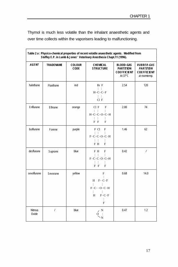
Thymol is much less volatile than the inhalant anaesthetic agents and
over time collects within the vaporisers leading to malfunctioning.
Table 2 a : Physico-chemical properties of recent volatile anaesthetic agents. Modified from Steffey E.P. in Lumb & Jones’ Veterinary Anesthesia Chapt.11 (1996).
AGENT TRADENAME COLOUR CODE
CHEMICAL STRUCTURE
BLOOD-GAS PARTITION
COEFFICIENT At 37°C
RUBBER-GAS PARTITION
COEFFICIENT at roomtemp.
halothane Fluothane red Br F ¦ ¦
H-C-C-F ¦ ¦ Cl F
2.54 120
Enflurane Ethrane orange Cl F F ¦ ¦ ¦
H-C-C-O-C-H ¦ ¦ ¦ F F F
2.00 74
Isoflurane Forene purple F Cl F ¦ ¦ ¦
F-C-C-O-C-H ¦ ¦ ¦ F H F
1.46 62
desflurane Suprane blue F H F ¦ ¦ ¦
F-C-C-O-C--H ¦ ¦ ¦ F F F
0.42 /
sevoflurane Sevorane yellow F ¦
H F- C-F ¦ ¦
F-C--O-C-H ¦ ¦
H F-C-F ¦ F
0.68 14.0
Nitrous Oxide
/ blue N O ¦ N
0.47 1.2

CHAPTER 1
18
Table 2 b : Physico-chemical properties of recent volatile anaesthetic agents. Modified from Steffey E.P. in Lumb & Jones’ Veterinary Anesthesia Chapt.11 (1996).
AGENT TRADENAME BOILING POINT
VAPOUR PRESSURE
at 20°C at 24°C
% METABOLISATION
halothane Fluothane 50.2 °C 243 mmHg 288 mmHg
20-25
Enflurane Ethrane 57 °C 172 mmHg 207 mmHg
2.4
Isoflurane Forene 49 °C 240 mmHg 286 mmHg
0.17
desflurane Suprane 23.5 °C 664 mmHg /
0.02
sevoflurane Sevorane 59 °C 160 mmHg 197 mmHg
3.0
Nitrous Oxide
/ -89 °C / /
0.004
ANAESTHETIC PROPERTIES OF SEVOFLURANE
* Inhalation Induction (Figure 1)
The aim in administering an inhalation anaesthetic agent to a
patient is to achieve an adequate partial pressure of anaesthetic in the
brain to cause a desired level of central nervous system depression.
The rate of change of anaesthetic depth is of obvious clinical
importance and is directly dependent upon the rate of change in
anaesthetic tensions in the various media in which it is taken up
before reaching the brain. Inhalation anaesthetics move down a series
of partial pressure gradients from regions of higher tension to those of
lower tension until equilibrium is established over the several
compartments. The anaesthetic agent travels from vaporizer to
breathing circuit, from circuit to lungs, from lungs to arterial blood, and
CHAPTER 1
19
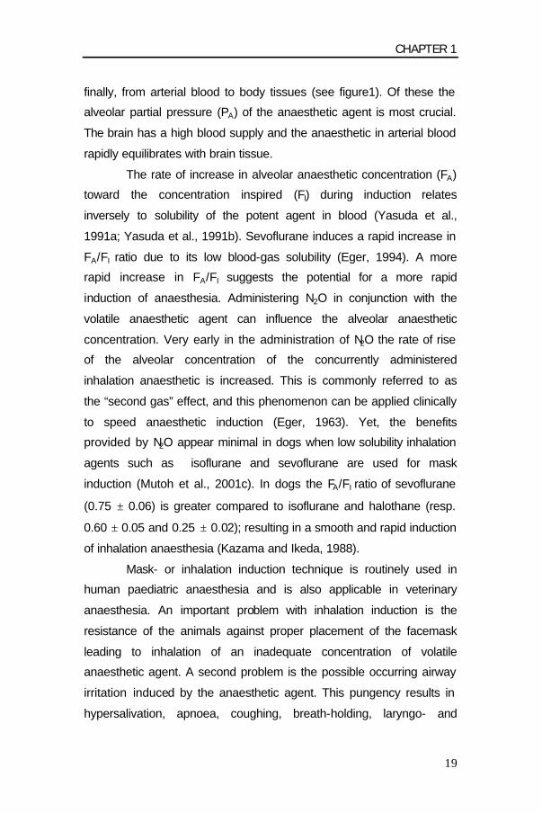
finally, from arterial blood to body tissues (see figure1). Of these the
alveolar partial pressure (PA) of the anaesthetic agent is most crucial.
The brain has a high blood supply and the anaesthetic in arterial blood
rapidly equilibrates with brain tissue.
The rate of increase in alveolar anaesthetic concentration (FA)
toward the concentration inspired (FI) during induction relates
inversely to solubility of the potent agent in blood (Yasuda et al.,
1991a; Yasuda et al., 1991b). Sevoflurane induces a rapid increase in
FA/FI ratio due to its low blood-gas solubility (Eger, 1994). A more
rapid increase in FA/FI suggests the potential for a more rapid
induction of anaesthesia. Administering N2O in conjunction with the
volatile anaesthetic agent can influence the alveolar anaesthetic
concentration. Very early in the administration of N2O the rate of rise
of the alveolar concentration of the concurrently administered
inhalation anaesthetic is increased. This is commonly referred to as
the “second gas” effect, and this phenomenon can be applied clinically
to speed anaesthetic induction (Eger, 1963). Yet, the benefits
provided by N2O appear minimal in dogs when low solubility inhalation
agents such as isoflurane and sevoflurane are used for mask
induction (Mutoh et al., 2001c). In dogs the FA/FI ratio of sevoflurane
(0.75 ± 0.06) is greater compared to isoflurane and halothane (resp.
0.60 ± 0.05 and 0.25 ± 0.02); resulting in a smooth and rapid induction
of inhalation anaesthesia (Kazama and Ikeda, 1988).
Mask- or inhalation induction technique is routinely used in
human paediatric anaesthesia and is also applicable in veterinary
anaesthesia. An important problem with inhalation induction is the
resistance of the animals against proper placement of the facemask
leading to inhalation of an inadequate concentration of volatile
anaesthetic agent. A second problem is the possible occurring airway
irritation induced by the anaesthetic agent. This pungency results in
hypersalivation, apnoea, coughing, breath-holding, laryngo- and

CHAPTER 1
20
bronchial spasms and increased airway secretions. These undesirable
responses result from irritation of the mucosa of the nasal passages,
pharynx and larynx, which may impair smooth induction of
anaesthesia and lead to airway obstruction and associated hypoxia
and hypercapnia in dogs, cats and humans. Mutoh et al. (1995)
described that inhalation induction with 2.5 MAC isoflurane in dogs
was accompanied with relatively more struggling compared to
induction with 2.5 MAC sevoflurane. Upper-airway administration of
sevoflurane, halothane and isoflurane with concentrations used for
mask induction induced milder reflex inhibition of breathing with
sevoflurane. Lack of respiratory reflexes attributable to stimulation of
the nasal passages may contribute to speed of onset and promote a
smoother induction with sevoflurane (Mutoh et al., 2001a; Mutoh et
al., 2001b).
The pungency of sevoflurane parallels that of halothane; this
makes them the less pungent volatile anaesthetic agents. Both
inhalant anaesthetics can be applied for mask induction in human and
small animal anaesthesia (Sarner et al., 1995; Lerman et al., 1996;
Blair et al., 2000). The differences in induction speed and airway
irritability have not been confirmed in cats. Hikasa et al. (1996) did not
see any difference in induction speed between halothane, isoflurane
and sevoflurane. Possible explanations for these different findings
could be the administered premedication and the slow and gradual
induction technique applied. Sevoflurane mask induction is suitable in
feline practice because of its good quality of induction in most cats
and dogs (Johnson et al., 1998; Tzannes et al., 2000; Mutoh et al.,
2001c; Lerche et al., 2002). Desflurane on the other hand is not
recommended for inhalation induction in paediatric anaesthesia due to
its high pungency with laryngeal spasms, coughing and increased
airway secretions (Eger, 1994).

CHAPTER 1
21
Fig.1: The flow pattern of inhaled anaesthetic agents during anaesthetic induction and recovery.
FD VAPORIZER METABOLITES
FI FE
FA LUNGALVEOLI METABOLISATION
B – G Fa ARTERIAL BLOOD VENOUS BLOOD Fv B - T
OTHER SYSTEMS
CENTRAL NERVOUS SYSTEM CLINICAL EFFECT FD: delivered anaesthetic concentration by the vaporizer FA: alveolar anaesthetic concentration FI: inspiratory anaesthetic concentration FE: expiratory anaesthetic concentration Fa: arterial anaesthetic concentration Fv: venous anaesthetic concentration B – G: blood – gas solubility B – T: blood – tissue solubility
CHAPTER 1
22
* Maintenance of anaesthesia
Maintenance of a constant level of anaesthesia with an
inhalant anaesthetic agent may be equated to the maintenance of a
constant alveolar anaesthetic concentration. A precise control of
anaesthetic depth on basis of vaporizer settings is desirable in clinical
practice. The difference between the concentration of anaesthetic
agent delivered (FD) from a vaporizer and the FA may be used to
define the degree of control of the anaesthetic level obtained with an
inhalant agent during maintenance of anaesthesia (Eger, 1994). A
ratio of FD/FA that approaches 1.0 indicates precise control, and
deviations from 1.0 less control. The FD/FA ratio depends on the
anaesthetic agent, the anaesthetic system (rebreathing degree) and
the fresh gas flow. Anaesthetic uptake and rebreathing determine the
proximity of FD/FA to 1.0: a smaller uptake (lower solubility and greater
tissue equilibration) and diminished rebreathing (i.e., a higher inflow
rate, a higher fresh gas flow rate) provide a value closer to 1.0.
Furthermore, cardiac output and alveolar ventilation have an important
influence on the FD/FA ratio of a volatile anaesthetic agent.
The low blood-gas solubility of sevoflurane even in
combination with an economical flow rate of 1-2 L/min permits to
estimate the alveolar anaesthetic concentration from the delivered
concentration by the vaporizer.
The use of an agent-specific analyser facilitates a precision
over the control of maintenance of anaesthesia, regardless of
solubility by measuring inspiratory and end tidal anaesthetic gases.
* Recovery from anaesthesia
Recovery from inhalation anaesthesia depends on solubility
and concentration of the volatile agent, duration of anaesthesia and

CHAPTER 1
23
metabolisation percentage (Lerman et al., 1996). The lower solubility
of sevoflurane permits a more rapid decrease in FA at the end of
anaesthesia. Its low fat solubility assures a rapid elimination
regardless of anaesthesia duration.
In humans a positive correlation (r = 0.517) was found for
isoflurane between total anaesthetic exposure or dose (MAC-hours,
see further) and recovery time. After sevoflurane anaesthesia MAC-
awake (the average of the bracketing alveolar anaesthetic
concentration that allows and prevents the response to verbal
command during recovery from anaesthesia; Stoelting et al., 1970)
was independent of anaesthetic duration in adults (Campbell et al.,
1995). Hikasa et al. (1996) showed that recovery times in cats were
significantly shorter after 90 minutes of sevoflurane anaesthesia
compared to halothane anaesthesia, but only slightly shorter
compared to isoflurane anaesthesia.
Inhalation anaesthetic agents are not chemically inert. They
undergo varying degrees of metabolism primarily in the liver, but also
to a lesser degree in the lung, kidney and intestinal tract (Rehder et
al., 1967; Holaday et al., 1970). Especially methoxyflurane and to a
lesser extent halothane have longer recovery times caused by their
extended metabolisation (Carpenter et al., 1986).
More recent volatile anaesthetic agents have shorter
emergence times greatly due to their low extent of biotransformation
(see table 2). Important elimination routes from the body are the
lungs, and of minor clinical importance through faeces, urine,
transpiration, percutaneous loss and eventually through the surgical
site (Stoelting and Eger, 1969; Fassoulaki et al., 1991; Lockhart et al.,
1991).
CHAPTER 1
24
Nevertheless, in comparison with isoflurane and desflurane,
sevoflurane still has a relatively high metabolisation percentage, yet it
has a rapid recovery. This is probably related to the low fat solubility of
sevoflurane resulting in low deposition of the anaesthetic agent in
body fat tissue. Body fat tissue functions as depot for the volatile
anaesthetic agent during elimination.
The pharmacokinetic profile of sevoflurane resulting in rapid
emergence times is especially useful after ambulatory anaesthesia in
human anaesthesia. Time intervals from stopping the delivery of the
anaesthetic to specific emergence and recovery parameters (e.g.,
time to extubation, opening of the eyes, emergence, orientation,
response to commands, etc.) are shorter when compared to
anaesthesia using volatile agents with higher blood-gas solubilities
(e.g. isoflurane, enflurane) (Frink et al., 1992; Smith et al., 1992;
Campbell et al., 1995; Eriksson et al., 1995; Philip et al., 1996; Aono
et al., 1997; Ebert et al., 1998; Song et al., 1998 ; Robinson et al.,
1999). In comparison with halothane time interval between end of
anaesthesia and response to commands is reduced with 33% after
sevoflurane anaesthesia (Lerman et al., 1996).
Fast recovery from sevoflurane, however, is likely to be
accompanied by postoperative delirium, which is considered due to
the early appearance of pain (Naito et al., 1991; Lerman et al., 1996;
Aono et al., 1997). Especially in children who did not receive any
analgesic or regional anaesthesia, the incidence of agitation and
excitement during emergence from sevoflurane was greater than the
incidence after halothane or propofol anaesthesia (Lerman et al.,
1996; Beskow and Westrin, 1999; Picard et al., 2000). The excitement
was probably due to inadequate postoperative analgesia and a fast
recovery of cognitive functions (Lerman, 1995). In animals early pain
CHAPTER 1
25
perception will probably occur if inadequate postoperative analgesia is
provided.
MINIMUM ALVEOLAR CONCENTRATION (MAC) (Table 3)
MAC is defined as the minimum alveolar concentration of a
volatile anaesthetic agent that prevents a reaction on a standardised
pain stimulus (a haemostatic forceps clamped on the tail or a
standardised electric pulse) in 50% of a population (Merkel and Eger,
1963; Laster et al., 1993). Thus MAC corresponds to the effective
dose50 or ED50; half of the subjects are anaesthetized and half have
not yet reached that level (De Jong and Eger, 1975, Quasha et al.,
1980).
MAC-values are used to compare the anaesthetic potency of
different volatile anaesthetic agents. The term potency refers to the
quantity of an inhalant anaesthetic that must be administered to cause
a desired effect (e.g. general anaesthesia). Equipotent doses are
useful for comparing effects of inhalation anaesthetics on vital organs.
MAC-values from volatile anaesthetic agents are inversely
related to their oil-gas solubility (Lerman, 1993). The anaesthetic
potency of a volatile anaesthetic agent is also inversely related with
the MAC-value. Sevoflurane has an intermediate anaesthetic potency
and a low oil-gas solubility leading to a relatively high MAC-value.
Halothane and isoflurane are relatively more potent volatile
anaesthetic agents with a high oil-gas partition coefficient and a low
MAC-value.

CHAPTER 1
26
Table 3 : Minimal alveolar concentration (MAC) of volatile anaesthetic agents in dogs, cats and humans.
AGENT MAC-VALUE*
SPECIES REFERENCES
Halothane 0.77 0.89 1.19
man dog cat
Saidman et al., 1967 Kazama et al., 1988
Drummond et al., 1983
Enflurane 1.68 2.06 2.37
man dog cat
Gion et al., 1971 Steffey and Howland, 1978
Drummond et al., 1983
Isoflurane 1.15 1.39 1.63
man dog cat
Stevens et al., 1975 Steffey and Howland, 1977 Steffey and Howland, 1977
Desflurane 6.00/ 7.25 7.20 9.79
man dog cat
Rampil et al., 1991 Doorley et al., 1988
McMurphy et al., 1995
Sevoflurane 1.71 2.36 2.58
man dog cat
Katoh and Ikeda, 1987 Kazama et al., 1988 Scheller et al., 1990
In a single species the variability in MAC is generally small
and is only minimally influenced by age, gender, body temperature,
pregnancy, administered premedication, duration of anaesthesia and
N2O administration (Saidman and Eger, 1964; Palahniuk et al., 1974;
Steffey et al., 1977; Heard et al., 1986; Katoh et al., 1987; Glosten et
al., 1990; Ewing et al., 1993; Katoh et al., 1994). In humans a marked
decrease in sevoflurane MAC is observed with increasing age, except
for a small rise in MAC between birth and the age of 6 months (Katoh
et al., 1993a; Nakajima et al., 1993; Inomata et al., 1994). Nitrous
oxide (60% end tidal) reduces the MAC-value of sevoflurane with 60%
in adults and with 24% in young children (Lerman et al., 1994). Hence,
CHAPTER 1
27
N2O is frequently used in combination with volatile anaesthetic agents
since less volatile agent is needed and fewer side effects occur. To
get important benefits of N2O, it is usually administered in high-
inspired concentrations. Nitrous oxide has less value in the
anaesthetic management of animals because the anaesthetic potency
of N2O is only half that found for humans (Eger et al., 1965; Steffey et
al., 1974; DeYoung et al., 1980; Hornbein et al., 1982). MAC-values of
several inhalant anaesthetic agents were determined in dogs and cats
(see Table 3). The addition of 66% inspired nitrous oxide reduces the
mean end tidal halothane concentration with 39%, with 26% for
isoflurane and with 23% for sevoflurane in cats (McMurphy and
Hodgson, 1995; Hikasa et al., 1996).
Fentanyl, a potent and short acting narcotic analgetic agent,
has a low hypnotic effect and reduces in a dose-dependent manner
the MAC-awake of sevoflurane. In contrast, morfine is a less potent
and longer acting opioid with less influence on the MAC-awake of
sevoflurane (Katoh et al., 1993b).

CHAPTER 1
28
REFERENCES Aida, H., Y. Mizuno, S. Hobo, K. Yoshida, and T. Fujinaga,1994: Determination of the minimum alveolar concentration (MAC) and physical response to sevoflurane inhalation in horses. Journal of Veterinary Medical Science 56, 1161-1165. Aono, J., W. Ueda, K. Mamiya, E. Takimoto, and M. Manabe,1997: Greater incidence of delirium during recovery from sevoflurane anesthesia in preschool boys. Anesthesiology 87, 1298-1300. Artusio, J.F., A. Van Poznak, R.E. Hunt, F.M. Tiers, and M. Alexander, 1960: A clinical evaluation of methoxyflurane in man. Anesthesiology 21, 512. Beskow, A., and P. Westrin, 1999: Sevoflurane causes more postoperative agitation in children than does halothane. Acta Anaesthesiologica Scandinavica 43, 536-541. Blair, J.M., D.A. Hill, I.M. Bali, and J.P.H. Fee, 2000: Tracheal intubating conditions after induction with sevoflurane 8% in children. A comparison with two intravenous techniques. Anaesthesia 55, 774-778. Campbell, C., M.L. Nahrwold, and D.D. Miller, 1995: Clinical comparison of sevoflurane and isoflurane when administered with nitrous oxid for surgical procedures of intermediate duration. Canadian Journal of Anaesthesiology 42, 884-890. Carpenter, R.L., E.I.II Eger, B.H. Johnson, J.D. Unadkat, and L.B. Sheiner, 1986: The extent of metabolism of inhaled anesthetics in humans. Anesthesiology 65, 201-205. Dadd, G.H.,1854: The modern horse doctor. Boston: JP Jewett. De Jong, R.H., and E.I.II Eger, 1975: MAC expanded: AD50 and AD95 values of common inhalation anesthetics in man. Anesthesiology 42, 408-419. Doorley, B.M., S.J. Waters, R.C. Terrell, and J.L. Robinson, 1988: MAC of I-653 in beagle dogs and new zealand white rabbits. Anesthesiology 69, 89-91. Drummond, J.C., M.M. Todd, and H.M. Shapiro, 1983: Minimum alveolar concentrations for halothane, enflurane and isoflurane in the cat. Journal of the American Veterinary Medical Association 182, 1099-1101. Ebert, T.J., B.J. Robinson, T.D. Uhrich, A. Mackenthun, and P.J. Pichotta, 1998: Recovery from sevoflurane anesthesia. A comparison to isoflurane and propofol anesthesia. Anesthesiology 89, 1524-1531. Eger, E.I. II, 1963: The effect of inspired concentration on the rate of rise of alveolar concentration. Anesthesiology 24, 153-157.
CHAPTER 1
29
Eger, E.I.II, 1993: New inhalational agents - desflurane and sevoflurane. Canadian Journal of Anaesthesiology 40, R3-R5. Eger, E.I. II, 1994: New inhaled anesthetics. Anesthesiology 80, 906-922. Eger, E.I. II, P. Ionescu, and D. Gong, 1998: Circuit absorption of halothane, isoflurane, and sevoflurane. Anesthesia & Analgesia 86, 1070-1074. Eriksson, H., J. Haasio, and K. Korttila, 1995: Recovery from sevoflurane and isoflurane anaesthesia after outpatient gynaecological laparoscopy. Acta Anaesthesiologica Scandinavica 39, 377-380. Ewing, K.K., H.O. Mohammed, J.M. Scarlett, and C.E. Short, 1993: Reduction of isoflurane anesthetic requirement by medetomidine and its restoration by atipamezole in dogs. American Journal of Veterinary Research 54, 294-299. Fassoulaki, A., S.H. Lockhart, B.A. Freire, N. Yasuda, E.I.II Eger, R.B. Weiskopf, and B.H. Johnson, 1991: Percutaneous loss of desflurane, isoflurane and halothane in humans. Anesthesiology 74, 479-483. Frink, E.J.Jr., T.P. Malan, M. Atlas, L.M. Dominguez, J.A. DiNardo, and B.R.Jr. Brown, 1992: Clinical comparison of sevoflurane and isoflurane in healthy patients. Anesthesia & Analgesia 74, 241-245. Gion, H., and L.J. Saidman, 1971: The minimum alveolar concentration of enflurane in man. Anesthesiology 35, 361-364. Glosten, B., E. Faure, J. Lichtor, J. Apfelbaum, M. Roizen, M. Robert, S. Bedwell, and L. Karl, 1990: Desflurane MAC is decreased but recovery time is unaltered following premedication with midazolam (0,05 mg/ kg). Anesthesiology 73, A346. Hall, L.W., and K.W. Clarke, 1991: General pharmacology of the inhalational anesthetics. In: Veterinary Anesthesia. Baillière Tindall, 9th edition, 98-111. Heard, D.J., A.I. Webb, and R.T. Daniels, 1986: Effect of acepromazine on the anesthetic requirement of halothane in the dog. American Journal of Veterinary Research 47, 2113-2116. Hikasa, Y., H. Kawanabe, K. Takase, and S. Ogasawara, 1996: Comparisons of sevoflurane, isoflurane and halothane anesthesia in spontaneously breathing cats. Veterinary Surgery 25, 234-243. Holaday, D.A., S. Rudofsky, and P.S. Treuhaft, 1970: Metabolic degradation of methoxyflurane in man. Anesthesiology 33, 579-593. Inomata, S., S. Watanabe, M. Taguchi, and M. Okada, 1994: End-tidal sevoflurane concentration for tracheal intubation and minimum alveolar concentration in pediatric patients. Anesthesiology 80, 93-96.

CHAPTER 1
30
Jackson, C.P., 1853: Etherisation of animals. Report of the Commissioner of Patents for the Year 1853. Washington, DC: Beverly Tucker, Senate Printer. Johnson, R.A., . Striler, D.C. Sawyer, and D.B. Brunson, 1998: Comparison of isoflurane with sevoflurane for anesthesia induction and recovery in adult dogs. American Journal of Veterinary Research 59, 478-481. Jones, R.M., 1990: Desflurane or sevoflurane: inhalation anesthetics for this decade? British Journal of Anaesthesia 65, 527-536. Katoh, T., K. Ikeda, 1987: The minimum alveolar concentration (MAC) of sevoflurane in humans. Anesthesiology 66, 301-303. Katoh, T., Y. Suguro, T. Ikeda, T. Kazama, and K. Ikeda, 1993a: Influence of age on awakening concentrations of sevoflurane and isoflurane. Anesthesia & Analgesia 76, 348-352. Katoh, T., S. Suguro, T. Kimura, and K. Ikeda, 1993b: Morphine does not affect the awakening concentration of sevoflurane. Canadian Journal of Anaesthesia 40, 825-828. Katoh, T., T. Uchiyama, and K. Ikeda, 1994: Effect of fentanyl on awakening concentration of sevoflurane. British Journal of Anaesthesia 73, 322-325. Kazama, T., and K. Ikeda, 1988: Comparison of MAC and the rate of rise of alveolar concentration of sevoflurane with halothane and isoflurane in the dog. Anesthesiology 68, 435-437. Laster, M.J., J. Liu, E.I.II Eger, and S. Taheri, 1993: Electrical stimulation as a substitute for the tail clamp in the determination of minimum alveolar concentration. Anesthesia & Analgesia 76, 1310-1312. Lerche, P., W.W. Muir, and T.L. Grubb, 2002: Mask induction of anaesthesia with isoflurane or sevoflurane in premedicated cats. Journal of Small Animal Practice 43, 12-15. Lerman, J., 1993: Sevoflurane and desflurane in paediatric patients. Current Opinion on Anaesthesiology 6, 527-531. Lerman, J., 1995: Sevoflurane in pediatric anesthesia. Anesthesia & Analgesia 81, 4-10. Lerman, J., P.J. Davis, L.G. Welborn, R.J. Orr, M. Rabb, R. Carpenter, E. Motoyama, R. Hannallah, and C.M. Haberkern, 1996: Induction, recovery and safety characteristics of sevoflurane in children undergoing ambulatory surgery. A comparison with halothane. Anesthesiology 84, 1332-1340. Lerman, J., N. Sikich, S. Kleinman, and S. Yentis, 1994: The pharmacology of sevoflurane in infants and children. Anesthesiology 80, 814-824.

CHAPTER 1
31
Lockhart, S.L., N. Yasuda, N. Peterson, M.J. Laster, S. Taheri, R.B. Weiskopf, and E.I.II Eger, 1991: Comparison of percutaneous losses of sevoflurane and isoflurane in humans. Anesthesia & Analgesia 72, 212-215. Mazze, R.I., G.L. Shue, and S.H. Jackson, 1971: Renal dysfunction associated with methoxyflurane anesthesia. A randomized, prospective clinical evaluation. Journal of the American Medical Association 216, 278. McMurphy, R.M., and D.S. Hodgson, 1995: The minimum alveolar concentration of desflurane in cats. Veterinary Surgery 24, 453-455. Merkel, G., and E.I.II Eger, 1963: A comparative study of halothane and halopropane anesthesia including method for determining equipotency. Anesthesiology 24, 346-357. Mutoh, T., R. Nishimura, H. Kim, S. Matsunaga, T. Kadosawa, M. Mochiruki, and N. Sasaki, 1995: Rapid inhalation induction of anesthesia by halothane, enflurane, isoflurane and sevoflurane and their cardiopulmonary effects in dogs. Journal of Veterinary Medical Science 57, 1007-1013. Mutoh, T., A. Kanamura, H. Tsubone, R. Nishimura, and N. Sasaki, 2001a: Respiratory reflexes in response to upper-airway administration of sevoflurane and isoflurane in anesthetized, spontaneously breathing dogs. Veterinary Surgery 30, 87-96. Mutoh, T., A. Kanamura, H. Suzuki, H. Tsubone, R. Nishimura, and N. Sasaki, 2001b: Respiratory reflexes in spontaneously breathing anesthetized dogs in response to nasal administration of sevoflurane, isoflurane or halothane. American Journal of Veterinary Research 62, 311-319. Mutoh, T., R. Nishimura, and N. Sasaki, 2001c: Effects of nitrous oxide on mask induction of anaesthesia with sevoflurane or isoflurane in dogs. American Journal of Veterinary Research 62, 1727-1733. Naito, Y., S. Tamai, K . Shingu, R. Fujimori, and K. Mori, 1991: Comparison between sevoflurane and halothane for paediatric ambulatory anaesthesia. British Journal of Anaesthesia 67, 387-389. Nakajima, R., Y. Nakajima, and K. Ikeda, 1993: Minimum alveolar concentration of sevoflurane in elderly patients. British Journal of Anaesthesia 70, 273-275. Oshima, E., N. Urabe, K. Shingu, and K. Mori, 1985: Anticonvulsant actions of enflurane on epilepsy models in cats. Anesthesiology 63, 29-40. Palahniuk, R.J., S.M. Shnider, and E.I.II Eger, 1974: Pregnancy decreases the requirement for inhaled anaesthetic agents. Anesthesiology 41, 82-83. Philip, B.K., S.K. Kallar, M.S. Bogetz, M.S. Scheller, and B.V. Wetchler, 1996): Sevoflurane Multicenter Ambulatory Group. A multicenter comparison of maintenance and recovery with sevoflurane or isoflurane for adult ambulatory anesthesia. Anesthesia & Analgesia 83, 314-319.

CHAPTER 1
32
Picard, V., L. Dumont, and M. Pellegrini, 2000: Quality of recovery in children: sevoflurane versus propofol. Acta Anaesthesiologica Scandinavica 44, 307-310. Quasha, A.L., E.I.II. Eger, and J.H. Tinker, 1980: Determination and applications of MAC. Anesthesiology 53, 315-334. Rampil, I.J., S.H. Lockhart, M.S. Zwass, N. Peterson, N. Yasuda, E.I.II Eger, R.B. Weiskopf, and M.C. Damask,1991: Clinical characteristics of desflurane in surgical patients - Minimum alveolar concentration. Anesthesiology 74, 429-433. Rehder, K., J. Forbes, H. Alter, O. Hessler, and A. Stier, 1967: Halothane biotransformation in man: A quantitative study. Anesthesiology 28, 711-715. Robinson, B.J., T.D. Uhrich, and T.J. Ebert, 1999: A review of recovery from sevoflurane anaesthesia: Comparisons with isoflurane and propofol including meta-analysis. Acta anaesthesiologica Scandinavica 43, 185-190. Saidman, L.J., and E.I.II Eger, 1964: Effect of nitrous oxide and of narcotic premedication on the alveolar concentration of halothane required for anesthesia. Anesthesiology 25, 302-306. Saidman, L.J., E.I.II Eger, E.S. Munson, A.A. Babad, and M. Muallem, 1967): Minimum alveolar concentrations of methoxyflurane, halothane, ether and cyclopropane in man: Correlation with theories of anesthesia. Anesthesiology 28, 994-1002. Sarner, J.B., M. Levine, P.J. Davis, J. Lerman, D.R. Cook, and E.K. Motoyama, 1995: Clinical characteristics of sevoflurane in children. A comparison with halothane. Anesthesiology 82, 38-46. Scheller, M.S., K. Nakakimura, J.E. Fleischer, and M.H. Zornow, 1990: Cerebral effects of sevoflurane in the dog: Comparison with isoflurane and enflurane. British Journal of Anaesthesia 65, 388-392. Short, C.E., 1987: Inhalant anesthetics. In: Principles & Practice of Veterinary Anesthesia. Williams & Wilkins,Baltimore, 70-90. Smith, I., Y. Ding, and P.F. White, 1992: Comparison of induction, maintenance, and recovery characteristics of sevoflurane –N2O and propofol-sevoflurane-N2O with propofol-isoflurane N2O anesthesia. Anesthesia & Analgesia 74, 253-259. Song, D., G.P. Joshi, and P.F. White, 1998: Fast-track eligibility after ambulatory anesthesia: a comparison of desflurane, sevoflurane and propofol. Anesthesia & Analgesia 86, 267-273. Steffey, E.P., and D. Howland, 1977: Isoflurane potency in the dog and cat. American Journal of Veterinary Research 38, 1833-1836.

CHAPTER 1
33
Steffey, E.P., and D.Jr. Howland, 1978: Potency of enflurane in dogs: Comparison with halothane and isoflurane. American Journal of Veterinary Research 39, 673-677. Steffey, E.P., 1996: Pharmacology: inhalation anesthetics. In: Lumb & Jones’ Veterinary Anesthesia. Williams & Wilkins, 3td edition, Baltimore, 297-329. Steffey, E.P., R. Martucci, D. Howland, J.H. Asling, and J.H. Eisele, 1977): Meperidine-halothane interaction in dogs. Canadian Anaesth Society Journal 24, 459-467. Stevens, W.C., W.M. Dolan, R.D. Gibbons, A. White, E.I.II Eger, R.D. Miller, R.H. De Jong, R.M. Elashoff, 1975: Minimum alveolar concentrations (MAC) of isoflurane with and without nitrous oxide in patients of various ages. Anesthesiology 42, 197-200. Stevens, J.E., E. Oshima, and K. Mori, 1983: Effects of nitrous oxide in the epileptogenic property of enflurane in cats. British Journal of Anaesthesia 55, 145-154. Stoelting, R.K., and E.I.II Eger, 1969: The effects of ventilation and anaesthetic solubility on recovery from anaesthesia: An in vivo and analog analysis before and after equilibration. Anesthesiology 30, 290-296. Stoelting, R.K., D.E. Longnecker, and E.I.II Eger, 1970: Minimum alveolar concentrations in man on awakening from methoxyflurane, halothane, ether and fluroxene anaesthesia: MAC-awake. Anesthesiology 33, 5-9. Suckling, C.W., 1957: Some chemical and physical features in the development of fluothane. British Journal of Anesthesia 29, 466-472. Targ, A., N. Yasuda, and E.I.II Eger, 1989a: Solubility of I-653, sevoflurane, isoflurane, and halothane in plastics and rubber composing a conventional anesthetic circuit. Anesthesia & Analgesia 68, 218-225. Targ, A., N. Yasuda, E.I.II Eger, G. Huang, G. Vernice, R. Terrell, and D. Koblin, 1989b: Halogenation and anesthetic potency. Anesthesia & Analgesia 68, 599-602. Thurmon, J.C., W.J. Tranquilli, and G.J. Benson, 1996 : History and outline of animal anaesthesia. Chapt. 1, Lumb & Jones’ Veterinary Anesthesia. Williams & Wilkins, third edition, Baltimore, 1-4. Tzannes, S., M. Govendir, S. Zaki, Y. Miyaki, P. Packiarajah, and R. Malik, 2000: The use of sevoflurane in a 2:1 mixture of nitrous oxide and oxygen for rapid mask induction of anaesthesia in the cat. Journal of Feline Medicine and Surgery 2, 83-90. Vickers, M.D., F.G. Wood-Smith, and H.C. Stewart, 1978: General anaesthetics. In: Drugs in anesthetic practice. Butterworth group, 5th edition, London, 120-171.
CHAPTER 1
34
Wade, J.G., and W.C. Stevens, 1981: Isoflurane: An anesthetic for the Eighties? Anesthesia & Analgesia 60, 666-682. Wallin, R.F., B.M. Regan, M.D. Napoli, and I.J. Stern, 1975: Sevoflurane: a new inhalational anesthetic agent. Anesthesia & Analgesia 54, 758. Yasuda, N., S. Lockhart, E.I.II Eger, R. Weiskopf, B. Johnson, B. Freire, and A. Fassoulaki, 1991a: Kinetics of desflurane, isoflurane, and halothane in humans. Anesthesiology 74, 489-498. Yasuda, N., S. Lockhart, E.I.II Eger, R. Weiskopf, J. Liu, M. Laster, S. Taheri, and N. Peterson, 1991b: Comparison of kinetics of sevoflurane and isoflurane in humans. Anesthesia & Analgesia 72, 316-324. Young, C.J., and J.L. Apfelbaum, 1995: Pharmacology of outpatient anaesthesia in the year 2000. Anesthetic agents for ambulatory surgery into the twenty-first century - Do the new drugs really make a difference? Acta Anaesthesiologica Scandinavica, 75-83.

CHAPTER 2
35
CHAPTER 2
SEVOFLURANE: INFLUENCES ON BODY SYSTEMS.
ECONOMIC CONSIDERATIONS.
I. Polis1, F. Gasthuys2, L. Van Ham1 1 Department of Small Animal Medicine and Clinical Biology 2 Department of Surgery and Anaesthesia of Domestic Animals Ghent University, Faculty of Veterinary Medicine, Salisburylaan 133, B-9820 Merelbeke, Belgium
Adapted from:
I.Polis, F. Gasthuys, L. Van Ham (1999). Sevoflurane: een nieuw inhalatieanestheticum voor hond en kat. Deel 2 Vlaams Diergeneeskundig Tijdschrift 68: 267-272.

CHAPTER 2
36

CHAPTER 2
37
SUMMARY
In this chapter the influences of sevoflurane on the different
vital systems are discussed. It shows that sevoflurane has little
influence on brain perfusion and intracranial pressure. Cardiac output
only decreases at high sevoflurane concentrations and coronary
circulation is maintained. Sevoflurane induces a dose dependent
respiratory depression and lacks airway pungency. Its low
metabolisation percentage and minimal influence on total liver
perfusion make it extremely useful for patients with liver dysfunction.
Nevertheless, for animals with renal insufficiency some caution is
adviced, since the compound A and fluoride issues merit further
investigation. At this moment a possible renal toxicity has only been
proved in rats.
Furthermore, the economic considerations on the use of
sevoflurane in clinical practice are discussed. In the near future the
use of sevoflurane will be affordable in veterinary practice.
INTRODUCTION
The chemical and physical properties of sevoflurane
concerning induction, maintenance and recovery from anaesthesia
were discussed in the first chapter.
Furthermore, the knowledge of the influences on the different
vital systems is of primary importance for using a new inhalant
anaesthetic agent since this can lead to the prevention and treatment
of potential side effects during anaesthesia. Therefore, the effects of
sevoflurane on central nervous system, cardiovascular system,

CHAPTER 2
38
respiratory system, in addition to hepatic and renal effects together
with economic considerations are discussed.
EFFECTS ON CENTRAL NERVOUS SYSTEM
Cerebral perfusion (cerebral blood flow: CBF) is influenced by
the so-called cerebral autoregulation mechanism (Brian, 1998). The
cerebral autoregulation is a sensitive physiologic mechanism keeping
CBF constant within a cerebral perfusion pressure between 50 and
150 mm Hg protecting the brain against acute changes in arterial
blood pressure.
All traditional inhalant anaesthetic agents (halothane,
isoflurane and enflurane) decrease cerebral vascular resistance
leading to an increased intracranial pressure (Hörmann et al., 1997).
The abolishment of cerebral autoregulation in a dose-dependent
matter by inhalation anaesthetic agents can be a problem during
intracranial surgery or in patients with head trauma. Between agents a
great difference exists in influencing degree of cerebral perfusion and
pressure (Ogawa et al., 1997). Halothane and enflurane influence
cerebral autoregulation in humans by dilation of cerebral vessels and
increase in CBF (Miletich et al., 1976). In healthy sevoflurane
anaesthetized patients cerebral autoregulation remains well preserved
unto 1.5 MAC (Summors et al., 1999). Moreover, even in patients with
ischaemic cerebrovascular diseases autoregulation is not disturbed at
0.88 MAC sevoflurane (Kitaguchi et al., 1993; Cho et al., 1996; Gupta
et al., 1997). It can be concluded that in common with other volatile
anaesthetic agents, sevoflurane has a ”weak” intrinsic, dose-
dependent cerebral vasodilatory effect (Bundgaard et al., 1998).
However, this effect is less than that reported for halothane, isoflurane
and desflurane at equipotent anaesthetic concentrations. Because of
CHAPTER 2
39
this weak intrinsic vasodilatory action, sevoflurane is unlikely to cause
a significant increase in intracranial pressure. Sevoflurane has
therefore a haemodynamic profile favouring its use in neuro-
anaesthesia (Matta et al., 1999).
In dogs the different inhalant anaesthetic agents have specific
influences on cerebral perfusion. Halothane decreases cerebral
vascular resistance leading to increased cerebral perfusion (Theye
and Michenfelder, 1968). Isoflurane and enflurane induce a dose-
related decrease in cerebral vascular resistance (Cucchiara et al.,
1974; Michenfelder and Cucchiara, 1974; Artru, 1983; Scheller et al.,
1990). The same goes for the two recently developed inhalation
anaesthetics, sevoflurane and desflurane: in dogs a dose-mediated
decrease in cerebral vascular resistance occurred associated with an
increased cerebral perfusion (Scheller et al., 1990; Lutz et al., 1990).
Desflurane reduces cerebral vascular resistance with 67% between
0.5 and 2 MAC; however, at higher concentrations between 1.5 and 2
MAC a further increase of CBF is limited by occurring hypotension
(Lutz et al., 1990). The same phenomenon is seen during isoflurane
anaesthesia. In general, the degree of occurring cerebral
vasodilatation during inhalation anaesthesia is as follows: desflurane >
halothane > enflurane > isoflurane ≈ sevoflurane (Todd and
Drummond, 1984; Lutz et al., 1990; Takahashi et al., 1993).
Besides the direct influence of the volatile anaesthetic agent
on CBF, the indirect role of carbon dioxide (CO2) on brain perfusion
has to be taken into account. Carbon dioxide is a potent cerebral
vasodilator. Hypercapnia exhausts the cerebral vasodilator response
to changes in perfusion pressure reducing the autoregulatory capacity
(Raichle and Stone, 1972). In contrast, hypocapnia increases cerebral
vascular tone resulting in improved cerebral autoregulation (Paulson

CHAPTER 2
40
et al., 1972). During inhalation anaesthesia hypercapnia (increase in
PaCO2) often occurs due to hypoventilation leading to cerebral
vasodilatation accompanied by increased CBF and intracranial
pressure. During brain surgery a decreased brain perfusion is
advisable and can be achieved by hyperventilation of the patients
(Cold et al., 1998). The low arterial CO2 concentration induces
cerebral vasoconstriction and a decreased CBF. In humans for every
change in PaCO2 with 1 mm Hg CBF alters with 1-2 ml/100 g/min
(Pickard et al., 1977). If PaCO2 decreases from 35-40 mm Hg to 20-25
mm Hg CBF decreases with 40-50%. On the other hand, a further
decrease in PaCO2 has no influence on CBF (Alexander et al., 1968).
Cats have a mean cortical blood flow of 86 ml/100 g/min; a difference
of 1.7 ml in CBF was observed after a change in PaCO2
with 1 mm Hg
(Sato et al., 1984).
In humans cerebrovascular reaction on changes in PaCO2
remain unaffected during sevoflurane and desflurane anaesthesia
leading to a beneficial decreased CBF and intracranial pressure with
hypocapnia (Kitaguchi et al., 1993; Ornstein et al., 1993; Cho et al.,
1996; Nishiyama et al., 1997; Bundgaard et al., 1998; Mielck et al.,
1999). Another study showed that hypocapnia induced reduction of
intracranial pressure was slightly more effective during the
administration of isoflurane than sevoflurane (Nishiyama et al.,
1999a). Hypocapnia can also be used in dogs to achieve an effective
decrease in CBF and intracranial pressure at 1 and 2 MAC isoflurane
and sevoflurane (McPherson et al., 1989; Takahashi et al., 1993).
However, when using halothane or enflurane even at low
concentrations in dogs cerebral vasoconstriction induced by
hypocapnia can be abolished (Artru 1983; Ogawa et al., 1997). On the
other hand, in cats CBF can be reduced by hyperventilation with

CHAPTER 2
41
hypocapnia during halothane and isoflurane anaesthesia (Drummond
and Todd, 1985). In conclusion, cerebral pressure autoregulation and
CO2 –responsiveness during brain surgery are best preserved in
sevoflurane or isoflurane anaesthetized hyperventilated patients.
In humans sevoflurane, isoflurane and desflurane induce a
depression in electroencephalogram (EEG) activity without the
occurrence of epileptiform activity (Eger et al., 1971; Rampil et al.,
1991; Kuroda et al., 1996;). Recently, periodic epileptiform discharges
were observed on EEG during single-breath sevoflurane induction.
The epileptiform EEG activity was of short duration and led to no
untoward effects after anaesthesia in healthy patients (Vakkuri et al.,
2000). A study in cats showed that sevoflurane suppresses central
nervous system background activities but has little effect on the
reactive properties of the brain in light stages (2% sevoflurane), and
facilitates them in relatively deep (5% sevoflurane) stages of
anaesthesia. These data support the hypothesis that sevoflurane may
have convulsive properties in cats similar to enflurane (Osawa et al.,
1994). With enflurane at high concentrations seizure activity was seen
on EEG, especially during hypocapnia (Neigh et al., 1971). As in
human anaesthesia enflurane induced seizure activity on EEG during
auditory stimulation in dogs when used at concentrations above 1
MAC (Scheller et al., 1990). Desflurane differs from the other
anaesthetics in that the effect of higher concentrations of desflurane
on EEG activity may be limited with time (Lutz et al., 1990). In healthy
dogs no epileptiform activity was registered on EEG during
sevoflurane and isoflurane anaesthesia and this during normocapnia,
hypocapnia as well as during intense auditory stimulation (Scheller et
al., 1990).
CHAPTER 2
42
Sevoflurane has a haemodynamic profile favouring its use in
neuro-anaesthesia due to its minimal influence on brain perfusion and
CO2 – responsiveness, both in human and veterinary medicine
(Baker, 1997). Furthermore, the fast and smooth recovery from
anaesthesia after sevoflurane is useful for a rapid postoperative
neurological evaluation of the patient. Special attention should
therefore be given to postoperative analgesia as one of the main
causes for postoperative excitation.
EFFECTS ON CARDIOVASCULAR SYSTEM
Like all other volatile anaesthetic agents sevoflurane induces
a dose-dependent cardiovascular depression. The influence of
sevoflurane on several cardiovascular parameters will be discussed:
heart rate (HR), cardiac output (CO), myocardial contractility, coronary
circulation and systemic blood pressure. Comparable to other volatile
anaesthetics a relatively stable heart rate has been reported during
sevoflurane anaesthesia in humans, even in children with congenital
heart disease (Ebert et al., 1995; Malan et al., 1995; Rivenes et al.,
2001). A stable heart rate is favourable for myocardial oxygen
consumption and for myocardial perfusion time. However, an increase
in heart rate was reported during sevoflurane anaesthesia in dogs
from 1.2 MAC on (Bernard et al., 1990; Mutoh et al., 1997). The
increased heart rate was mainly due to baroreceptor-reflex induced by
systemic hypotension. In dogs and humans sevoflurane has less
negative influence on baroreceptor-reflex function than isoflurane
(Tanaka and Nishikawa, 1999). No difference in compromising
baroreceptor-reflex was observed between sevoflurane and isoflurane
when increasing the MAC above 2 (Bernard et al., 1990). Arterial
baroreflex function is an important neural control system for

CHAPTER 2
43
maintaining cardiovascular stability. Halothane has less influence on
heart rate in small animals, a slight increase was observed in dogs,
while a small decrease occurred in cats. On the other hand,
desflurane and isoflurane induced a non dose-dependent increase in
heart rate in dogs (Grandy et al., 1989; Merin et al., 1991; Pagel et al.,
1991a; Clarke et al., 1996;).
Sympathetic nerve stimulation (e.g. tachycardia, hypertension)
as reported to occur in humans after desflurane induction, is not
observed during sevoflurane mask induction (Ebert and Muzi, 1993;
Moore et al., 1994; Weiskopf et al., 1994; Ebert et al., 1995; Muzi et
al., 1996). The neurocirculatory excitation seen with rapid increases in
desflurane did not occur with sevoflurane. The airway irritation
associated with desflurane in humans may be involved in the marked
activation of the neuro-endocrine axis (Ebert and Muzi, 1993;
Weiskopf et al., 1994).
Volatile anaesthetic agents can sensitise the myocardium to
adrenaline-induced premature ventricular depolarisations presumably
due to the depression of sinus node automaticity, the slowing of atrio-
ventricular nodal and His-Purkinje’s conduction, and the
hyperpolarisation and shortening of the refractoriness of Purkinje’s
fibre (Atlee, 1985). In humans and dogs sevoflurane does not change
the sensitivity of the myocardium to the arrhythmogenic effect of
exogenously administered adrenaline (Imamura and Ikeda, 1987;
Hayashi et al., 1988; Navarro et al., 1994). The dose of adrenaline
required with sevoflurane is higher than that required with halothane
and enflurane, and similar to that with isoflurane in dogs (Imamura
and Ikeda, 1987; Hayashi et al., 1988). This was also reported in cats,
the effect of sevoflurane on the sensitisation of the feline myocardium
to the arrhythmogenic effect of adrenaline was significantly less than

CHAPTER 2
44
that of halothane and not different from isoflurane (Hikasa et al.,
1996).
In sevoflurane anaesthetized men myocardial depression
mainly occurs due to the negative inotropic property of sevoflurane,
although this is less pronounced compared to halothane (Malan et al.,
1995; Holzman et al., 1996; Rivenes et al., 2001). A dose-related
decreased myocardial contractility was also observed in dogs during
sevoflurane anaesthesia and was comparable with the depression
seen with isoflurane and desflurane (Bernard et al., 1990; Pagel et al.,
1991b; Harkin et al., 1994; Pagel et al., 1994; Hettrick et al., 1996).
Depression of myocardial contractility by sevoflurane may be due to a
block of the transmembrane calcium influx and is accompanied by a
decrease in stroke volume (Bernard et al., 1990; Hatakeyama et al.,
1993; Park et al., 1996). Cardiac output will only decrease from 2
MAC on, because the initial decrease in stroke volume at lower
sevoflurane concentrations is abolished by tachycardia (Bernard et al.,
1990; Lowe et al., 1996).
Global coronary circulation remains intact in sevoflurane
anaesthetized dogs even during myocardial ischaemia. Nevertheless,
a small decrease in coronary vascular resistance was observed in
dogs (Bernard et al., 1990). This might lead to the so-called “coronary
steal” effect. Coronary steal is defined as a marked redistribution of
myocardial blood flow from ischaemic to normal zones; this can lead
to exacerbation of myocardial ischaemia in patients with coronary
artery disease (Warltier et al., 1980; Gross and Warltier, 1981).
Isoflurane and to a lesser degree halothane, induce a coronary steal
effect in dogs because of their coronary vasodilating properties
(Buffington et al., 1987; Priebe, 1988). In contrast, sevoflurane lacks
potent coronary vasodilating properties in dogs, which are necessary

CHAPTER 2
45
to cause this effect (Kersten et al., 1994; Kitahata et al., 1999). Since
sevoflurane is a less potent coronary vasodilator than isoflurane, it
preserves coronary blood flow reserve and diminishes the potential for
coronary steal (Larach and Schuler, 1991; Hirano et al., 1992; Ebert et
al., 1997; Tomiyasu et al., 1999; Crystal et al., 2000).
In humans, as in companion animals sevoflurane induces a
dose-related hypotension partly due to decreased peripheral
resistance and partly to a reduced stroke volume (Ebert et al., 1995;
Malan et al., 1995; Lowe et al., 1996; Mutoh et al., 1997). Halothane,
isoflurane, desflurane and enflurane also induce a dose-dependent
decrease in arterial blood pressure in dogs and cats (Steffey and
Howland, 1977; Steffey and Howland, 1978; Frink et al., 1992c,
McMurphy and Hodgson, 1996).
In conclusion, cardiovascular influences of sevoflurane are
similar to those of isoflurane, but favourable to those of halothane.
Sevoflurane only decreases cardiac output during high concentrations
and offers protection against catecholamine induced arrhythmias.
Moreover, adequate coronary circulation is maintained offering
potential benefits for anaesthetizing cardiac patients.
EFFECTS ON RESPIRATORY SYSTEM
Sevoflurane induces a dose-related respiratory depression in
both humans and companion animals (Doi et al., 1986; Doi and Ikeda,
1987; Tamura et al., 1991; Mutoh et al., 1997). The depression in
ventilatory function is characterized by a decrease in tidal volume with
increasing depth of anaesthesia and a moderate increase in PaCO2.
The decrease in tidal volume is not adequately compensated for by an
increase in respiration rate, which leads to hypoventilation.
CHAPTER 2
46
Respiratory depression is mediated by central depression of the
medullar respiratory neurons and by a decrease in diaphragmatic
contractility (Doi et al., 1988; Ide et al., 1991; Ide et al., 1992).
Isoflurane induces a similar respiratory depression in dogs.
Tidal volume remains higher during enflurane anaesthesia compared
to sevoflurane, but respiratory rate is more decreased (Mutoh et al.,
1997). During halothane anaesthesia in dogs respiratory rate is higher
and tidal volume lower compared to sevoflurane (Mutoh et al., 1997).
From 1.4 MAC on sevoflurane anaesthesia is accompanied by a more
pronounced respiratory depression in humans compared to equipotent
concentrations of halothane (Doi and Ikeda, 1987). Dose-related
respiratory depression is also reported during desflurane anaesthesia
both in humans, dogs and cats (Lockhart et al., 1991; Clarke et al.,
1996; McMurphy and Hodgson, 1996).
Sevoflurane induces bronchodilation in dogs by inhibition of
histamine- or acetylcholine-induced bronchial muscle contractions
(Katoh and Ikeda, 1991). Isoflurane but mainly halothane abolished
histamine-induced bronchoconstriction in a dose-dependent manner
(Brown et al., 1993). In human anaesthesia sevoflurane may be a
worthwhile alternative to the traditional choice of halothane as an
adjunct to prevent and manage intraoperative bronchospasm (Rooke
et al., 1997). Sevoflurane is as effective as isoflurane in attenuating
bronchoconstriction associated with anaphylaxis in dogs and may be
a useful alternative for the other volatile agents in the treatment of
bronchospasm in asthma or anaphylaxis (Mitsuhata et al., 1994).
Lack of pungency is an important characteristic for volatile
anaesthetic agents used for mask induction. Airway reflexes such as
apnoea, breath-holding, laryngospasm and hypersecretion as well as

CHAPTER 2
47
excitement can occur during induction (Harvey, 1992). These
undesirable responses are believed to be the result of irritation of the
mucosa of the nasal passages, pharynx and larynx, which may impair
smooth induction of anaesthesia and lead to airway obstruction and
associated hypoxia and hypercapnia in dogs, cats and humans
(Yurino and Kimura, 1993; Steffey, 1994; Mutoh et al., 1995). The
degree of airway irritation varies with the type of inhalant (Doi and
Ikeda, 1993). In contrast with isoflurane, sevoflurane and halothane
cause less airway irritation, less stimulation of the cough reflex and
less reflex inhibition of breathing in both dogs and humans (Inomata et
al., 1994; Green, 1995; Kandasamy and Sivalingam, 2000; Klock et
al., 2001; Mutoh et al., 2001a; Mutoh et al., 2001b; Mutoh et al.,
2001c). Rapid induction of anaesthesia (sevoflurane > isoflurane >>
halothane) is of great importance in preventing excitation during mask
induction. The risk for cardiopulmonary problems and overdosage
increases during long inhalation inductions. Mask induction in healthy
dogs is fast and accompanied by less excitation when using
sevoflurane. However, isoflurane, enflurane and halothane are
associated with longer induction times and more resistance from the
animals on mask placement (Mutoh et al., 1995). Until now
sevoflurane is a very suitable volatile anaesthetic for inhalation
induction in humans and small animals (Doi and Ikeda, 1992; Doi and
Ikeda, 1993).
HEPATIC EFFECTS
All volatile anaesthetic agents are primarily metabolised in the
liver to a different extent. Normal liver functioning is necessary for
metabolisation and elimination of most volatile anaesthetic. In
contrast, metabolism of sevoflurane does not contribute to termination
of clinical drug effect, unlike more extensively metabolised drugs as
CHAPTER 2
48
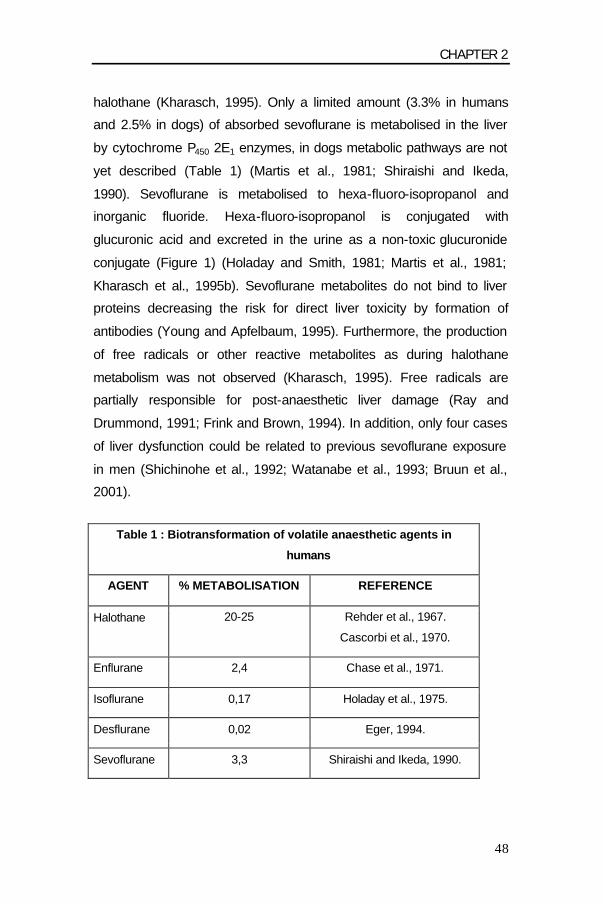
halothane (Kharasch, 1995). Only a limited amount (3.3% in humans
and 2.5% in dogs) of absorbed sevoflurane is metabolised in the liver
by cytochrome P450 2E1 enzymes, in dogs metabolic pathways are not
yet described (Table 1) (Martis et al., 1981; Shiraishi and Ikeda,
1990). Sevoflurane is metabolised to hexa-fluoro-isopropanol and
inorganic fluoride. Hexa-fluoro-isopropanol is conjugated with
glucuronic acid and excreted in the urine as a non-toxic glucuronide
conjugate (Figure 1) (Holaday and Smith, 1981; Martis et al., 1981;
Kharasch et al., 1995b). Sevoflurane metabolites do not bind to liver
proteins decreasing the risk for direct liver toxicity by formation of
antibodies (Young and Apfelbaum, 1995). Furthermore, the production
of free radicals or other reactive metabolites as during halothane
metabolism was not observed (Kharasch, 1995). Free radicals are
partially responsible for post-anaesthetic liver damage (Ray and
Drummond, 1991; Frink and Brown, 1994). In addition, only four cases
of liver dysfunction could be related to previous sevoflurane exposure
in men (Shichinohe et al., 1992; Watanabe et al., 1993; Bruun et al.,
2001).
Table 1 : Biotransformation of volatile anaesthetic agents in
humans
AGENT % METABOLISATION REFERENCE
Halothane 20-25 Rehder et al., 1967.
Cascorbi et al., 1970.
Enflurane 2,4 Chase et al., 1971.
Isoflurane 0,17 Holaday et al., 1975.
Desflurane 0,02 Eger, 1994.
Sevoflurane 3,3 Shiraishi and Ikeda, 1990.

CHAPTER 2
49
Post-anaesthetic liver damage can also be caused by local
hypoxaemia due to inadequate hepatic circulation. Total liver
perfusion is assured by the hepatic arterial blood flow and the portal
venous blood flow. During sevoflurane anaesthesia in dogs a
decrease in hepatic arterial circulation was reported at 2 MAC. Arterial
circulation remained constant at lower anaesthetic concentrations. On
the other hand, portal venous circulation decreased at 1.5 and 2 MAC.
In conclusion, total liver perfusion only decreased at high sevoflurane
concentrations (2 MAC) (Frink et al., 1992c). During isoflurane
anaesthesia in dogs hepatic arterial blood flow remained constant at 2
MAC, while portal venous perfusion only decreased slightly. This
resulted in a constant total liver perfusion even at higher anaesthetic
levels of isoflurane (Bernard et al., 1992). Halothane and to a lesser
extent enflurane induced a marked decrease in hepatic arterial and
portal venous circulation in dogs (Frink et al., 1992c).
A recent study reported that isoflurane induced an increase in
serum levels of liver enzymes more frequently than did sevoflurane 3
to 14 days after anaesthesia (Nishiyama et al., 1999b). Standard
hepatocellular enzymes were within normal range, while a clinically
non-significant increase of indirect bilirubine was reported after
sevoflurane anaesthesia in men (Frink et al., 1992a; Newman et al.,
1994; Ebert et al., 1998; Ebert and Arain, 2000; Suttner et al., 2000).
Even in patients with minimal or no hepatic metabolic capacity, such
as those with diminished enzyme activity or with intrinsic liver disease,
recovery from sevoflurane anaesthesia should not be affected
significantly (Kharasch et al., 1995b). Moreover, sevoflurane and
isoflurane only give a decrease in total liver perfusion at higher
concentrations, which are seldom necessary in clinical practice using
balanced anaesthesia protocols. In conclusion, in patients with liver
malfunctioning sevoflurane or isoflurane are preferably used, since
CHAPTER 2
50
both volatile anaesthetic agents have a low metabolisation degree
(sevoflurane: 3.3% and isoflurane: 0.17%) and are thought to be less
hepatotoxic compared to halothane and enflurane.
RENAL EFFECTS
The introduction of sevoflurane into clinical human
anaesthesia has been clouded by concerns about the potential risk of
nephrotoxicity after its use. Two theoretical sources for the
nephrotoxicity after sevoflurane are the plasma concentration of
inorganic fluoride, an in vivo metabolite of sevoflurane and the so
called ”compound A”, an in vitro degradation product of sevoflurane in
the presence of soda lime and baralyme. The metabolism of
sevoflurane involves enzymatic breakdown leading to the generation
of fluoride ions. Fluoride ions are potentially toxic and can cause renal
failure (Mazze, 1984). Because evidence of methoxyflurane renal
dysfunction was not observed when peak fluoride concentrations were
less than 50 µmol/l, this concentration was considered to be the
threshold of fluoride nephrotoxicity (Cousins and Mazze, 1973). In
clinical anaesthesia with sevoflurane some transient fluoride plasma
concentrations of more than 50 µmol/l were measured in humans
(Frink et al., 1992a; Kobayashi et al., 1992; Stickler et al., 1994;
Munday et al., 1995). And yet, no clinically relevant kidney failure was
reported in humans. Even in patients with pre-existing renal
impairment no further deterioration occurred after sevoflurane
anaesthesia (Melotte et al., 1994; Nuschler et al., 1994). For children
and obese persons the use of sevoflurane did not increase the risk for
potential renal toxicity, although, obesity was reported to increase the
fluoride production (Frink et al., 1993; Levine et al., 1996).
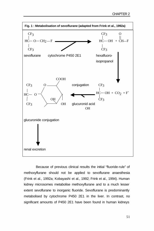
CHAPTER 2
51
Fig. 1 : Metabolisation of sevoflurane (adapted from Frink et al., 1992a)
CF3 CF3 O
HC O CH2 F HC OH + CH F
CF3 CF3
sevoflurane cytochrome P450 2E1 hexafluoro-
isopropanol
COOH
CF3 O conjugation CF3
HC O HC OH + CO2 + F-
OH CF3 CF3 OH glucuronid acid
OH
glucuronide conjugation
renal excretion
Because of previous clinical results the initial “fluoride-rule” of
methoxyflurane should not be applied to sevoflurane anaesthesia
(Frink et al., 1992a; Kobayashi et al., 1992; Frink et al., 1994). Human
kidney microsomes metabolise methoxyflurane and to a much lesser
extent sevoflurane to inorganic fluoride. Sevoflurane is predominantly
metabolised by cytochrome P450 2E1 in the liver. In contrast, no
significant amounts of P450 2E1 have been found in human kidneys

CHAPTER 2
52
(de Waziers et al., 1990; Kharasch et al., 1995c). Therefore, human
renal fluoride concentrations after sevoflurane anaesthesia are
considerably lower than serum fluoride concentrations. Hence, serum
fluoride concentration after sevoflurane is probably of little importance
for renal damage, even when the 50 µmol/l threshold is exceeded
(Kharasch et al., 1995a).
In a study on biotransformation of sevoflurane in dogs (2.5 %)
maximum serum fluoride concentrations were considerably lower than
those associated with nephrotoxicity in rats; respectively 18.5 µmol/l
and 20.0 µmol/l after 3 and 4% sevoflurane exposures (Martis et al.,
1981). These fluoride concentrations were not expected to induce
renal damage.
Of more concern than fluoride production during sevoflurane
metabolisation, is its interaction with CO2 absorbents, which generates
several degradation products. In modern anaesthesia, low fresh gas
flows (< 2 L/min) are common practice in order to reduce costs of
volatile anaesthetics and to avoid environmental pollution as well as to
preserve heat and humidify the inspired gas. Yet, all volatile
anaesthetics can be degraded by the lime in the circle absorber
system. Desflurane, enflurane and isoflurane react with dry
absorbents forming CO (Fang et al., 1995). Sevoflurane on the other
hand reacts with absorbents by formation of degradation products
called compounds A-E (Cunningham et al., 1996). The amounts of
compounds B, C, D and E were negligible whereby compounds C, D
and E were only found in in vitro studies using closed containers filled
with sevoflurane and sodalime (Wallin et al., 1975; Hanaki et al.,
1987). The most significant substance is compound A, a vinyl ether,
which has dose-dependent nephrotoxic properties inducing tubular
necrosis in rats. Clinically significant effects of this degradation in

CHAPTER 2
53
humans are still controversial (Bito et al., 1997; Kharasch et al., 1997;
Mazze and Jamison, 1997). Exposure of rats to high sevoflurane
concentrations is also detrimental for liver, lungs and central nervous
system (Gonsowski et al., 1994a). Compound A production and
accumulation in a circle absorber system is dependent on sevoflurane
concentration, the type of absorber material (baralyme or soda lime),
the water content of the absorbent, absorbent temperature, freshness
of the CO2-absorbent, CO2-production, fresh gas flow rates and type
of anaesthetic machine (Bito and Ikeda, 1991; Liu et al., 1991; Frink et
al., 1992b; Wong and Lerman, 1992; Ruzicka et al., 1994; Osawa and
Shinomura, 1998; Bito et al., 1998; Goeters et al., 2001; Yamakage et
al., 2001;). In the former absorbents, NaOH and KOH appear to
enhance the production of compound A and CO by degrading volatile
anaesthetics (Stabernack et al., 2000). New absorbents containing
Ca(OH)2 or Li(OH)2 could eliminate any potential hazards from the
toxic compounds by decreasing the production of compound A and
CO (Higuchi et al., 2000; Yamakage et al., 2000). The new material is
an effective carbon dioxide absorbent and is chemically unreactive
with sevoflurane, enflurane, isoflurane and desflurane (Murray et al.,
1999; Mchaourab et al., 2001).
The lethal concentration in 50% (LC50) of compound A in rats
equaled 331 ppm, 203 ppm and 127 ppm for a 3-h, 6-h and 12-h
exposure period (Gonsowski et al., 1994a en b). In a recent study on
low-flow sevoflurane anaesthesia in dogs concentrations of compound
A in the anaesthetic circuit were less than values reported to produce
renal toxicosis and death in rats (Muir and Gadawski, 1998). In all
reported sevoflurane studies compound A concentration remained far
below the toxic margin in humans (Frink et al., 1992b; Bito and Ikeda,
1994; Frink et al., 1994; Bito and Ikeda, 1995; Kharasch et al., 1997;
Kharasch and Jubert, 1999; Igarashi et al., 1999). Moreover, Rolly
CHAPTER 2
54
and Versichelen (1998) found amounts of compound A of 6 to 9 ppm
using soda-lime as CO2-absorbent. In a recent study using a high-flow
(7.0 l/min) closed-circuit PhysioFlex apparatus (Dräger, Lübeck,
Germany) with soda-lime and computer-controlled liquid injection of
sevoflurane compound A concentrations were significantly lower (6
ppm) than in conventional (14.3 ppm), valve-based machines during
closed-circuit conditions. Lower absorbent temperatures, resulting
from the high flow appear to account for the lower compound A
formation (Versichelen et al., 2000). Furthermore, recent studies
revealed that humans may be less susceptible to compound A renal
toxicity than rats, since the beta-lyase pathway responsible for
nephrotoxic metabolites of compound A is 10- to 30-fold less active in
humans than in rats (Altuntas and Kharasch, 2001; Altuntas and
Kharasch, 2002)
Renal functioning is mainly assessed by changes in serum
creatinine or blood urea nitrogen (BUN). These conventional markers
do not evaluate tubular function and are insensitive measures of
glomerular filtration (Shemesh et al., 1985). They are usually within
the normal range after “high-flow” (Frink et al., 1994; Higuchi et al.,
1994; Obata et al., 2000) and “low-flow” sevoflurane anaesthesia
(Kharasch et al., 1997; Bito et al., 1997; Eger et al., 1997; Higuchi et
al., 1998; Groudine et al., 1999; Obata et al., 2000). Even in patients
with moderately impaired renal function low-flow sevoflurane
anaesthesia had similar effects on renal function compared to
isoflurane (Higuchi et al., 2001). In specific experimental studies
several specific and sensitive biomarkers were used for evaluation of
renal function. These biomarkers bind on enzymes released with
kidney damage. Despite the sensitive tests no difference in renal
enzyme excretion between sevoflurane and isoflurane was found (Bito
et al., 1997; Kharasch et al., 1997; Kharasch et al., 2001). Eger et al.
CHAPTER 2
55
(1997; 1999) on the other hand, associated low flow (2 l/min during 8
hours) sevoflurane in humans with a transient renal injury in the
glomerulus, the proximale and distale tubuli, in contrast to desflurane
anaesthesia where no renal damage was observed. However, the
reliability of urinary biomarkers as indicator for clinical significant
kidney injuries in humans is still controversial (Baines, 1994).
The Food and Drug Administration (FDA) approved the clinical
use of sevoflurane regarding some precautions. A minimal flow of 2
l/min for exposures greater than 1 hour should be respected in semi-
closed systems. Until more information on degradation (compound A)
and metabolisation (fluoride) of sevoflurane is available, sevoflurane is
not recommended for using in patients with impaired kidney function
(Mazze and Jamison, 1995; 1997).
ECONOMIC CONSIDERATIONS
Do the benefits of sevoflurane compensate for the associated
higher costs in comparison to halothane and isoflurane in clinical
anaesthesia practice? Cost considerations are of increasing
importance when choosing anaesthetic techniques and drugs.
Different factors have an influence on the price of volatile
anaesthetics. The immediate cost of an inhaled anaesthetic results
from an interplay between 4 main factors: 1/ the cost per ml of liquid
anaesthetic, 2/ the volume of vapour that results from each ml of
liquid, 3/ the effective potency of the anaesthetic, 4/ the fresh gas flow.
First of all, there is the price imposed by the manufacturer
(Weiskopf and Eger, 1993). At this moment the price of 1 ml of
sevoflurane is a decisive factor for its use in veterinary practice. This
price is still considerably high in comparison with halothane and to a

CHAPTER 2
56
lesser extent with isoflurane. The purchasing cost will probably be
reduced in the near future, since sevoflurane is rapidly gaining ground
in human anaesthesia at the expense of halothane and isoflurane.
Secondly, the amount of vapour produced from 1 ml liquid
anaesthetic influences anaesthetic costs. The amount of vapour
produced is a function of the specific gravity and molecular weight of
the volatile anaesthetic, respectively 168 g and 1.467 g/ml for
desflurane, 184.5 g and 1.50 g/ml for isoflurane and 200 g and 1.505
g/ml for sevoflurane. The amount of produced vapour from 1 ml
decreases with 7% as follows: desflurane > isoflurane > sevoflurane
(Eger, 1994).
Furthermore, there are the factors inherent to the used
anaesthetic agent, as anaesthetic potency and blood-gas solubility of
the volatile anaesthetic agent (Bach et al., 1997; Philip, 1997). A lower
blood-gas solubility of an anaesthetic accords the same level of
control at a lower fresh gas flow rate than is achieved at a higher fresh
gas flow with a more soluble anaesthetic (Weiskopf and Eger, 1993).
The anaesthetic potency of sevoflurane is smaller compared to
halothane and isoflurane, but sevoflurane has a substantially lower
blood-gas partition coefficient. Low blood-gas solubility permits a rapid
and precise control of anaesthetic depth during anaesthesia induction
and maintenance, even with a relatively low flow. Halothane and
isoflurane need higher, less economic fresh gas flows to adjust
anaesthetic depth. Finally, the delivered concentration of the
anaesthetic by the vaporiser is responsible for the anaesthetic
consumption and not the alveolar concentration. The difference
between both is wasted. The low blood-gas solubility of sevoflurane
decreases the difference between inspired and end tidal concentration
leading to less waste and better control of anaesthesia.

CHAPTER 2
57
As already mentioned, the applied fresh gas flow is also of
great importance for anaesthetic cost savings. The main waste with
an inhalant anaesthetic is the one induced by unnecessarily high
carrier gas flows (Rosenberg et al., 1994). The lower the fresh gas
flow, the less volatile anaesthetic is wasted (Camu and Van De Velde,
1997; Suttner and Boldt, 2000). In closed circuit only a fresh gas flow
that supplies vapours required by the patient is used. This produces
the least cost, but also the least control of anaesthetic depth.
Nevertheless, the FDA suggested recently a minimal flow of 1 l/min for
exposures up to 1 hour in semi-closed systems to reduce the potential
risk for renal damage by accumulation of degradation products of
sevoflurane (Mazze and Jamison, 1997; Gentz and Malan, 2001).
Another benefit of sevoflurane, especially in human
anaesthesia is its fast recovery which could result in decreased
hospitalisation costs. For veterinary anaesthesia, however, the capital
costs include the expense of agent-specific vaporisers and of
converting or purchasing gas analysers. To reduce expenses an
enflurane vaporiser could be used for sevoflurane, since both
anaesthetics have a similar vapour pressure, 172 mm Hg for
enflurane and 160 mm Hg for sevoflurane (chapter 1 table 2). In
addition, the anaesthetic potency of both anaesthetics is similar: MAC
of enflurane in dogs is 2.06 and 2.36 for sevoflurane (chapter 1 table
3). When using an enflurane vaporiser type Enfluratec 4 for
sevoflurane administration the MAC-output is reduced with 21 % to 31
% in comparison with a specific sevoflurane vaporiser (Abel and
Eisenkraft, 1996). Nevertheless, the use of an enflurane vaporiser for
sevoflurane is not recommendable, since the limited vaporiser output
could lead to insufficient surgical anaesthetic depth. In short term
second-hand sevoflurane vaporisers will be available for veterinary
anaesthesia.

CHAPTER 2
58
CONCLUSION
Considering the multiple benefits of sevoflurane in veterinary
anaesthesia, there will certainly be a future for sevoflurane in
anaesthesia of dogs and cats. In patients with renal impairment some
precautions are advised, since more experience and studies on the
compound A and fluoride issues are necessary. Due to a rapid
advance of sevoflurane in human anaesthesia price reductions are
expected in the near future justifying the use of sevoflurane in modern
veterinary practice.

CHAPTER 2
59
REFERENCES
Abel, M., and J.B. Eisenkraft, 1996: Performance of erroneously filled sevoflurane, enflurane, and other agent-specific vaporizers. Journal of Clinical Monitoring 12, 119-125. Alexander, F.C., T.C. Smith, G. Strobel, G.W. Stephen, and H. Wollman, 1968: Cerebral carbohydrate metabolism of man during respiratory and metabolic alkalosis. Journal of Applied Physiology 24, 66-72. Altuntas, T.G., and E.D. Kharasch, 2001: Glutathione S-conjugation of the sevoflurane degradation product, fluoromethyl-2,2-difluoro-1-(trifluoromethyl)vinyl ether (compound A) in human liver, kidney and blood in vitro. Toxicology and Applied Pharmacology 177, 85-93. Altuntas, T.G., and E.D. Kharasch, 2002: Biotransformation of L-cysteine S-conjugates and N-acetyl-L-cysteine S-conjugates of the sevoflurane degradation product fluoromethyl-2,2-difluoro-1-(trifluoromethyl)vinyl ether (compound A) in human kidney in vitro: interindividual variability in N-acetylation, N-deacetylation, and beta-lyase-catalyzed metabolism. Drug metabolism and disposition 30, 148-154. Artru, A.A., 1983: Relationship between cerebral blood volume and CSF pressure during anesthesia with halothane or enflurane in dogs. Anesthesiology 58, 533-539. Atlee, J.L., 1985: Anaesthesia and cardiac electrophysiology. European Journal of Anesthesiology 2, 215-256. Bach, A., H. Böhrer, H. Schmidt, J. Motsch, and E. Martin, 1997: Ökonomische aspekte beim einsatz moderner inhalationsanästhetika am beispiel des sevofluran. Der Anaesthesist 46, 21-28. Baines, A.D.,1994: Strategies and criteria for developing new urinalysis tests. Kidney Intern 46 (suppl. 47), 137-141. Baker, K.Z., 1997: Desflurane and sevoflurane are valuable additions to the practice of neuroanesthesiology: Pro. J. Neurosurgical Anesthesiology 9, 66-68. Bernard, J.-M., M.-F. Doursout, P. Wouters, C.J. Hartley, R.G. Merin, and J.E. Chelly, 1992: Effects of sevoflurane and isoflurane on hepatic circulation in the chronically instrumented dog. Anesthesiology 77, 541-545. Bernard, J.-M., P.F. Wouters, M.-F. Doursout, B. Florence, J.E. Chelly, and R.G. Merin, 1990: Effects of sevoflurane and isoflurane on cardiac and coronary dynamics in chronically instrumented dogs. Anesthesiology 72, 659-662. Bito, H., and K. Ikeda, 1991: Sevoflurane in closed circuit anesthesia. Anesthesiology 75, A343.

CHAPTER 2
60
Bito, H., and K. Ikeda, 1994: Closed-circuit anesthesia with sevoflurane in humans. Effects on renal and hepatic function and concentrations of breakdown products with soda lime in the circuit. Anesthesiology 80, 71-76. Bito, H., and K. Ikeda, 1995: Degradation products of sevoflurane during low-flow anaesthesia. British Journal of Anaesthesia 74, 56-59. Bito, H., Y. Ikeuchi, and K. Ikeda, 1997: Effects of low-flow sevoflurane anesthesia on renal function. Comparison with high-flow sevoflurane anesthesia and low-flow isoflurane anesthesia. Anesthesiology 86, 1231-1237. Bito, H., Y. Ikeuchi, and K. Ikeda, 1998: Effects of the water content of soda lime on compound A concentration in the anesthesia circuit in sevoflurane anesthesia. Anesthesiology 88, 66-71. Brian, J.E., 1998 : Carbon dioxide and the cerebral circulation. Anesthesiology 88, 1365-1386. Brown, R.H., E.A. Zerhouni, and C.A. Hirshman, 1993: Comparison of low concentrations of halothane and isoflurane as bronchodilators. Anesthesiology 78, 1097-1101. Bruun, L.S., S. Elkjaer, D. Bitsch-larsen, and O. Andersen, 2001: Hepatic failure in a child after acetaminophen and sevoflurane exposure. Anesthesia & Analgesia 92, 1446-1448. Buffington, C.W., J.L. Romson, and A. Levin, 1987: Isoflurane induces coronary steal in a canine model of chronic occlusion. Anesthesiology 66, 289-292. Bundgaard, H., G. von Oettingen, K.M. Larsen, U. Landsfelt, K.A. Jensen, E. Nielsen, and G.E. Cold, 1998: Effects of sevoflurane on intracranial pressure, cerebral blood flow and cerebral metabolism. A dose-response study in patients subjected to craniotomy for cerebral tumours. Acta Anaesthesiologica Scandinavica 42, 621-627. Camu, F., and A. Van De Velde, 1997: Cost containment in inhalation anesthesia: the best way. Acta Anaesthesiologica Belgica 48, 155-160. Cascorbi, H.F., D.A. Blake, and M. Helrich, 1970: Differences in the biotransformation of halothane in man. Anesthesiology 32, 119-123. Chase, R.E., D.A. Holaday, V. Fiserova-Bergerova, L.J. Saidman, and F.E. Mack, 1971: The biotransformation of ethrane in man. Anesthesiology 35, 262-267. Cho, S., T. Fujigaki, Y. Uchiyama, M. Fukusaki, O. Shibata, and K. Sumikawa, 1996: Effects of sevoflurane with and without nitrous oxide on human cerebral circulation. Transcranial doppler study. Anesthesiology 85, 755-760.
CHAPTER 2
61
Clarke, K.W., H.I. Alibhai, Y.H. Lee, and R.A. Hammond, 1996: Cardiopulmonary effects of desflurane in the dog during spontaneous and artificial ventilation. Research in Veterinary Science 61, 82-86. Cold, G.E., H. Bundgaard, G. von Oettingen, K.A. Jensen, U. Landsfeldt, and K.M. Larsen, 1998: ICP during anaesthesia with sevoflurane: a dose-response study. Effect of hypocapnia. Acta Neurochir. Suppl. 71, 279-281. Cousins, M.J., and R.I. Mazze, 1973: Methoxyflurane nephrotoxicity: a study of a dose response in man. Journal of the American Medical Association 225, 1611-1616. Crystal, G.J., X. Zhou, J. Gurevicius, E.A. Czinn, M.R. Salem, S. Alam, A. Piotrowski, and G. Hu, 2000: Direct coronary vasomotor effects of sevoflurane and desflurane in in situ canine hearts. Anesthesiology 92, 1103-1113. Cucchiara, R.F., R.A. Theye, and J.D. Michenfelder, 1974: The effects of isoflurane on canine cerebral metabolism and blood flow. Anesthesiology 40, 571-574. Cunningham, D.D., S. Huang, J. Webster, J. Mayoral, and R.W. Grabenkort, 1996: Sevoflurane degradation to compound A in anaesthesia breathing systems. British Journal of Anaesthesia 77, 537-543. de Waziers, I., P.H. Cugnenc, C.S. Yang, J.P. Leroux, and P.H. Beaune, 1990: Cytochrome P-450 isoenzymes, epoxide hydrolase and glutathione transferases in rat and human hepatic and extrahepatic tissues. Journal of Pharmacologic Experimental Therapeutics 253, 387-394. Doi, M., and K. Ikeda,1987: Respiratory effects of sevoflurane. Anesthesia & Analgesia 66, 241-244. Doi, M., and K. Ikeda, 1992: Sevoflurane irritates airway least among four anesthetic agents: halothane, enflurane, isoflurane and sevoflurane. Anesthesiology 77, A335. Doi, M., and K. Ikeda, 1993: Airway irritation produced by volatile anesthetics during brief inhalation: comparison of halothane, enflurane, isoflurane, and sevoflurane. Canadian Journal of Anesthesiology 40, 122-126. Doi, K., T. Kasaba, and Y. Kosaka, 1988: A comparative study of the depressive effects of halothane and sevoflurane on medullary respiratory neurons in cats. Japanese Journal of Anesthesiology 37, 1466-1477. Doi, M., T. Katoh, T. Takii, M. Yura, and K. Ikeda, 1986: The respiratory effects of sevoflurane in dogs. Japanese Journal of Clinical Pharmacology and Therapeutics 17, 103-104. Drummond, J.C., and M.M. Todd,1985: The response of the feline cerebral circulation to PaCO2 during anesthesia with isoflurane and halothane and during sedation with nitrous oxide. Anesthesiology 62, 268-273.
CHAPTER 2
62
Ebert, T.J., and S.R. Arain, 2000: Renal responses to low-flow desflurane, sevoflurane and propofol in patients. Anesthesiology 93, 1401-1406. Ebert, T.J., L.D. Messana, T.D. Uhrich, and T.S. Staacke, 1998: Absence of renal and hepatic toxicity after four hours of 1.25 minimum alveolar anesthetic concentration sevoflurane anesthesia in volunteers. Anesthesia & Analgesia 86, 693-694. Ebert, T.J., and M. Muzi, 1993: Sympathetic hyperactivity during desflurane anesthesia in healthy volunteers: a comparison with isoflurane. Anesthesiology 79, 444-453. Ebert, T.J., M. Muzi, and C.W. Lopatka, 1995: Neurocirculatory responses to sevoflurane in humans. A comparison to desflurane. Anesthesiology 83, 88-95. Ebert, T.J., E.D. Kharasch, G.A. Rooke, A. Shroff, M. Muzi and the Sevoflurane Ischemia Study Group, 1997: Myocardial ischaemia and adverse cardiac outcomes in cardiac patients undergoing noncardiac surgery with sevoflurane and isoflurane. Anesthesia & Analgesia 85, 993-999. Eger, E.I.II, 1994: New inhaled anesthetics. Anesthesiology 80, 906-922. Eger, E.I.II, J. Cantillo, I. Gratz, E. Deal, D. Vekeman, R. McDougall, M. Afshar, A. Zafeiridis, and G. Larijani, 1999: Dose of compound A, not sevoflurane, determines changes in the biochemical markers of renal injury in healthy volunteers. Anesthesia & Analgesia 88, 437-445. Eger, E.I.II, D.D. Koblin, T. Bowland, P. Ionescu, M.J. Laster, Z. Fang, D. Gong, J. Sonner, and R.B. Weiskopf, 1997: Nephrotoxicity of sevoflurane versus desflurane anesthesia in volunteers. Anesthesia & Analgesia 84, 160-168. Eger, E.I.II, W.C. Stevens, and T.H. Cromwell, 1971: The electroencephalogram in man anesthetized with Forane. Anesthesiology 35, 504-508. Fang, Z.X., E.I.II Eger, M.J. Laster, B.S. Chortkoff, L. Kandel, and P. Ionescu, 1995: Carbon monoxide production from degradation of desflurane, enflurane, isoflurane, halothane and sevoflurane by soda lime and baralyme. Anesthesia & Analgesia 80, 1187-1193. Frink, E.J.,and B.R. Brown, 1994: Sevoflurane. Anesthetic Pharmacology Review 2, 61-67. Frink, E.J., H. Ghantous, T.P. Malan, S. Morgan, J. Fernando, A.J. Gandolfi, and B.R. Brown, 1992a: Plasma inorganic fluoride with sevoflurane anesthesia: correlation with indices of hepatic and renal function. Anesthesia & Analgesia 74, 231-235.

CHAPTER 2
63
Frink, E.J., T.P. Malan, E.A. Brown, S. Morgan, and B.R. Brown, 1993: Plasma inorganic fluoride levels with sevoflurane anesthesia in morbidly obese and nonobese patients. Anesthesia & Analgesia 76, 1333-1337. Frink, E.J., T.P. Malan, J. Isner, E.A. Brown, S. Morgan, and B.R. Brown, 1994: Renal concentrating function with prolonged sevoflurane or enflurane anesthesia in volunteers. Anesthesiology 80, 1019-1025. Frink, E.J., T.P. Malan, S.E. Morgan, E.A. Brown, M. Malcomson, and B.R. Brown, 1992b: Quantification of the degradation products of sevoflurane in two CO2-absorbants during low-flow anesthesia in surgical patients. Anesthesiology 77, 1064-1069. Frink, E.J., S.E. Morgan, A. Coetzee, P.F. Conzen, and B.F. Brown, 1992c: The effects of sevoflurane, halothane, enflurane, and isoflurane on hepatic blood flow and oxygenation in chronically instrumented greyhound dogs. Anesthesiology 76, 85-90. Gentz, B.A., and T.P.Jr. Malan, 2001: Renal toxicity with sevoflurane: a storm in a teacup? Drugs 61, 2155-2162. Goeters, C., C. Reinhardt, E. Gronau, R. Wüsten, T. Prien, J. Baum, S. Vrana, and H. Van Aken, 2001: Minimal flow sevoflurane and isoflurane anaesthesia and impact on renal function. European Journal of Anaesthesiology 18, 43-50. Gonsowski, C.T., M.J. Laster, E.I.II Eger, L.T. Ferrell, and R.L. Kerschmann, 1994a: Toxicity of compound A in rats. Effect of a 3-hour administration. Anesthesiology 80, 556-565. Gonsowski, C.T., M.J. Laster, E.I.II Eger, L.T. Ferrell, and R.L. Kerschmann, 1994b: Toxicity of compound A in rats. Effect of increasing duration of administration. Anesthesiology 80, 566-573. Grandy, J.L., D.S. Hodgson, C.I. Dunlop, C.R. Curtis, and R.B. Heath, 1989: Cardiopulmonary effects of halothane anesthesia in cats. American Journal of Veterinary Research 50, 1729-1732. Green, W.B., 1995: The ventilatory effects of sevoflurane. Anesthesia & Analgesia 81, 23-26. Gross, G.J., D.C. Warltier, 1981: Coronary steal in four models of single or multiple vessel obstruction in dogs. American Journal of Cardioliology 48, 84-92. Groudine, S.B., R.J. Fragen, E.D. Kharasch, T.S. Eisenman, E.J. Frink, and S. McConnell, 1999: Comparison of renal function following anesthesia with low-flow sevoflurane and isoflurane. Journal of Clinical Anesthesia 11, 201-207.
CHAPTER 2
64
Gupta, S., K. Heath, and B.F. Matta, 1997: Effect of incremental doses of sevoflurane on cerebral pressure autoregulation in humans. British Journal of Anaesthesia 79, 469-472. Hanaki, C., K. Fujii, M. Morio, and T. Tashima, 1987: Decomposition of sevoflurane by soda lime. Hiroshima Journal of Medicine Science 36, 61-67. Harkin, C.P., P.S. Pagel, J.R. Kersten, D.A. Hettrick, and D.C. Warltier, 1994: Direct negative inotropic and lusitropic effects of sevoflurane. Anesthesiology 81, 156-167. Harvey, R.C., 1992: Precautions when using mask induction. Veterinary Clinics of North American Small Animal Practice 22, 310-311. Hatakeyama, N., Y. Ito, and Y. Momose, 1993: Effects of sevoflurane, isoflurane, and halothane on mechanical and electrophysiologic properties of canine myocardium. Anesthesia & Analgesia 76, 1327-1332. Hayashi, Y., K. Sumikawa, C. Tashiro, A. Yamatodani, and I. Yoshiya, 1988: Arrhythmogenic threshold of epinephrine during sevoflurane, enflurane and isoflurane anesthesia in dogs. Anesthesiology 69, 145-147. Hettrick, D.A., P.S. Pagel, and D.C. Warltier, 1996: Desflurane, sevoflurane, and isoflurane impair canine left ventricular-arterial coupling and mechanical efficiency. Anesthesiology 85, 403-413. Higuchi, H., Y. Adachi, S. Arimura, M. Kanno, and T. Satoh, 2000: Compound A concentrations during low-flow sevoflurane anesthesia correlate directly with the concentration of monovalent bases in carbon dioxide absorbents. Anesthesia & Analgesia 91, 434-439. Higuchi, H., Y. Adachi, H. Wada, M. Kanno, and T. Satoh, 2001: The effects of low-flow sevoflurane and isoflurane anesthesia on renal function in patients with stable moderate renal insufficiency. Anesthesia & Analgesia 92, 650-655. Higuchi, H., S. Arimura, H. Sumikura, T. Satoh, and M. Kanno, 1994: Urine concentrating ability after prolonged sevoflurane anaesthesia. British Journal of Anaesthesia 73, 239-240. Higuchi, H., S. umita, H. Wada, T. Ura, T. Ikemoto, T. Nakai, M. Kanno, and T. Satoh, 1998: Effects of sevoflurane and isoflurane on renal function and on possible markers of nephrotoxicity. Anesthesiology 89, 307-322. Hikasa, Y., C. Okabe, K. Takase, and S. Ogasawara, 1996: Ventricular arrhythmogenic dose of adrenaline during sevoflurane, isoflurane, and halothane anaesthesia either with or without ketamine or thiopentone in cats. Research in Veterinary Science 60, 134-137.
CHAPTER 2
65
Hirano, M., T. Fujigaki, O. Shibata, and K. Sumikawa, 1992: A comparison of coronary hemodynamics during isoflurane and sevoflurane in dogs. Anesthesiology 77 (3A), A614. Holaday, D.A., V. Fiserova-Bergerova, I.P. Latto, and M.A. Zumbiel, 1975: Resistance of isoflurane to biotransformation in man. Anesthesiology 43, 325-332. Holaday, D.A., and F.R. Smith, 1981: Clinical characteristics and biotransformation of sevoflurane in healthy human volunteers. Anesthesiology 54, 100-106. Holzman, R.S., M.E. van der Velde, S.J. Kaus, S.C. Body, S.D. Colan, L.J. Sullivan, and S.G. Soriano, 1996: Sevoflurane depresses myocardial contractility less than halothane during induction of anaesthesia in children. Anesthesiology 85, 1260-1267. Hörmann, C., C. Kolbitsch, and A. Benzer, 1997: The role of sevoflurane in neuro-anesthesia practice. Acta Anaesthesiologica Scandinavica 111, 148-150. Ide, T., T. Kochi, S. Isono, and T. Mizuguchi, 1991: Diaphragmatic function during sevoflurane anaesthesia in dogs. Canadian Journal of Anaesthesiology 38, 116-120. Ide, T., T. Kochi, S. Isono, and T. Mizuguchi, 1992: Effect of sevoflurane on diaphragmatic contractility in dogs. Anesthesia & Analgesia 74, 739-746. Igarashi, M., H. Watanabe, H. Iwasaki, and A. Namiki, 1999: Clinical evaluation of low-flow sevoflurane anaesthesia for paediatric patients. Acta Anaesthesiologica Scandinavica 43, 19-23. Imamura, S., and K. Ikeda, 1987: Comparison of the epinephrine induced arrhytmogenic effect of sevoflurane with isoflurane and halothane. Journal of Anesthesiology 1, 62-68. Inomata, S., S. Watanabe, M. Taguchi, and M. Okada, 1994: End-tidal sevoflurane concentration for tracheal intubation and minimum alveolar concentration in pediatric patients. Anesthesiology 80, 93-96. Kandasamy, R., and P. Sivalingam, 2000: Use of sevoflurane in difficult airways. Acta Anaesthesiologica Scandinavica 44, 627-629. Katoh, T., and K. Ikeda, 1991: Effect of sevoflurane on bronchoconstriction caused by histamine or acetylcholine. Anesthesiology 75, A973. Kersten, J.R., A.P. Brayer, P.S. Pagel, J.P. Tessmer, and D.C. Warltier, 1994: Perfusion of ischemic myocardium during anesthesia with sevoflurane. Anesthesiology 81, 995-1004. Kharasch, E.D., 1995: Biotransformation of sevoflurane. Anesthesia & Analgesia 81, 27-38.
CHAPTER 2
66
Kharasch, E.D., E.J. Frink, A. Artru, P. Michalowski, G.A. Rooke and W. Nogami, 2001: Long-duration low-flow sevoflurane and isoflurane effects on postoperative renal and hepatic function. Anesthesia & Analgesia 93, 1511-1520. Kharasch, E.D., E.J. Frink, R. Zager, T.A. Bowdle, A. Artru, and W.M. Nogami, 1997: Assessment of low-flow sevoflurane and isoflurane effects on renal function using sensitive markers of tubular toxicity. Anesthesiology 86, 1238-1253. Kharasch, E.D., D.C. Hankins, and K.E. Thummel, 1995a: Human kidney methoxyflurane and sevoflurane metabolism. Intrarenal fluoride production as a possible mechanism of methoxyflurane nephrotoxicity. Anesthesiology 82, 689-699. Kharasch, E.D., and C. Jubert, 1999: Compound A uptake and metabolism to mercapturic acids and 3,3,3-trifluoro-2-fluoromethoxypropanoic acid during low-flow sevoflurane anesthesia: biomarkers for exposure, risk assessment, and interspecies comparison. Anesthesiology 91, 1267-1278. Kharasch, E.D., M.D. Karol, C. Lanni, and R. Sawchuk, 1995b: Clinical sevoflurane metabolism and disposition. I. Sevoflurane and metabolite pharmacokinetics. Anesthesiology 82, 1369-1378. Kharasch, E.D., A.S. Armstron, K. Gunn, A. Artru, and M.D. Karol, 1995c: Clinical sevoflurane metabolism and disposition. II. The role of cytochrome P450 2E1 in fluoride and hexafluoroisopropanol formation. Anesthesiology 82, 1379-1388. Kitaguchi, K., H. Ohsumi, M. Kuro, T. Nakajima, and H. Hayashi, 1993: Effects of sevoflurane on cerebral circulation and metabolism in patients with ischemic cerebrovascular disease. Anesthesiology 79, 704- 709. Kitahata, H., S. Kawahito, J. Nozaki, H. Kimura, K. Tanaka, T. Kitagawa, and S. Oshita, 1999: Effects of sevoflurane on regional myocardial blood flow distribution. Quantification with myocardial contrast echocardiography. Anesthesiology 90, 1436-1445. Klock, P.A.Jr., E.G. Czeslick, J.M. Klafta, A. Ovassapian, and J. Moss, 2001: The effect of sevoflurane and desflurane on upper airway reactivity. Anesthesiology 94, 963- 967. Kobayashi, Y., R. Ochiai, J. Takeda, H. Sekiguchi, and K. Fukushima, 1992: Serum and inorganic fluoride concentrations after prolonged inhalation of sevoflurane in humans. Anesthesia & Analgesia 74, 753-757. Kuroda, Y., M. Murakami, J. Tsuruta, T. Murakawa, and T. Sakabe, 1996: Preservation of the ratio of cerebral blood flow/ metabolic rate for oxygen during prolonged anesthesia with isoflurane, sevoflurane, and halothane in humans. Anesthesiology 84, 555-561.

CHAPTER 2
67
Larach, D.R., and H.G. Schuler, 1991: Direct vasodilation by sevoflurane, isoflurane, and halothane alters coronary flow reserve in the isolated rat heart. Anesthesiology 75, 268-278. Levine, M.F., J. Sarner, J. Lerman, P. Davis, N. Sikich, K. Maloney, E. Motoyama, and D.R. Cook, 1996: Plasma inorganic fluoride concentrations after sevoflurane anesthesia in children. Anesthesiology 84, 348-353. Lockhart, S.H., I.J. Rampil, N. Yasuda, E.I.II Eger, and R.B. Weiskopf, 1991: Depression of ventilation by desflurane in humans. Anesthesiology 74, 484-488. Lowe, D., D.A. Hettrick, P.S. Pagel, and D.C. Warltier, 1996: Influence of volatile anesthetics on left ventricular afterload in vivo. Differences between desflurane and sevoflurane. Anesthesiology 85, 112-120. Liu, J., M.J. Laster, E.I.II Eger, and S. Taheri, 1991: Absorption and degradation of sevoflurane and isoflurane in a conventional anesthetic circuit. Anesthesia & Analgesia 72, 785-798. Lutz, L.J., J.H. Milde, and L.N. Milde, 1990: The cerebral functional metabolic and hemodynamic effects of desflurane in dogs. Anesthesiology 73, 125-131. Malan, T.P., J.A. Di Nardo, R.J. Isner, E.J. Frink, M. Goldberg, P.E. Fenster, E.A. Brown, R. Depa, L.C. Hammond, and H. Mata, 1995: Cardiovascular effects of sevoflurane compared with those of isoflurane in volunteers. Anesthesiology 83, 918-928. Martis, L., S. Lynch, M.D. Napoli, and E.F. Woods, 1981: Biotransformation of sevoflurane in dogs and rats. Anesthesia & Analgesia 60, 186-191. Matta, B.F., K.J. Heath, K. Tipping, and A.C. Summors, 1999: Direct cerebral vasodilatory effects of sevoflurane and isoflurane. Anesthesiology 91, 677-680. Mazze, R.I., 1984: Fluorinated anesthetic nephrotoxicity: An update. Canadian Anaesthesiology Society Journal 31 (suppl.), 16-22. Mazze, R.I., and R.L. Jamison, 1995: Renal effects of sevoflurane. Anesthesiology 83, 443-445. Mazze, R.I., and R.L. Jamison, 1997: Low-flow (1 L/ min) sevoflurane. Is it safe? Anesthesiology 86, 1225-1227. Mchaourab, A., S.R. Arain, and T.J. Ebert, 2001: Lack of degradation of sevoflurane by a new carbon dioxide absorbent in humans. Anesthesiology 94, 1007-1009. McMurphy, R.M., and D.S. Hodgson, 1996: Cardiopulmonary effects of desflurane in cats. American Journal of Veterinary Research 57, 367-370.

CHAPTER 2
68
McPherson, R.W., J.E. Brian, and R.J. Traystman, 1989: Cerebrovascular responsiveness to carbon dioxide in dogs with 1,4% and 2,8% isoflurane. Anesthesiology 70, 843-850. Melotte, A., M. Verhaegen, P. Conzen, H. Van Aken, and K. Peter, 1994: Plasma inorganic fluoride levels after sevoflurane or enflurane anesthesia in patients with renal impairment. Anesthesiology 81, A368. Merin, R.G., J.M. Bernard, M.F. Doursout, M. Cohen, and J.E. Chelly, 1991: Comparison of the effects of isoflurane and desflurane on cardiovascular dynamics and regional blood flow in the chronically instrumented dog. Anesthesiology 74, 568-574. Michenfelder, J.D., and R.F. Cucchiara, 1974: Canine cerebral oxygen consumption during enflurane anesthesia and its modification during induced seizures. Anesthesiology 40, 575-580. Mielck, F., H. Stephan, A. Weyland, and H. Sonntag, 1999: Effects of one minimum alveolar anesthetic concentration sevoflurane on cerebral metabolism, blood flow, and CO2 reactivity in cardiac patients. Anesthesia & Analgesia 89, 364-369. Miletich, D.J., A.D. Ivankovich, R.F. Albrecht, C.R. Reimann, R. Rosenberg, and E.D. Mc Kissic, 1976: Absence of autoregulation of cerebral blood flow during halothane and enflurane anesthesia. Anesthesia & Analgesia 55, 100-109. Mitsuhata, H., J. Saitoh, R. Shimizu, H. Takeuchi, N. Hasome, and Y. Horiguchi, 1994: Sevoflurane and isoflurane protect against bronchospasms in dogs. Anesthesiology 81, 1230-1234. Moore, M.A., R.B. Weiskopf, E.I.II Eger, M. Noorani, L. Mc Kay, and M. Damask, 1994: Rapid 1% increases of end-tidal desflurane concentration to greater than 5% transiently increase heart rate and blood pressure in humans. Anesthesiology 81, 94-98. Muir, W.W. 3rd, and J. Gadawski, 1998: Cardiorespiratory effects of low-flow and closed circuit inhalation anesthesia, using sevoflurane delivered with an in-circuit vaporizer and concentrations of compound A. American Journal of Veterinary Research 59, 603-608. Munday, I.T., P.A. Stoddart, R.M. Jones, J. Lytle, and M.R. Cross, 1995: Serum fluoride concentration and urine osmolality after enflurane and sevoflurane anesthesia in male volunteers. Anesthesia & Analgesia 81, 353-359. Murray, J.M., C.W. Renfrew, A. Bedi, C.B. McCrystal, D.S. Jones, and J.P. Fee, 1999: Amsorb: A new carbon dioxide absorbent for use in anesthetic breathing systems. Anesthesiology 91, 1342-1348.
CHAPTER 2
69
Mutoh, T., A. Kanamaru, H. Suzuki, H. Tsubone, R. Nishimura, and N. Sasaki, 2001a: Respiratory reflexes in spontaneously breathing anesthetized dogs in response to nasal administration of sevoflurane, isoflurane, or halothane. American Journal of Veterinary Research 62, 311-319. Mutoh, T., A. Kanamaru, H. Tsubone, R. Nishimura, and N. Sasaki, 2001b: Respiratory reflexes in response to upper-airway administration of sevoflurane and isoflurane in anesthetized, spontaneously breathing dogs. Veterinary Surgery 30, 87-96. Mutoh, T., K. Kojima, K. Takao, R. Nishimura, and N. Sasaki, 2001c: Comparison of sevoflurane with isoflurane for rapid mask induction in midazolam and butorphanol-sedated dogs. Journal of Veterinary Medicine A 48, 223-230. Mutoh, T., R. Nishimura, H.-Y. Kim, S. Matsunaga, T. Kadosawa, M. Mochizuki, and N. Sasaki, 1995: Rapid inhalation induction of anesthesia by halothane, enflurane, isoflurane and sevoflurane and their cardiopulmonary effects in dogs. Journal of Veterinary Medical Science 57, 1007-1013. Mutoh, T., R. Nishimura, H.-Y.Kim, S. Matsunaga, and N. Sasaki, 1997: Cardiopulmomary effects of sevoflurane, compared with halothane, enflurane and isoflurane, in dogs. American Journal of Veterinary Research 58, 885-890. Muzi, M., C.W. Lopatka, and T.J. Ebert, 1996: Desflurane-mediated neurocirculatory activation in humans. Effects of concentration and rate of change on responses. Anesthesiology 84, 1035-1042. Navarro, R., R.B. Weiskopf, M.A. Moore, S. Lockhart, E.I.II Eger, D. Koblin, G. Lu, and C. Wilson, 1994: Humans anesthetized with sevoflurane or isoflurane have similar arrhytmic response to epinephrine. Anesthesiology 80, 545-549. Neigh, J.L., J.K. Garman, and J.R. Harp, 1971: The electroencephalographic pattern during anesthesia with Ethrane: effects of depth of anesthesia, PaCO2, and nitrous oxide. Anesthesiology 35, 482-487. Newman, P.J., A.C. Quinn, G.M. Hall, and R.M. Grounds, 1994: Circulating fluoride changes and hepatorenal function following sevoflurane anaesthesia. Anaesthesia 49, 936-939. Nishiyama, T., N. Sugai, and K. Hanaoka, 1997: Cerebrovascular CO2 reactivity in elderly and younger adult patients during sevoflurane anaesthesia. Canadian Journal of Anaesthesiology 44, 160-164. Nishiyama, T., T. Matsukawa, T. Yokoyama, and K. Hanaoka, 1999a: Cerebrovascular carbon dioxide reactivity during general anaesthesia: a comparison between sevoflurane and isoflurane. Anesthesia & Analgesia 89, 1437-1441.
CHAPTER 2
70
Nishiyama, T., T. Yokoyama, and K. Hanaoka, 1999b: Effects of sevoflurane and isoflurane anesthesia on arterial ketone body ratio and liver function. Acta Anaesthesiologic Scandinavica 43, 347-351. Nuschler, M., P.F. Conzen, A. Melotte, H. Van Aken, and K. Peter, 1994: Renal function after sevoflurane versus enflurane anesthesia in patients with renal impairment. Anesthesiology 81, A362. Obata, R., H. Bito, M. Ohmura, G. Moriwaki, Y. Ikeuchi, T. Katoh, and S. Sato, 2000: The effects of prolonged low-flow sevoflurane anesthesia on renal and hepatic function. Anesthesia & Analgesia 91, 1262-1268. Ogawa, K., M. Yamamoto, K. Mizumoto, and Y. Hatano, 1997: Volatile anaesthetics attenuate hypocapnia-induced constriction in isolated dog cerebral arteries. Canadian Journal of Anaesthesiology 44, 426-432. Ornstein, E., W. Young, L. Fleischer, and N. Ostapkovich, 1993: Desflurane and isoflurane have similar effects on cerebral blood flow in patients with intracranial mass lesions. Anesthesiology 79, 498-502. Osawa, M., K. Shingu, M. Murakawa, T. Adachi, J. Kurata, N. Seo, T. Murayama, S. Nakao, and K. Mori, 1994: Effects of sevoflurane on central nervous system electrical activity in cats. Neurosurgical Anesthesia 79, 52-57. Osawa, M., and T. Shinimura, 1998: Compound A concentration is decreased by cooling anaesthetic circuit during low-flow sevoflurane anaesthesia. Canadian Journal of Anaesthesiology 45, 1215-1218. Pagel, P.S., J.P. Kampine, W.T. Schmeling, and D.C. Warltier, 1991a: Comparison of the systemic and coronary hemodynamic actions of desflurane, isoflurane, halothane, and enflurane in the chronically instrumented dog. Anesthesiology 74, 539-551. Pagel, P.S., J.P. Kampine, W.T. Schmeling, and D.C. Warltier, 1991b: Influence of volatile anaesthetics on myocardial contractility in vivo: desflurane versus isoflurane. Anesthesiology 74, 900-907. Pagel, P.S., J.R. Kersten, D.A. Hettrick, and D.C. Warltier, 1994: Negative inotropic and lusiotropic actions of sevoflurane in chronically instrumented dogs. Anesthesia & Analgesia 78, 332. Park, W.K., J.J. Pancrazio, C. Kook Suh, and C. Lynch, 1996: Myocardial depressant effects of sevoflurane. Mechanical and electrophysiologic actions in vitro. Anesthesiology 84, 1166-1176. Paulson, O.B., J. Olesen, and M.S. Christensen, 1972: Restoration of autoregulation of cerebral blood flow by hypocapnia. Neurology 22, 286-293. Philip, B.K., 1997: New approaches to anesthesia for day case surgery. Acta Anaesthesiologica Belgica 48, 167-174.

CHAPTER 2
71
Pickard, J.D., J.E. Rose, M.B.D. Cooke, I.M. Blair, and A. Strathdee, 1977: The effect of salicylate on cerebral blood flow in man. Acta Neurologica Scandinavica 64 (suppl.), 422-423. Priebe, H.J., 1988: Isoflurane causes more severe regional myocardial dysfunction than halothane in dogs with a critical coronary artery stenosis. Anesthesiology 69, 72-83. Raichle, M.E., and H.L. Stone, 1972: Cerebral blood flow autoregulation and graded hypercapnia. European Neurology 6, 1-5. Rampil, I.J., S.H. Lockhart, E.I.II Eger, N. Yasuda, R.B. Weiskopf, and M.K. Cahalan: 1991: The electroencephalographic effects of desflurane in humans. Anesthesiology 74, 434-439. Ray, D.C., and G.B. Drummond, 1991: Halothane hepatitis. British Journal of Anaesthesia 67, 84-99. Rehder, K., J. Forbes, H. Alter, O. Hessler, and A. Stier, 1967: Halothane biotransformation in man: A quantitative study. Anesthesiology 28, 711-715. Rivenes, S.M., M.B. Lewin, S.A. Stayer, S.T. Bent, H.M. Schoenig, E.D. McKenzie, C.D. Fraser, and D.B. Andropoulos, 2001: Cardiovascular effects of sevoflurane, isoflurane, halothane, and fentanyl-midazolam in children with congenital heart disease: an echocardiographic study of myocardial contractility and hemodynamics. Anesthesiology 94, 223-229. Rolly, G., L. Versichelen, 1998: Formation of compound A with sevoflurane. Update in low flow and closed circuit anesthesia, abstract book, Annual Meeting of the Association for Low Flow Anaesthesia, september 18-19. Rooke, G.A., J.-H. Choi, and M.J. Bishop, 1997: The effect of isoflurane, halothane, sevoflurane and thiopental/ nitrous oxide on respiratory system resistance after tracheal intubation. Anesthesiology 86, 1294-1299. Rosenberg, M.K., P. Bridge, and M. Brown, 1994: Cost comparison: A desflurane- versus a propofol-based general anesthetic technique. Anesthesia & Analgesia 79, 852-855. Ruzicka, J.A., J.C. Hidalgo, J.H. Tinker, and M.T. Baker, 1994: Inhibition of volatile sevoflurane degradation product formation in an anesthesia circuit by a reduction in soda lime temperature. Anesthesiology 81, 238-244. Sato, M., G. Pawlik, and W-D. Heiss, 1984: Comparative studies of regional CNS blood flow autoregulation and responses to CO2 in the cat. Stroke 15, 91-97. Scheller, M.S., K. Nakakimura, J.E. Fleischer, and M.H. Zornow, 1990: Cerebral effects of sevoflurane in the dog: comparison with isoflurane and enflurane. British Journal of Anaesthesia 65, 388-392.

CHAPTER 2
72
Shemesh, O., H. Golbetz, J.P. Kriss, and B.D. Meyers, 1985: Linmitations of creatinine as a filtration marker in glomerulopathic patients. Kidney Int. 28, 830-838. Shichinohe, Y., Y. Masuda, H. Takahashi, M. Kotaki, T. Omote, M. Shichinohe, and A. Namiki, 1992: A case of postoperative hepatic injury after sevoflurane anesthesia. Masui 41, 1802-1805. Shiraishi, Y., and K. Ikeda, 1990: Uptake and biotransformation of sevoflurane in humans: A comparative study of sevoflurane with halothane, enflurane, and isoflurane. Journal of Clinical Anesthesiology 2, 381-386. Stabernack, C.R., R. Brown, M.J. Laster, R. Dudziak, and E.I.II Eger, 2000: Absorbents differ enormously in their capacity to produce compound A and carbon monoxide. Anesthesia & Analgesia 90, 1428-1435. Steffey, E.P., 1994: Inhalation anaesthesia. In: Hall L.W., Taylor P.M., eds. Anaesthesia of the cat. London: Baillière Tindall; 157-193. Steffey, E.P., and D. Jr. Howland, 1977: Isoflurane potency in the dog and cat. American Journal of Veterinary Research 38, 1833-1836. Steffey, E.P., and D. Jr. Howland, 1978: Potency of enflurane in dogs: comparison with halothane and isoflurane. American Journal of Veterinary Research 39, 673-677. Stickler, T., C. Callan, J. Sayre, K. Blahunka, and P. Prokocimez, 1994: Incidence of inorganic fluoride concentrations greater than or equal to µmol/l in sevoflurane comparative clinical studies. Anesthesiology 81 (Suppl.), A1283-A1283. Summors, A.C., A.K. Gupta, and B.F. Matta, 1999: Dynamic cerebral autoregulation during sevoflurane anesthesia: a comparison with isoflurane. Anesthesia & Analgesia 88, 341-345. Suttner, S.W., and J. Boldt, 2000: Low-flow anaesthesia. Does it have potential pharmacoeconomic consequences? Pharmacoeconomics 17, 585-590. Suttner, S.W., C.C. Schmidt, J. Boldt, I. Huttner, B. Kumle, and S.N. Piper, 2000: Low-flow desflurane and sevoflurane anesthesia minimally affect hepatic integrity and function in elderly patients. Anesthesia & Analgesia 91, 206-212. Takahashi, H., K. Murata, and K. Ikeda, 1993: Sevoflurane does not increase intracranial pressure in hyperventilated dogs. British Journal of Anaesthesia 71, 551-555. Tamura, C., M. Doi, and K. Ikeda, 1991: Hypoxic ventilatory response in cats lightly anesthetized with ketamine: effects of halothane and sevoflurane in low concentrations. Journal of Anesthesiology 5, 233-238.

CHAPTER 2
73
Tanaka, M., and T. Nishikawa, 1999: Sevoflurane speeds recovery of baroreflex control of heart rate after minor surgical procedures compared with isoflurane. Anesthesia & Analgesia 89, 284-289. Theye, R.A., and J.D. Michenfelder, 1968: The effect of halothane on canine cerebral metabolism. Anesthesiology 29, 1113-1118. Tomiyasu S., T. Hara, H. Hasuo, H. Ureshino, and K. Sumikawa, 1999: Comparative analysis of systemic and coronary hemodynamics during sevoflurane- and isoflurane-induced hypotension in dogs. Journal of Cardiovascular Pharmacology 33, 741-747. Todd, M.M., and J.C. Drummond, 1984: A comparison of the cerebrovascular and metabolic effects of halothane and isoflurane in the cat. Anesthesiology 60, 276-282. Vakkuri, A., V. Jäntti, M. Särkelä, L. Lindgren, K. Korttila, and A. Yli-Hankala, 2000: Epileptiform EEG during sevoflurane mask induction: Effect of delaying the onset of hyperventilation. Acta Anaesthesiologica Scandinavica 44, 713-719. Versichelen, L.F., G. Rolly, M.P. Bouche, J.F. Van Boxclaer, M.M. Struys, C. Van Der Herten, A.P. De Leenheer, and E.P. Mortier, 2000: In vitro compound A formation in a computer-controlled closed-circuit anesthetic apparatus. Comparison with a classical valve circuit. Anesthesiology 93, 1064-1068. Wallin, R.F., B.M. Regan, M.D. Napoli, and I.J. Stern, 1975: Sevoflurane: A new inhalation anesthetic agent. Anesthesia & Analgesia 54, 758-765. Warltier, D.C., G.J. Gross, and H.L. Brooks, 1980: Coronary-steal induced increase in myocardial infarct size after pharmacologic coronary vasodilation. American Journal of Cardioliology 46, 83-90. Watanabe, K., S. Hatakenaka, K. Ikemune, Y. Chigyo, T. Kubozono, and T. Arai, 1993: A case of suspected liver disfunction induced by sevoflurane anesthesia. Masui 42, 902-905. Weiskopf, R.B., and E.I.II Eger, 1993: Comparing the costs of inhaled anesthetics. Anesthesiology 79, 1413-1418. Weiskopf, R.B., M.A. Moore, E.I.II Eger, M. Noorani, L. McKay, B. Chortkoff, P.S. Hart, and M. Damask, 1994: Rapid increase in desflurane concentration is associated with greater transient cardiovascular stimulation than with rapid increase in isoflurane concentration in humans. Anesthesiology 80, 1035-1045. Wong, D.T., and J. Lerman, 1992: Factors affecting the rate of disappearance of sevoflurane in baralime. Canadian Journal of Anaesthesia 39, 366-369.

CHAPTER 2
74
Yamakage, M., A. Kimura, X. Chen, N. Tsujiguchi, Y. Kamada, A. Namiki, 2001: Production of compound A under low-flow anesthesia is affected by type of anesthetic machine. Canadian Journal of Anesthesia 48 (5), 435-438. Yamakage, M., S. Yamada, X. Chen, S. Iwasaki, N. Tsujiguchi, and A. Namiki, 2000: Carbon dioxide absorbents containing potassium hydroxide produce much larger concentrations of compound A from sevoflurane in clinical practice. Anesthesia & Analgesia 91, 220-224. Young, C.J., and J.L. Apfelbaum, 1995: Pharmacology of outpatient anaesthesia in the year 2000. Anesthetic agents for ambulatory surgery into the twenty-first century. Do the new drugs really make a difference? Acta Anaesthesiologica Scandinavica, 75-83.
Yurino, M., and H. Kimura, 1993: Vital capacity rapid inhalation induction technique: comparison of sevoflurane and halothane. Canadian Journal Anaesthesiology 40, 440-443.

INTRODUCTION TO CHAPTERS 3 & 4
75
INTRODUCTION TO CHAPTERS 3/ 4

INTRODUCTION TO CHAPTERS 3 & 4
76
INTRODUCTION TO CHAPTERS 3 & 4
77
In these chapters the clinical aspect of the anaesthetic
protocol is emphasised. The majority of articles describe recovery
times and cardiorespiratory influences of sevoflurane anaesthesia in
studies with an experimental anaesthetic set-up. In contrast, the
amount of information gathered under clinical circumstances with a
standard anaesthetic protocol is rather limited. In most experimental
studies on sevoflurane in dogs the anaesthetic protocol is reduced to
the strict minimum to eliminate potential influences of other
anaesthetic drugs. Therefore, premedication and induction with
intravenous anaesthetic agents are excluded. Instead, anaesthesia is
induced and maintained solely by the volatile anaesthetic agent
(sevoflurane). In the present work the anaesthetic protocol included a
standard premedication, since preanaesthetics drugs are of major
importance under clinical circumstances.
Premedication decreases stress before induction of
anaesthesia, facilitates manipulations and contributes to a smooth
induction and recovery from anaesthesia. Moreover, opioids are often
included in premedication reducing the severity and duration of post-
operative pain. The so-called “pre-emptive analgesia” is of major
importance for the patient. For these reasons premedication with a
neurolept-analgesic mixture including droperidol and fentanyl can be
justified. Influences of droperidol on recovery times and
cardiorespiratory parameters are probably not neglectable because of
its long-lasting action, and hence can not be ruled out. Fentanyl, on
the other hand, has a rapid onset of effect (1-2 minutes and is short-
acting (20-30 minutes). Accordingly, no effects on recovery times and
only initial influences on cardiorespiratory parameters could be
expected.
INTRODUCTION TO CHAPTERS 3 & 4
78
Induction of anaesthesia is mainly achieved by intravenous
anaesthetics because of their rapid onset of action and ease of
administration. Propofol was preferred in the present study due to its
unique pharmacokinetic properties and clinical advantages. It has a
specific extrahepatic metabolism and is rapidly cleared from the body.
After bolus injection, plasma concentrations decrease rapidly due to
redistribution of the drug to the brain and other highly perfused
tissues. It provides a rapid and smooth induction of anaesthesia and a
fast excitement-free recovery with no hangover-effect. Although the
elimination half-life of propofol is long (330 minutes in dogs), this is not
clinically relevant and should not influence recovery times after 1 hour
of inhalation anaesthesia. Influences on cardiopulmonary parameters
under sevoflurane anaesthesia during spontaneous and controlled
ventilation might be observed the first 30 minutes of the standardised
anaesthetic period. This was not considered a problem since surgical
preparation of the dogs was performed during the first hour of
anaesthesia.
Chapter 3 discusses the recovery times after clinical
sevoflurane anaesthesia compared with more frequently used inhalant
anaesthetic agents (isoflurane and halothane). The aim was to
investigate the advantage of the low blood-gas solubility of
sevoflurane compared to the other volatile anaesthetic agents for the
recovery times in premedicated dogs. In chapter 4 cardio-pulmonary
parameters of sevoflurane anaesthesia during spontaneous and
controlled ventilation at different MAC values are described using a
clinical anaesthetic protocol including premedication and intravenous
induction.
79
CHAPTER 3
RECOVERY TIMES AND EVALUATION OF CLINICAL HEMODYNAMIC
PARAMETERS OF SEVOFLURANE, ISOFLURANE AND HALOTHANE
ANAESTHESIA IN MONGREL DOGS.
I. Polis1, F. Gasthuys2, L. Van Ham1, H. Laevens3
1 Department of Small Animal Medicine and Clinical Biology; 2 Department of Surgery and Anaesthesia of Domestic Animals; 3 Department of Reproduction, Obstetrics and Herd Health, Faculty of Veterinary Medicine, Ghent University, Salisburylaan 133, B- 9820 Merelbeke, Belgium
Adapted from: I. Polis, F. Gasthuys, L. Van Ham, H. Laevens (2001). Recovery times and evaluation of clinical hemodynamic parameters of sevoflurane, isoflurane and halothane anaesthesia in mongrel dogs. Journal of Veterinary Medicine A 48, 401-411.

CHAPTER 3
80
CHAPTER 3
81
SUMMARY
In the present study the influence of 3 volatile agents
{halothane, isoflurane and sevoflurane} in oxygen at 2 concentrations
(1.5 and 2 MAC) in 6 dogs on non-invasive cardio-respiratory
parameters (heart and respiratory rates, non-invasive blood pressures
at 15, 30, 60 minutes and after extubation) and on the recovery times
(appearance of the first eyelid reflex, emergence time) after clinical
anaesthesia was studied (cross-over design). After premedication with
fentanyl-droperidol (5 µg/kg and 0.25 mg/kg IM) and induction with
propofol (5 mg/kg IV) six dogs were randomly anaesthetised for one
hour for a standard neurologic stimulation test.
A wide individual variation in respiration rate (induced by an
initial hyperpnea) was observed in the 1.5 MAC protocols, without
significant differences between the 3 different volatile agents. Heart
rate was significantly lower during 1.5 and 2 MAC halothane when
compared to isoflurane and sevoflurane. An increase from 1.5 to 2
MAC induced significant decreases in diastolic (DAP) and mean
arterial blood pressure in all groups without significant changes in the
systolic arterial pressures. Only DAP in sevoflurane protocol was
significantly lower at 1.5 and 2 MAC compared to halothane. Time had
no significant influences on the non-invasive blood pressures in all
protocols. Extubation induced a significant increase of all parameters
in all protocols. Time for a first eyelid reflex was significantly longer
after 2 MAC compared to the 1.5 MAC protocol. There was no
significant difference between the 3 anaesthetic agents. Although
emergence time was longest for halothane at both anaesthetic
concentrations, no significant difference in emergence time was
observed for the 3 volatile agents.
CHAPTER 3
82
INTRODUCTION
Sevoflurane (CFH2-O-CH(CF3)2) is a volatile anaesthetic
agent developed in the early seventies (Wallin et al., 1975). This drug
has specific characteristics including a low blood/gas partition
coefficient (0.69) (Strum and Eger, 1987). The low solubility partly
contributes to a rapid induction of and emergence from anaesthesia
compared to other volatile anaesthetics such as halothane, enflurane
and isoflurane (Smith et al., 1992; Lerman et al., 1996; Steffey, 1996;
Aono et al., 1997; Ebert et al., 1998). Sevoflurane has a low pungency
and produces little to no airway irritability compared to isoflurane but
especially to desflurane. This positive effect results in little to no
airway responses (coughing or laryngeal spasm), allowing a smooth
mask induction in children and small animals (Doi and Ikeda, 1992;
Doi and Ikeda, 1993; Inomata et al., 1994; Lerman et al., 1996; Muzi
et al., 1996).
A lot of research has been performed in man, in particular
about the emergence time of sevoflurane compared to other fast
acting anaesthetic agents such as isoflurane, desflurane and propofol
(Smith et al., 1992; Ebert et al., 1998; Song et al., 1998). Only a
limited number of reports on this subject are available in small
animals. Johnson et al. (1998) reported the induction and recovery
characteristics of sevoflurane, in comparison with isoflurane in adult
unpremedicated experimental dogs using mask induction. However,
mask induction without premedication is seldom used under clinical
circumstances. In clinical trials sevoflurane was compared to
halothane after propofol or thiopentone induction (Oliva et al., 2000).
The present study was performed to determine the differences in
emergence times and related parameters during and after a clinical

CHAPTER 3
83
standard anaesthesia in spontaneously breathing adult dogs.
Commonly used non-invasive cardio-respiratory parameters were also
compared. The anaesthetic protocol included a standard neurolept-
analgesic premedication and propofol induction.
MATERIALS AND METHODS
The study was approved by the Ethical committee of the
Faculty of Veterinary Medicine, Ghent University (filenumber: 97/12).
Six adult male mongrel dogs weighing 18 to 35 kg from 3 to 7 years
were used for the study. The dogs were dewormed and vaccinated at
a regular basis. Clinical examination and a blood analysis (standard
kidney and liver function tests) confirmed the health status of the
animals before the study. No specific medication altering anaesthetic
or analgesic requirements were administered at least 1 month before
the experiments.
Study Protocol
The dogs were randomly assigned to 6 anaesthetic protocols
of 1 hour (including halothane (Halo), isoflurane (Iso), and sevoflurane
(Sevo), each at 1.5 and 2 MAC) using a cross over design. The
minimum interval between 2 successive studies was 2 weeks. Food
but not water was withheld for 12 hours before each experiment. Body
weight, heart (HR) and respiratory (RR) rates were obtained in the
unpremedicated dogs.
Premedication and Induction of Anaesthesia
The dogs were premedicated 30 minutes before induction of
anaesthesia with fentanyl-droperidol IM (respectively 5 µg/kg and 0.25
mg/kg of body weight BWT) (Thalamonal, Janssen-Cilag, Berchem,
Belgium). Induction of anaesthesia was performed using propofol (5

CHAPTER 3
84
mg/kg of BWT) (Rapinovet, Mallinckrodt Veterinary, Stockholm,
Sweden) intravenously over 15-20 seconds. After loss of the
swallowing reflex the dogs were orally intubated (endo-tracheal tube 9
or 12 mm ID, Rüsch, Germany).
Maintenance of Anaesthesia
A circle system (Titus, Dräger, Lübeck, Germany) with an
agent specific calibrated precision out of circuit vaporiser (Vapor
19,3, Dräger, Lübeck, Germany) was used throughout the study.
The soda lime (Dräger Sorb 800, Dräger, Lübeck, Germany) was
renewed before each experiment and the whole system was air dried
between the experiments. The anaesthetic circuit was flushed with
100% oxygen for 5 minutes before the experiment and afterwards
filled with the desired inspiratory anaesthetic concentration of the
specific volatile agent using a fresh gas flow of 2 l/min oxygen. The
intubated dogs were connected to an anaesthetic circuit and
anaesthesia was continued for 1 hour. No intravenous infusions were
administered during the trial.
Measurements and Monitoring
A multi anaesthetic gas analyser (Capnomac Ultima, Datex
Engstrom Instrumentarium Corp., Helsinki, Finland) was calibrated
(Quick Cal TM calibration gas, Datex Engstrom Instrumentation
Corp., Helsinki, Finland) before each experiment. Inspiratory oxygen
fraction (FiO²), end-tidal anaesthetic agent percentage (AA %), end-
tidal carbon dioxide percentage (CO²ET %) and RR were recorded
continuously (sampling of 200 ml/min at Y-piece, sample scavenged).
The vaporizer settings were adjusted throughout the experiment to
maintain an end-tidal anaesthetic concentration of 1.5 and 2 MAC of
each specific volatile agent. The used MAC values throughout the

CHAPTER 3
85
study were 2.36 vol% for Sevo, 1.39 for Iso and 0.89 for Halo (Steffey
and Howland, 1977; Steffey and Howland, 1978; Kazama et al.,
1988). HR and peripheral haemoglobin saturation (SpO² %) were
monitored continuously using a pulse oxymeter (N-20PA Nellcor
Puritan Bennett, Pleasanton, California, USA) with the probe placed
on the tongue.
Non-invasive systolic and diastolic arterial blood pressures
(SAP, DAP) were obtained after 15, 30 and 60 minutes of anaesthesia
using a Doppler flow detector. The indirect blood pressures were
obtained by placing a paediatric cuff above a Doppler device
(Ultrasonic Doppler Flow Detector, Model 811-B, Parks Medical
Electronics, Inc., Aloha, Oregon U.S.A.) around the radial area of the
foreleg of the dogs.
A standardised neurologic stimulation test was performed
during the anaesthesia in each anaesthetized dog. Briefly, motor
evoked potentials were recorded from the extensor carpi radialis
muscle of the forelimb after magnetic stimulation of the radial nerve,
and from the cranial tibial muscle of the hindlimb after magnetic
stimulation of the sciatic nerve. Reference values were established for
onset latencies and peak-to-peak amplitudes of these potentials. After
1 hour of anaesthesia the dogs were allowed to recover while
breathing ambient air in a quiet room without stimulation. The time
period between the disconnection of the circuit and the reappearance
of the first eyelid reflex was recorded. Emergence time was defined as
the time from end of anaesthesia to extubation without external
stimulus. HR, RR, SAP and DAP were recorded immediately after
extubation.

CHAPTER 3
86
Statistical analysis
Mean arterial blood pressure (MAP) was calculated using the
following equation: MAP = 0.33 (SAP-DAP) + DAP (Trim, 1994).
Continuous dependent variables, i.e. HR, RR, MAP, SAP and
DAP were checked for normality and, if necessary, transformed in
order to obtain a normal distribution. A loge-transformation was
necessary for RR. Statistical evaluation was done with repeated
measures ANOVA (PROC MIXED, SAS 6.12, SAS Institute Inc.).
Time was considered a repeated measure, treatment and dose fixed
effects, and dog a random effect nested within treatment and dose.
Dependent variables expressing a fraction, i.e. FiO², end-tidal
anaesthetic agent percentage, and SpO² %, were evaluated with
repeated measures ANOVA (GLIMMIX Macro, SAS 6.12, SAS
Institute Inc.), using the same model as described for continuous
dependent variables.
The effect of treatment and dose on the time to a positive
eyelid reflex and a positive swallowing reflex following discontinuation
of anaesthesia was analysed with ANOVA (PROC MIXED, SAS 6.12;
SAS Institute Inc.). A p-value < 0.05 was considered significant. The
results of the different recorded and calculated parameters are
expressed as mean values ± standard deviation.
RESULTS
The magnetic motor evoked potentials protocol had no effect
on the different parameters investigated during the different
anaesthetic protocols. HR did not change significantly over time for
CHAPTER 3
87
each individual volatile agent. There were no significant differences in
HR between the 2 anaesthetic concentrations of each agent and
between the Sevo and Iso protocols. HR during 1.5 and 2 MAC Halo
was significantly lower compared to similar Sevo and Iso protocols
(table 1A and 1B).
A significant decrease in RR over time was present in the 1.5
MAC protocols, whereby large individual variations were observed.
Although much lower and stable RR’s were present in the 2 MAC
protocols, no significant differences were observed between the
different agents at 1.5 and 2 MAC concentrations. Extubation induced
a significant increase in RR in all protocols (table 1A and 1B).
No specific differences were observed for DAP over time for
each agent. DAP in 1.5 MAC protocols was significantly different from
2 MAC protocols for all agents (p = 0.04). DAP of the 1.5 and 2 MAC
Sevo was significantly lower than Halo (p = 0.01), but not significantly
different from the Iso protocol. Extubation induced a significant
increase in DAP in all protocols (table 1A and 1B). Although no
specific differences were observed for SAP over time between the
different agents and between the different anaesthetic concentrations,
the lowest SAP was recorded at 2 MAC Sevo. Extubation induced a
significant increase in SAP in all protocols (table 1A and 1B). No
specific differences were present for calculated MAP over time of the
different agents; the lowest MAP was recorded during Sevo
anaesthesia. The MAP at 1.5 MAC was significantly higher than MAP
in 2 MAC protocols for all agents. Extubation induced a significant
increase in MAP in all protocols (table 1A and 1B).
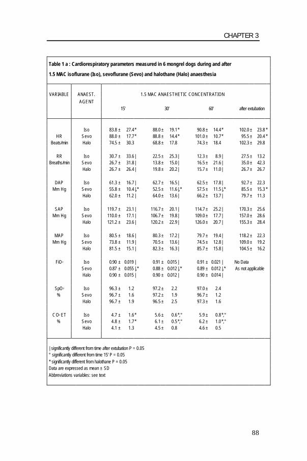
CHAPTER 3
88
Table 1 a : Cardiorespiratory parameters measured in 6 mongrel dogs during and after
1.5 MAC isoflurane (Iso), sevoflurane (Sevo) and halothane (Halo) anaesthesia VARIABLE ANAEST. 1.5 MAC ANAESTHETIC CONCENTRATION
AGENT 15' 30' 60' after extubation
Iso 83.8 ± 27.4* 88.0 ± 19.1* 90.8 ± 14.4* 102.0 ± 23.8 * HR Sevo 88.0 ± 17.7* 88.8 ± 14.4* 101.0 ± 10.7* 95.5 ± 20.4 *
Beats/min Halo 74.5 ± 30.3 68.8 ± 17.8 74.3 ± 18.4 102.3 ± 29.8
RR Iso 30.7 ± 33.6| 22.5 ± 25.3| 12.3 ± 8.9 | 27.5 ± 13.2 Breaths/min Sevo 26.7 ± 31.8| 13.8 ± 15.0| 16.5 ± 21.6| 35.0 ± 42.3
Halo 26.7 ± 26.4| 19.8 ± 20.2| 15.7 ± 11.0| 26.7 ± 26.7
DAP Iso 61.3 ± 16.7| 62.7 ± 16.5| 62.5 ± 17.8| 92.7 ± 22.3 Mm Hg Sevo 55.8 ± 10.4|,* 52.5 ± 11.6|,* 57.5 ± 11.5|,* 85.5 ± 15.3 *
Halo 62.0 ± 11.2 | 64.0 ± 13.6| 66.2 ± 13.7| 79.7 ± 11.3
SAP Iso 119.7 ± 23.1| 116.7 ± 20.1| 114.7 ± 25.2| 170.3 ± 25.6 Mm Hg Sevo 110.0 ± 17.1| 106.7 ± 19.8| 109.0 ± 17.7| 157.0 ± 28.6
Halo 121.2 ± 23.6| 120.2 ± 22.9| 126.0 ± 20.7 | 155.3 ± 28.4
MAP Iso 80.5 ± 18.6| 80.3 ± 17.2| 79.7 ± 19.4| 118.2 ± 22.3 Mm Hg Sevo 73.8 ± 11.9| 70.5 ± 13.6| 74.5 ± 12.8| 109.0 ± 19.2
Halo 81.5 ± 15.1| 82.3 ± 16.3| 85.7 ± 15.8| 104.5 ± 16.2
FiO ² Iso 0.90 ± 0.019 | 0.91 ± 0.015 | 0.91 ± 0.021 | No Data Sevo 0.87 ± 0.055 |,* 0.88 ± 0.012 |,* 0.89 ± 0.012 |,* As not applicable Halo 0.90 ± 0.015 | 0.90 ± 0.012 | 0.90 ± 0.014 |
SpO ² Iso 96.3 ± 1.2 97.2 ± 2.2 97.0 ± 2.4 % Sevo 96.7 ± 1.6 97.2 ± 1.9 96.7 ± 1.2 Halo 96.7 ± 1.9 96.5 ± 2.5 97.3 ± 1.6
CO² ET Iso 4.7 ± 1.6* 5.6 ± 0.6*,° 5.9 ± 0.8*,° % Sevo 4.8 ± 1.7* 6.1 ± 0.5*,° 6.2 ± 1.0*,° Halo 4.1 ± 1.3 4.5 ± 0.8 4.6 ± 0.5
| significantly different from time after extubation P = 0.05 ° significantly different from time 15' P = 0.05 * significantly different from halothane P = 0.05 Data are expressed as mean ± SD Abbreviations variables: see text
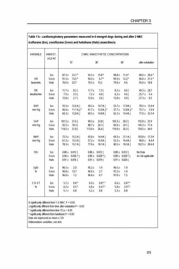
CHAPTER 3
89
Table 1 b : cardiorespiratory parameters measured in 6 mongrel dogs during and after 2 MAC
isoflurane (Iso), sevoflurane (Sevo) and halothane (Halo) anaesthesia
VARIABLE ANAEST. 2 MAC ANAESTHETIC CONCENTRATION AGENT 15' 30' 60' after extubation
Iso 87.3 ± 23.7 * 92.5 ± 15.8* 98.8 ± 11.4* 98.0 ± 28.6 *
HR Sevo 91.3 ± 13.5 * 94.3 ± 6.7* 99.3 ± 12.2* 96.0 ± 21.5 * beats/min. Halo 70.0 ± 23.7 70.3 ± 15.2 79.0 ± 9.6 93.0 ± 18.8
RR Iso 11.7 ± 8.2 | 11.7 ± 7.3 | 8.2 ± 4.6 | 49.3 ± 28.7
breaths/min. Sevo 7.5 ± 3.5 | 7.2 ± 4.0 | 6.2 ± 4.6 | 25.7 ± 5.4 Halo 12.8 ± 2.7 | 12.0 ± 3.6 | 12.8 ± 4.9 | 27.3 ± 8.5
DAP Iso 55.3 ± 12.6 #,| 49.2 ± 10.7#,| 52.7 ± 17.8#,| 78.5 ± 13.6 # mm Hg Sevo 46.0 ± 11.1 #,|,* 41.7 ± 12.4#,|,* 37.7 ± 12.8#,|,* 75.7 ± 7.6 #
Halo 60.3 ± 12.0 #,| 60.5 ± 14.8#,| 62.3 ± 14.4#,| 77.0 ± 22.5 #
SAP Iso 107.2 ± 21.6 | 98.3 ± 23.8| 103.3 ± 28.2| 155.8 ± 25.9 mm Hg Sevo 92.5 ± 18.3 | 88.7 ± 26.7| 83.0 ± 24.1| 145.3 ± 17.4
Halo 114.5 ± 21.8 | 113.0 ± 26.4| 119.0 ± 26.3| 154.5 ± 44.9
MAP Iso 72.5 ± 15.2 #,| 65.8 ± 14.4#,| 69.3 ± 21.1#,| 103.8 ± 17.3 # mm Hg Sevo 61.3 ± 13.2 #,| 57.2 ± 16.9#,| 52.5 ± 16.4#,| 98.8 ± 8.4 #
Halo 78.3 ± 15.1 #,| 77.8 ± 18.1#,| 80.5 ± 18.3#,| 102.5 ± 28.6 #
FiO ² Iso 0.88 ± 0.012 | 0.88 ± 0.012 | 0.89 ± 0.012 | No Data Sevo 0.90 ± 0.036 *,| 0.89 ± 0.028 *,| 0.89 ± 0.020 *,| As not applicable Halo 0.91 ± 0.016 | 0.91 ± 0.019 | 0.91 ± 0.024 |
SpO ² Iso 96.5 ± 2.0 95.2 ± 1.9 96.5 ± 1.9 % Sevo 90.8 ± 13.7 96.0 ± 2.7 97.2 ± 1.9 Halo 96.8 ± 1.2 96.8 ± 0.7 97.0 ± 1.5
CO² ET Iso 5.7 ± 0.8 * 6.0 ± 0.8*,° 6.6 ± 0.8*,° % Sevo 6.2 ± 0.5 * 6.8 ± 0.4*,° 5.8 ± 2.9*,° Halo 5.1 ± 0.8 5.2 ± 0.8 5.3 ± 0.8
# significantly different from 1.5 MAC P = 0.05 | significantly different from time after extubation P = 0.05 ° significantly different from time 15' p = 0.05 * significantly different from halothane P = 0.05 Data are expressed as mean ± SD Abbreviations variables: see text
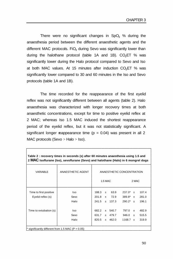
CHAPTER 3
90
There were no significant changes in SpO² % during the
anaesthesia period between the different anaesthetic agents and the
different MAC protocols. FiO² during Sevo was significantly lower than
during the halothane protocol (table 1A and 1B). CO²ET % was
significantly lower during the Halo protocol compared to Sevo and Iso
at both MAC values. At 15 minutes after induction CO²ET % was
significantly lower compared to 30 and 60 minutes in the Iso and Sevo
protocols (table 1A and 1B).
The time recorded for the reappearance of the first eyelid
reflex was not significantly different between all agents (table 2). Halo
anaesthesia was characterized with longer recovery times at both
anaesthetic concentrations, except for time to positive eyelid reflex at
2 MAC; whereas Iso 1.5 MAC induced the shortest reappearance
period of the eyelid reflex, but it was not statistically significant. A
significant longer reappearance time (p = 0.04) was present in all 2
MAC protocols (Sevo > Halo > Iso).
Table 2 : recovery times in seconds (s) after 60 minutes anaesthesia using 1.5 and 2 MAC Isoflurane (Iso), sevoflurane (Sevo) and halothane (Halo) in 6 mongrel dogs
VARIABLE ANAESTHETIC AGENT ANAESTHETIC CONCENTRATION
1.5 MAC 2 MAC
Time to first positive Iso 188.3 ± 63.9 237.0* ± 107.4
Eyelid reflex (s) Sevo 201.8 ± 72.9 389.8* ± 281.0
Halo 241.5 ± 137.3 290.2* ± 196.1
Time to extubation (s) Iso 682.2 ± 540.7 797.0 ± 482.9
Sevo 631.7 ± 479.7 946.0 ± 515.5
Halo 820.5 ± 462.0 1168.7 ± 319.9
* significantly different from 1.5 MAC (P = 0.05)
CHAPTER 3
91
Sevo 1.5 MAC induced a faster extubation time but not in the
2 MAC protocol (table 2). At both MAC protocols the longest
extubation time was after Halo anaesthesia; but this difference was
not significant. Overall, no significant differences were calculated
between the different agents and the different MAC protocols.
DISCUSSION
In experimental studies there are quite a lot of differences in
cardio-respiratory parameters and emergence times between
isoflurane, sevoflurane and halothane (Bernard et al., 1990; Merin et
al., 1991; Pagel et al., 1991; Ebert et al., 1998). However clinically
those differences are not always clear (Oliva et al., 2000; Tacke et al.,
2000). To simulate clinical anaesthesia, the dogs in this study were
premedicated and induction was done with an intravenous anaesthetic
agent. But premedication and induction certainly influence the cardio-
respiratory and recovery parameters. The dogs received a neurolept-
analgesic mixture, fentanyl and droperidol, as premedicant agent.
Droperidol is a butyrophenone tranquilizer causing sedation with
decreased motor activity and tranquilization similar to acepromazine
(Thurmon et al., 1996). Furthermore it gives a high incidence of post-
operative sedation, which can influence the recovery profile
(Bissonnette et al., 1999). Fentanyl is an opioid agonist with a short
duration of action; the peak effect lasts less than 30 minutes.
Droperidol has a long duration of action that extends beyond the
analgesic effects of fentanyl (Moore and Dundee, 1961). The drug
combination of droperidol and fentanyl induces an intense analgesic
effect of relatively short duration. In dogs, this mixture produces
sedation, analgesia, immobilization, respiratory depression and/or
CHAPTER 3
92
panting, α1-adrenergic blockade, a decrease in blood pressure and
bradycardia (Thurmon et al., 1996).
In the present study anaesthesia was induced with propofol, a
short acting induction agent, with rapid metabolisation (Shafer et al.,
1988). Since propofol has a fast redistribution from highly perfused
tissues (e.g. brain) into less-well perfused tissues, plasma levels of
propofol decline rapidly (Shafer, 1993). And because of its high
metabolic clearance rate, which is approximately ten times faster than
that of thiopentone, we can assume that the emergence time is
minimally influenced by propofol (Shafer, 1993; Smith et al., 1994).
For those reasons we chose propofol as induction agent. Oliva et al.
(2000) compared sevoflurane with halothane recovery after propofol
and thiopentone induction. Shorter extubation times after propofol
induction with both inhalant anaesthetics were reported. But induction
technique will have progressively less influence on post-anaesthetic
recovery as the duration of anaesthesia increases, mainly because
the washout period of an anaesthetic agent is determined by the
duration of anaesthesia and its oil/gas partition coefficient (Stoelting
and Eger, 1969).
Equipotent doses using minimum alveolar concentration
multiples were used in this study to allow a comparison not only of the
recovery time, but also of clinically useful non-invasively measured
cardio-respiratory parameters in dogs. Monitoring is essential during
clinical anaesthesia in dogs. We preferred non-invasive techniques
including anaesthetic gas analysis, capnography, pulse oxymetry and
non-invasive blood pressure measurements. Although the Doppler
blood pressure technique is less accurate than invasive techniques, it
is certainly acceptable under clinical circumstances (Stepien, 2000).
CHAPTER 3
93
In the present study heart rate during halothane anaesthesia
was significantly lower than during isoflurane and sevoflurane
anaesthesia at both anaesthetic concentrations. There was no
significant difference in heart rate between isoflurane and sevoflurane
anaesthesia. In non-premedicated dogs sevoflurane induces a rise in
heart rate from 1.2 MAC on (Bernard et al., 1990; Mutoh et al., 1997).
Activation of the baroreceptor-reflex, induced by a decreased arterial
blood pressure, was reported to be mainly responsible for this rise in
heart rate. Isoflurane also gives an increased heart rate in non-
premedicated dogs (Merin et al., 1991). In contrast, halothane has
little influence on heart rate in dogs (Pagel et al., 1991). These
findings might be an indication for a less depressant effect to
baroreceptor-reflex function with Sevo and Iso compared to Halo
(Bernard et al., 1990; Pagel et al., 1991).
All volatile anaesthetic agents cause a dose-related
respiratory depression in dogs (Doi et al., 1986; Mutoh et al., 1997).
This decrease in respiratory rate results in hypoventilation with
increasing CO²ET %. The respiratory depression is also characterized
by a decrease in tidal volume for all anaesthetic agents (Doi et al.,
1986; Mutoh et al., 1997). In our study respiration rate decreased in all
anaesthetic protocols whereby 2 MAC induced a lower respiration rate
compared to 1.5 MAC. However, there was a clear difference between
the protocols; halothane anaesthesia was accompanied by higher
respiration rates compared to isoflurane and sevoflurane. This finding
was also reflected in a lower CO²ET % during halothane anaesthesia.
It is also consistent with literature since halothane was reported to
induce a smaller decrease in respiration rate (Mutoh et al., 1997).
During the initial 15 minutes of anaesthesia at 1.5 MAC with all
CHAPTER 3
94
agents, there was a large individual variation in respiration rate. This
variation was probably due to the premedication with fentanyl-
droperidol, which can cause panting in dogs. This initial tachypnoea
can be a problem for achieving a stable anaesthesia at a low
anaesthetic concentration. Panting did not occur at 2 MAC, probably
because a deeper anaesthetic stage was reached faster.
Volatile anaesthetic agents induce a dose-related decrease in
arterial blood pressure (Steffey and Howland, 1977; Frink et al.,
1992). This decrease in blood pressure is partly caused by a
decreased peripheral vascular resistance and partly by a decrease in
stroke volume (Malan et al., 1995; Lowe et al., 1996; Mutoh et al.,
1997). MAP and DAP at 1.5 MAC were significantly higher than MAP
and DAP in 2 MAC protocols for all agents. SAP was also higher at
1.5 MAC, but the difference was not significant. At both anaesthetic
concentrations the MAP, SAP and DAP were lowest for sevoflurane.
Only DAP from sevoflurane at both anaesthetic concentrations was
significantly lower compared to DAP from isoflurane and halothane.
This can be attributed to a decrease in peripheral vascular resistance
as mentioned in literature (Mutoh et al., 1997). In contrast, the
hypotension induced by halothane anaesthesia occurs mainly
because of direct myocardial depression (Paddleford, 1999; Stowe et
al., 1991). Oliva et al. (2000) found also a decrease in arterial blood
pressure during sevoflurane anaesthesia compared to base line
values. In our study blood pressure is lower during sevoflurane
anaesthesia compared to halothane and isoflurane. Because of this
greater decrease in arterial blood pressure with sevoflurane, heart
rate in compensation was probably higher than during halothane
anaesthesia, due to the baroreceptor-reflex (Pagel et al., 1991).
CHAPTER 3
95
The FiO² during sevoflurane anaesthesia was significantly
lower compared to halothane and isoflurane anaesthesia. This can
easily be explained by the lower anaesthetic potency of sevoflurane.
The MAC from sevoflurane 2.36 vol% is considerably higher than the
MAC from halothane and isoflurane (resp. 0.89 and 1.39) (Steffey et
al., 1977; Kazama et al., 1988). To reach the same anaesthetic depth
a higher concentration of sevoflurane is necessary and the FiO² will
be lower. SpO² % was constant during the anaesthesia period with all
agents and with both MAC values. This could be expected considering
the high, inspired oxygen fractions.
Recovery from inhalation anaesthesia is underlies several
influences. Anaesthetic recovery is mostly influenced by the blood/gas
partition coefficient of the anaesthetic agent. A low blood/gas partition
coefficient allows more rapid drug elimination and results in a shorter
emergence time. The blood/gas partition coefficient of halothane and
isoflurane in man is respectively 2.54 and 1.46. The blood/gas
solubility of sevoflurane (0.68) is much lower and similar to that for
nitrous oxide (0.47) (Strum and Eger, 1987; Steffey, 1996). This
indicates that the anaesthetic recovery of sevoflurane would be more
rapid than that with the other two inhalant anaesthetics. Based on
blood/gas solubility the length of the emergence time would increase
in this order: sevoflurane < isoflurane < halothane.
Of minor importance is the oil/gas partition coefficient. It has
an influence on potency and washout speed of volatile agents. The
oil/gas partition coefficient is 47 for sevoflurane, 91 for isoflurane and
224 for halothane (Steward et al., 1973; Wallin et al., 1975; Strum and
Eger, 1987). The higher the oil/gas partition coefficient, the greater the
potency of the anaesthetic agent. With a smaller oil/gas partition

CHAPTER 3
96
coefficient, there is less transfer and storage of anaesthetic into the
lipid tissue. The washout speed of the inhalation anaesthetic will be
much higher in contrast with anaesthetics with a higher oil/gas
partition coefficient (Steffey, 1996).
Low metabolism can facilitate anaesthetic recovery but only in
a limited way. The metabolisation percentages in man for halothane,
isoflurane and sevoflurane are respectively 20-25%, 0.17% and 3%
(Cascorbi et al., 1970; Holaday et al., 1975; Eger, 1994). The smaller
the metabolisation percentage of an inhalation anaesthetic agent, the
faster the anaesthetic recovery will be (Carpenter et al., 1987).
Other factors such as alveolar ventilation, cardiac output and
duration of anaesthesia have also an influence on recovery from
inhalation anaesthesia (Stoelting and Eger, 1969; Carpenter et al.,
1987). Even the rebreathing circuit itself can reduce the rate of
recovery, because at the end of anaesthesia it is still containing some
anaesthetic agent in the rubber parts of the system. To prevent this
negative influence on recovery times, we disconnected the patient
from the anaesthetic system at the end of the anaesthesia period.
Factors also influencing inhalation anaesthetic elimination
from the body are percutaneous loss and intertissue diffusion of
agents. However, these influences are of little clinical importance
(Lockhart et al., 1991; Carpenter et al., 1987).
Possible parameters for evaluation of anaesthesia recovery in
dogs are eyelid and swallowing reflexes and, time to sternal
recumbency. As was expected in our study the time for a first positive
eyelid reflex was significantly shorter at the lower anaesthetic dosage
CHAPTER 3
97
of the three anaesthetic agents. Surprisingly at both anaesthetic
concentrations the time for a first positive eyelid reflex was the
shortest with isoflurane. On the other hand at 1.5 MAC the time to a
first positive eyelid reflex was longest for halothane and at 2 MAC it
was longest for sevoflurane. But the differences between the three
anaesthetic agents were not significant, since wide individual
variations were present between individuals per anaesthetic agent. As
mentioned above this is easily explained by the greater blood/gas and
oil/gas solubility of halothane. The washout of anaesthetic from alveoli
of more soluble anaesthetics is more gradually in time, than the
washout from less soluble anaesthetics (Eger, 1992). For sevoflurane
at 2 MAC the time for a first positive eyelid reflex was longer than for
the other anaesthetic agents. This could be attributed to a decrease in
cardiac output and a decrease in ventilation (Steffey, 1996). The
respiration rate for sevoflurane at 2 MAC was the lowest and heart
rate higher than with halothane. This higher heart rate might be
attributed to the low arterial blood pressure and a low cardiac output
at this stage of anaesthesia, and together with the low respiration rate
this could explain why the time for a first positive eyelid reflex is
longest for sevoflurane at 2 MAC. During halothane anaesthesia at 2
MAC the respiration rate was higher and heart rate much lower,
together with a higher arterial blood pressure than during sevoflurane
anaesthesia. This can be an explanation for a shorter time to a
positive eyelid reflex with halothane at 2 MAC, although the blood/ gas
solubility of halothane is much higher than for sevoflurane.
Furthermore, it is more difficult to show the kinetic advantages of less
soluble anaesthetics, as sevoflurane, after anaesthetic exposures of
less than 1 hour compared to anaesthetic exposures of more than 1
hour (Eger and Johnson, 1987). After short-duration anaesthetic
exposures a minimal difference in recovery will exist between any of

CHAPTER 3
98
the volatile anaesthetics because there will be little time to saturate
tissue groups. It also has been reported in rodents that the differences
in times to recovery endpoints between anaesthetics are smaller when
low concentrations of the anaesthetics are used (Eger and Johnson,
1987). In addition there are the residual effects of droperidol and
fentanyl, exerting an effect on cognitive functioning. This might nullify
any kinetic advantage of the less-soluble anaesthetic sevoflurane over
isoflurane and halothane.
There were no statistically significant differences in
emergence times between the 3 anaesthetic agents, although the
emergence time was the longest for halothane at both anaesthetic
concentrations. Like for the eyelid reflex this can be explained by the
high blood/ gas and oil/gas solubility of halothane in combination with
its higher metabolisation percentage (20-25%) (Cascorbi et al., 1970).
At 1.5 MAC the emergence time was the shortest for sevoflurane, but
at 2 MAC isoflurane had a shorter emergence time, although the
difference was not significant and very small. In conclusion, clinically
there is little difference in emergence times between halothane,
isoflurane, and sevoflurane in premedicated dogs after 1 hour of
inhalation anaesthesia.

CHAPTER 3
99
REFERENCES Aono, J., W. Ueda, K. Mamiya, E. Takimoto, and M. Manabe, 1997: Greater incidence of delirium during recovery from sevoflurane anesthesia in preschool boys. Anesthesiology 87, 1298-1300. Bernard, J.-M., P.F. Wouters, M.-F. Doursout, B. Florence, J.E. Chelly, and R.G. Merin, 1990: Effects of sevoflurane and isoflurane on cardiac and coronary dynamics in chronically instrumented dogs. Anesthesiology 72, 659-662. Bissonnette, B., H. Swan, P. Ravussin, and V. Un, 1999: Neuroleptanesthesia: current status. Canadian Journal of Anaesthesiology 46, 154-168. Carpenter, R.L., E.I.II Eger, B.H. Johnson, J.D. Unadkat, and L.B. Sheiner, 1987: Does the duration of anesthetic administration affect the pharmacokinetics or metabolism of inhaled anesthetics in humans? Anesthesia & Analgesia 66, 1-8. Cascorbi, H.F., D.A. Blake, and M. Helrich, 1970: Differences in the biotransformation of halothane in man. Anesthesiology 32, 119-123. Doi, M., and K. Ikeda, 1992: Sevoflurane irritates airway least among four anesthetic agents: halothane, enflurane, isoflurane and sevoflurane. Anesthesiology 77(3A), A335. Doi, M., and K. Ikeda, 1993: Airway irritation produced by volatile anesthetics during brief inhalation: Comparison of halothane, enflurane, isoflurane and sevoflurane. Canadian Journal of Anaesthaesiology 40, 122-126. Doi, M., T. Katoh, T. Takii, M. Yura, and K. Ikeda, 1986: The respiratory effects of sevoflurane in dogs. Japanese Journal of Clinical Pharmacologic Therapy 17, 103-104. Ebert, T.J., B.J. Robinson, T.D. Uhrich, A. Mackenthun, and P.J. Pichotta, 1998: Recovery from sevoflurane anesthesia. A comparison to isoflurane and propofol anesthesia. Anesthesiology 89, 1524-1531. Eger, E.I.II, 1992: Desflurane animal and human pharmacology: Aspects of kinetics, safety, and MAC. Anesthesia & Analgesia 75, 3-9. Eger, E.I.II, 1994: New Inhaled Anesthetics. Anesthesiology 80, 906-922. Eger, E.I.II, and B.H. Johnson, 1987: Rates of awakening from anesthesia with I-653, halothane, isoflurane, and sevoflurane: A test of the effect of anesthetic concentration and duration in rats. Anesthesia & Analgesia 66, 977-982.

CHAPTER 3
100
Frink, E.J., S.E. Morgan, A. Coetzee, P.F. Conzen, and B.F. Brown, 1992: The effects of sevoflurane, halothane, enflurane, and isoflurane on hepatic blood flow and oxygenation in chronically instrumented greyhound dogs. Anesthesiology 76, 85-90. Holaday, D.A., V. Fiserova-Bergerova, I.P. Latto, and M.A. Zumbiel, 1975: Resistance of isoflurane to biotransformation in man. Anesthesiology 43, 325-332. Inomata, S., S. Watanabe, M. Taguchi, and M. Okada, 1994: End-tidal sevoflurane concentration for tracheal intubation and minimum alveolar concentration in pediatric patients. Anesthesiology 80, 93-96. Johnson, R.A., E. Striler, D.C. Sawyer, and D.B. Brunson, 1998: Comparison of isoflurane with sevoflurane for anesthesia induction and recovery in adult dogs. American Journal of Veterinary Research 59, 478-481. Kazama, T., and K. Ikeda, 1988: Comparison of MAC and the rate of rise of alveolar concentration of sevoflurane with halothane and isoflurane in the dog. Anesthesiology 68, 435-437. Lerman, J., P.J. Davis, L.G. Welborn, R.J. Orr, M. Rabb, R. Carpenter, E. Motoyama, R. Hannallah, and C.M. Haberkern, 1996: Induction, recovery, and safety characteristics of sevoflurane in children undergoing ambulatory surgery. A comparison with halothane. Anesthesiology 84, 1332-1340. Lockhart, S.L., N. Yasuda, N. Peterson, M.J. Laster, S. Taheri, R.B. Weiskopf, and E.I.II Eger, 1991: Comparison of percutaneous losses of sevoflurane and isoflurane in humans. Anesthesia & Analgesia 72, 212-215. Lowe, D., D.A. Hettrick, P.S. Pagel, and D.C. Warltier, 1996: Influence of volatile anesthetics on left ventricular afterload in vivo. Differences between desflurane and sevoflurane. Anesthesiology 85, 112-120. Malan, T.P., J.A. Di Nardo, R.J. Isner, E.J. Frink, M. Goldberg, P.E. Fenster, E.A. Brown, R. Depa, R., L.C. Hammond, and H. Mata, 1995: Cardiovascular effects of sevoflurane compared with those of isoflurane in volunteers. Anesthesiology 83, 918-928. Merin, R.G., J.-M. Bernard, M.-F. Doursout, M. Cohen, and J.E. Chelly, 1991: Comparison of the effects of isoflurane and desflurane on cardiovascular dynamics and regional blood flow in the chronically instrumented dog. Anesthesiology 74, 568-574. Moore, J., and J.W. Dundee, 1961: Alterations in response to somatic pain associated with anesthesia. VII. The effects of nine phenothiazine derivates. British Journal of Anaesthesia 33, 422. Mutoh, T., R. Nishimura, H.-Y. Kim, S. Matsunaga, and N. Sasaki, 1997: Cardiopulmonary effects of sevoflurane, compared with halothane, enflurane
CHAPTER 3
101
and isoflurane, in dogs. American Journal of Veterinary Research 58, 885-890. Muzi, M., B.J. Robinson, T.J. Ebert, and T.J. O’Brien, 1996: Induction of anesthesia and tracheal intubation with sevoflurane in adults. Anesthesiology 85, 536-543. Oliva, V.N.L.S., A.J.A. Aguiar, F.R. Eugênio, A.L. Andrade, C.R.C.B. Guimarães, and S.H.V. Perri, 2000: Sevoflurane or halothane anaesthesia after propofol or thiopentone induction for orthopedic surgery in dogs. Proc. 7th World Congress of Veterinary Anaesthesia Berne Sept. 20-23, 134-135. Paddleford, R.R., 1999: Anesthetic Agents. In: Paddleford R.R. (ed), Manual of Small Animal Anesthesia 2nd edn. pp. 31-77. W.B. Saunders Company, Philadelphia. Pagel, P.S., J.P. Kampine, W.T. Schmeling, and D.C. Warltier, 1991: Comparison of the systemic and coronary hemodynamic actions of desflurane, isoflurane, halothane, and enflurane in the chronically instrumented dog. Anesthesiology 74, 539-551. Shafer, A.,V.A. Doze, S.L. Shafer, and P.F. White,1988: Pharmacokinetics and pharmacodynamics of propofol infusions during general anesthesia. Anesthesiology 69, 348-356. Shafer, S.L., 1993: Advances in propofol pharmacokinetics and pharmacodynamics. Journal of Clinical Anesthesia 5(suppl. 1), 14-21. Smith, I., Y. Ding, and P.F. White, 1992: Comparison of induction, maintenance, and recovery characteristics of sevoflurane-N²O and propofol-sevoflurane-N²O with propofol-isoflurane-N²O anesthesia. Anesthesia & Analgesia 74, 253-259. Smith, I., P.F. White, M. Nathanson, and R. Gouldson, 1994: Propofol. An update on its clinical use. Anesthesiology 81, 1005-1043. Song, D., G.P. Joshi, and P.F. White, 1998: Fast-track eligibility after ambulatory anesthesia: A comparison of desflurane, sevoflurane and propofol. Anesthesia & Analgesia 86, 267-273. Steffey, E.P., 1996: Inhalation Anesthetics. In: Thurmon, J.C., W.J. Tranquilli, G.J. Benson (eds), Lumb & Jones’ Veterinary Anesthesia. 3rd edn. pp. 297-329. Williams & Wilkins, Baltimore. Steffey, E.P., and D. Howland, 1977: Isoflurane potency in the dog and cat. American Journal of Veterinary Research 38, 1833-1836. Stepien, R.L., 2000: Blood pressure measurement in dogs and cats. In Practice, 136-145.
CHAPTER 3
102
Steward, A., P.R. Allott, A.L. Cowles, and W.W. Mapleson, 1973: Solubility coefficients for inhaled anaesthetics for water, oil and biological media. British Journal of Anaesthesia 45, 282-293. Stoelting, R.K., and E.I.II Eger, 1969: The effects of ventilation and anesthetic solubility on recovery from anesthesia: An in vivo and analog analysis before and after equilibration. Anesthesiology 30, 290-296. Stowe, D.F., S.M. Monroe, J. Marijic, Z.J. Bosnjak, and J.P. Kampine, 1991: Comparison of halothane, enflurane, and isoflurane with nitrous oxide on contractility and oxygen supply and demand in isolated hearts. Anesthesiology 75, 1062-1074. Strum, D.P., and E.I.II Eger, 1987: Partition coefficients for sevoflurane in human blood, saline, and olive oil. Anesthesia & Analgesia 66, 654-656. Tacke, S., H. Xiong, and E. Schimke, 2000: Sevoflurane anaesthesia in dogs after premedication with L-methadone, diazepam and propofol in comparison to halothane and isoflurane. Proc. 7th World Congress of Veterinary Anaesthesia Berne Sept. 20-23, 79. Thurmon, J.C., W.J. Tranquilli, G. Benson (eds), 1996: Preanesthetics and anesthetic adjuncts. In: Lumb & Jones’ Veterinary Anesthesia. 3rd edn, pp. 186. Williams & Wilkins, Baltimore. Trim, C.M., 1994: Monitoring the anaesthetized cat. In: Hall, L.W., and P.M. Taylor (eds), Anaesthesia of the Cat, pp. 194-223. Baillière Tindal, London. Wallin, R.F., B.M. Regen, M.D. Napoli, and I.J. Stern, 1975: Sevoflurane: a new inhalational anesthetic agent. Anesthesia & Analgesia 54, 758-765.
CHAPTER 4
103
CHAPTER 4
THE INFLUENCE OF VENTILATION MODE (SPONTANEOUS
VENTILATION, IPPV AND PEEP) ON CARDIOPULMONARY
PARAMETERS IN SEVOFLURANE ANAESTHETIZED DOGS.
I. Polis 1, F. Gasthuys 2, H. Laevens 3, L. Van Ham 1 and A. De Rick1
1 Department of Small Animal Internal Medicine and Clinical Biology, 2 Department of Surgery and Anaesthesia of Domestic Animals, 3 Department of Reproduction, Obstetrics and Herd Health; Veterinary Epidemiology Unit, Ghent University, Faculty of Veterinary Medicine, Salisburylaan 133, 9820 Merelbeke, Belgium.
Adapted from: I. Polis, F. Gasthuys, H. Laevens, L. Van Ham, A. De Rick. The influence of ventialtion mode (spontaneous ventilation, IPPV and PEEP) on cardiopulmonary parameters in sevoflurane anaesthetized dogs. Journal of Veterinary Medicine A 48, 619-630.

CHAPTER 4
104

CHAPTER 4
105
SUMMARY
The purpose of this study was to investigate the
cardiopulmonary influences of sevoflurane (Sevo) in oxygen at 2
anaesthetic concentrations (1.5 and 2 MAC) during spontaneous and
controlled ventilation in dogs. After premedication with fentanyl-
droperidol (5 µg/kg and 0.25 mg/kg IM) and induction with propofol (6
mg/kg IV) 6 dogs were anaesthetized for 3 hours. Three types of
ventilation were compared: spontaneous ventilation (SpV), intermittent
positive pressure ventilation (IPPV), and positive end expiratory
pressure ventilation (PEEP, 5 cm H²O).
Heart rate, haemoglobin oxygen saturation, arterial blood
pressures, right atrial and pulmonary arterial pressure, pulmonary
capillary wedge pressure and cardiac output were measured. End
tidal CO² percentage, inspiratory oxygen fraction, respiration rate and
tidal volume were recorded using a multi gas analyzer and a
respirometer. Acid-base and blood gas analyses were performed.
Cardiac index, stroke volume, stroke index, systemic and pulmonary
vascular resistance, left and right ventricular stroke work index were
calculated.
Increasing the anaesthetic concentration during sevoflurane
anaesthesia with spontaneous ventilation induced a marked
cardiopulmonary depression; on the other hand, HR increased
significantly, but the increases were clinically not relevant.
The influences of artificial respiration on cardiopulmonary
parameters during 1.5 MAC sevoflurane anaesthesia were minimal. In
contrast, PEEP ventilation during 2 MAC concentration had more
CHAPTER 4
106
pronounced negative influences, especially on right cardiac
parameters. In conclusion, at 1.5 MAC, a surgical anaesthesia level,
sevoflurane can be used safely in healthy dogs during spontaneous
and controlled ventilation (IPPV and PEEP of 5 cm H²O).
INTRODUCTION
Sevoflurane (CFH²-O-CH(CF³)²) is a recently developed
volatile anaesthetic agent currently used in human anaesthesia.
Sevoflurane has a low blood-gas partition coefficient of 0.69 (Strum
and Eger, 1987). This low blood-gas solubility contributes to a more
rapid induction of, and emergence from anaesthesia compared to
halothane, enflurane, and isoflurane (Smith et al., 1992; Lerman et al.,
1996; Aono et al., 1997; Ebert et al., 1998). The low blood-gas
solubility also permits an easier control in anaesthetic depth. The
minimum alveolar concentration (MAC) value of sevoflurane in dogs is
2.36 % (Kazama and Ikeda, 1988).
A lot of research has been performed in man, but only a
limited number of reports on cardiopulmonary parameters during
sevoflurane anaesthesia are available in small animals (Bernard et al.,
1990; Oliva et al., 2000; Tacke et al., 2000). Mutoh et al. (1997)
reported the cardiopulmonary effects of sevoflurane, in comparison to
halothane, enflurane, and isoflurane in adult unpremedicated dogs
using mask induction. However, mask induction without premedication
is seldom used under clinical circumstances. The principal goal of the
present study was to evaluate the effects of spontaneous and
controlled ventilation (IPPV and PEEP) on cardiopulmonary
parameters in sevoflurane anaesthetized premedicated dogs.
CHAPTER 4
107
MATERIALS AND METHODS
Instrumentation and measurements
The study was approved by the Ethical Committee of the
Faculty of Veterinary Medicine, Ghent University (filenumber: 97/12).
Six, ASA I male mongrel dogs weighing 29 ± 7.30 kg (mean ±
standard deviation) from 3 to 7 years were used for the study. The
dogs were vaccinated and dewormed at a regular base. Clinical
examination and a blood analysis confirmed the health status of the
animals before the study. No specific medication altering anaesthetic
or analgesic requirements were administered at least 1 month before
the experiments.
Food, but not water was withheld for 12 hours before each
experiment. The dogs were premedicated 30 minutes before induction
with fentanyl-droperidol IM (respectively 5 µg/kg and 0,25 mg/kg of
body weight BWT) (Thalamonal, Janssen-Cilag, Berchem, Belgium).
Induction of anaesthesia was performed using propofol (6 mg/kg of
BWT) (Rapinovet, Mallinckrodt Veterinary, Stockholm, Sweden)
intravenously over 15-20 seconds. After loss of swallow reflex the
dogs were orally intubated (endotracheal tube 9 to 12 mm ID, Rüsch,
Germany).
A circle system (Titus, Dräger, Lübeck, Germany) with a
precision out of circuit vaporiser (quick lock system) (Vapor 19,3,
Dräger, Lübeck, Germany) was used throughout the study. The soda
lime (Dräger Sorb 800, Dräger, Lübeck, Germany) was renewed
before each experiment. The whole system was air dried between the
experiments. The anaesthetic circuit was flushed with 100% oxygen
for 5 minutes before the experiment and afterwards filled with
sevoflurane 1.5 MAC (1.5 × 2.36 % (Kazama and Ikeda, 1988)) using

CHAPTER 4
108
a fresh gas flow of 2 L/min oxygen. No intravenous infusions were
administered during the trial.
Monitoring included a calibrated (Quick Cal Calibration Gas,
Datex-Ohmeda Corp., Helsinki, Finland) multi anaesthetic gas
analyser (Capnomac Ultima, Datex Engstrom Instrumentation Corp.,
Helsinki, Finland) for determination of the following parameters:
inspiratory anaesthetic agent concentration (FiAA %), inspiratory
oxygen fraction (FiO²), end tidal CO² concentration (ET CO² %) and
respiratory rate (RR). Samples were taken at the Y-part (Straight
Adapter®, Datex-Engstrom Instrumentarium Corp., Helsinki, Finland)
with a rate of 200 mL/min and were scavenged. Tidal volume (TV)
was monitored with a respirometer (Volumeter, Dräger, Lübeck,
Germany). TV and RR were adjusted if necessary during the
experiment to keep a normal PaCO2 level between 35 and 45 mm Hg
(Hartsfield, 1996). The vaporizer settings were adjusted throughout
the experiment to maintain an end-tidal anaesthetic concentration of
1.5 and 2 MAC sevoflurane by monitoring the inspiratory sevoflurane
concentration. Heart rate (HR) and peripheral haemoglobin saturation
(SpO²%) were monitored continuously using a pulse oximeter (N-
20PA Portable Pulse Oximeter, Nellcor Puritan Bennett Inc.,
Pleasanton, CA, U.S.A.) with the probe placed on the tongue.
During the first hour of anaesthesia the dogs were instrumented
for the experiment. A thermodilution catheter (Swan-Ganz catheter,
7.5 french, American Edwards Laboratories, Santa Ana, U.S.A.) was
placed in the left jugular vein of the dog through an introducer
(Percutaneous Sheath Introducer Set, Arrow, Reading, U.S.A.). The
thermodilution catheter was advanced into the pulmonary artery using
the characteristic pressure waveforms on the display of the pressure
monitor (Hellige Servomed SMV 104, Germany). The proximal port
of the thermodilution catheter was positioned in the right atrium. The
CHAPTER 4
109
distal port and thermistor were positioned in the pulmonary artery in a
way that by inflating the balloon of the catheter wedge position was
achieved. Mean (MPAP), systolic (SPAP), diastolic (DPAP) pulmonary
artery pressure, right atrial pressure (RAP) and pulmonary capillary
wedge pressure (PCWP) were measured by connecting the
thermodilution catheter to the pressure transducer (Monitoring-set,
Vascumed N.V., Ghent, Belgium) using an extension tube
(Lectrocath, 150 cm, cap. 1.60 mL, Vygon, Ecouen, France) filled
with heparinised saline (5 I.U. heparin per ml). The pressure
transducer was placed at the level of the heart of the dog.
The thermodilution catheter was connected to the cardiac ouput
computer (COM-1, American Edwards Laboratories, Santa Ana,
U.S.A.) and a closed injectate delivery system (CO-set+, Model 93-
610, Baxter Healthcare Corporation, Edwards Critical –Care Division,
Irvine, U.S.A.). Several cardiac output (CO) determinations were
performed using 5 ml of a saline 0.9% solution at room temperature
injected into the right atrium. The mean value from three results close
to each other was used as actual value. Blood temperature of the dog
and injection temperature of the saline solution were measured using
the cardiac output computer.
A catheter (VasocanBraunüle, 22 gauge, B.Braun,
Melsungen, Germany) was surgically placed into the right femoral
artery. The catheter was connected to the pressure transducer
(Monitoring-set, Vascumed N.V., Ghent, Belgium) by means of
extension tubing filled with heparinised saline. The pressure
transducer was placed at the level of the heart of the dog. The mean
(MAP), systolic (SAP) and diastolic arterial blood pressure (DAP) were
measured using a calibrated blood pressure monitor (Hellige

CHAPTER 4
110
Servomed SMV 104, Germany). Arterial blood was collected in
heparinized 2 ml syringes and stored on ice for measurement of blood
gas tensions and acid-base balance (PCV, pH, pCO², pO², plasma
bicarbonate concentration (HCO³-), and standard base excess (SBE))
with a blood gas analyzer calibrated at 37°C (ABL5, Radiometer
Copenhagen, Denmark).
Experimental Design
The dogs were anaesthetised during the instrumentation
period at a concentration of 1.5 MAC sevoflurane breathing
spontaneously (SpV) before the first measurements were done. These
measurements were used as baseline values. Afterwards the
concentration of sevoflurane was increased to 2 MAC and a
stabilisation period of 20 minutes was respected before the next
measurements. Then the dogs were ventilated using IPPV (Ventilog 3,
Dräger, Lübeck, Germany), the anaesthetic concentration was
reduced back to 1.5 MAC and afterwards increased to 2 MAC.
Between each alteration in anaesthetic concentration or ventilation
pattern, 20 minutes of stabilisation time was respected before new
measurements were done. At the end the dogs were ventilated with a
PEEP of 5 cm H²O and a concentration of 1.5 MAC and 2 MAC. Again
20 minutes of stabilisation time was respected before each
measurement. The entire experimental protocol was finished after 180
minutes (Table1).
After the experiment, catheters were removed and
postoperative analgesia (buprenorphine 10 µg/kg IM q 6h
(Temgesic, Schering-Plough, Hull, England) was administered
before transferring the dogs to the recovery room. The dogs received
amoxycillin clavulanate (8.75 mg/kg/day SC, SynuloxReady-To-Use,

CHAPTER 4
111
Pfizer Animal Health) during 5 days to prevent wound infection.
Anaesthesia and recovery were uneventful.
Table 1: Time Schedule.
TIME SEVO CONCENTRATION ACTION (minutes) VENTILATION PATTERN
T0 1.5 MAC SpV induction instrumentation
T60 measurement 1
2 MAC SpV
T80 measurement 2
1.5 MAC IPPV
T100 measurement 3
2 MAC IPPV
T120 measurement 4
1.5 MAC PEEP
T140 measurement 5
2 MAC PEEP
T160 measurement 6
Calculations
Calculated values were determined as follows: (Gross et al., 1990)
Body surface area (BSA; m²)
BSA = Body Weight (g) ²/³ x 10.1
104

CHAPTER 4
112
Cardiac index (CI; L/min/m²)
CI = CO
BSA
Stroke volume (SV; mL/beat)
SV = CO x 1000
HR
Stroke index (SI; mL/beat/m²)
SI = SV
BSA
Systemic vascular resistance (SVR; dynes.sec/cm5)
SVR = MAP – RAP x 80
CO
Pulmonary vascular resistance (PVR; dynes.sec/ cm5)
PVR = MPAP – PCWP x 80
CO
Left ventricular stroke work index (LVSWI; g.m/m²)
LVSWI = 1.36 (MAP – PCWP) x SI
100
Right ventricular stroke work index (RVSWI; g.m/m²)
RVSWI = 1.36 (MPAP – RAP) x SI
100
Statistical analysis
The results of the different recorded parameters are
expressed as mean ± standard deviation. Statistical analysis for each
parameter was done with repeated measures analysis of variance.
Continuous dependent variables were analysed with Proc Mixed (SAS
v8, SAS Institute Inc., SAS Campus Drive, Cary, NC, USA).
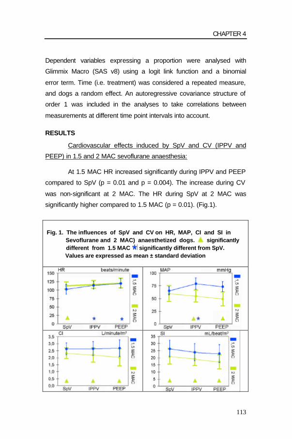
CHAPTER 4
113
Dependent variables expressing a proportion were analysed with
Glimmix Macro (SAS v8) using a logit link function and a binomial
error term. Time (i.e. treatment) was considered a repeated measure,
and dogs a random effect. An autoregressive covariance structure of
order 1 was included in the analyses to take correlations between
measurements at different time point intervals into account.
RESULTS
Cardiovascular effects induced by SpV and CV (IPPV and
PEEP) in 1.5 and 2 MAC sevoflurane anaesthesia:
At 1.5 MAC HR increased significantly during IPPV and PEEP
compared to SpV (p = 0.01 and p = 0.004). The increase during CV
was non-significant at 2 MAC. The HR during SpV at 2 MAC was
significantly higher compared to 1.5 MAC (p = 0.01). (Fig.1).
Fig. 1. The influences of SpV and CV on HR, MAP, CI and SI in Sevoflurane and 2 MAC) anaesthetized dogs. significantly different from 1.5 MAC significantly different from SpV. Values are expressed as mean ± standard deviation
CHAPTER 4
114
MAP, SAP, and DAP increased significantly during the IPPV
1.5 MAC Sevo protocol compared to SpV (p = 0.01). On the other
hand, 2 MAC IPPV and PEEP induced a non-significant decrease of
these parameters. Blood pressures were significantly lower at 2 MAC
Sevo CV compared to 1.5 MAC Sevo (p = 0.0001) (Fig.1).
In the 1.5 MAC protocol only RAP during IPPV was
significantly lower compared to PEEP (p = 0.02). RAP during 2 MAC
PEEP was significantly higher than SpV and IPPV (p = 0.05 and p =
0.03). RAP of 2 MAC CV was significantly higher compared to 1.5
MAC.
MPAP in the 1.5 MAC protocol increased significantly during
IPPV and PEEP compared to SpV (IPPV was also significantly
different from PEEP) (p = 0.001). The MPAP of 2 MAC Sevo also
increased during CV; but the differences were only significant for
PEEP compared to SpV and IPPV (p = 0.001). The MPAP of the 1.5
MAC SpV was significantly lower than 2 MAC (p = 0.02). SPAP and
DPAP during 1.5 MAC increased when CV was applied; PEEP was
significantly different from SpV for both parameters (p = 0.01) and
from IPPV for DPAP (p = 0.01). The increase of SPAP and DPAP was
non-significant in the 2 MAC protocol. 2 MAC during SpV induced a
significant increase in SPAP and DPAP compared to 1.5 MAC (p =
0.04).
At 1.5 MAC PCWP increased significantly during CV, this
increase was significant during PEEP compared to SpV (p = 0.007)
and for PEEP compared to IPPV (p = 0.007). At 2 MAC only the
increase in PCWP during PEEP compared to IPPV was significant (p
= 0.02). 2 MAC during SpV and IPPV induced a significant increase in
PCWP compared to 1.5 MAC (p = 0.0009 and p = 0.03).

CHAPTER 4
115
Fig. 2 : The influences of SpV and CV on LVSWI, RVSWI, SVR and PVR in sevoflurane (1.5 and 2 MAC) anaesthetized dogs. significantly different from 1.5 MAC ; significantly different from SpV. Values are expressed as mean ± standard deviation
CV induced no significant changes in CO and CI, SV, SI and
LVSWI in both MAC protocols. 2 MAC anaesthesia induced signicant
lower values compared to 1.5 MAC for these parameters. (Fig. 1 and
2). CV induced a non-significant rise in RVSWI and SVR during the
1.5 MAC protocol. On the contrary at 2 MAC there was a slight
decrease in RVSWI and SVR during CV compared to SpV. During 2
MAC CV there was a significant decrease in RVSWI and SVR
compared to 1.5 MAC (p = 0.006). (Fig. 2). CV induced a non-
significant increase in PVR in both MAC protocols; only PEEP was
significantly different from SpV (p = 0.04). (Fig. 2).

CHAPTER 4
116
Table 2 : Influences of SpV and CV on cardiovascular parameters in sevoflurane
(1.5 and 2 MAC) anaesthetized dogs.
VAR VENT SEVO CONCENTRATION VAR VENT SEVO CONCENTRATION
1,5 MAC 2 MAC 1,5 MAC 2 MAC
HR SpV 102 ± 14 113 ± 10.8 * CO SpV 2.5 ± 0.6 2.2 ± 0.5 *
beats IPPV 114 ± 10.8 ° 116 ± 11.6 L/min IPPV 2.5 ± 0.7 2 ± 0.5 * per min. PEEP 119 ± 14.1 ° 121 ± 14.5 PEEP 2.5 ± 0.7 1.9 ± 0.7 *
MAP SpV 65 ± 8.1 59 ± 15.6 CI SpV 2.62 ± 0.43 2.31 ± 0.38 *
mm Hg IPPV 79 ± 11.2 ° 55 ± 11.7 * L/min/m² IPPV 2.64 ± 0.5 2.18 ± 0.51 * PEEP 73 ± 9.5 49 ± 13.7* PEEP 2.67 ± 0.59 2.02 ± 0.62 *
SAP SpV 92 ± 11 84 ± 19.5 SV SpV 24.91 ± 7.83 19.69 ± 6.22 * mm Hg IPPV 112 ± 18 ° 78 ± 21.6 * mL/beat IPPV 22.24 ± 7.01 17.72 ± 5.23 *
PEEP 99 ± 23 70 ± 25.5 * PEEP 21.46 ± 7.12 16.07 ± 6.06 *
DAP SpV 54 ± 7.5 50 ± 12.8 SI SpV 26.28 ± 5.93 20.86 ± 5.4 * mm Hg IPPV 66 ± 10.9 ° 44 ± 10 * mL/beat/m² IPPV 23.59 ± 6.04 18.97 ± 4.71 *
PEEP 59 ± 10 42 ± 10.3 * PEEP 22.79 ± 6.28 16.94 ± 4.95 *
RAP SpV 5.8 ± 3.4 6 ± 3.3 | LVSWI SpV 20.46 ± 5.23 14.39 ± 6.05 * mm Hg IPPV 5.2 ± 3.5 | 6.3 ± 3.7 *| g*m/m² IPPV 23.09 ± 7.68 12.31 ± 5.07 *
PEEP 6.5 ± 3.2 7.5 ± 3.6 * PEEP 19.89 ± 7.22 9.24 ± 5.29 *
MPAP SpV 11.3 ± 3.4 13 ± 4 * RVSWI SpV 2.03 ± 0.79 1.89 ± 0.93 mm Hg IPPV 13.2 ± 4 °| 13 ± 4.1 | g*m/m² IPPV 2.67 ± 1.15 1.8 ± 0.68 *
PEEP 15.3 ± 3.3 ° 15 ± 3.1° PEEP 2.79 ± 0.99 1.81 ± 0.65 *
SPAP SpV 18 ± 4.1 20 ± 5.2 * SVR SpV 2015.7 ± 571.2 2022.8 ± 625.23
mm Hg IPPV 20.5 ± 3.9 20 ± 4.5 | dynes*sec/cm5 IPPV 2511.3 ± 746 1948.6 ± 411.63* PEEP 22.7 ± 2.8 ° 23 ± 5.1 PEEP 2243.4 ± 599.7 1807.7 ± 524.49*
DPAP SpV 5.8 ± 3.5 7.5 ± 3.8 * PVR SpV 125.42 ± 47.58 121.2 ± 43.03
mm Hg IPPV 6.7 ± 3.3 | 7.5 ± 4 dynes*sec/cm5 IPPV 162.23 ± 40.2 154.1 ± 36.48 PEEP 9.5 ± 4.9 ° 9.5 ± 5 PEEP 171.49 ± 45.08° 216.67 ± 124.45°
PCWP SpV 7.7 ± 3.7 9.3 ± 4.3 * mm Hg IPPV 8.3 ± 4.2 | 9.3 ± 3.5 *|
PEEP 10.2 ± 3.1 ° 11 ± 4
Data are expressed as mean ± standard deviation
* significantly different from 1,5 MAC p = 0,05
| significantly different from PEEP p = 0,05
° significantly different from SpV p = 0,05
Abbreviations of variables: see text

CHAPTER 4
117
Effects of SpV and CV (IPPV and PEEP) on blood gas
variables and respiratory parameters in 1.5 and 2 MAC sevoflurane
anaesthesia: The 1.5 MAC CV protocol induced a decrease in PaCO²
compared to SpV, this decrease was only significant for IPPV
compared to SpV (p = 0.05). At 2 MAC there was also a decline in
PaCO² during CV, this decrease was significant for IPPV and PEEP
compared to SpV (resp. p = 0.0004 and p = 0.01). PaCO² during SpV
was significantly higher at 2 MAC sevoflurane compared to 1.5 MAC
(p = 0.01). There was no significant difference in PaO² at any
anaesthesia stage.
There was a significant rise in pH during CV compared to SpV
(p = 0.003), only the rise during PEEP compared to SpV at 1.5 MAC
was not significant. pH was significantly lower at 2 MAC compared to
1.5 MAC anaesthetic concentration during SpV (p = 0.009). SBC
increased significantly during IPPV and PEEP compared to SpV at 2
MAC (resp. p = 0.04 and p = 0.03). There was no significant
difference in PCV at any anaesthesia stage. There were significant
but small changes in blood temperature during 2 MAC compared to
1.5 MAC (p = 0.0002).
The inspiratory pressure increased significantly during CV
compared to SpV at both anaesthetic concentrations (p = 0.0001) and
there was also a significant increase in inspiratory pressure during
PEEP compared to IPPV (p = 0.0001). TV remained constant in all
anaesthesia stages, but with large individual variations. There was no
significant difference in RR between the 3 different types of
ventilation, but RR was significantly lower at 2 MAC compared to 1,5
MAC during SpV (p = 0.02). ETCO²% decreased significantly during
CV compared to SpV at both MAC protocols. The 2 MAC SpV

CHAPTER 4
118
protocol induced a significant increase in ETCO²% compared to 1.5
MAC (p = 0.004).
Table 3: Influences of SpV and CV on pH, blood gas analysis and respiratory parameters in sevoflurane anaesthetized dogs. .
VARIABLE VENT SEVOFLURANE CONCENTRATION 1.5 MAC 2 MAC
BLOODTEMP. SpV 38.7 ± 0.5 38.8 ± 0.4 * (°C) IPPV 38.8 ± 0.4 | 38.9 ± 0.4 * |
PEEP 39 ± 0.5 ° 39.1 ± 0.5 * °
PCV SpV 35 ± 8.9 36 ± 8.4 IPPV 35 ± 7.5 34 ± 9.5 PEEP 36 ± 10.6 36 ± 9.8
pH SpV 7.255 ± 0.06 7.212 ± 0.08 * IPPV 7.312 ± 0.03 ° 7.303 ± 0.02 ° PEEP 7.295 ± 0.02 7.287 ± 0.03 °
PaCO2 SpV 47 ± 9.9 56 ± 15.6 * (mm Hg) IPPV 40 ± 4.6 ° 40 ± 2.4 °
PEEP 43 ± 1.5 44 ± 3.5 ° PaO2 SpV 530 ± 28.5 513 ± 62.1
(mm Hg) IPPV 551 ± 28.1 539 ± 22.5 PEEP 480 ± 109.3 547 ± 15.1
RR SpV 14 ± 14.3 9 ± 6.6 * (breaths/min.) IPPV 16 ± 3.6 14 ± 2.9
PEEP 14 ± 1.9 15 ± 1.9 TV SpV 451 ± 230.7 453 ± 181.5 (ml) IPPV 402 ± 116.9 402 ± 91.1
PEEP 420 ± 116.9 390 ± 105.2
Pinsp. SpV -0.3 ± 0.8 0 ± 1.3 (mm Hg) IPPV 9 ± 1°| 10 ± 1.4 °|
PEEP 14 ± 2.3 ° 13 ± 2 ° CO2 % ET SpV 6.3 ± 1.2 7.5 ± 1.8 *
IPPV 5.3 ± 0.3 ° 5.4 ± 0.6 ° PEEP 5.6 ± 0.6° 5.6 ± 0.8 °
SBC SpV 18.83 ± 1.47 18.5 ± 1.76 (mEq/ l) IPPV 19.8 ± 1.17 19.8 ± 1.17 °
PEEP 20 ± 0.63 20 ± 0.89 °
Data are expressed as mean ± standard deviation
* significantly different from 1, 5 MAC (p = 0,05)
° significantly different from SpV (p = 0,05)
| significantly different from PEEP (p = 0,05)
Abbreviations of variables: see text

CHAPTER 4
119
DISCUSSION
Sevoflurane is a recently developed inhalational anaesthetic
agent, which is presently very popular in human anaesthesia (Eger,
1994; Young and Apfelbaum, 1995). Until now, few studies with
sevoflurane in dogs under clinical anaesthesia circumstances are
available (Oliva et al., 2000; Tacke et al., 2000). The cardiopulmonary
influences of sevoflurane in experimental dogs were already reported
but the design of these studies included no premedication but a mask
induction (Bernard et al., 1990; Mutoh et al., 1997). The present study
was performed to investigate the cardiopulmonary effects in 1.5 and 2
MAC sevoflurane anaesthetized dogs using a clinical protocol
including standard premedication and induction. Moreover, the
influences of different modes of controlled ventilation (IPPV and
PEEP) were compared with the spontaneous breathing pattern.
Spontaneous ventilation (SpV) is mostly used during clinical
anaesthesia in dogs. However, this mode of breathing is often
accompanied with hypoventilation. A moderate increase in PaCO² has
certainly beneficial effects on the occurring cardiovascular depression,
whereby the increased PaCO² is a potential stimulator of the
sympathetic nerve system (Cullen and Eger, 1974). Artificial
respiration (AR) overcomes the occurring hypoventilation. AR is also
required when intrathoracic surgery is necessary or the patient is
curarized.
Inhalation anaesthetics including sevoflurane induce a dose-
dependent cardiopulmonary depression in all animals (Aida et al.,
1996; Bernard et al., 1992; Grosenbaugh and Muir, 1998). The MAC
of sevoflurane in dogs has been established to be 2.36 volume %
(Kazama and Ikeda, 1988). One and a half MAC is generally accepted
CHAPTER 4
120
as the standard to allow most surgical interventions. However, some
cases require a higher MAC when no supplementary analgesics or
related drugs are administered during anaesthesia.
Basically, sevoflurane produces a dose dependent
cardiopulmonary depression with systemic hypotension what is partly
explained by an occuring peripheral vasodilatation. The influences of
sevoflurane without premedication on the cardiopulmonary
parameters were already intensively studied, whereby different MAC
values were compared with the awake status in experimental dogs
(Bernard et al., 1990; Frink et al., 1992; Harkin et al., 1994; Mutoh et
al., 1997). In the present study only the changes induced by an
increase in MAC were investigated. Fentanyl and propofol are
relatively fast acting and short lasting agents. Interactions of these
drugs after the instrumentation period of one hour with the sevoflurane
protocol in the present study were not expected. On the other hand,
although these drugs have a short half-life, it could not be excluded
that their interfering effects were of longer duration. Droperidol has
relatively long lasting effects. A possible influence of droperidol can
therefore not be ruled out (Bissonnette et al., 1999). A fluid rate of 4 to
8 ml/ kg/ hour is generally accepted for the maintenance of a stable
water balance during anaesthesia (Giesecke and Egberth, 1985). No
fluids were administered in the present study. Nevertheless, the
thermodilution method includes the administration of different boli of
saline. Overall, an estimated quantity of 2 to 4 ml/ kg/ hour was
administered during the whole experimental period. The influences of
this relatively low fluid administration in this study can probably be
neglected.
Two papers reported the cardiopulmonary influences of
sevoflurane when increasing the MAC in spontaneous breathing dogs
CHAPTER 4
121
(Harkin et al., 1994; Mutoh et al., 1997). Overall, the cardiopulmonary
depression was characterized with non-significant decreases in
cardiac output and index, stroke volume and index and pressure work
index and a significant decrease in left ventricular stroke pressure.
The decrease in mean and systolic arterial blood pressure only
changed significantly in the study of Mutoh et al. (1997). All these
findings were similar in the present study, although significant
changes were observed or calculated mainly in the pulmonary
pressures, the cardiac output and stroke volume and the LVSWI.
Surprisingly, a significant increase in HR was also observed in the
present study. This is in contrast to the literature, where a constant or
a slight decrease in HR was reported (Harkin et al., 1994; Mutoh et
al., 1997). The reason for this is not clear. Although the observed
increase in HR was not very high, it was obvious and constant (about
10 %) in all dogs. Most likely the increased HR might be related to
different factors. First of all, Mutoh et al. (1997) reported that
sevoflurane in dogs induced a stimulation of the baroreceptor-reflex
due to a dose dependent decrease in blood pressure. Secondly, the
increase in PaCO² in men resulted in a sympathic stimulation (Cullen
and Eger, 1974). Both factors might explain the observed slight, but
significant increase in HR.
In our study significant decreases in SI, CI, and LVSWI with
an increasing MAC value were seen. The decrease in SV and SI
could be related to a decreased preload, an increased afterload, a
decreased contractility or a combination of those (Suga et al., 1985).
PCWP and SVR can be used as measures for preload and afterload,
respectively (Muir and Mason, 1996; Suga et al., 1985). In the present
study PCWP increased significantly, while the SVR remained
constant. Mutoh et al. (1997) only found non-significant increases in
RAP and PCWP, while PAP and SVR remained constant by
CHAPTER 4
122
increasing the sevoflurane MAC. Thus, decreased contractility is the
most obvious explanation for the depression of cardial function by
increasing the MAC value. A reduced myocardial contractility is often
compensated by an increase in end diastolic pressure (Kittleson,
1988). This phenomenon most likely occurred in the present study
since PCWP and PAP’s which are good reflections of the end diastolic
pressure, increased (Brutsaert et al., 1985). PVR and RVSWI reflect
the right ventricular afterload (Kaplan, 1986). Little changes occurred
in the spontaneous breathing dogs by increasing the sevoflurane
concentration.
Cardiopulmonary influences of sevoflurane were investigated
in chronically instrumented dogs ventilated with IPPV (Bernard et al.,
1990; Frink et al., 1992). Several MAC multiples (1.2, 1.5 and 2 MAC)
were compared with the awake values in these studies. Bernard et al.
(1990) reported significant decreases in CO, arterial pressures and
SV by increasing the MAC from 1.2 to 2 MAC. The systemic vascular
resistance remained constant. The same trend was noticed by Frink et
al. (1992) when increasing the MAC from 1.5 to 2 MAC; although no
significancy was observed. This is in agreement with the findings in
the present study. An increase in MAC from 1.5 to 2 MAC in IPPV
ventilated dogs induced a severe cardiopulmonary depression.
Arterial pressures were significantly lower during the high MAC
protocol. This decrease was more pronounced compared to the same
MAC protocol in spontaneously ventilated dogs. The CO, CI, SV, SI,
LVSWI and RVSWI decreased significantly. These observed
decreases were more accentuated but not significantly different from
the spontaneously breathing dogs. Right cardiac pressures increased
also with increasing anaesthetic concentration; although only the RAP
and PCWP were significantly different in the IPPV protocol.
CHAPTER 4
123
Changing from SpV to IPPV using 1.5 MAC in our study
surprisingly induced a small increase of arterial blood pressures and
HR. In the same low MAC protocol little to no influences on the other
cardiac parameters were observed by using IPPV. Data concerning
the influences of AR on arterial blood pressure and vascular
resistance in animals are conflicting. In horses a severe impact on the
cardiopulmonary system using IPPV was reported (Aida et al., 1996).
However, experiments in rats demonstrated a significant rise in
arterial blood pressure and systemic vascular resistance when
artificial respiration was applied (Sellden et al., 1986). Apparently, the
influences of IPPV in smaller body weights were almost non-existing
whereby a clear reason for our findings were not obvious. The same
tendency was observed when changing from IPPV to PEEP
ventilation. Right cardiac pressures increased when changing from
SpV to IPPV at 1.5 MAC sevoflurane; but only the increase in MPAP
was significant. During PEEP ventilation at 1.5 MAC the increases in
right cardiac pressures were even more pronounced and only the
increase in RAP was not significant. Overall, the existing
cardiopulmonary depression was only slightly influenced by the
different pressure ventilation patterns in the 1.5 MAC protocols.
The situation changed completely when MAC was increased
from 1.5 to 2. Artificial respiration with 2 MAC induced a severe impact
on the arterial pressures and most of the cardiac related parameters.
CO, CI, SV, SI, LVSWI and RVSWI decreased significantly. PEEP
induced a more pronounced negative impact than IPPV, but the
difference between both ventilation patterns was not significant. The
right cardiac pressures increased while the arterial pressures were
clearly lowered. These findings were according to those reported in
the literature (Cassidy et al., 1978; Pinsky, 1990; Smiseth et al.,
1996). The main patho-physiologic mechanism for these

CHAPTER 4
124
cardiovascular side effects of PEEP is a decreased venous return due
to the increased intrathoracic pressure (Versprille, 1990) and a
decreased coronary blood flow inversely related with the PEEP level
(Jacobs and Venus, 1983). We did not find significant differences in
CO, CI, SV, SI, LVSWI and RVSWI between IPPV and PEEP in this
study.
In the present study, pulmonary vascular resistance increased
during AR compared to spontaneous ventilation at both anaesthetic
concentrations, the difference was only significant for the PEEP
ventilation mode. On the contrary, systemic vascular resistance
decreased when changing from spontaneous to controlled ventilation
at 2 MAC. Apparently, the reported vasodilating properties of
sevoflurane were more pronounced at the higher anaesthetic
concentration when combined with artificial breathing. An analogue
slight influence was reported in men using 3 cm H²O PEEP; a steeper
fall was observed when PEEP was increased to 10 cm H²O.
Increasing PEEP above 10 cm H²O had only minor effects (Schreuder
et al., 1982). The same tendancy was observed in dogs under
controlled ventilation whereby a stepwise increase in PEEP induced a
proportional decrease in CO (Sykes et al., 1970; Scharf et al., 1977).
In the present study AR was applied during a relatively short period of
20 minutes. The influence on the cardiopulmonary parameters was
also reported to be proportionally depending on the length of the
period in which positive pressure was applied (Shawley, 1987).
Therefore, to lessen cardiovascular compromise, the amount of time
either inspiratory or expiratory pressure is applied must be minimized.
It could have been possible that significant changes occurred if the
length of the observation period was enlarged.

CHAPTER 4
125
Arterial carbon dioxide pressure is the most frequently used
index of respiratory system response to general anaesthetics. All
contemporary inhalation anaesthetics depress alveolar ventilation and
as a consequence increase PaCO² in a dose-related fashion (Green,
1995). Sevoflurane is a more potent ventilatory depressant than
halothane (Green, 1995), and the characteristics of the ventilatory
depression associated with sevoflurane are similar to that of isoflurane
(Fourcade et al., 1971). As expected, in our study a significant rise in
PaCO² and CO²ET % occurred when increasing Sevo concentration in
spontaneous breathing dogs. This is consistent with the results of
Mutoh et al. (1997).
The present study showed that in anaesthetized
spontaneously ventilating dogs increasing MAC values of sevoflurane
from 1.5 to 2 induced a pronounced cardiopulmonary depression
together with a significant increase in HR. However, this increase had
little clinical consequence. The increased HR could be explained by
the baroreceptor-reflex and/ or by sympathetic stimulation. The
influences of artificial respiration on cardiopulmonary parameters at
1.5 MAC anaesthetic concentration were relatively minimal. On the
other hand, AR during 2 MAC sevoflurane anaesthesia had severe
negative influences, especially on all cardiac parameters. PEEP
ventilation had a greater impact than IPPV.
In conclusion, sevoflurane anaesthesia at 1.5 MAC in
premedicated healthy dogs induced a relatively moderate
cardiopulmonary depression during spontaneous and controlled
ventilation (IPPV and PEEP of 5 cm H²O) and can be used safely.
Increasing the MAC from 1.5 to 2 caused a marked cardiopulmonary
depression. Higher concentrations of sevoflurane should better be
avoided during all ventilation modes in dogs.
CHAPTER 4
126
REFERENCES Aida, H., Y. Mizuno, S. Hobo, K. Yoshida, and T. Fujinaga, 1996: Cardiovascular and pulmonary effects of sevoflurane anesthesia in horses. Veterinary Surgery 25, 164-170. Aono, J., W.Ueda, K. Mamiya, E. Takimoto, and M. Manabe, 1997: Greater incidence of delirium during recovery from sevoflurane anesthesia in preschool boys. Anesthesiology 87, 1298-1300. Bernard, J.-M., P.F. Wouters, M.-F. Doursout, B. Florence, J.E. Chelly, and R.G. Merin, 1990: Effects of sevoflurane and isoflurane on cardiac and coronary dynamics in chronically instrumented dogs. Anesthesiology 72, 659-662. Bernard, J.-M., M.-F. Doursout, P.F. Wouters,C.J. Hartley, R.G. Merin, and J.E. Chelly, 1992: Effects of sevoflurane and isoflurane on hepatic circulation in the chronically instrumented dog. Anesthesiology 77, 541-545. Bissonnette, B., H. Swan, P. Ravussin, and V. Un, 1999: Neuroleptanesthesia : current status. Canadian Journal of Anaesthesiology 46, 154-168. Brutsaert, D.L., F.E. Rademakers, S.U. Sys, T.C. Gillebert, and P.R. Housmans,1985: Analysis of relaxation in the evaluation of the ventricular function of the heart. Progress in Cardiovascular Disease 28, 143-163. Cassidy, S.S., C.H.Jr. Robertson, A.K. Pierce, and R.L. Johnson, 1978: Cardiovascular effects of positive end-expiratory pressure in dogs. Journal of Applied Physiology 44, 743-750. Cullen, D.J., and E.I.II Eger, 1974: Cardiovascular effects of carbon dioxide in man. Anesthesiology 41, 345. Ebert, T.J., B.J. Robinson, T.D. Uhrich, A. Mackenthun, and P.J. Pichotta, 1998: Recovery from sevoflurane anesthesia. A comparison to isoflurane and propofol anesthesia. Anesthesiology 89, 1524-1531. Eger, E.I.II, 1994: New inhaled anesthetics. Anesthesiology 80, 906-922. Fourcade, H.E., W.C. Stevens, C.P. Larson, T.H.Cromwell, S.H. Bahlman, R.F. Hickey, et al.,1971: The ventilatory effects of Forane, a new inhaled anesthetic. Anesthesiology 35, 26-31. Frink, E.J., S.E. Morgan, A. Coetzee, P.F. Conzen, and B.R. Brown, 1992: The effects of sevoflurane, halothane, enflurane, and isoflurane on hepatic blood flow and oxygenation in chronically instrumented greyhound dogs. Anesthesiology 76, 85-90.
CHAPTER 4
127
Giesecke, A.H., and L.D. Egbert, 1985: Perioperative fluid therapy-crystalloids. In: Anesthesia, ed. R. Miller, pp. 1313-1328. Churchill Livingstone, New York. Grosenbaugh, D.A., and W.W. Muir, 1998: Cardiorespiratory effects of sevoflurane, isoflurane, and halothane anesthesia in horses. American Journal of Veterinary Research 59, 101- 106. Green, W.B., 1995: The ventilatory effects of sevoflurane. Anesthesia & Analgesia 81, 23-26. Gross, M.E., W.J. Tranquilli, J.C. Thurmon, G.J.Benson, and W.A. Olson, 1990: Hemodynamic effects of intravenous Midazolam-Xylazine-Butorphanol in dogs. Veterinary Surgery 19, 173-180. Harkin, C.P., P.S. Pagel, J.R. Kersten, D.A. Hettrick, and D.C. Warltier, 1994: Direct negative inotropic and lusitropic effects of sevoflurane. Anesthesiology 81, 156-167. Hartsfield, S.M., 1996: Airway Management and Ventilation. In: Thurmon J.C., Tranquilli W.J., Benson G.J., eds. Lumb & Jones’ Veterinary Anesthesia. 3rd Ed., Baltimore, The Williams & Wilkins Co Inc., 515-556. Jacobs, H.K., and B. Venus, 1983: Left ventricular regional myocardial blood flows during controlled positive pressure ventilation and PEEP in dogs. Critical Care Medicine 11, 872-875. Kaplan, J.A., 1986: Cardiovascular Physiology. In: Miller R.D., ed. Anesthesia. 2nd ed. New York: Churchill Livingstone Inc, 1165- 1194. Kazama, T., and K. Ikeda, 1988: Comparison of MAC and the rate of rise of alveolar concentration of sevoflurane with halothane and isoflurane in the dog. Anesthesiology 68, 435-437. Kittleson, M.D., 1988: Management of heart failure: Concepts, Therapeutic Strategies, and Drug Pharmacology. In: Fox P.R. (ed.), Canine and Feline Cardiology. Churchill Livingstone, New York, pp. 171-204. Lerman, J., P.J. Davis, L.G. Welborn, R.J. Orr, M. Rabb, R. Carpenter, E. Motoyama, R. Hannallah, and C.M. Haberkern, 1996: Induction, recovery, and safety characteristics of sevoflurane in children undergoing ambulatory surgery. A comparison with halothane. Anesthesiology 84, 1332-1340. Muir, W.W., and D. Mason, 1996: Cardiovascular System. In: Thurmon J.C., Tranquilli W.J., Benson G.J., eds. Lumb & Jones’ Veterinary Anesthesia. 3rd Ed., Baltimore, The Williams & Wilkins Co Inc., 62-114. Mutoh, T., R. Nishimura, H.-Y. Kim, S. Matsunaga, and N. Sasaki 1997: Cardiopulmonary effects of sevoflurane, compared with halothane, enflurane,
CHAPTER 4
128
and isoflurane, in dogs. American Journal of Veterinary Research 58, 885-890. Oliva, V.N.L.S., A.J.A. Aguiar, F.R. Eugênio, A.L. Andrade, C.R.C.B. Guimarães, and S.H.V. Perri, 2000: Sevoflurane or halothane anaesthesia after propofol or thiopentone induction for orthopedic surgery in dogs. Schatzmann U. (Ed.), Proc. 7th World Congress of Veterinary Anaesthesia Berne Sept. 20-23, 134-135. Pinsky, M.R., 1990: The effects of mechanical ventilation on the cardiovascular system. Critical Care Clinics 6, 663-678. Scharf, S.M., P. Caldini, and R.H.Jr. Ingram, 1977: Cardiovascular effects of increasing airway pressure in the dog. American Journal of Physiology 232, H35-H43. Schreuder, J.J., J.R.C. Jansen, J.M.Bogaard, and A. Versprille, 1982: Hemodynamic effects of PEEP applied as a ramp. Journal of Applied Physiology 53, 1239-1247. Sellden, H., H. Sjövall, and S.E. Ricksten, 1986: Sympathetic nerve activity and haemodynamics during mechanical ventilation with positive end-expiratory pressure in rats. Acta Physiologica Scandinavica 127, 51-60. Shawley, R.V., 1987: Controlled ventilation and pulmonary function. In: Principles & Practice of Veterinary Anaesthesia. Short C.E. (eds). Williams & Wilkins, Baltimore. Pp. 419-425. Smiseth, O.A., C.R.Thompson, H. Ling, M. Robinson, and R.T. Miyagishima, 1996: A potential clinical method for calculating transmural left ventricular filling pressure during positive end-expiratory pressure ventilation: an intraoperative study in humans. Journal of the American College of Cardioliology 27, 155-160. Smith, I., Y.Ding, and P.F. White, 1992: Comparison of induction, maintenance, and recovery characteristics of sevoflurane-N²O and propofol-
sevoflurane- N²O with propofol-isoflurane- N²O anesthesia. Anesthesia &
Analgesia 74, 253-259. Steffey, E.P., 1996: In Lumb & Jones’ Veterinary Anesthesia. Third Edition. Chapter 11: Inhalation Anesthetics table 11-4, 301. Strum, D.P., and E.I.II Eger, 1987: Partition coefficients for sevoflurane in human blood, saline, and olive oil. Anesthesia & Analgesia 66, 654-656. Suga, H., Y. Igarashi, O. Yamada, and Y. Goto, 1985: Mechanical efficiency of the left ventricle as a function of preload, afterload, and contractility. Heart Vessels 1, 3-8.

CHAPTER 4
129
Sykes, M.K., A.P. Adams, W.E.I. Finley, P.W. McCormick, and A. Economides, 1970. The effect of variations in end-expiratory inflation pressure on cardio-respiratory function of normo-, hypo- and hypervolaemic dogs. British Journal of Anaesthesia 42, 669-677. Tacke, S., H. Xiong, and E. Schimke, 2000: Sevoflurane anaesthesia in dogs after premedication with L-methadone, diazepam and propofol in comparison to halothane and isoflurane. Schatzmann U. (Ed.), Proc. 7th World Congress of Veterinary Anaesthesia Berne Sept. 20-23, 79. Versprille, A., 1990: The pulmonary circulation during mechanical ventilation. Acta Anaesthaesiologica Scandinavica 34, S94, 51-62. Young, C.J., J.L. Apfelbaum, 1995: Pharmacology of outpatient anaesthesia in the year 2000. Anesthetic agents for ambulatory surgery into the twenty-first century. Do the new drugs really make a difference? Acta Anaesthesiologica Scandinavica, 75-83.

INTRODUCTION TO CHAPTER 5
130

INTRODUCTION TO CHAPTER 5
131
INTRODUCTION TO CHAPTER 5

INTRODUCTION TO CHAPTER 5
132

INTRODUCTION TO CHAPTER 5
133
Since the application of thoracoscopy for diagnostic
procedures is under development in veterinary clinical practice, the
search for an adequate anaesthetic technique has a high priority.
Anaesthesia during thoracoscopy has to deal with several specific
problems such as ventilation-to-perfusion mismatches, lung
atelectasis, hypoxemia, reduced hypoxic pulmonary vasoconstriction,
etc.
During thoracoscopy one lung is entirely collapsed to create
an optimal visualization and working space in the hemi-thorax. This
can be achieved by two possible ventilation techniques: one lung
ventilation (OLV) with passive lung collapse or two lung ventilation
(TLV) with active lung collapse by gas insufflation. During OLV
selective ventilation of one lung is applied using specific intubation
techniques and materials such as double-lumen tubes, endobronchial
intubation or bronchial blockers. During TLV the lung collapse is
induced by gas insufflation in the hemi-thorax and can be performed
with a standard endotracheal intubation technique.
Both ventilation techniques induce hypoxemia since one lung
is entirely collapsed and regions of atelectasis develop in the
ventilated lung. The atelectasis creates ventilation-to-perfusion
mismatches due to intra-pulmonary shunting. However, active
vasoconstrictive mechanisms in the non-ventilated lung reduce the
blood flow and minimize the shunt. This is the so-called “hypoxic
pulmonary vasoconstriction” reflex (HPV). Another problem arises
since the majority of inhalation anaesthetics inhibit HPV. Sevoflurane,
was reported to not inhibit HPV in dogs.
INTRODUCTION TO CHAPTER 5
134
This absence of HPV inhibition justifies the use of sevoflurane
in the examined anaesthetic protocol applying two lung ventilation with
different levels of CO2-insufflation. TLV was chosen since numerous
technical problems arise using bronchial blockers or double lumen
tubes during the one lung ventilation technique. Different levels of
CO2-insufflation were investigated in search for the insufflation
pressure with the least pronounced influence on cardiopulmonary
parameters in combination with an adequate visualization of the hemi-
thorax.

CHAPTER 5
135
CHAPTER 5
THE EFFECTS OF INTRATHORACIC PRESSURE DURING
CONTINUOUS TWO-LUNG VENTILATION FOR THORACOSCOPY
ON THE CARDIORESPIRATORY PARAMETERS IN
SEVOFLURANE ANAESTHETIZED DOGS.
I. Polis1, F. Gasthuys2, I. Gielen3, B. Van Ryssen3, H. van Bree3, H. Laevens4,
L. De Rijcke3
1 Department of Small Animal Medicine and Clinical Biology 2 Department of Surgery and Anaesthesia of Domestic Animals 3 Department of Medical Imaging of Domestic Animals 4 Department of Reproduction, Obstetrics and Herd Health, Veterinary Epidemiology Unit, Ghent University, Faculty of Veterinary Medicine, Salisburylaan 133, 9820 Merelbeke, Belgium.
Adapted from: I. Polis, F. Gasthuys, I. Gielen, B. Van Ryssen, H. Van Bree, H. Laevens, L. De Rijcke. The effects of intrathoracic pressure during two-lung ventilation for thoracoscopy on the cardiorespiratory parameters in sevoflurane anaesthetized dogs. Journal of Veterinary Medicine A, accepted.

CHAPTER 5
136
CHAPTER 5
137
SUMMARY
The cardiopulmonary effects of different levels of carbon
dioxide insufflation (3, 5 and 2 mm Hg) under two-lung ventilation
were studied in 6 sevoflurane (1.5 MAC) anaesthetized dogs during
left sided thoracoscopy.
An arterial catheter, Swan-Ganz catheter and multi-
anaesthetic gas analyser were used to monitor the cardiopulmonary
parameters during the experiment. Baseline data were obtained
before intrathoracic pressure elevation and the measurements were
repeated at several intervals after left lung collapse induced by
insufflation with carbon dioxide gas. The used intrapleural pressure
levels were 3, 5 and 2 mm Hg.
Arterial blood pressures, cardiac index, stroke index, left and
right ventricular stroke work index, arterial haemoglobin saturation,
arterial oxygen tension and systemic vascular resistance decreased
significantly during hemithorax insufflation, whereas heart rate, right
atrial pressure, mean, systolic and diastolic pulmonary arterial
pressure, pulmonary capillary wedge pressure, pulmonary vascular
resistance and arterial carbon dioxide tension significantly increased
during intrapleural pressure elevation.
Although carbon dioxide insufflation into the left hemithorax
with an intrapleural pressure of 2 to 5 mm Hg compromises cardiac
functioning in 1.5 MAC sevoflurane anaesthetized dogs, it can be an
efficacious adjunct for thoracoscopic procedures. Intrathoracic view
was satisfactory with an intrapleural pressure of 2 mm Hg. Therefore,
the intrathoracic pressure rise during thoracoscopy with two-lung
CHAPTER 5
138
ventilation should be kept as low as possible. Additional insufflation
periods should be avoided, since a more rapid and more severe
cardiopulmonary depression can occur.
INTRODUCTION
Compared with traditional surgery, the advantages of
minimally invasive techniques for diagnosis and treatment of
intrathoracic lesions have been well established in humans (Rodgers
and Talbert, 1976; Toy and Smoot, 1992; Marchandise et al., 1993;
Miller, 1993; Perrault et al., 1993; Tanguilig et al., 1993).
Thoracoscopy is the examination of the chest cavity with an
endoscope. A lot of diagnostic and therapeutic applications of
thoracoscopy have been described and are presently used in human
medicine. The use of thoracoscopy in veterinary medicine is only
gradually starting. Although the technique can be performed in
sedated standing horses, this is not possible in small animals such as
dogs, pigs and sheep (Vachon and Fischer, 1998; Peroni et al., 2000).
General anaesthesia is necessary to perform thoracoscopy in these
smaller animals (Fujita et al., 1993; Jones et al., 1993; Faunt et al.,
1998).
Specific problems in particular compromised ventilation occur
during general anaesthesia for thoracoscopy. Thoracoscopic
procedures require an immobilized and collapsed lung, facilitating
intrathoracic viewing and working space. This can be achieved by two
methods: the so-called one lung ventilation (OLV) with passive lung
collapse or two lung ventilation (TLV) with lung compression induced
by gas insufflation in one hemithorax. In the one-lung ventilation
technique only one lung is selectively ventilated. This technique is
CHAPTER 5
139
intensively used in human anaesthesia and needs specific intubation
techniques with double-lumen tubes, endobronchial intubation or
bronchial blockers. In all techniques bronchoscopic confirmation of an
adequate tube placement is strongly recommended and can be
considered as a disadvantage (Smith et al., 1986; Benumof, 1993).
Gas (mostly carbon dioxide) insufflation is routinely used for
laparoscopy and has been proved to be a safe, effective technique
allowing the creation of intra-abdominal space for surgical
interventions (Ishizaki, 1993). The same technique can be used for
thoracoscopy.
Both techniques (OLV and TLV) include significant risks for
the patient and require intensive attention from the anaesthetist. The
greatest risk during thoracoscopy is hypoxaemia, because one entire
lung is collapsed and non functional. Even more, atelectasis can
develop in the ventilated lung (Cohen et al., 1988). This complex
phenomenon leads to significant ventilation-to-perfusion mismatches
due to intrapulmonary shunting. Hypoxic pulmonary vasoconstriction
(HPV) is a physiologic response in the lung that decreases the shunt
fraction. Many factors, including the use of volatile anaesthetic agents,
can reduce the magnitude of HPV (Ishibe et al., 1993).
In veterinary medicine only two papers describe the technique
of TLV with gas insufflation in experimental dogs and pigs (Jones et
al., 1993; Faunt et al., 1998). In the present study several items were
investigated. First of all, the possible use of sevoflurane as
anaesthetic agent for thoracoscopy was evaluated. Furthermore, the
influence of low-pressure (2, 3, and 5 mm Hg) intrathoracic insufflation
of CO2 on cardiorespiratory parameters during non-selective intubation
with TLV was examined.
CHAPTER 5
140
MATERIALS AND METHODS
The study was approved by the Ethical Committee of the
Faculty of Veterinary Medicine, Ghent University (filenumber: 97/12).
Six ASA I mongrel male dogs weighing 31.5 ± 5.8 kg (mean ±
standard deviation) aged 3 to 7 years were used in the study. The
dogs were vaccinated and dewormed on a regular basis. Clinical
examination and a blood analysis (blood chemistry and haematology)
confirmed the health status of the animals before the study. Chest
radiographs revealed no abnormalities. No specific medication altering
anaesthetic or analgesic requirements were administered at least 1
month before the experiments.
Food but not water was withheld for 12 hours before each
experiment. The dogs were premedicated with 5 micrograms
fentanyl/kg and 0,25 mg droperidol/kg of body weight (Thalamonal,
Janssen-Cilag, Berchem, Belgium) 30 minutes before induction of
anaesthesia. Induction of anaesthesia was performed using propofol
(6 mg/kg of BWT) (Rapinovet, Mallinckrodt Veterinary, Stockholm,
Sweden) intravenously over 15 to 20 seconds. After loss of the
swallow reflex the dogs were orally intubated (endotracheal tube 12
mm ID, Rüsch, Germany).
An anaesthesia machine (Titus, Dräger, Lübeck, Germany)
with a circle system and a precision out of circuit vaporiser (Vapor
19,3, Dräger, Lübeck, Germany) was used in the study. The soda
lime (Dräger Sorb 800, Dräger, Lübeck, Germany) was renewed
before each experiment. The whole system was air dried between the
experiments. The anaesthetic circuit was flushed with 100% oxygen
for 5 minutes before the experiment. Sevoflurane (Sevorane, Abbott,
Ottignies, Belgium) (1.5 MAC x 2.36 vol%) (Kazama and Ikeda, 1988)

CHAPTER 5
141
and 2 L oxygen/min fresh gas flow was used. The dogs were
mechanically ventilated (IPPV) using a time-cycled ventilator (Ventilog
3, Dräger, Lübeck, Germany) and a tidal volume of 10 mL/kg. The
respiratory rate was adjusted to maintain end-tidal CO2 concentration
between 5 and 6.5 %. No intravenous infusions were administered
during the trial. The dogs were placed in right lateral recumbency.
Monitoring included a calibrated (Quick Cal Calibration Gas,
Datex-Ohmeda Corp., Helsinki, Finland) multi anaesthetic gas
analyser (Capnomac Ultima, Datex Engstrom Instrumentation Corp.,
Helsinki, Finland) for determination of the following parameters:
inspiratory anaesthetic agent concentration (FiAA %), inspiratory
oxygen fraction (FiO2), end tidal CO2 concentration (ET CO2 %) and
respiratory rate (RR). Samples were taken at the Y-piece with a rate of
200 mL/min and were scavenged. Tidal volume was monitored with a
respirometer (Volumeter, Dräger, Lübeck, Germany). The vaporizer
settings were adjusted throughout the experiment to maintain an end-
tidal anaesthetic concentration of 1.5 MAC sevoflurane. Heart rate
(HR) and haemoglobin saturation (SpO2 %) were monitored
continuously using a pulse oximeter (N-20PA Portable Pulse
Oximeter, Nellcor Puritan Bennett Inc., Pleasanton, CA, U.S.A.) with
the probe placed on the tongue.
During the first hour of anaesthesia the dogs were
instrumented for cardiopulmonary monitoring. A thermodilution
catheter (Swan-Ganz catheter, 7.5 french, American Edwards
Laboratories, Santa Ana, U.S.A.) was placed in the left jugular vein
through an introducer (Percutaneous Sheath Introducer Set, Arrow,
Reading, U.S.A.). The thermodilution catheter was advanced into the
pulmonary artery using the characteristic pressure waveforms on the
display of the haemodynamic monitor (Hellige Servomed SMV 104,

CHAPTER 5
142
Germany). The proximal port of the thermodilution catheter was
positioned in the right atrium. The distal port and thermistor were
positioned in the pulmonary artery in a way that by inflating the
balloon at the catheter tip the wedge position was reached. The
thermodilution catheter was connected to the pressure transducer
(Monitoring-set, Vascumed N.V., Ghent, Belgium) with an extension
tube (Lectrocath, 150 cm, cap. 1.60 mL, Vygon, Ecouen, France)
filled with heparinised saline (5 I.U. heparin per ml) to measure mean
(MPAP), systolic (SPAP), diastolic (DPAP) pulmonary artery pressure,
right atrial pressure (RAP) and pulmonary capillary wedge pressure
(PCWP). The pressure transducer was placed at the heart level of the
dog.
The thermodilution catheter was connected to the cardiac
ouput computer (COM-1, American Edwards Laboratories, Santa
Ana, U.S.A.) and a closed injectate delivery system (CO-set+, Model
93-610, Baxter Healthcare Corporation, Edwards Critical Care
Division, Irvine, U.S.A.). Several cardiac output (CO) determinations
were performed using 5 ml of a saline 0.9% solution at room
temperature injected into the right atrium. The mean value of 3
measurements close to each other was regarded as actual value.
Blood temperature of the dog and injection temperature of the saline
solution were measured using the cardiac output computer.
A catheter (VasocanBraunüle, 22 gauge, B.Braun,
Melsungen, Germany) was surgically placed into the right femoral
artery. The catheter was connected to the pressure transducer
(Monitoring-set, Vascumed N.V., Ghent, Belgium) by an extension
tube filled with heparinised saline. The mean (MAP), systolic (SAP)
and diastolic arterial blood pressure (DAP) were monitored using a
calibrated haemodynamic monitor (Hellige Servomed SMV 104,

CHAPTER 5
143
Germany). Arterial blood was collected in heparinised syringes and
stored on ice (maximum during 15 minutes) for measurement of blood
gas tensions (pCO2, pO2) and acid-base parameters (pH, standard
bicarbonate (HCO3-), standard base excess (SBE)) and PVC with a
blood gas analyser calibrated at 37°C (ABL 5, Radiometer
Copenhagen, Denmark).
Experimental Design
The type of intervention and timing are given in Table 1. The
dogs were anaesthetized for 1 hour at a concentration of 1.5 MAC
sevoflurane with controlled ventilation (IPPV) before base line
measurements were recorded (control 1). Afterwards intrathoracic
pressure was raised to 3 mm Hg by intrapleural insufflation of CO2 (4
L/min) through a 10-mm cannula (Thoracoscopic cannula; Richard
Wolf GmbH, Knittlingen, Germany) placed in the dorsal third of the
eight intercostal space. A high-flow, pressure-limited insufflating
device (Insufflator; Richard Wolf GmbH, Knittlingen, Germany) was
connected to the cannula and maintained a constant pressure.
Measurements were done after 5, 10 and 15 minutes (measurements
2, 3 and 4). After 20 minutes both lungs were expanded and a
stabilisation period of 10 minutes was respected. For the following
measurements intrathoracic pressure was increased to 5 and 2 mm
Hg; measurements of the different parameters were recorded 5
minutes after reaching intrathoracic pressure (measurements 6 and
8). Between each intrathoracic pressure a stabilisation period of 10
minutes was respected followed by a control measurement (control 2
and 3). After final lung re-expansion, the dogs were ventilated during
15 minutes with positive end expiratory pressure ventilation (PEEP to
5 cm H2O) and final recordings were performed (control 4) (Table 1).
All instruments and catheters were removed during this period.
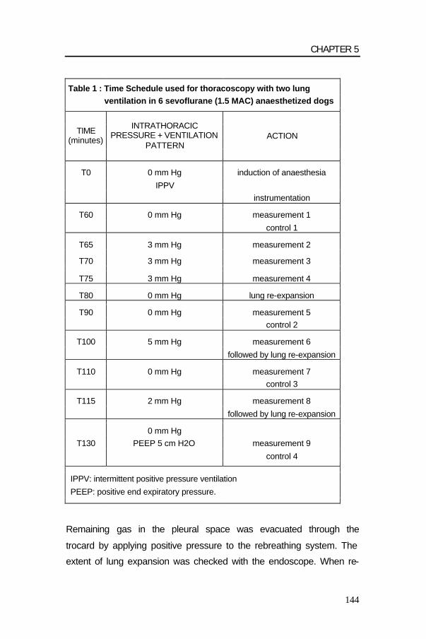
CHAPTER 5
144
Table 1 : Time Schedule used for thoracoscopy with two lung ventilation in 6 sevoflurane (1.5 MAC) anaesthetized dogs
TIME (minutes)
INTRATHORACIC PRESSURE + VENTILATION
PATTERN ACTION
T0 0 mm Hg induction of anaesthesia
IPPV instrumentation
T60 0 mm Hg measurement 1
control 1
T65 3 mm Hg measurement 2
T70 3 mm Hg measurement 3
T75 3 mm Hg measurement 4
T80 0 mm Hg lung re-expansion
T90 0 mm Hg measurement 5 control 2
T100 5 mm Hg measurement 6
followed by lung re-expansion
T110 0 mm Hg measurement 7 control 3
T115 2 mm Hg measurement 8
followed by lung re-expansion
0 mm Hg T130 PEEP 5 cm H2O measurement 9
control 4
IPPV: intermittent positive pressure ventilation
PEEP: positive end expiratory pressure.
Remaining gas in the pleural space was evacuated through the
trocard by applying positive pressure to the rebreathing system. The
extent of lung expansion was checked with the endoscope. When re-
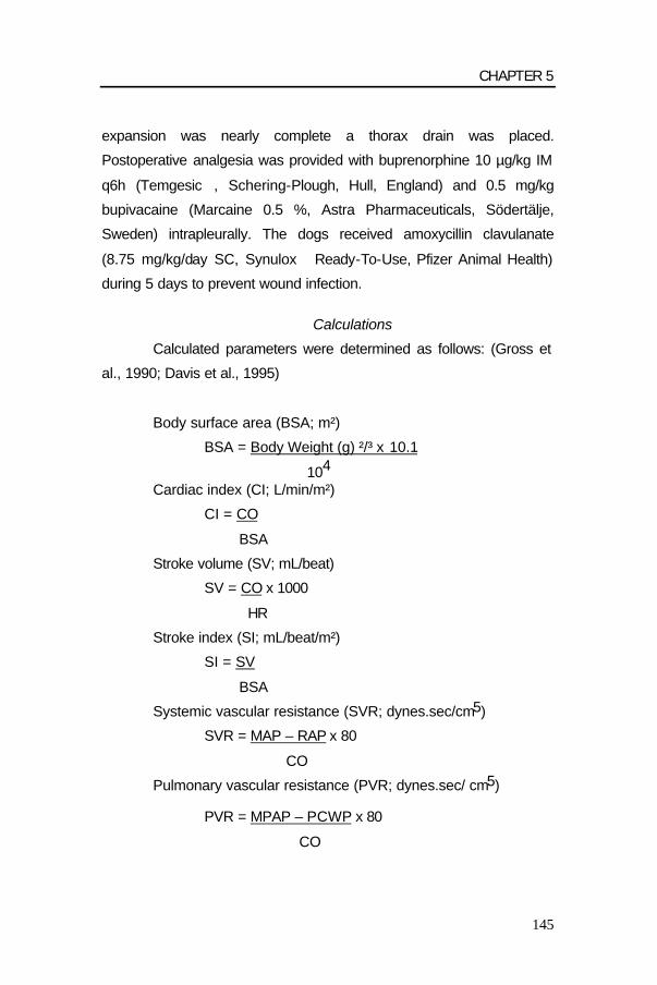
CHAPTER 5
145
expansion was nearly complete a thorax drain was placed.
Postoperative analgesia was provided with buprenorphine 10 µg/kg IM
q6h (Temgesic, Schering-Plough, Hull, England) and 0.5 mg/kg
bupivacaine (Marcaine 0.5 %, Astra Pharmaceuticals, Södertälje,
Sweden) intrapleurally. The dogs received amoxycillin clavulanate
(8.75 mg/kg/day SC, Synulox Ready-To-Use, Pfizer Animal Health)
during 5 days to prevent wound infection.
Calculations
Calculated parameters were determined as follows: (Gross et
al., 1990; Davis et al., 1995)
Body surface area (BSA; m²)
BSA = Body Weight (g) ²/³ x 10.1
104
Cardiac index (CI; L/min/m²)
CI = CO
BSA
Stroke volume (SV; mL/beat)
SV = CO x 1000
HR
Stroke index (SI; mL/beat/m²)
SI = SV
BSA
Systemic vascular resistance (SVR; dynes.sec/cm5)
SVR = MAP – RAP x 80
CO
Pulmonary vascular resistance (PVR; dynes.sec/ cm5)
PVR = MPAP – PCWP x 80
CO
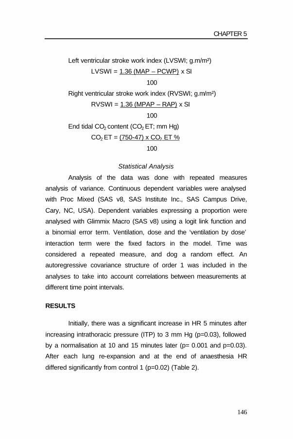
CHAPTER 5
146
Left ventricular stroke work index (LVSWI; g.m/m²)
LVSWI = 1.36 (MAP – PCWP) x SI
100
Right ventricular stroke work index (RVSWI; g.m/m²)
RVSWI = 1.36 (MPAP – RAP) x SI
100
End tidal CO2 content (CO2 ET; mm Hg)
CO2 ET = (750-47) x CO2 ET %
100
Statistical Analysis
Analysis of the data was done with repeated measures
analysis of variance. Continuous dependent variables were analysed
with Proc Mixed (SAS v8, SAS Institute Inc., SAS Campus Drive,
Cary, NC, USA). Dependent variables expressing a proportion were
analysed with Glimmix Macro (SAS v8) using a logit link function and
a binomial error term. Ventilation, dose and the ‘ventilation by dose’
interaction term were the fixed factors in the model. Time was
considered a repeated measure, and dog a random effect. An
autoregressive covariance structure of order 1 was included in the
analyses to take into account correlations between measurements at
different time point intervals.
RESULTS
Initially, there was a significant increase in HR 5 minutes after
increasing intrathoracic pressure (ITP) to 3 mm Hg (p=0.03), followed
by a normalisation at 10 and 15 minutes later (p= 0.001 and p=0.03).
After each lung re-expansion and at the end of anaesthesia HR
differed significantly from control 1 (p=0.02) (Table 2).
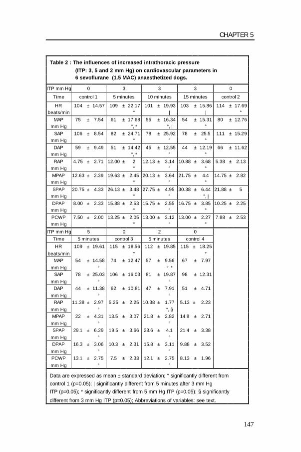
CHAPTER 5
147
Table 2 : The influences of increased intrathoracic pressure
(ITP: 3, 5 and 2 mm Hg) on cardiovascular parameters in 6 sevoflurane (1.5 MAC) anaesthetized dogs.
ITP mm Hg 0 3 3 3 0
Time control 1 5 minutes 10 minutes 15 minutes control 2
HR 104 ± 14.57 109 ± 22.17 101 ± 19.93 103 ± 15.86 114 ± 17.69 beats/min ° | | °
MAP 75 ± 7.54 61 ± 17.68 55 ± 16.34 54 ± 15.31 80 ± 12.76 mm Hg °, * °, | °
SAP 106 ± 8.54 82 ± 24.71 78 ± 25.92 78 ± 25.5 111 ± 15.29 mm Hg ° ° °
DAP 59 ± 9.49 51 ± 14.42 45 ± 12.55 44 ± 12.19 66 ± 11.62 mm Hg °, * ° °
RAP 4.75 ± 2.71 12.00 ± 2 12.13 ± 3.14 10.88 ± 3.68 5.38 ± 2.13 mm Hg ° ° ° MPAP 12.63 ± 2.39 19.63 ± 2.45 20.13 ± 3.64 21.75 ± 4.4 14.75 ± 2.82
mm Hg ° ° ° SPAP 20.75 ± 4.33 26.13 ± 3.48 27.75 ± 4.95 30.38 ± 6.44 21.88 ± 5
mm Hg ° ° °, | DPAP 8.00 ± 2.33 15.88 ± 2.53 15.75 ± 2.55 16.75 ± 3.85 10.25 ± 2.25
mm Hg ° ° ° PCWP 7.50 ± 2.00 13.25 ± 2.05 13.00 ± 3.12 13.00 ± 2.27 7.88 ± 2.53 mm Hg ° ° °
ITP mm Hg 5 0 2 0 Time 5 minutes control 3 5 minutes control 4 HR 109 ± 19.61 115 ± 18.56 112 ± 19.85 115 ± 18.25
beats/min ° ° MAP 54 ± 14.58 74 ± 12.47 57 ± 9.56 67 ± 7.97
mm Hg ° °, * SAP 78 ± 25.03 106 ± 16.03 81 ± 19.87 98 ± 12.31
mm Hg ° ° DAP 44 ± 11.38 62 ± 10.81 47 ± 7.91 51 ± 4.71
mm Hg ° ° RAP 11.38 ± 2.97 5.25 ± 2.25 10.38 ± 1.77 5.13 ± 2.23
mm Hg ° °, § MPAP 22 ± 4.31 13.5 ± 3.07 21.8 ± 2.82 14.8 ± 2.71
mm Hg ° ° SPAP 29.1 ± 6.29 19.5 ± 3.66 28.6 ± 4.1 21.4 ± 3.38
mm Hg ° ° DPAP 16.3 ± 3.06 10.3 ± 2.31 15.8 ± 3.11 9.88 ± 3.52
mm Hg ° ° PCWP 13.1 ± 2.75 7.5 ± 2.33 12.1 ± 2.75 8.13 ± 1.96 mm Hg ° °
Data are expressed as mean ± standard deviation; ° significantly different from
control 1 (p=0.05); | significantly different from 5 minutes after 3 mm Hg
ITP (p=0.05); * significantly different from 5 mm Hg ITP (p=0.05); § significantly
different from 3 mm Hg ITP (p=0.05); Abbreviations of variables: see text.

CHAPTER 5
148
MAP, SAP and DAP decreased significantly when ITP increased
to 3, 5 and 2 mm Hg compared to control 1(p<0.0001). MAP
continued to decrease during the 15-minute period of ITP of 3 mm Hg,
but this was only significantly different between 5 and 10 minutes
(p=0.05). The 5 mm Hg ITP induced the lowest MAP compared to 2
and 3 mm Hg at 5 minutes after ITP elevation (p=0.007 and p=0.04).
There was no significant difference in SAP between 2, 3 and 5 mm Hg
ITP; again 5 mm Hg ITP produced the lowest DAP. Ten minutes after
lung re-expansion and the end of the experiment MAP, SAP and DAP
returned to baseline values (control 1) (Table 2 and Figure 1).
RAP, MPAP, SPAP, DPAP and PCWP increased significantly
during ITP rise to 3, 5 and 2 mm Hg compared to control 1. During 3
mm Hg ITP RAP, MPAP, DPAP and PCWP remained relatively
constant during the entire period. SPAP increased significantly after
15 minutes of ITP 3 mm Hg compared to 5 minutes after pressure
elevation (p=0.03). RAP was significantly lower at 2 mm Hg ITP
compared to 3 mm Hg. MPAP, SPAP, DPAP and PCWP were not
significantly different between the 3 ITP levels. There were no
significant differences in these right heart parameters after lung re-
expansion and at the end of anaesthesia compared to values before
ITP rise (control 1) (Table 2 and Figure 1).
Initially, CO, CI, SV and SI decreased significantly during 3
mm Hg ITP compared to control 1. However, these parameters
increased gradually afterwards. The differences in CO, SV and SI
between 5 and 15 minutes of 3 mm Hg ITP were significant (p=0.04,
p=0.006 and p=0.02). SV and SI were significantly lower at 5 and 2
mm Hg ITP compared to values before ITP elevation (control 1).
There was no significant difference in CO, CI, SV and SI between the
3 ITP levels. On the contrary, these parameters returned to control

CHAPTER 5
149
values (control 1) after ITP elevation to 5 and 2 mm Hg (control 3 and
4). There was a significant overshoot of CO and CI after the first lung
re-expansion (p=0.04), but this was less pronounced after 5 and 2 mm
Hg of ITP. SV and SI returned to baseline (control 1) after lung re-
expansion. At the end of anaesthesia these parameters were not
significantly different from values before starting ITP increase (control
1) (Table 2 continued and Figure 1).
LVSWI decreased significantly after ITP increase to 3, 5 and 2
mm Hg compared to control 1. There were no significant differences in
LVSWI between 5 and 15 minutes in the 3 mm Hg protocol. On the
other hand, RVSWI increased non-significantly at 5 and 2 mm Hg ITP
compared to control 1; RVSWI increased significantly only between 5
and 15 minutes of 3 mm Hg ITP (p=0.03). LVSWI and RVSWI were
not significantly different between the 3 pressure levels and returned
to the start values (control 1) after lung re-expansion and at the end of
anaesthesia (Table 2 continued).
PVR increased significantly after ITP of 3, 5 and 2 mm Hg
compared to control 1 and remained constant during the entire period
of 3 mm Hg ITP. There was no significant difference between the 3
mm Hg ITP levels. PVR decreased to base line values (control 1) after
lung re-expansion and at the end of anaesthesia (Table 2 continued).
Five minutes after ITP 3 mm Hg SVR remained constant, but
decreased significantly after 10 and 15 minutes compared to 5
minutes post and before pressure elevation (control 1) (p<0.0001).
SVR was also significantly lower compared to control 1 during 5 and 2
mm Hg ITP. There was a significant difference between 3 and 5 mm

CHAPTER 5
150
Table 2 (continued) : The influences of increased intrathoracic pressure (ITP: 3, 5 and 2 mm Hg) on cardiovascular parameters in 6 sevoflurane anaesthetized dogs
ITP mm Hg 0 3 3 3 0
Time control 1 5 minutes 10 minutes 15 minutes control 2
CO 2.68 ± 0.38 1.85 ± 0.72 2.07 ± 0.88 2.35 ± 0.81 3.28 ± 0.7 L/min ° ° °, | °
CI 2.70 ± 0.45 1.87 ± 0.64 2.05 ± 0.76 2.33 ± 0.66 3.33 ± 0.91 L/min/m² ° ° ° °
SV 26.11 ± 4.22 16.86 ± 3.87 19.96 ± 5.79 22.49 ± 5.51 28.88 ± 4.89 mL/beat ° ° °, |
SI 26.72 ± 7.71 17.07 ± 4.84 20.02 ± 5.5 22.56 ± 5.19 29.63 ± 8.69 mL/beat/m² ° ° °, |
LVSWI 24.86 ± 10.22 11.50 ± 5.87 12.12 ± 7.62 12.91 ± 6.72 29.38 ± 12.23 g*m/m² ° ° ° RVSWI 2.89 ± 1.09 1.85 ± 0.96 2.26 ± 1.35 3.46 ± 2.58 3.92 ± 2.13 g*m/m² ° ° |
PVR 156.7 ± 37.34 299.3 ± 93.2 293.4 ± 106 290.9 ± 85.9 167.4 ± 39.72 dynes*sec/cm5 ° ° °
SVR 2114 ± 323.7 2118 ± 353 1671 ± 194 1461 ± 267 1831 ± 169.8 dynes*sec/cm5 ° °, | °, | °
SpO2 98 ± 1.69 95 ± 2.14 94 ± 3.07 92 ± 4.75 96 ± 2.39 % ° ° °, |
ITP mm Hg 5 0 2 0 Time 5 minutes control 3 5 minutes control 4 CO 2.40 ± 1.03 3.19 ± 0.55 2.51 ± 0.54 2.91 ± 0.53
L/min ° ° CI 2.37 ± 0.87 3.22 ± 0.67 2.54 ± 0.58 2.92 ± 0.49
L/min/m² SV 21.30 ± 5.62 27.87 ± 3.29 22.39 ± 5.97 25.52 ± 3.72
mL/beat ° ° SI 21.33 ± 5.00 28.49 ± 6.81 23.33 ± 7.52 25.97 ± 5.89
mL/beat/m² ° ° LVSWI 12.47 ± 6.79 25.76 ± 7.91 14.37 ± 5.24 21.00 ± 3.59 g*m/m² ° ° RVSWI 3.34 ± 2.25 3.28 ± 1.43 3.61 ± 1.39 3.46 ± 1.22 g*m/m²
PVR 298.0 ± 77.67 154.2 ± 38.8 305.7 ± 72.7 185.3 ± 37.4 dynes*sec/cm5 ° °
SVR 1440 ± 179.5 1719 ± 102 1501 ± 143 1724 ± 308 dynes*sec/cm5 °, § ° ° °
SpO2 92 ± 4.28 97 ± 1.92 91 ± 4.41 97 ± 1.92 % ° °
Data are expressed as mean ± standard deviation; ° significantly different from control 1 (p=0.05); | significantly different from 5 minutes after 3 mm Hg ITP (p=0.05); § ignificantly different from 3 mm Hg ITP (p=0.05); Abbreviations: see text

CHAPTER 5
151
Hg ITP (p=0.004). SVR values did not return to baseline values
(control 1) after lung re-expansion and at the end of anaesthesia, but
remained significantly lower (Table 2 continued).
SpO2 % decreased significantly during ITP rise of 3, 5 and 2
mm Hg compared to control 1 (p<0.0001) and continued decreasing
significantly between 5 and 15 minutes of ITP of 3 mm Hg. There
were no significant differences between the 3 pressure levels. SpO2 %
returned to normal values (control 1) after lung re-expansion and at
the end of anaesthesia (Table 2 continued).
Fig. 1a : The influences of increased ITP (3, 5 and 2 mm Hg) on PaO2 and cardiovascular parameters in 6 sevoflurane (1.5 MAC) anaesthetized dogs. significantly different from control value 1; significantly different from 5 minutes after 3 mm Hg ITP; significantly different from 5 mm Hg ITP. Values are expressed as mean ± standard deviation.
Blood temperature decreased slightly but significantly in time
(p<0.0001). PCV changed significantly over time with a significant
decrease at the end of anaesthesia (p=0.003). The pH decreased
significantly from control 1 during the whole anaesthesia duration
CHAPTER 5
152
(p<0.0001). There were no significant differences between the 3 ITP
levels. PaCO2 increased significantly during 3, 5 and 2 mm Hg ITP
compared to control 1 (p<0.0001) and continued increasing
progressively during 3 mm Hg ITP. There were no significant
differences between the 3 different ITP levels. PaCO2 decreased after
lung re-expansion. However, PaCO2 at the end of anaesthesia was
significantly higher compared to control 1 (p=0.008) (Table 3).
PaO2 decreased significantly during the 3 pressure levels
(p<0.0001) compared to control 1. The decrease in PaO2 during 3 mm
Hg ITP was significant between 5 and 15 minutes after elevation of
intrathoracic pressure (p=0.02). PaO2 was significantly lower at 5 and
2 mm Hg compared to PaO2 after 5 minutes of 3 mm Hg ITP (p=0.01
and p=0.03). There was no significant difference in PaO2 between 5
and 2 mm Hg ITP elevation. After lung re-expansion and at the end of
anaesthesia PaO2 still remained significantly lower compared to
control 1 (p<0.0001) (Table 3 and Figure 1).
Inspiratory pressure (Pinsp.) in dogs significantly increased
with IPPV and addition of ITP at 3, 5 and 2 mm Hg of CO2 -insufflation.
There were no significant differences between the 3 different ITP
levels. Pinsp. returned to base line values (control 1) after termination
of lung re-expansion of dogs in the 3 and 5 mm Hg ITP group but not
in dogs treated with 5 cm H2O PEEP (p=0.04). There were no
significant changes in TV, CO2 ET % and RR during the entire
protocol. Standard bicarbonate concentration (SBC) decreased
significantly from before ITP elevation (control 1) at 3, 5 and 2 mm Hg.
There were no significant differences between the 3 different pressure
levels. SBC returned to base line value (control 1) after 5 and 2 mm
Hg ITP, but remained low after the longer period of 3 mm Hg ITP
elevation (p=0.008) (Table 3).

CHAPTER 5
153
Table 3 : The influences of increased intrathoracic pressure (ITP: 3, 5 and 2 mm Hg) on cardiovascular parameters in 6 sevoflurane anaesthetized dogs
ITP mm Hg 0 3 3 3 0
Time control 1 5 minutes 10 minutes 15 minutes control 2
BLOODT. 37.24 ± 0.55 36.95 ± 0.69 36.95 ± 0.75 36.91 ± 0.74 36.70 ± 0.77 (°C) PCV 31.01 ± 3.52 32.16 ± 4.69 32.16 ± 4.36 32.20 ± 3.89 31.86 ± 4.19
pH 7.30 ± 0.02 7.23 ± 0.06 7.18 ± 0.05 7.14 ± 0.06 7.23 ± 0.05 ° °, | °, | °
PaCO2 40.38 ± 3.16 50.38 ± 6.19 59.75 ± 13.77 67.13 ± 10.95 47.13 ± 5.51 (mm Hg) ° ° °
PaO2 507 ± 58.37 212 ± 56.31 153 ± 60.66 122 ± 62.26 296 ± 78.64 (mm Hg) ° ° °, | °
RR 12.50 ± 1.85 13.25 ± 2.31 13.25 ± 2.12 14.13 ± 4.05 13.13 ± 2.42 (breaths/min)
TV 336 ± 30.68 338 ± 51.27 359 ± 27.48 346 ± 51.23 341 ± 31.37 (mL)
Pinsp. 9.13 ± 1.55 21.00 ± 2.78 21.38 ± 3.54 22.38 ± 2.00 8.88 ± 2.30 (mm Hg) ° ° ° CO2 % ET 5.53 ± 0.32 5.74 ± 0.97 5.98 ± 1.31 6.24 ± 1.06 5.46 ± 0.41
SBC 19.13 ± 1.46 18.25 ± 1.16 17.88 ± 1.13 17.75 ± 1.58 17.75 ± 1.58
(mEq/L) ° ° ° ° ITP mm Hg 5 0 2 0
Time 5 minutes control 3 5 minutes control 4 BLOODT. 36.78 ± 0.83 36.6 ± 0.82 36.6 ± 0.83 36.45 ± 0.92
(°C) PCV 32.28 ± 4.66 33.01 ± 4.6 29.96 ± 1.81 30.39 ± 2.32
° pH 7.17 ± 0.05 7.24 ± 0.04 7.17 ± 0.04 7.23 ± 0.07
° ° ° ° PaCO2 61.38 ± 7.11 47.5 ± 6.23 60.29 ± 5.82 50.43 ± 7.76
(mm Hg) ° ° ° PaO2 131 ± 63.94 334 ± 116.9 150 ± 95.75 328 ± 109.1
(mm Hg) °, | ° °, | ° RR 14.25 ± 5.09 13.13 ± 2.42 14.13 ± 2.47 13.38 ± 2.92
(breaths/min) TV 324 ± 100.3 348 ± 40.27 358 ± 30.59 330 ± 37.03
(mL) Pinsp. 22.50 ± 2.88 9.38 ± 2.26 21.63 ± 2.77 11.50 ± 3.02
(mm Hg) ° ° ° CO2 % ET 5.71 ± 0.52 5.41 ± 0.41 5.51 ± 0.94 5.73 ± 0.66
SBC 17.88 ± 1.46 18.75 ± 1.58 18.00 ± 1.41 18.71 ± 1.8
(mEq/L) ° °
Data are expressed as mean ± standard deviation; ° significantly different from control 1 (p=0.05); | significantly different from 5 minutes after 3 mm Hg ITP (p=0.05); Abbreviations of variables: see text.

CHAPTER 5
154
DISCUSSION
Minimally invasive surgery techniques, including thoracoscopy
are quickly gaining ground in human and veterinary medicine. Until
recently, few studies were available about the applicable ventilation
techniques for anaesthesia of dogs, pigs, sheep and horses during
thoracoscopy (Fujita et al., 1993; Jones et al., 1993; Faunt et al.,
1998; Vachon and Fischer, 1998; Peroni et al., 2000). Thoracoscopy
requires general anaesthesia in dogs, sheep and pigs, whereas the
technique can be performed in sedated standing horses. The basic
procedure involves the introduction of a rigid or flexible fibreoptic
endoscope into the thoracic cavity, and the use of air or another gas
to create a pneumothorax allowing visualization of intrathoracic
structures. If the animals are placed in lateral recumbency, it is
necessary to collapse the upper lung. This can be achieved by two
possible ventilation techniques: one lung ventilation (OLV) with
passive lung collapse or two lung ventilation (TLV) with active lung
collapse by gas insufflation.
During OLV selective ventilation of one lung is applied for
which specific intubation including double-lumen tubes, endobronchial
intubation or bronchial blockers is necessary. Positioning is technically
difficult in dogs and sheep (Muneyuki et al., 1983; Fujita et al., 1993;
Cantwell et al., 2000). Due to malpositioning (up to 30 %) in men
routine bronchoscopy after double-lumen tube or bronchial blocker
placement is certainly recommended. However, bronchoscopy is
expensive, time consuming and not universally available (Kleine et al.,
1998). Another technical problem is the limited use of commercially
available tube systems for animals with smaller body weights such as
cats and toy breed dogs. Furthermore, the potentially more
CHAPTER 5
155
pronounced cardiopulmonary impact of OLV and TLV with gas
insufflation in animals with smaller body weights has not yet been
investigated.
Abdominal insufflation with gas is routine for laparoscopic
procedures to produce the required viewing space. This insufflation
has certainly adverse cardiopulmonary effects. The effects vary with
the gas used, the presence or absence of mechanical ventilation, and
the pressure and duration of insufflation (Johannsen et al., 1989;
Windberger et al., 1994). With TLV for thoracoscopy insufflating the
thorax minimally enhances the optical cavity; however, it must be
applied with extreme caution. TLV with gas insufflation is seldom used
in human anaesthesia for thoracoscopy because of occurring
haemodynamic impairment (Brock et al., 2000).
Both ventilation techniques induce hypoxaemia since one lung
is entirely collapsed and regions of atelectasis develop in the
ventilated lung (Cohen et al., 1988). This induces ventilation-perfusion
mismatches due to intra-pulmonary shunting. However, active
vasoconstrictive mechanisms in the non-ventilated lung reduce the
blood flow and minimize the shunt. This is the so-called hypoxic
pulmonary vasoconstriction reflex. Several techniques can be applied
to improve oxygenation during thoracoscopy: selective continuous
airway pressure (CPAP) to the non-ventilated lung, positive end
expiratory pressure (PEEP) to the ventilated lung, a combination of
both or the use of high frequency ventilation to the non-dependent
lung (Alfery et al., 1981; Benumof, 1982; Nakatsuka et al., 1988; Fujita
et al., 1993).

CHAPTER 5
156
Until now only two studies described TLV with gas insufflation
in smaller animals. Faunt et al. (1998) examined the cardiopulmonary
effects of TLV with N2O insufflation for thoracoscopy in isoflurane
anaesthetized dogs. In this study intrathoracic pressures were not
measured, but were stated to be below 10 mm Hg. TLV with sustained
pneumothorax was well tolerated in these clinically healthy dogs. In
another study the effects on haemodynamic parameters of 5, 10 and
15 mm Hg of CO2 insufflation during TLV were evaluated in isoflurane
anaesthetized pigs (Jones et al., 1993). Routinely used positive
pressure insufflation during thoracoscopy was not recommended
because of the significant haemodynamic compromise in this
experiment.
In the present study the cardiopulmonary effects of varying
degrees of intrathoracic pressure elevation after CO2-insufflation
during 1.5 MAC sevoflurane anaesthesia in continuous two-lung
ventilated dogs were examined. Conventional intubation was chosen
to avoid the laborious difficulties of endobronchial intubation or
bronchial blocker placement and to skip the need for specialized
equipment. Three different levels of intrathoracic pressure elevation of
the left hemi-thorax (3, 5 and 2 mm Hg) after CO2-insufflation were
evaluated during IPPV. The intrathoracic pressure level that induced a
sufficient visualisation into the thorax and the least pronounced
cardiopulmonary side effects was tested, since only one short
communication about the maximum intrathoracic pressure increase (3
to 10 mm Hg) during thoracoscopy in dogs is available (Daly et al.,
1999).
Inhalation anaesthetics including sevoflurane induce a dose-
dependent cardiopulmonary depression in all animals (Bernard et al.,

CHAPTER 5
157
1992; Aida et al., 1996; Grosenbaugh and Muir, 1998). The MAC of
sevoflurane in dogs has been established to be 2.36 volume %
(Kazama and Ikeda, 1988). One and a half MAC is generally accepted
as the standard to allow most surgical interventions. However, some
cases require a higher MAC when no supplementary analgesics or
related drugs are administered during anaesthesia. Furthermore,
several studies showed that 4% sevoflurane does not inhibit the
hypoxic pulmonary vasoconstriction reflex in dogs (Domino et al.,
1986; Okutomi and Ikeda, 1990). This specific characteristic might be
justified in the present study because of the occurring ventilation-
perfusion mismatches.
Fentanyl and propofol are relatively fast acting and short
lasting agents. Interactions of these drugs after the instrumentation
period of one hour with sevoflurane protocol were not likely to be
present. However, although these drugs have a short half-life, it could
not be excluded that their interfering effects were of longer duration.
On the other hand, droperidol has relatively long lasting effects. A
possible influence of droperidol can therefore not be ruled out
(Bissonnette et al., 1999).
A fluid rate of 4 to 8 ml/ kg/ h is generally accepted for the
maintenance of a stable water balance during anaesthesia (Giesecke
and Egbert, 1985). No fluids were administered in the present study.
Nevertheless, the thermodilution method includes the administration
of different boli of saline. Overall, an estimated quantity of 2 to 4 ml/
kg/ h was administered during the whole experimental period. The
influences of this relatively low fluid administration in this study were
probably neglectable.

CHAPTER 5
158
In the present study all direct cardiac parameters (blood
pressures, CO, SV, SI, LVSWI) initially decreased significantly during
ITP increase to 3, 5 and 2 mm Hg. Afterwards, there was a gradual
correction of these parameters probably induced by the occurring
hypercapnia (Walley et al., 1990). After lung re-expansion there was a
clear overshoot in cardiac output and cardiac index related to the
increased HR at that moment, since SV remained constant during this
period. Heart rate, on the other hand, increased at the start of ITP
increase at every level and increased even more after lung
insufflation. The tachycardia could result from a sympathetic
stimulation induced by a stress response related to the increase in
PaCO2 and/ or from stimulation of the baroreceptor-reflex due to a
dose dependent decrease in blood pressure induced by sevoflurane in
dogs (Cullen and Eger, 1974; Mutoh et al., 1997). These findings were
in contrast with the results of Faunt et al. (1998). In this study an
increase in CI, stroke volume index, MPAP and total peripheral
vascular resistance was observed. Jones et al. (1993) and Daly et al.
(1999), reported decreases in CO and arterial blood pressure in an
analogue protocol, while HR remained relatively constant. The
decrease in direct cardiac parameters in our study could be
subsequent to the reduced venous return due to increased
intrathoracic pressure, as described in humans, and/ or to a
decreased myocardial contractility induced by sevoflurane
anaesthesia (Mutoh et al., 1997; Brock et al., 2000; Polis et al., 2001).
The reduced venous return is comparable with the one induced by a
tension pneumothorax (Conolly, 1993; Light, 1994).
Right heart parameters (RAP, PAP, PCWP) and PVR
increased significantly during intrathoracic pressure elevation at every
level compared to values before CO²-insufflation in the present study.
This was consistent with previous studies (Jones et al., 1993; Faunt et
CHAPTER 5
159
al., 1998). In contrast with the other right heart parameters, RVSWI
decreased significantly during the first 10 minutes of 3 mm Hg ITP and
then increased significantly after 15 minutes compared to 5 minutes
after ITP rise. RVSWI increased at 5 and 2 mm Hg IT pressure
compared to before CO2-insufflation. The initial decrease in RVSWI is
difficult to explain, since an increase was expected. This finding might
be related to the position of the Swan-Ganz catheter either in the left
or the right lung. The exact position of the catheter in the lung
(ventilated or collapsed) was not checked. The increase in RAP, PAP,
PCWP and PVR can be explained by the rise in pulmonary tissue
pressure that leads to a decrease in pulmonary perfusion after CO2-
insufflation, as well as by hypoxic vasoconstriction in the collapsed
pulmonary parenchyma (Ohtsuka, 1999). Large increases in
intrathoracic pressure are presumed to decrease venous return and
increase pulmonary vascular pressures so that stroke work and
cardiac output are compromised, leading to hypotension and
hypoperfusion (Lenaghan et al., 1969; Conolly, 1993).
SVR decreased significantly after CO2-insufflation and
remained low during the entire procedure. This could be partly
explained by the vasodilating properties of sevoflurane, but also by
the occurring hypercapnia during thoracoscopy. In a previous study
SVR remained constant in 1.5 MAC sevoflurane anaesthetized dogs.
A significant decrease in SVR was observed only at 2 MAC
sevoflurane (Polis et al., 2001). The decreased SVR is consistent with
the study of Faunt et al. (1998) where a small decrease in SVR was
found. Jones et al. (1993) observed a constant SVR at low ITP (5 mm
Hg), an increased SVR at 10 mm Hg and a decreased SVR at 15 mm
Hg.
SpO2 and PaO2 decreased significantly after CO2-insufflation,
whereby the decrease was more rapid and pronounced after

CHAPTER 5
160
consecutive ITP elevations compared to the first pressure rise. SpO2
returned quickly to base line value after lung insufflations, but PaO2
remained significantly lower even at the end of anaesthesia. This was
also reported in analogue studies (Faunt et al., 1998; Daly et al.,
1999). The phenomenon might be explained by an increased amount
of blood flow due to the effect of gravity in the underlying lung, while
its lung volume is decreased by compression of the mediastinal
weight. This results in inefficient oxygenation of circulating blood due
to ventilation/perfusion mismatch and blood shunting.
PaCO2 increased significantly during CO2-insufflation and
continued to increase during the 15-minute period of intrathoracic
pressure of 3 mm Hg despite constant ventilator settings. The
increase could be related to the induced capnothorax (Peden and
Prys-Roberts, 1993). In the study of Faunt et al. (1998) PaCO2
increased also, although insufflation was done with N2O, there was
found no evidence of N2O resorption from the thorax. Hypercapnia
causes acidemia, resulting in decreased pH. In the present study the
calculated P(a-A) CO2 -gradient did not increase. The use of
capnography and pulse oximetry is certainly recommended during
thoracoscopic interventions because indirect moment-to-moment
indications of PaCO2 and haemoglobin saturation with oxygen are
provided.
In conclusion, thoracoscopic procedures in sevoflurane (1.5
MAC) anaesthetized dogs with low pressure (2 mm Hg) CO2-
insufflation into one hemithorax allows safe visualization of the
intrathoracic space for short periods. The thoracoscopic procedure
should be accomplished in one short episode of CO2-insufflation since
additional insufflation periods can lead to more rapidly occurring and
more pronounced cardiopulmonary depression.

CHAPTER 5
161
REFERENCES
Aida, H., Y. Mizuno, S. Hobo, K. Yoshida, and T. Fujinaga, 1996: Cardiovascular and pulmonary effects of sevoflurane anesthesia in horses. Veterinary Surgery 25, 164-170. Alfery, D.D., J.L. Benumof, and F.R. Trousdale, 1981: Improving oxygenation during one-lung ventilation in dogs: The effects of positive end-expiratory pressure and blood flow restriction to the nonventilated lung. Anesthesiology 55, 381-385. Benumof, J.L., 1982: One-lung ventilation: Which lung should be PEEPed? Anesthesiology 56, 161-163. Benumof, J.L., 1993: The position of a double-lumen tube should be routinely determined by fibreoptic bronchoscopy. Cardiothoracic Vascular Anesthesia 7, 513-514. Bernard, J.-M., M.-F. Doursout, P.F. Wouters, C.J. Hartley, R.G. Merin, and J.E. Chelly, 1992: Effects of sevoflurane and isoflurane on hepatic circulation in the chronically instrumented dog. Anesthesiology 77, 541-545. Bissonnette, B., H. Swan, P. Ravussin, and V. Un, 1999: Neuroleptanesthesia : current status. Canadian Journal of Anaesthesiology 46, 154-168. Brock, H., R. Rieger, C. Gabriel, W. Pölz, W. Moosbauer, and S. Necek, 2000: Haemodynamic changes during thoracoscopic surgery. The effects of one-lung ventilation compared with carbon dioxide insufflation. Anaesthesia 55, 10-16.
Cantwell, S.L., T. Duke, P.J. Walsh, A.M. Remedios, D. Walker, and J.G. Ferguson, 2000: One-lung versus two-lung ventilation in the closed-chest anesthetized dog: A comparison of cardiopulmonary parameters. Veterinary Surgery 29, 365-373.
Cohen, E., J.B. Eisenkraft, D.M. Thys, P.A. Kirschner, and J.A. Kaplan, 1988: Oxygenation and hemodynamic changes during one-lung ventilation: effects of CPAP10, PEEP10, and CPAP10/PEEP10. Journal of Cardiothoracic
Anesthesia 2, 34-40.
Connolly, J.P., 1993: Hemodynamic measurements during a tension pneumothorax. Critical Care Medicine 21, 294-296. Cullen, D.J., and E.I.II Eger, 1974: Cardiovascular effects of carbon dioxide in man. Anesthesiology 41, 345. Daly, C., K.M. Tobias, A. Tobias, and R. Keegan, 1999: Effect of intrathoracic insufflation on cardiac output, heart rate, arterial and central venous

CHAPTER 5
162
pressures, and arterial oxyhemoglobin saturation. Scientific Presentation Abstracts, 9th Annual ACVS Symposium, Sept. 30- Oct. 3, San Francisco. Davis, P.D., G.D. Parbrook, and G.N.C. Kenny, 1995: Measurement of (H+) and CO2. In: Basic Physics and measurement in anaesthesia. 4th Ed. Butterworth-Heinemann, Oxford. Domino, K.B., L. Borowec, C.M. Alexander, J.J. Williams, L. Chen, C. Marshall, and B.E. Marshall, 1986: Influence of isoflurane on hypoxic pulmonary vasoconstriction in dogs. Anesthesiology 64, 423-429. Faunt, K.K., L.A. Cohn, B.D. Jones, and J.R. Dodam, 1998: Cardiopulmonary effects of bilateral hemithorax ventilation and diagnostic thoracoscopy in dogs. American Journal of Veterinary Research 59 (11), 1494-1498. Fujita, Y., T. Yamasaki, M. Takaori, and K. Sekioka, 1993: Sevoflurane anaesthesia for one-lung ventilation with PEEP to the dependent lung in sheep: effects on right ventricular function and oxygenation. Canadian Journal of Anaesthesiology 40 (12), 1195-1200. Giesecke, A.H., and L.D. Egbert, 1985: Perioperative fluid therapy-crystalloids. In: Anesthesia, ed. R. Miller, pp. 1313-1328. Churchill Livingstone, New York. Grosenbaugh, D.A., and W.W. Muir, 1998: Cardiorespiratory effects of sevoflurane, isoflurane, and halothane anesthesia in horses. American Journal of Veterinary Research 59, 101- 106. Gross, M.E., W.J. Tranquilli, J.C. Thurmon, G.J. Benson, and W.A. Olson, 1990: Hemodynamic effects of intravenous Midazolam-Xylazine-Butorphanol in dogs. Veterinary Surgery 19, 173-180. Ishibe, Y., X. Gui, H. Uno, Y. Shiokawa, T. Umeda, and K. Suekane, 1993: Effects of sevoflurane on hypoxic pulmonary vasoconstriction in the perfused rabbit lung. Anesthesiology 79, 1348-1353. Ishizaki, Y., Y. Bandai, K. Shimomura, H. Abe, Y. Ohtomo, and Y. Idezuki, 1993: Safe intraabdominal pressure of carbon dioxide pneumoperitoneum during laparoscopic surgery. Surgery 114, 549-554.
Johannsen, G., M. Andersen, and B. Juhl, 1989: The effect of general anaesthesia on the haemodynamic events during laparoscopy with CO2 insufflation. Acta Anaesthesiologica Scandinavica 33, 132-136. Jones, D., G. Graeber, G. Tanguilig, G. Hobbs, and G.F. Murray, 1993: Effects of insufflation on hemodynamics during thoracoscopy. Annales of Thoracic Surgery 55, 1379-1382.
CHAPTER 5
163
Kazama, T., and K. Ikeda, 1988: Comparison of MAC and the rate of rise of alveolar concentration of sevoflurane with halothane and isoflurane in the dog. Anesthesiology 68, 435-437. Kleine, U., W. Karzai, F. Bloos, M. Wohlfarth, R. Gottschall, H. Fritz, M. Gugel, and A. Seifert, 1998: Role of fibreoptic bronchoscopy in conjunction with the use of double-lumen tubes for thoracic anaesthesia. A prospective study. Anesthesiology 88, 346-350. Lenaghan, R., Y.J. Silva, and A.J. Walt, 1969: Hemodynamic alterations associated with expansion rupture of the lung. Archives of Surgery 99, 339-343. Light, R.W., 1994: Tension pneumothorax. Internal Care Medicine 20, 468-469. Marchandise, F.X., O. Vandenplas, J. Wallon, and C. Francis, 1993 : Thoracoscopy in the diagnosis and management of chronic pleural effusions. Acta Clinica Belgica 48, 5-10. Miller, J.I.Jr., 1993: The present role and future considerations of video-assisted thoracoscopy in general thoracic surgery. Annales of Thoracic Surgery 56, 804-806. Muneyuki, M., K. Konishi, R. Horiguchi, S. Tsujimoto, M. Saito, S. Sakakura, and A. Konishi, 1983: Effects of altering lung ventilation on cardiopulmonary functions in dogs. Anesthesiology 58, 353-356. Mutoh, T., R. Nishimura, H.-Y. Kim, S. Matsunaga, and N. Sasaki, 1997: Cardiopulmonary effects of sevoflurane, compared with halothane, enflurane, and isoflurane, in dogs. American Journal of Veterinary Research 58, 885-890. Nakatsuka, M., L. Wetstein, and R. Keenan, 1988: Unilateral high-frequency jet ventilation during one-lung ventilation for thoracotomy. Annales of Thoracic Surgery 46, 654-660. Ohtsuka, T., K. Imanaka, M. Endoh, T. Kohno, J. Nakajima, Y. Kotsuka, and S. Takamoto, 1999: Hemodynamic effects of carbon dioxide insufflation under single-lung ventilation during thoracoscopy. Annales of Thoracic Surgery 68, 29-33. Okutomi, T., and K. Ikeda, 1990: Sevoflurane has no inhibitory effect on hypoxic pulmonary vasoconstriction (HPV) in dogs. Journal of Anesthesiology 4, 123-130. Peden, C.J., and C. Prys-Roberts, 1993: Capnothorax: implications for the anaesthetist. Anaesthesia 48, 664-666.

CHAPTER 5
164
Peroni, J.F., N.E. Robinson, J.A. Stick, and F.J. Derksen, 2000: Pleuropulmonary and cardiovascular consequences of thoracoscopy performed in healthy standing horses. Equine Veterinary Journal 32, 280-286. Perrault, L.P., J. Gregoire, and A. Page, 1993: Video-assisted thoracoscopy and thoracic surgery: the first 50 patients. Annales de Chirurgie 47, 838-843. Polis, I., F. Gasthuys, H. Laevens, L. Van Ham, and A. De Rick (2001): The influence of ventilation mode (spontaneous ventilation, IPPV and PEEP) on cardiopulmonary parameters in sevoflurane anaesthetized dogs. Journal of Veterinary Medicine A. 48, 619-630. Rodgers, B.M., and J.L. Talbert, 1976: Thoracoscopy for diagnosis of intrathoracic lesions in children. Journal of Pediatric Surgery 11, 703-708. Smith, G.B., N.P. Hirsch, and J. Ehrenwerth, 1986: Placement of double-lumen endobronchial tubes. British Journal of Anaesthesiology 58, 1317-1320. Tanguilig, G.G., D.R. Jones, and G.M. Graeber, 1993: Video-assisted thoracoscopy: a major advance in diagnosis and treatment of intrathoracic pathology. West Virginia Medical Journal 89, 230-232. Toy, F.K., R.T.Jr. Smoot, 1992: Preliminary experience with thoracoscopic surgery. Journal of Laparoendoscopic Surgery 2, 303-309. Vachon, A.M., and A.T. Fischer, 1998: Thoracoscopy in the horse: diagnostic and therapeutic indications in 28 cases. Equine Veterinary Journal 30, 467-475. Walley, K., T. Lewis, and L. Wood, 1990: Acute respiratory acidosis decreases left ventricular contractility but increases cardiac output in dogs. Circulation Research 67, 628-635. Windberger, U., H. Siegl, and R. Woisetschager, 1994: Hemodynamic changes during laparoscopic surgery. European Surgical Research 26, 1-9.

INTRODUCTION TO CHAPTERS 6/ 7/ 8
165
INTRODUCTION TO CHAPTERS 6/ 7/ 8

INTRODUCTION TO CHAPTERS 6/ 7/ 8
166
INTRODUCTION TO CHAPTERS 6/ 7/ 8
167
The use of sevoflurane is still rather expensive for veterinary
practice. Therefore, methods inducing a decreased consumption of
inhalant anaesthetic agent can be indicated to reduce anaesthetic
costs. This can be achieved by reduction of fresh gas flows and/or by
combination of inhalation anaesthesia with analgesic drugs. A
“balanced anaesthesia” regimen combining potent opioids with
sevoflurane should result in a MAC reducing effect and less
anaesthetic agent. During the last decade, treatment of animal pain,
its recognition, alleviation and subsequent prevention has gained
increasing attention. This trend provides a relevant argument in
support of adequate pain relief and the application of “balanced
anaesthesia” techniques in general practice.
In order to obtain a method for beneficial cost optimisation and
adequate pain relief when using sevoflurane, premedication with a
long-acting sufentanil formulation was investigated. Hence, 40 dogs
were enclosed in a study combining sevoflurane with sufentanil LA
administered at different time intervals before induction of
anaesthesia. The emphasis in this study was put on the
antinociceptive and sedative effects of sufentanil LA and the dosage
reducing effect on sevoflurane anaesthesia in dogs, since “pre-
emptive analgesia” is nowadays considered as the keystone for post-
operative pain relief (Chapter 8). In addition, potential influences on
cardio-respiratory parameters when using this drug combination were
evaluated in chapter 7. The administration of a long-acting formulation
of sufentanil should facilitate pre- per- and post-operative analgesia in
veterinary practice, because a single intramuscular injection could
provide satisfactory pain relief during 24 hours.

INTRODUCTION TO CHAPTERS 6/ 7/ 8
168
To perform the study repeated blood pressure measurements
and arterial blood sampling were necessary. Multiple femoral artery
punctures were unfeasible and therefore catheter and vascular access
port implantation was imposed (Chapter 6). Two possible techniques
were considered: conventional externalised or totally implantable
catheter systems. Conventional externalised catheter systems have a
rate of complications that limits its clinical and animal experimental
use. These complications have led to the development of the totally
implantable catheter system. The system consists of a titanium port
and a poly-urethane catheter. The port is implanted subcutaneously
and can be accessed by a hypodermic needle. The totally implantable
system gives only limited infection risks compared to the use of
externalised catheters and was therefore preferred in our study.
169
CHAPTER 6
ARTERIAL CATHETERISATION AND VASCULAR ACCESS PORT
IMPLANTATION FOR BLOOD SAMPLING AND CONTINUOUS
BLOOD PRESSURE MEASUREMENT IN DOGS.
I. Polis1, Y. Moens2, F. Gasthuys3, M. Tshamala1
1Department of Small Animal Medicine and Clinical Biology, Faculty of Veterinary Medicine, Ghent University, Salisburylaan 133, 9820 Merelbeke, Belgium; 2Department of Clinical Veterinary Sciences, Section Anaesthesiology, Faculty of Veterinary Medicine, University of Berne, Länggasstrasse 124, CH-3012 Berne, Switserland; 3Department of Surgery and Anaesthesia of Domestic Animals, Faculty of Veterinary Medicine, Ghent University, Salisburylaan 133, 9820 Merelbeke, Belgium

CHAPTER 6
170
CHAPTER 6
171
SUMMARY
In the present study a modified method using coated
polyurethane catheters into the femoral artery and a titanium vascular
access port (VAP) with a silicone membrane was described in forty
dogs. This device allowed repeated arterial blood pressure
measurement and blood sampling in conscious and anaesthetised
dogs for an average period of 2 weeks. Minor clinical influences nor
catheter extraction were observed. On the other hand, infection with
Pseudomonas aeruginosa induced by a contaminated flush solution
was diagnosed in 4 dogs. These dogs recovered rapidly after an
appropriate antibiotic therapy.
It was concluded that the described arterial catheterisation
technique with vascular access port over a two weeks period was
suitable and technically feasible for experimental protocols in dogs.
INTRODUCTION
Anaesthetic studies in experimental dogs often require
repetitive blood sampling and blood pressure monitoring in
unrestrained animals over a relatively long period of time. The
repetitive puncture of arteries and veins or multiple consecutive
peripheral catheter placement is accompanied by technical problems
and stress responses, but also by iatrogenically induced damage of
the blood vessels including thrombosis and sclerosis (Mesfin et al.,
1988; Bagley and Flanders, 1990; Endres et al., 1990; Grosse-
Siestrup and Lajous-Petter, 1990). Therefore, these methods are only
suitable for the prelevation of a limited number of blood samples.
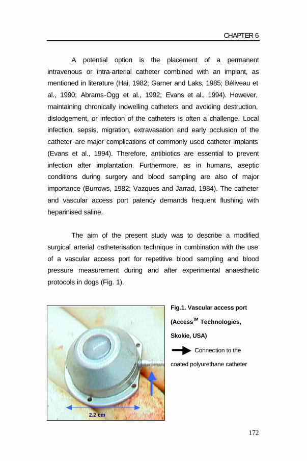
CHAPTER 6
172
A potential option is the placement of a permanent
intravenous or intra-arterial catheter combined with an implant, as
mentioned in literature (Hai, 1982; Garner and Laks, 1985; Béliveau et
al., 1990; Abrams-Ogg et al., 1992; Evans et al., 1994). However,
maintaining chronically indwelling catheters and avoiding destruction,
dislodgement, or infection of the catheters is often a challenge. Local
infection, sepsis, migration, extravasation and early occlusion of the
catheter are major complications of commonly used catheter implants
(Evans et al., 1994). Therefore, antibiotics are essential to prevent
infection after implantation. Furthermore, as in humans, aseptic
conditions during surgery and blood sampling are also of major
importance (Burrows, 1982; Vazques and Jarrad, 1984). The catheter
and vascular access port patency demands frequent flushing with
heparinised saline.
The aim of the present study was to describe a modified
surgical arterial catheterisation technique in combination with the use
of a vascular access port for repetitive blood sampling and blood
pressure measurement during and after experimental anaesthetic
protocols in dogs (Fig. 1).
Fig.1. Vascular access port
(AccessTM Technologies,
Skokie, USA)
Connection to the
coated polyurethane catheter
2.2 cm

CHAPTER 6
173
Materials and Methods
The study was approved by the Ethical committee of the
Faculty of Veterinary Medicine, Ghent University (filenumber:
39/2000). Forty adult female Beagles weighing 11.97 ± 1.40 kg (mean
± SD) from 1 to 2 years old were used in the study. The dogs were
dewormed and vaccinated before the experiment. Clinical and blood
examination one week before and the day of the experiment
confirmed the good health status of the animals. Animals had free
access to tap water and commercial dog food (Advance TM Adult,
Master Foods N.V., Belgium).
Anaesthetic protocol
The dogs were fasted 12 hours before surgical intervention.
Acepromazine (0.05 mg/kg Placivet® 2%, Codifar N.V., Wommelgem,
Belgium) was administered intramuscularly as premedication.
Anaesthesia was induced 30 minutes later using propofol (4-6 mg/kg
Rapinovet®, Mallinckrodt Veterinary, Aalst, Belgium) administered
intravenously to effect. The dogs were intubated (endotracheal tube 6
mm ID, Rüsch, Germany) and connected to a commercial anaesthetic
machine (Titus, Dräger, Lübeck, Germany) with a circle system
delivering 1 L/min of oxygen. The dogs breathed spontaneously
throughout the surgical procedure. An anaesthetic gas analyser
(Capnomac Ultima, Datex Engstrom Instrumentation Corp., Helsinki,
Finland) and pulse oximeter (N-20PA Portable Pulse Oximeter,
Nellcor Puritan Bennett Inc., Pleasanton, CA, U.S.A.) were used for
non-invasive monitoring. No intravenous infusions were administered
during anaesthesia.
Anaesthesia was maintained with propofol administered
intravenously by an infusion pump (Ohmeda 9000 Syringe pump®,

CHAPTER 6
174
Ohmeda, West Yorkshire, UK) at a dosage of 0.5 mg/kg/min.
Analgesia and anaesthesia of the hind quarters was assured with an
epidural technique using a 50/50 (v/v) mixture of lidocain 2%
(Xylocaine® 2%, Astra Zeneca, Brussels, Belgium) and bupivacain
0.5% (Marcaine® 0.5%, Astra Zeneca, Brussels, Belgium) at a dosage
of 1 ml/4 kg of body weight. Two dogs received half of the epidural
dosage because the subarachnoidal space was punctured. For
additional postoperative analgesia, the dogs were treated with
carprofen (4 mg/kg PO) during 3 days, starting the day before surgery.
The dog received a single injection of amoxicillin long acting (15
mg/kg SC) (Duphamox LA, Fort Dodge Animal Health Benelux,
Brussel, Belgium) the day of catheter implantation.
Surgical procedure
After the dogs had been placed in left lateral recumbency, the
right inguinal area, the medial side of the right knee and the backside
of the dogs were surgically prepared. A 60 cm coated polyurethane
catheter (Hydrocoat TM Catheter 3.5 Fr; Access TM Technologies,
Skokie, USA) with a bead at 8 cm from the catheter tip and filled with
heparinised saline (5 IU/ ml of Heparine®, Leo, Belgium) was used for
arterial catheterisation. The catheter tip was transected obliquely and
was grasped with a forceps (Semkin dressing forceps) for better
handling. The catheter was occluded at the free end with a
haemostatic clamp to prevent bleeding when inserting it to the femoral
artery. A 4 cm incision was made through skin and subcutis above the
femoral groove. The femoral artery was exposed by separation of the
sartorius and gracilis muscles starting in the femoral triangle. The
artery was freed from the femoral nerve and vein by blunt dissection
(Fig. 2). Hence, the femoral artery was elevated and occluded during
the surgical handling by gentle pulling on 2 sutures (Safil® green 3/0,
B/Braun, Melsungen, Germany) positioned under the artery (Fig. 3).

CHAPTER 6
175
Topical lidocain was administered to counteract the induced iatrogenic
vasospasm of the femoral artery during handling. A small hole was
made into the artery with a 19 gauge needle (19 G x 1”; 1.1 x 25 mm;
Terumo Europe N.V., Leuven, Belgium) and the catheter tip was
inserted into the artery towards the aorta for about 8 cm (Fig. 4). The
proximal suture was used to fix the artery around the inserted catheter
(Fig. 5). An additional suture was placed immediately behind the bead
to assure an adequate ligation of the artery and the distal suture was
used to ligate the femoral artery to the bead.
Fig.2. Surgical exposition
of the femoral artery
1/ femoral vein and nerve
2/ femoral artery
Fig.3. Elevation of the
femoral artery by
2 stay sutures
1
2

CHAPTER 6
176
Fig.4. Insertion of the
catheter into the
femoral artery
Fig.5. Ligation of the
femoral artery around the
inserted catheter
A second skin incision was made medial of the knee. The
catheter was pulled subcutaneous from the femoral triangle to the
knee with an atraumatic forceps (straight Rochester-Carmalt
Haemostatic forceps). The catheter was secured using a single
subcutaneous suture at the medial level of the knee. Finally a
paramedian incision was made proximal from the ilium on the back of
the dog. A small amount of subcutaneous fat was prelevated allowing
bead
femoral artery
proximal suture
bead

CHAPTER 6
177
3
2
1
the formation of a suitable pocket for the titanium vascular access port
(Access TM Technologies, Skokie, USA). The catheter was tunnelled
further subcutaneously from the knee to the backside of the dog with
the same atraumatic forceps (Fig. 6). The vascular port was attached
to the catheter allowing enough length for a so called tension loop
around the port (Fig. 7-8). Catheter patency was tested using
heparinised saline and a Huber point needle (Posi-Grip Huber Point
Needle 22 gauge x 0-3/4’’, Access TM Technologies, Skokie, USA)
placed through the silicone membrane of the vascular access port.
The vascular access port was sutured with 3 single sutures (Safil®
green 3/0, B/Braun, Melsungen, Germany) into the subcutaneous
tissue.
After a final control of permeability, the catheter was filled with
heparinised saline (200 IU/ml) and all skin incisions were closed with
Safil® (fig. 9). The catheters and implants were removed 17.9 ± 8.7
days after implantation (mean ± SD) under a standardised isoflurane
anaesthesia.
Fig. 6. Subcutaneous tunneling from the medial knee incision to the lumbar incision
1/ Subcutaneous tunnelling
2/ Top of the atraumatic forceps
3/ Katheter
LATERAL
MEDIAL
KN
EE
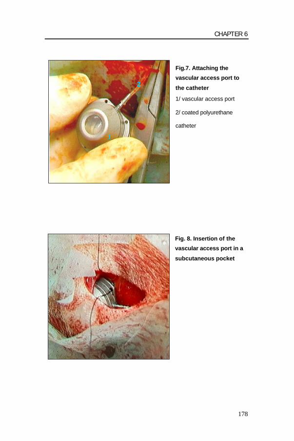
CHAPTER 6
178
Fig.7. Attaching the
vascular access port to
the catheter
1/ vascular access port
2/ coated polyurethane
catheter
Fig. 8. Insertion of the
vascular access port in a
subcutaneous pocket
1
2
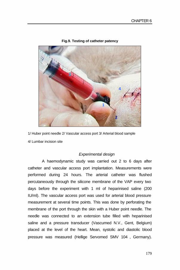
CHAPTER 6
179
Fig.9. Testing of catheter patency
1/ Huber point needle 2/ Vascular access port 3/ Arterial blood sample
4/ Lumbar incision site
Experimental design
A haemodynamic study was carried out 2 to 6 days after
catheter and vascular access port implantation. Measurements were
performed during 24 hours. The arterial catheter was flushed
percutaneously through the silicone membrane of the VAP every two
days before the experiment with 1 ml of heparinised saline (200
IU/ml). The vascular access port was used for arterial blood pressure
measurement at several time points. This was done by perforating the
membrane of the port through the skin with a Huber point needle. The
needle was connected to an extension tube filled with heparinised
saline and a pressure transducer (Vascumed N.V., Gent, Belgium)
placed at the level of the heart. Mean, systolic and diastolic blood
pressure was measured (Hellige Servomed SMV 104, Germany).
1
2
3
4

CHAPTER 6
180
Arterial blood for blood gas analysis was sampled using the same
method.
Clinical observation and examination
A clinical examination was done and rectal temperature of the
dogs was recorded on a daily basis.
RESULTS
Anaesthesia and surgery were uneventful. The surgical
implantation time ranged from 35 to 75 minutes. All 40 dogs tolerated
the sampling procedures (14 samples in 24 hours) well without
external signs of discomfort. Blood sampling and blood pressure
measurement were successful and easy to perform in all dogs.
Several complications were encountered. An inadvertent
implantation of the catheter into the femoral vein was diagnosed in
one dog. This dog had a swelling of the hind leg and venous blood
(PaO2: 33 mm Hg) could be sampled from the vascular access port.
The catheter was removed and a period of one month was respected
before a new catheter could be implanted into the femoral artery of the
same side without problems. Another dog was lame in the surgically
treated hind leg the day of the experiment, however without pain or
swelling. Lameness disappeared spontaneously within 4 days without
therapy. One dog removed the sutures of the inguinal wound without
damaging the arterial catheter. The wound was sutured again under
local anaesthesia with lidocain (Xylocaine 2%, Astra, Brussel,
Belgium). Finally, a seroma occurred around the VAP in one of the
dogs and was surgically treated by placing a Penrose drain (Penrose
Tubing, Sherwood Medical, Tullamore, Ireland) after removal of the

CHAPTER 6
181
VAP. The swelling caused no difficulties for blood sampling or blood
pressure measurement.
Mean rectal temperature of all dogs was 39.3 ± 0.48°C on the
day of the experiment (2 to 6 days after catheter implantation). Four
dogs had fever (more than 40.0°C) one to two days after catheter
implantation. However, on clinical examination no signs of lameness
or inflammation on the catheter site were observed in these dogs.
Samples for bacteriologic examination were taken from the
heparinised flush solution used in these dogs. Contamination of the
solution with Pseudomonas aeruginosa was found by a bacteriologic
culture. An appropriate antimicrobial therapy guided by an antibiogram
was done for 5 to 10 days with enrofloxacine SC (5 mg/kg/day,
Baytril 5%, Bayer, Leverkusen, Germany) and resulted in full clinical
recovery. The arterial catheter and vascular access port of one of
these dogs showed no bacteriological growth after surgical removal.
The catheters remained patent in all animals for at least 4.2 ±
2.2 days (mean ± SD). After the experiment the catheters were not
further flushed in order to have an obstructed catheter by time of
removal 17.9 ± 8.7 days after implantation (mean ± SD). This
facilitated catheter removal, which could be done without an additional
ligation of the femoral artery. After removal of catheters and vascular
access ports skin incisions healed rapidly. No specific problems
induced by the ligation of the femoral artery were encountered over
time.

CHAPTER 6
182
DISCUSSION
In the present study a permanent arterial catheter and totally
implantable vascular access port system were inserted into the
femoral artery of dogs for repetitive arterial blood sampling and
invasive arterial blood pressure measurement at several time intervals
during experimental procedures. The surgical technique was slightly
modified from previously published studies (Garner and Laks, 1985;
Grosse-Siestrup and Lajous-Petter, 1990; Evans et al., 1994).
Coated polyurethane catheters in combination with titanium
vascular access ports were used in the present study. Compared to
polyvinyl chloride, silicone and Teflon catheters, heparinised
polyurethane were proven to be the least thrombogenic of all
materials (Solomon et al., 1987). Moreover, silicone carries a greater
risk for subcutaneous infection and induces a greater inflammation
and kinking risk (Sheretz et al., 1995). In the present study, the
catheter was inserted for about 8 cm into the femoral artery.
Immediately after surgical handling of the artery a marked collapse of
the vessel was observed making the insertion of the catheter difficult.
Topical lidocain was administered to counteract the iatrogenically
induced vasospasm (Wadstrom and Gerdin, 1991; Kim et al., 1996).
To facilitate catheter implantation the catheter tip was cut off obliquely
in combination with an opening in the vessel wall. Efficiency of
catheter placement improved with experience. No problems of the
hind limb vascularisation were observed after occlusion of the distal
part of the femoral artery. Most likely a sufficient amount of collateral
vessels assured an adequate circulation of the hind limb rapidly
(Schaper and Ito, 1996). All catheters remained patent during the
experiment. The incision wounds healed rapidly in the majority of the

CHAPTER 6
183
dogs, particularly the femoral wound. This was also reported in similar
studies (Hai, 1982).
The vascular access port consisted of a titanium base with
multiple holes for securing it to the surrounding tissue and a central
silicone diaphragm for puncturing. Multiple membrane punctures are
guaranteed without vascular access port leakage when using Huber
point needles with off-centre tips. The port was secured on the lumbar
region away from the femoral incision in a separate pocket to
minimize the potential for incisional swelling, pain or inflammation
from interfering with multiple port punction. The lumbar port position
was also facilitating blood sampling. In other studies the vascular
access port was implanted in the right hemi-cervical region of the
dogs facilitating access to the jugular vein (Evans et al., 1994).
Blood sampling and blood pressure measurement was easy
requiring only minimal restraint. In most trained animals one person
could do the blood sampling; the assistance of a second person was
only necessary in nervous dogs. For blood pressure measurement
previous flushing with heparinised saline was advisable to get a sharp
blood pressure waveform on the haemodynamic monitor.
The rectal temperature remained within acceptable ranges in
most dogs. However, some dogs had a transient slightly increased
temperature after catheter implantation. The impact of anaesthesia
and surgery and the implantation of a foreign body (catheter and VAP)
had certainly an impact on the body temperature. A slight transient
increase in body temperature was also observed in similar studies
after intravenous catheterisation in pigs (Van Leengoed et al., 1987;
Pijpers et al., 1989). Four dogs had an abnormal and persistent

CHAPTER 6
184
increase in body temperature (> 40°C) without signs of lameness or
local infection around the femoral artery or vascular access port. In
human studies the most likely mechanism for catheter infection was
reported to be caused by the normal bacterial skin flora, which can
gain access during implantation of the catheter (Eykeyn, 1984). The
aetiology of the infection in the present study was a contamination of
the heparinised saline solution with Pseudomonas aeruginosa,
although the flush solution was prepared weekly and stored
aseptically. After an appropriate antibiotic treatment with enrofloxacine
complete recovery was obtained. However, the anaesthetic
experiment was postponed for several days until the body
temperature of the dogs returned to normal values. Heparin solution
was renewed daily to prevent this phenomenon. Occasionally and
successfully treated bacteraemia were also observed in similar
studies describing totally implantable catheter systems in dogs
(Grosse-Siestrup and Lajous-Petter, 1990).
Other problems such as suture removement and seroma
formation were of minor clinical importance and were easily treated
using standard techniques (Garner and Laks, 1985). Since the distal
part of the femoral artery was occluded, the artery could probably not
be reused for the same purpose afterwards.
In conclusion, the above described modified arterial
catheterisation technique with vascular access port is suitable and
technically feasible for experimental haemodynamic protocols in dogs.
This catheter system can be applied to improve the well-being of
experimental animals, to facilitate experimental work, to simplify serial
blood sampling. However, proper handling and aseptic blood sampling
procedures are prerequisites.

CHAPTER 6
185
REFERENCES
Abrams-Ogg, A.C.G., S.A. Kruth, R.F. Carter, V.E.O. Valli, S. Kamel-Reid, and I.D. Dubé, 1992: The use of an implantable central venous (Hickman) catheter for long-term venous access in dogs undergoing bone marrow transplantation. Canadian Journal of Veterinary Research 56, 382-386. Bagley, R.S., and J.A. Flanders,1990: The use of totally implantable vascular access systems. Compendium on Continuing Education for Practising Veterinarians 12, 22-27. Béliveau, L., P. Fortier, F. Péronnet, F. Trudeau, and R. Nadeau, 1990 : A method for long-term catheterization of various capacitance vessels in dogs. Laboratory Animal Science 40, 97-100. Burrows, C..F., 1982: Inadequate skin preparation as a cause of intravenous catheter-related infection in the dog. Journal of the American Veterinary Medical Association 180 , 747-749. Endres, D.R., R. Akimoto, M. Lavelle-Jones, and H.E. Wahlstrom, 1990 : A simple method of maintaining chronic vascular access in the dog. Journal of Investigational Surgery 3, 267-278. Evans, K.L., D.D. Smeak, C.G. Couto, A.S. Hammer, and J.S. Gaynor, 1994: Comparison of two indwelling central venous access catheters in dogs undergoing fractionated radiotherapy. Veterinary Surgery 23, 135-142. Eykeyn, S.J., 1984: Infection and intravenous catheters. Journal of Antimicrobial Chemotherapy 14, 203-208. Garner, D., and M.M. Laks, 1985: New implanted chronic catheter device for determining blood pressure and cardiac output in conscious dogs. American Journal of Physiology 249, 681-684. Grosse-Siestrup, C., A.M. Lajous-Petter, 1990: Totally implantable catheter system in the dog. Journal of Investigational Surgery 3, 373-385. Hai, N.P., 1982 : Technical notes on long-term vascular access for more than 12 months in conscious dogs. Journal of Pharmacological Methods 7, 57-64. Kim, D.C., D. Chen, V.J. Perrotta, C.A. Shuman, W.W. Wendling, C. Haraka, and A. Mitra, 1996: Porcine gastroepiploic artery as an in vitro experimental model to study vasodilators in microsurgery. Annales of Plastic Surgery 36, 502-507. Mesfin, G.M., M.J. Higgins, W.P. Brown, and D. Rosnick, 1988: Cardiovascular complications of chronic catheterization of the jugular vein in the dog. Veterinary Pathology 25, 492-502.

CHAPTER 6
186
Pijpers, A., E.N. Noordhuizen-Stassen, S.A. Goedegebuure, O.A. van Dobbenburgh, M. Roosendaal, A.H.M. Cornelissen, and J.H.M. Verheijden, 1989: Intravenous catheterisation of conventinal pigs without application of antimicrobial agents. Veterinary Quarterly 11, 216-221. Schaper, W., and W.D. Ito, 1996: Molecular mechanisms of coronary collateral vessel growth. Circulation Research 79, 911-919. Sheretz, R.J., W.A. Carruth, R.D. Morosk, M.A. Espeland, R.A. Johnston, and D.D. Solomon, 1995: Contribution of vascular catheter material to the pathogenesis of infection: the enhanced risk of silicone in vivo. Journal of Biomedical Material Research 29, 635-645. Solomon, D.D., W.L. Arnold, N.D. Martin, and D.J. Lentz, 1987: An in vivo model for the evaluation of catheter thrombogenicity. Journal of Biomedical Material Research 21, 43-57. Van Leengoed, L.A.M.G., P. De Vrey, and J.H.M. Verheijden, 1987: Intravenous catheterisation in pigs: an evaluation of two methods. Journal of Veterinary Medicine 34, 649-656. Vazques, R.M., and M.M. Jarrad, 1984: Care of the central venous catheterisation site: the use of transparent polyurethane film. Parenteral and Enteral Nutrition 8, 181-186. Wadstrom, J., and B. Gerdin, 1991: Modulatory effects of topically administered lidocaine and pentobarbital on traumatic vasospasm in the rabbit ear artery. British Journal of Plastic Surgery 44, 341-347.
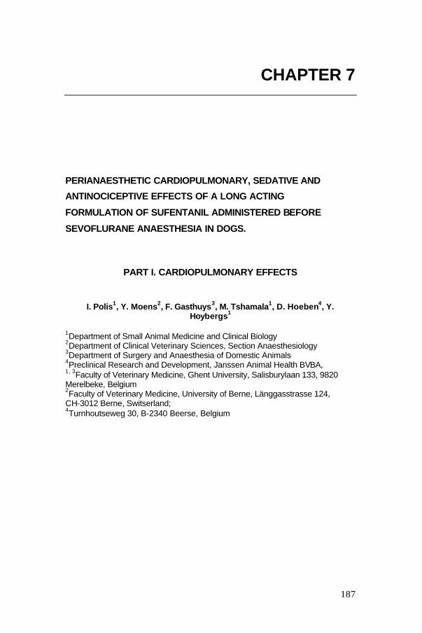
187
CHAPTER 7
PERIANAESTHETIC CARDIOPULMONARY, SEDATIVE AND
ANTINOCICEPTIVE EFFECTS OF A LONG ACTING
FORMULATION OF SUFENTANIL ADMINISTERED BEFORE
SEVOFLURANE ANAESTHESIA IN DOGS.
PART I. CARDIOPULMONARY EFFECTS
I. Polis1, Y. Moens2, F. Gasthuys3, M. Tshamala1, D. Hoeben4, Y. Hoybergs1
1Department of Small Animal Medicine and Clinical Biology 2Department of Clinical Veterinary Sciences, Section Anaesthesiology 3Department of Surgery and Anaesthesia of Domestic Animals 4Preclinical Research and Development, Janssen Animal Health BVBA, 1, 3Faculty of Veterinary Medicine, Ghent University, Salisburylaan 133, 9820 Merelbeke, Belgium 2Faculty of Veterinary Medicine, University of Berne, Länggasstrasse 124, CH-3012 Berne, Switserland; 4Turnhoutseweg 30, B-2340 Beerse, Belgium

CHAPTER 7
188
CHAPTER 7
189
SUMMARY
The purpose of the present study was to evaluate the
haemodynamic effects of sufentanil long acting premedication in
sevoflurane anaesthetized dogs in an open randomised study. Forty
dogs were divided over 5 parallel groups of 8 dogs each. Two control
groups were used: one group of dogs (A) received sufentanil long
acting (50 µg/kg IM) and a second group (B) the sufentanil vehicle
followed by a standard inhalation anaesthesia of 90 minutes. After
premedication with sufentanil long acting immediately before (C0), 15
minutes (D15) or 30 minutes (E30) before induction with thiopental (IV)
the dogs were anaesthetized for 90 minutes with sevoflurane in
oxygen.
Heart rate (HR), direct mean arterial blood pressure (MAP),
respiration rate (RR), arterial oxygen haemoglobin saturation (SaO2)
and sevoflurane concentration were measured every 10 minutes
during anaesthesia and at 2, 4 and 24 hours after initiation of
anaesthesia. Acid-base and blood gas analyses were performed at
the same time points.
The present study showed that premedication with sufentanil
LA followed by sevoflurane anaesthesia enhanced moderately some
cardiopulmonary side effects accompanying clinically adjusted
sevoflurane anaesthesia. The addition of sufentanil LA as
premedication caused a decrease in heart rate and an increase in
PaCO2, while MAP was well maintained. The clinical importance is
probably limited during inhalation anaesthesia where a high inspired
oxygen fraction resulted in high PaO2 and SaO2 levels. Temporary
support of ventilation with IPPV however might be occasionally
necessary. Thirty minutes between sufentanil LA premedication and
CHAPTER 7
190
induction of anaesthesia might be preferable, since less respiratory
depression occurred in group E30. In the post-anaesthetic period the
bradycardia persisted and was still present after 24 hours. Although
RR was lower then the control group without sufentanil pretreatment,
PaCO2 and PaO2 were within an acceptable range in the
postanaesthetic period up to 24 hours.
INTRODUCTION
The search for an optimal method to control acute pre-, per-
and post-operative pain in veterinary medicine is still ongoing. Opioids
are still of enormous therapeutic importance as the drug of choice for
the treatment of moderate to severe pain. The major action of opioids
is analgesia by binding to specific receptors (µ, ?, d, s, e) localised
mainly in the brain and spinal cord (Pert and Snyder, 1973; Stein,
1993). In general, the clinically most effective opioids act selectively at
µ receptors (Nolan, 2000). Intermittent administration of short-acting
opioids on an as-needed basis leads to peaks and troughs in drug
plasma level. Coupled to an inadequate dosage or too lengthy dosing
intervals this can lead to insufficient control of peri- and postoperative
pain (Oden, 1989; Sinatra, 1991). Pre-anaesthetic drugs classically
include anticholinergics, tranquilizers or a2-agonists, but nowadays
opioids are often included in the premedication protocol because of
their analgesic characteristics (Taylor, 1999). The clinical advantage is
a dosage reduction of the drugs used for general anaesthesia which
eventually leads to improved patient safety (Brunner et al., 1994;
Moon et al., 1995). Furthermore administration of analgesics before
the surgical stimulus arises (pre-emptive analgesia) is thought to be
important for better control of postoperative pain (Lascelles et al.,
1995).

CHAPTER 7
191
The problems associated with intermittent administration of
short-acting opioids might be overcome with the intramuscular
administration of a potent opioid in a long-acting formulation. Such a
drug could than be used as premedication, providing effective pre-
emptive analgesia over an extended period of time including the
postanaesthetic period. The opioid sufentanil is a short-acting thiamyl
analogue of the µ-agonist fentanyl (T 1/2 = 2 - 2.5 hours), but it is 11.5
times more potent (Brunner et al., 1994). It is used as an anaesthetic
supplement to provide analgesia in balanced anaesthesia protocols in
men. The clinical use of a balanced anaesthetic protocol using
sufentanil and midazolam in dogs has been reported (Hellebrekers
and Sap, 1991). A long acting formulation of sufentanil (on a medium chain
triglyceride base) has been investigated in several preclinical studies
(Engelen et al., 1996a; Engelen et al., 1996b; Engelen et al., 1996c;
Short and Vlaminck, 1998; Verbeeck et al., 1998). The dosage and
the pharmacokinetic profile were confirmed in a dose finding trial and
the safety was assessed in a tolerance study (Verbeeck et al., 1998;
Hoeben et al., 1999; Sterkens, 1999). After intramuscular
administration of 50 µg/kg BWT plasma levels of sufentanil very
rapidly increased and peak levels around 1.53 ± 0.45 ng/ml were
observed around 6 hours after injection. After this peak, the plasma
concentration of sufentanil slowly decreased. The T1/2 was 15.8 ± 5.1
hours. A dosage of 50 µg/kg of sufentanil LA provided plasma levels
between 0.85 and 1.5 ng sufentanil/ml for at least 12 hours (Short,
1996).
In these studies sufentanil LA induced not only sedation but
also the typical opioid side-effects such as reductions in heart rate,
arterial blood pressure, rectal temperature, respiratory rate, and an
increase in arterial carbon dioxide tension (Abdul-Rasool et al., 1989;

CHAPTER 7
192
Werner et al., 1991; Engelen et al., 1996a; Engelen et al., 1996b;
Engelen et al., 1996c; Short, 1996; Short and Vlaminck, 1998;
Verbeeck et al., 1998; Hoeben et al., 1999; Sterkens et al., 1999). A
classic anaesthetic technique to provide surgical anaesthesia in
veterinary medicine consists in an intravenous induction of
anaesthesia and subsequent maintenance by delivering a volatile
agent with oxygen. Thiopental, a short acting barbiturate and a popular veterinary
induction agent, is known to induce induction apnoea, respiratory
acidosis and hypoxaemia (Rawlings and Kolata, 1983; Muir, 1998a;
Muir, 1998b). It also affects the cardiovascular system with
hypotension, bradycardia followed by reflex tachycardia, hypertension
and arrhythmias (Muir, 1998a; Muir, 1998b). Sevoflurane is a
halogenated hydrocarbon developed by Wallin et al. (1975). In Europe
it is commercially available as an inhalant anaesthetic for men since
the early nineties. It is also used as a volatile anaesthetic in animals
and its use in this field is expected to increase in the future. Like all
volatile anaesthetics it also affects the cardiovascular and respiratory
system (Mutoh et al., 1997; Polis et al., 2001a; Polis et al., 2001b). In
a previous study in sevoflurane anaesthetized dogs dose dependent
decreases in blood pressure, cardiac output, stroke volume and
respiration rate in combination with an increase in arterial carbon
dioxide tension were observed (Polis et al., 2001a). Because the proposed therapeutic dose of sufentanil LA (50
µg/kg) is known to depress several cardiovascular and respiratory
parameters, the use of sufentanil LA premedication before thiopental
induction and sevoflurane as anaesthetic maintenance is expected to
enhance cardiovascular and respiratory side-effects of the general
anaesthetics. The present study was done to evaluate cardiovascular

CHAPTER 7
193
status and gas exchange during and following clinically conducted
sevoflurane anaesthesia when sufentanil LA was used as a
premedication administered at different time points before induction.
MATERIALS AND METHODS
The study was approved by the Ethical committee of the
Faculty of Veterinary Medicine, Ghent University (filenumber:
39/2000). Forty adult female Beagles weighing 11.97 ± 1.40 kg (mean
± SD) from 1 to 2 years old were used in the study. The dogs were
dewormed and vaccinated 1 month before the experiment. Clinical
examination one week before the experiment confirmed the good
health status of the animals.
Study Protocol
The present study was an open randomised study with 40
dogs divided over 5 parallel groups. The study was conducted in 40
phases; each phase consisted of 1 dog monitored 24 hours after the
administration of the drugs. No blinding was performed.
Table 1: Study groups involved in the study of sufentanil LA followed by sevoflurane anaesthesia in dogs (n=40).
STUDY No. of DOGS SUFENTANIL
GROUP (µg/kg)
Time interval between sufentanil/vehicle administration and initiation of inhalation
anaesthesia
A (control suf) 8 50 *
B (control sevo) 8 0 (vehicle) §
8 50 0
8 50 15
8 50 30
* Dogs receive no inhalation anaesthesia, but only sufentanil at T0
§ Dogs receive sufentanil-vehicle and inhalation anaesthesia at T0
Initiation of inhalation anaesthesia was defined as T0; except for group A Time expressed in minutes
C0
D15
E30

CHAPTER 7
194
The dogs in group A (control suf) received sufentanil LA at T0
without inhalation anaesthesia. The dogs from group B (control sevo)
received only the sufentanil-vehicle at T0 followed by inhalation
anaesthesia. In groups C0, D15 and E30 a time interval of respectively
0, 15 and 30 minutes between sufentanil LA administration and
anaesthesia induction was respected (Table 1).
Preparation of the dogs
A titanium vascular access port with a silicone membrane
(Access Technologies, Skokie, IL, USA) connected to the femoral
artery was surgically implanted 3 to 6 days before each experiment.
This device was used for blood sampling and blood pressure
measurement. Anaesthesia for this procedure consisted in a total
intravenous protocol with propofol combined with epidural anaesthesia
using lidocaine and bupivacaine. Analgesia was supplemented with
carprofen given at a dosage of 4 mg/kg orally for 3 days, starting the
evening before surgery. Patency of the vessel access port was
controlled by daily flushing with 1 ml of heparinised saline (200-300
I.U. heparin per ml). Three to five days after the experiment the port
was surgically removed under isoflurane anaesthesia.
Premedication and Induction of Anaesthesia
The dogs of groups A, C0, D15 and E30 received sufentanil long
acting formulation (sufentanil (0.5 mg/ml); Janssen Animal Health,
Beerse, Belgium) at a dosage of 50 µg/kg of BWT administered IM in
the lumbar muscles. The dogs of group B received only sufentanil-
vehicle. Anaesthesia (group B, C0, D15 and E30) was induced with
thiopental (Pentothal®, Abbott Laboratories Ltd., Queenborough, UK).
Four mg/kg was injected as a bolus and further dosing was slowly
done to effect. “Effect” was defined as the moment that eyeballs

CHAPTER 7
195
rotated ventrally and that intubation could be easily performed. Mean
injection dose was 13.3 ± 2.5 mg/kg of BWT.
Maintenance of Anaesthesia
The dogs were positioned in lateral recumbency and
connected to an anaesthetic machine with a circle system (Titus®,
Dräger, Lübeck, Germany) and a sevoflurane out of circuit vaporiser
(Vapor 19.3®, Dräger, Lübeck, Germany). The anaesthetic circuit was
flushed with 100% oxygen for 5 minutes before the experiment.
During the first 5 minutes of anaesthesia, a fresh gas flow of 2 l/min
O2 was used which was subsequently reduced to 1 l/min. The dogs
breathed spontaneously, but manual ventilation was performed when
marked respiratory depression occurred (PaCO2 > 55 mm Hg and/or
RR< 15 breaths/min). During anaesthesia the percentage of
sevoflurane was adjusted to obtain and maintain an anaesthetic depth
suitable to perform surgical interventions much like it would have been
done in clinical conditions. Such a level was thought to exist when the
eye-lid reflex was absent and the eyeballs were ventrally rotated.
Moreover a standardised pain stimulus was administered by clamping
the tail for 5 seconds every 10 minutes at a distance of approximately
3 cm from its top with a straight Rochester-Carmalt haemostatic
forceps (10 cm) closed to the first ratchet lock. If a reaction (a
movement, and/or an increase in HR of more than 15%) was
observed, vaporizer settings were adjusted upwards with 0.5% on the
vaporizer scale. If no reaction occurred, vaporizer settings were
decreased step wise. Depth of anaesthesia was also controlled by
observation of eye-lid reflex and position of the eyeballs and if
necessary adjusted. Anaesthesia was continued for 90 minutes. No
intravenous infusions were administered during the study.
CHAPTER 7
196
Measurements and monitoring
A calibrated multigas analyser including a pulse oximetry
unit (Quick Cal TM calibration gas® and Capnomac Ultima®, Datex
Engstrom Instrumentation Corp., Helsinki, Finland) was used to
monitor heart rate (HR), respiratory rate (RR), end tidal CO2%,
inspiratory and end tidal sevoflurane concentration and peripheral
oxygen haemoglobin saturation (SpO2%). MAP was recorded using a
blood pressure transducer (Vascumed N.V., Gent, Belgium)
connected to a blood pressure measuring device (Hellige Servomed
SMV 104, Germany) following standard calibration procedure. The
pressure transducer was connected to the vascular access port using
extension tubing filled with heparinised saline (200-300 I.U. heparin
per ml) and fitted with a special Huber point needle (Access
Technologies, Skokie, IL, USA) used to perforate the silicone
membrane of the vascular access port. Arterial blood was sampled
through the vascular access port. The arterial oxygen (PaO2 in mm
Hg) and carbon dioxide tension (PaCO2 in mm Hg), pH and arterial
oxygen saturation (SaO2 in %) were measured immediately after
sampling with a calibrated blood gas analyser and corrected for body
temperature (ABL 5, Radiometer Copenhagen, Denmark).
All parameters were measured immediately before
intramuscular administration of sufentanil LA or the vehicle,
immediately before inhalation anaesthesia (T0; except group A), and
every 10 minutes during 90 minutes of inhalation anaesthesia. The
same measurements were continued in the post anaesthetic period at
120 minutes (T120), 240 minutes (T240) and 24 hours (T1440).
At the end of inhalation anaesthesia, the dogs breathed
oxygen until extubation was possible. The dogs were transferred to
the recovery box. If the rectal temperature was lower than 36°C, an

CHAPTER 7
197
infrared heating source was installed in the recovery box. The
occurrence of critical events was recorded in the time period T0 to
T1440 when predetermined limits were surpassed. Critical values of
each parameter are represented in Table 2.
Table 2: Limits of the presence of critical events in the study of sufentanil LA followed by sevoflurane anaesthesia in dogs (n=40).
PARAMETER LIMIT CRITICAL VALUE
Heart Rate < 45 beats/min
Mean Arterial Blood Pressure < 75 mm Hg
Respiratory Rate < 15 breaths/min
PaCO2 > 55 mm Hg
PaO2 < 75 mm Hg
pH < 7.25
Arterial O2 Saturation < 90 %
Abbreviations : see text.
Statistical Analysis
The dogs were sorted according to decreasing body weight
and divided into 8 classes of 5 dogs each. In each class, the body
weights were comparable. Within each class the 5 dogs were
randomly allocated to the five different study groups and the 40
phases by a computer randomisation program. The statistical tests
were two-sided and used a 0.05 type I error (α=5%).
The StatXact software (StatXact 3 For Windows (1995), Users
Manual, CYTEL Software, Cambridge, MA 02139 USA) was used for
the Kruskal-Wallis one-way analysis of variance tests, for the
Wilcoxon Mann-Whitney U tests and for the Fisher’s exact tests
(Siegel,1977).

CHAPTER 7
198
The treatment groups A (control suf) and B (control sevo)
were control groups. No statistics were done for these groups, since
the effects of sufentanil LA and sevoflurane are studied previously in
dogs (Bernard et al., 1990; Engelen et al., 1996a; Engelen et al.,
1996b; Engelen et al., 1996c; Short, 1996; Short and Vlaminck, 1998;
Verbeeck et al., 1998; Hoeben et al., 1999; Sterkens et al., 1999). The
body weights of the dogs were compared by means of the Kruskal-
Wallis one-way analysis of variance test to check the randomisation
procedure.
To evaluate the homogeneity of the several treatment groups,
baseline comparisons (T0) was also performed on HR, MAP, RR, pH
and blood gases of each treatment group by means of the Kruskal-
Wallis one-way analysis of variance test. For each parameter (HR,
MAP, RR, PaCO2, PaO2, pH, SaO2) the following tests were
performed: statistical comparisons on the mean over time (T0 to T90)
was performed between the treatment groups C0, D15 and E30 by
means of the Kruskal-Wallis one-way analysis of variance test
followed by two-by-two Wilcoxon Mann-Whitney U tests. Each of the
treatment groups C0, D15 and E30 was compared with the control
groups A and B by Wilcoxon Mann-Whitney U tests.
The individual time points after inhalation anaesthesia (T120,
T240 and T1440) were analysed in the same way as the variable mean
over time. The number of dogs with a critical event was calculated.
The amounts of dogs with a critical parameter value were used to
compare the treatment groups by means of the Fisher’s exact test.

CHAPTER 7
199
RESULTS
Results for different measured variables are given in Table 3 and
4 and Figures 1 to 6. No significant differences in baseline values per
study group and per parameter were observed. The analgesic and
sedative effects of sufentanil LA and the side effects which occurred in
this experiment will be discussed in a second paper.
Anaesthesia period (T0 to T90)
HR was significantly lower in sufentanil treated inhalation
groups D15 and E30 compared to group B (control sevo) (p<0.01). HR
was significantly lower in group A (control suf) compared to groups C0
and D15 (p<0.05). Minimum and maximum (min-max) values for HR
were 52 (group A at T60) and 155 (group B at T0) beats/minute
respectively. MAP was similar in all inhalation groups (B, C0, D15 and
E30). MAP was lower in all inhalation groups compared to group A
and this difference was significant for groups C0, D15 and E30
(p<0.001). Min-max values for MAP were 36 (group B at T90) and 137
(group C0 at T0) mm Hg. RR was similar in all inhalation groups (B, C0,
D15, E30). RR was higher in group A compared to all inhalation groups
and this difference was significant for groups C0, D15 and E30 (p<0.001
and p<0.01). Min-max for RR were 2 (group D15 at T10) and 160
(group A at T20) breaths/minute. PaCO2 was higher in the sufentanil
treated inhalation groups compared to group B (control sevo) but this
was significant for group C0 and D15 only (p<0.01). PaCO2 was also
significantly higher in groups C0 and D15 compared to group E30
(p<0.01). PaCO2 was higher in all inhalation groups compared to
group A and this difference was significant for groups C0, D15 and E30
(p<0.001 and p<0.01). Min-max values of PaCO2 were 21 (group C0 at
T0) and 74 (group D15 at T10) mm Hg. PaO2 was similar in all inhalation
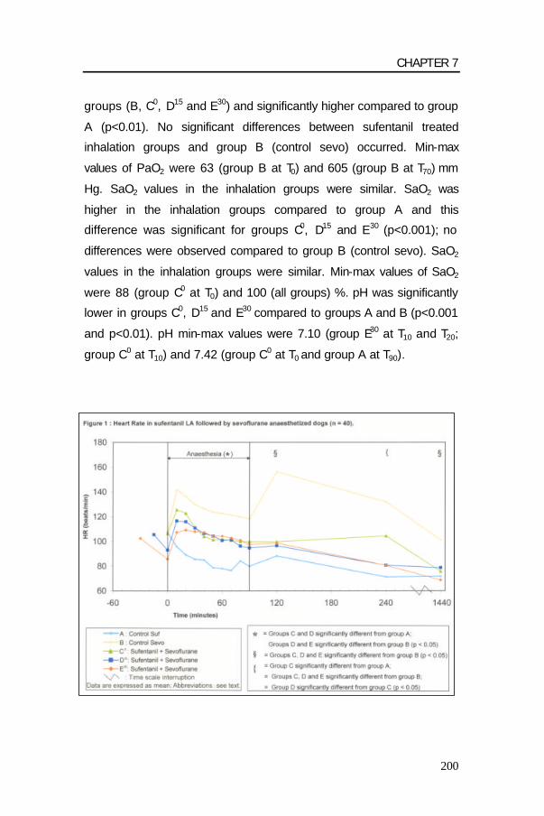
CHAPTER 7
200
groups (B, C0, D15 and E30) and significantly higher compared to group
A (p<0.01). No significant differences between sufentanil treated
inhalation groups and group B (control sevo) occurred. Min-max
values of PaO2 were 63 (group B at T0) and 605 (group B at T70) mm
Hg. SaO2 values in the inhalation groups were similar. SaO2 was
higher in the inhalation groups compared to group A and this
difference was significant for groups C0, D15 and E30 (p<0.001); no
differences were observed compared to group B (control sevo). SaO2
values in the inhalation groups were similar. Min-max values of SaO2
were 88 (group C0 at T0) and 100 (all groups) %. pH was significantly
lower in groups C0, D15 and E30 compared to groups A and B (p<0.001
and p<0.01). pH min-max values were 7.10 (group E30 at T10 and T20;
group C0 at T10) and 7.42 (group C0 at T0 and group A at T90).

CHAPTER 7
201
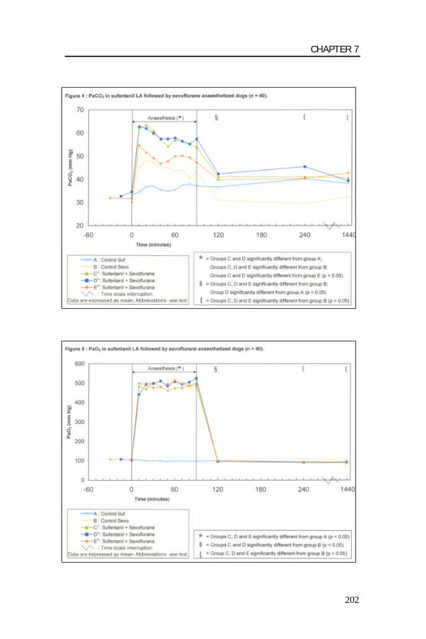
CHAPTER 7
202

CHAPTER 7
203
Post-anaesthetic period (T120 to T1440)
HR was significantly lower at all time points in the sufentanil
treated inhalation groups C0, D15 and E30 compared to group B
(control sevo) (p<0.05 to p<0.001). HR was not significantly different
between these groups except in group D15, where HR was
significantly lower compared to group C0 at time point T240 (p<0.05).
HR was not significantly different between sufentanil treated inhalation
groups C0, D15 and E30 and group A (control suf) except for time point
T240 where HR in group C0 was significantly higher (p<0.01). Min-max
values for HR were 44 (group E30 at T240) and 170 (group B at T120).
MAP was lower in all sufentanil treated groups compared to group B
(control sevo). At T120 this difference was statistically significant when
compared with groups C0 and E30 (p<0.01 and p<0.001). At T1440 MAP
in group C0 was significantly higher compared to group A (control suf).
Min-max values for MAP were between 71 (group A at T120) and 165
(group C0 at T1440) mm Hg.
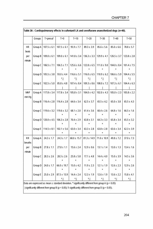
CHAPTER 7
204
Table 3A : Cardiopulmonary effects in sufentanil LA and sevoflurane anaesthetized dogs (n=40).
Groups T=presuf T=0 T=10 T=20 T=30 T=40 T=50
HR Group A 107.5 ± 6.1 107.5 ± 6.1 95.9 ± 7.7 89.3 ± 3.9 85.6 ± 5.6 85.0 ± 6.6 78.8 ± 5.7
beats
per Group B 109.0 ± 6.1 109.0 ± 6.1 141.8 ± 3.4 136.3 ± 3.3 129.9 ± 4.1 126.5 ± 3.7 123.8 ± 2.8
minute
Group C 106.5 ± 7.1 106.5 ± 7.1 125.6 ± 6.6 122.8 ± 6.5 111.0 ± 9.0 104.0 ± 8.4 101.4 ± 7.5
* * * * * *
Group D 105.5 ± 3.8 93.0 ± 4.4 116.6 ± 5.1 116.0 ± 6.5 110.9 ± 6.2 106.6 ± 5.9 104.4 ± 5.5
*,| *,| *,| *,| *,| *,|
Group E 102.5 ± 5.0 85.8 ± 4.8 107.4 ± 8.4 109.3 ± 8.6 108.0 ± 7.2 107.3 ± 6.1 104.4 ± 6.3
| | | | | |
MAP Group A 117.8 ± 3.4 117.8 ± 3.4 105.8 ± 3.1 104.0 ± 4.2 102.0 ± 4.3 105.0 ± 2.3 105.8 ± 2.2
mm Hg
Group B 116.4 ± 2.8 116.4 ± 2.8 64.6 ± 3.4 62.5 ± 3.7 65.5 ± 4.2 65.0 ± 3.8 65.5 ± 4.3
Group C 119.6 ± 3.2 119.6 ± 3.2 68.1 ± 2.0 61.4 ± 3.6 60.4 ± 2.6 64.8 ± 1.6 66.5 ± 1.6
* * * * * *
Group D 120.0 ± 4.5 106.3 ± 2.8 70.4 ± 2.9 63.8 ± 3.1 64.3 ± 3.3 65.8 ± 3.4 65.5 ± 3.2
* * * * * *
Group E 114.5 ± 8.1 102.1 ± 5.6 63.0 ± 3.4 63.3 ± 2.8 63.8 ± 2.8 63.6 ± 3.4 62.3 ± 3.9
* * * * * *
RR Group A 24.3 ± 1.7 24.3 ± 1.7 68.0 ± 15.7 81.3 ± 14.9 71.0 ± 10.9 49.8 ± 7.2 37.0 ± 7.3
breaths
per Group B 27.8 ± 1.1 27.8 ± 1.1 15.6 ± 2.4 12.9 ± 0.6 13.1 ± 1.4 13.0 ± 1.3 13.4 ± 1.6
minute
Group C 28.3 ± 2.6 28.3 ± 2.6 25.8 ± 5.8 17.1 ± 4.8 14.4 ± 4.0 15.0 ± 3.9 14.5 ± 3.6
* * * * * *
Group D 24.0 ± 1.7 66.0 ± 19.7 15.0 ± 4.2 11.9 ± 2.3 12.1 ± 1.9 12.6 ± 2.3 12.9 ± 2.6
* * * * * *
Group E 25.0 ± 2.9 87.5 ± 13.9 14.4 ± 2.4 12.3 ± 1.9 12.6 ± 1.9 13.6 ± 2.2 15.8 ± 4.1
*,| *,| *,| *,| *,| *,|
Data are expressed as mean ± standard deviation. * significantly different from group A (p < 0.05)
| significantly different from group B (p < 0.05); § significantly different from group D (p < 0.05).

CHAPTER 7
205
Table 3B : Cardiopulmonary effects in sufentanil LA and sevoflurane anaesthetized dogs (n=40).
Groups T=60 T=70 T=80 T=90 T=120 T=240 T=1440
HR Group A 78.0 ± 6.4 76.5 ± 5.8 84.3 ± 8.3 80.0 ± 6.7 88.3 ± 6.3 71.3 ± 5.8 71.8 ± 4.3
beats
per Group B 122.6 ± 3.5 121.3 ± 3.4 120.0 ± 3.6 118.3 ± 3.6 156.3 ± 3.7 132.0 ± 7.9 101.3 ± 7.5
minute
Group C 101.6 ± 7.5 101.0 ± 7.8 99.9 ± 7.9 99.9 ± 8.7 99.8 ± 9.2 104.5 ± 7.1 75.8 ± 5.3
* * * * | *,| |
Group D 100.9 ± 5.4 101.3 ± 6.7 96.3 ± 4.7 94.8 ± 4.3 96.5 ± 9.0 80.8 ± 5.3 78.8 ± 8.4
*,| *,| *,| *,| | | |
Group E 104.4 ± 6.1 102.8 ± 6.1 101.0 ± 5.3 97.5 ± 4.7 98.6 ± 7.2 80.6 ± 8.0 68.8 ± 5.5
| | | | | | |
MAP Group A 98.3 ± 2.7 96.1 ± 3.6 94.8 ± 3.3 94.3 ± 2.4 104.1 ± 6.9 113.1 ± 3.5 108.9 ± 4.4
mm Hg
Group B 67.4 ± 4.4 69.1 ± 4.6 68.5 ± 5.0 67.1 ± 5.1 132.4 ± 4.0 122.1 ± 2.9 126.3 ± 2.4
Group C 67.9 ± 2.4 64.6 ± 2.3 64.3 ± 2.9 64.3 ± 3.0 109.9 ± 4.5 118.0 ± 5.8 126.8 ± 6.4
* * * * | *
Group D 63.8 ± 3.4 65.0 ± 3.2 64.4 ± 3.3 66.8 ± 3.5 112.9 ± 7.4 110.3 ± 4.8 119.3 ± 7.1
* * * *
Group E 61.8 ± 2.5 62.0 ± 3.0 62.9 ± 3.2 62.4 ± 3.5 103.9 ± 4.1 109.6 ± 5.6 115.4 ± 8.0
* * * * |
RR Group A 39.0 ± 5.7 47.5 ± 10.8 42.3 ± 10.4 41.5 ± 6.7 43.0 ± 9.1 34.5 ± 14.0 22.5 ± 2.4
breaths
per Group B 13.9 ± 1.3 13.4 ± 1.2 13.1 ± 1.0 13.6 ± 1.1 30.6 ± 3.1 43.0 ± 16.8 40.8 ± 6.5
minute
Group C 16.4 ± 3.0 17.1 ± 3.0 17.4 ± 3.3 17.6 ± 3.5 25.0 ± 3.7 22.5 ± 2.8 23.3 ± 2.5
* * * * * |
Group D 13.9 ± 2.4 14.9 ± 2.5 14.8 ± 2.7 13.8 ± 2.0 27.5 ± 3.0 19.0 ± 1.5 23.3 ± 1.6
* * * * | |
Group E 13.0 ± 2.5 14.9 ± 3.3 13.6 ± 2.8 13.1 ± 2.5 20.8 ± 2.1 18.3 ± 1.8 22.3 ± 1.6
*,| *,| *,| *,| *,|, § | |
Data are expressed as mean ± standard deviation. * significantly different from group A (p < 0.05)
| significantly different from group B (p < 0.05); § significantly different from group D (p < 0.05).

CHAPTER 7
206
Table 4A : Acid-base balance and blood gas analysis in sufentanil LA and sevoflurane anaesthetized dogs (n=40).
T=control T=0 T=10 T=20 T=30 T=40 T=50
PaO2 A 105.6 ± 3.8 105.6 ± 3.8 104.3 ± 3.5 100.0 ± 2.9 100.1 ± 2.1 99.9 ± 4.1 97.4 ± 2.4 mm Hg B 107.3 ± 2.0 107.3 ± 2.0 493.5 ± 19.0 505.0 ± 20.3 497.4 ± 16.8 509.0 ± 14.7 516.8 ± 18.6
C 105.4 ± 6.4 105.4 ± 6.4 484.5 ± 12.4 469.9 ± 15.7 503.3 ± 16.5 481.9 ± 20.9 462.1 ± 20.1 * * * * * * D 107.8 ± 1.4 103.8 ± 2.2 442.3 ± 38.0 494.4 ± 11.5 498.1 ± 14.8 510.8 ± 10.2 484.9 ± 15.5 * * * * * * E 108.0 ± 2.1 105.0 ± 2.4 499.5 ± 12.9 488.0 ± 24.2 475.9 ± 25.0 480.1 ± 26.5 495.8 ± 16.0 * * * * * *
SaO2 A 96.6 ± 0.3 96.6 ± 0.3 96.5 ± 0.4 96.1 ± 0.4 96.4 ± 0.3 96.3 ± 0.5 96.0 ± 0.3
% B 97.1 ± 0.2 97.1 ± 0.2 100.0 ± 0.0 100.0 ± 0.0 100.0 ± 0.0 100.0 ± 0.0 100.0 ± 0.0 C 96.1 ± 1.2 96.1 ± 1.2 100.0 ± 0.0 100.0 ± 0.0 100.0 ± 0.0 100.0 ± 0.0 100.0 ± 0.0 * * * * * * D 97.3 ± 0.2 97.0 ± 0.2 99.9 ± 0.1 100.0 ± 0.0 100.0 ± 0.0 100.0 ± 0.0 100.0 ± 0.0 * * * * * * E 97.0 ± 0.2 96.9 ± 0.3 100.0 ± 0.0 100.0 ± 0.0 100.0 ± 0.0 100.0 ± 0.0 100.0 ± 0.0 * * * * * *
pH A 7.37 ± 0.01 7.37 ± 0.01 7.35 ± 0.01 7.34 ± 0.01 7.33 ± 0.01 7.34 ± 0.01 7.34 ± 0.01 B 7.34 ± 0.01 7.34 ± 0.01 7.26 ± 0.01 7.25 ± 0.01 7.25 ± 0.01 7.26 ± 0.01 7.27 ± 0.01 C 7.36 ± 0.01 7.36 ± 0.01 7.17 ± 0.02 7.16 ± 0.02 7.18 ± 0.02 7.20 ± 0.01 7.20 ± 0.01 *,| *,| *,| *,| *,| *,| D 7.36 ± 0.01 7.34 ± 0.01 7.17 ± 0.01 7.17 ± 0.01 7.19 ± 0.01 7.20 ± 0.01 7.21 ± 0.01 *,| *,| *,| *,| *,| *,| E 7.34 ± 0.01 7.31 ± 0.02 7.17 ± 0.02 7.19 ± 0.02 7.20 ± 0.01 7.22 ± 0.02 7.22 ± 0.02 *,| *,| *,| *,| *,| *,|
PaCO2 A 33.3 ± 0.9 33.3 ± 0.9 34.6 ± 0.9 36.9 ± 1.4 37.1 ± 1.3 35.5 ± 1.2 35.0 ± 1.4 mm Hg B 31.9 ± 0.7 31.9 ± 0.7 45.1 ± 3.2 48.1 ± 1.7 47.4 ± 1.9 45.9 ± 1.5 44.9 ± 1.7
C 32.8 ± 1.0 32.8 ± 1.0 34.6 ± 0.6 62.8 ± 1.9 62.0 ± 1.9 59.9 ± 1.2 57.5 ± 1.7 *,|,§ *,|,§ *,|,§ *,|,§ *,|,§ *,|,§ D 32.8 ± 1.0 34.6 ± 0.6 62.8 ± 3.1 62.0 ± 1.9 59.9 ± 1.2 57.5 ± 1.7 57.5 ± 1.7 *,|,§ *,|,§ *,|,§ *,|,§ *,|,§ *,|,§ E 32.0 ± 1.1 31.9 ± 1.9 54.8 ± 1.6 51.6 ± 2.8 49.3 ± 2.8 46.9 ± 3.3 47.9 ± 3.3 | | | | | |
Data are expressed as mean ± standard deviation. * significantly different from group A (p < 0.05)
| significantly different from group B (p < 0.05); § significantly different from group E (p < 0.05).

CHAPTER 7
207
Table 4B : Acid-base balance and blood gas analysis in sufentanil LA and sevoflurane anaesthetized dogs (n=40).
T=60 T=70 T=80 T=90 T=120 T=240 T=1440
PaO2 A 98.9 ± 2.5 98.9 ± 2.5 98.3 ± 3.0 98.1 ± 4.4 101.0 ± 3.0 94.9 ± 2.8 91.5 ± 2.5 mm Hg B 521.8 ± 14.4 513.6 ± 19.2 472.1 ± 27.2 519.9 ± 15.8 110.6 ± 1.3 107.3 ± 3.1 104.9 ± 2.6
C 475.4 ± 32.8 476.4 ± 13.9 487.8 ± 25.0 498.8 ± 13.7 95.3 ± 2.5 93.6 ± 1.9 88.3 ± 2.4 * * * * | | | D 508.0 ± 12.1 494.9 ± 16.4 505.1 ± 15.8 525.0 ± 10.2 96.1 ± 2.4 90.0 ± 1.3 93.5 ± 3.4 * * * * | | | E 513.5 ± 18.4 497.0 ± 19.6 488.0 ± 21.6 487.8 ± 22.3 101.4 ± 4.1 90.3 ± 3.0 89.0 ± 1.5 * * * * | |
SaO2 A 96.1 ± 0.4 96.3 ± 0.3 96.1 ± 0.4 96.4 ± 0.4 96.6 ± 0.3 96.5 ± 0.3 95.9 ± 0.6
% B 100.0 ± 0.0 100.0 ± 0.0 100.0 ± 0.0 100.0 ± 0.0 97.6 ± 0.2 97.1 ± 0.2 97.0 ± 0.2 C 100.0 ± 0.0 100.0 ± 0.0 100.0 ± 0.0 100.0 ± 0.0 96.0 ± 0.3 96.1 ± 0.4 95.5 ± 0.6 * * * * | D 100.0 ± 0.0 100.0 ± 0.0 100.0 ± 0.0 100.0 ± 0.0 96.4 ± 0.3 96.0 ± 0.2 96.1 ± 0.4
* * * * | | E 100.0 ± 0.0 100.0 ± 0.0 100.0 ± 0.0 100.0 ± 0.0 96.8 ± 0.5 96.1 ± 0.4 96.0 ± 0.3 * * * * |
pH A 7.33 ± 0.01 7.33 ± 0.01 7.34 ± 0.01 7.34 ± 0.01 7.32 ± 0.01 7.31 ± 0.01 7.34 ± 0.01 B 7.27 ± 0..01 7.28 ± 0.01 7.28 ± 0.02 7.28 ± 0.01 7.34 ± 0.01 7.35 ± 0.01 7.35 ± 0.01 C 7.20 ± 0.01 7.20 ± 0.01 7.21 ± 0.01 7.21 ± 0.01 7.28 ± 0.02 7.28 ± 0.01 7.31 ± 0.01 *,| *,| *,| *,| | | | D 7.21 ± 0.01 7.21 ± 0.01 7.22 ± 0.01 7.21 ± 0.01 7.28 ± 0.01 7.26 ± 0.01 7.33 ± 0.01 *,| *,| *,| *,| | *,| E 7.22 ± 0.02 7.22 ± 0.02 7.22 ± 0.02 7.23 ± 0.02 7.26 ± 0.01 7.26 ± 0.01 7.33 ± 0.01 *,| *,| *,| *,| *,| *,|
PaCO2 A 35.5 ± 1.0 37.5 ± 2.5 38.0 ± 3.0 37.1 ± 2.9 36.8 ± 2.0 40.3 ± 2.1 38.3 ± 2.0 mm Hg B 43.8 ± 1.5 44.1 ± 1.6 40.5 ± 2.2 43.0 ± 1.6 31.0 ± 0.7 29.5 ± 1.1 32.4 ± 1.0
C 57.5 ± 1.7 57.9 ± 1.2 56.5 ± 0.9 55.3 ± 1.5 57.5 ± 1.6 42.4 ± 1.8 39.4 ± 1.6 *,|,§ *,|,§ *,|,§ *,|,§ | | | D 57.9 ± 1.2 56.5 ± 0.9 55.3 ± 1.5 57.5 ± 1.6 42.4 ± 1.8 45.5 ± 1.7 39.4 ± 1.6 *,|,§ *,|,§ *,|,§ *,|,§ *,| | | E 50.0 ± 3.0 50.3 ± 2.1 49.5 ± 2.5 47.3 ± 2.2 39.9 ± 1.7 40.4 ± 3.0 42.8 ± 1.4 | | | | | | |
Data are expressed as mean ± standard deviation. * significantly different from group A (p < 0.05)
| significantly different from group B (p < 0.05); § significantly different from group E (p < 0.05).

CHAPTER 7
208
Following the end of inhalation anaesthesia RR increased in
all groups. RR was lower in sufentanil treated inhalation groups
compared to group B (control sevo). This difference was significant at
T120 for group E30 (p<0.05) , at T240 for groups D15 and E30 (p<0.01)
and at T1440 for groups C0, D15 and E30 (p<0.05 and p<0.01). There
was no significant difference between the sufentanil treated inhalation
groups; except at T120 where group E30 had a lower RR compared to
D15 (p<0.05). Respiratory rate in group A (control suf) was higher than
in the sufentanil treated inhalation groups in the post-anaesthetic
period. This difference was significant at T120 compared to groups C0
and E30 (p<0.05 and p<0.01). The min-max values for RR were 12 (1
dog in groups A, C0, D15 and E30 at T240) and 160 (group B at T240)
breaths/minute. Following the end of inhalation anaesthesia PaCO2
decreased rapidly in all groups until T120 and remained stable for the
remainder of the post-anaesthetic period. PaCO2 was significantly
higher in the sufentanil treated inhalation groups compared to group B
(control sevo) at all time points (p<0.001 and p<0.05). PaCO2 in
group A (control suf ) was similar as in the sufentanil treated inhalation
groups; except at T120 where PaCO2 was significantly higher in group
D15 compared to A (p<0.05). Min-max values for PaCO2 were 25
(group B at T240) and 54 (group D15 at T240). Following the end of
inhalation anaesthesia PaO2 decreased rapidly to baseline values in
all groups. PaO2 was lower in the sufentanil treated inhalation groups
compared to group B (control sevo). This difference was significant at
all time points (at T120 not for group E30) (from p<0.05 to p<0.001). No
significant differences existed between the sufentanil treated
inhalation groups (C0, D15 and E30) and group A (control suf). Min-max
values for PaO2 were 75 (group C0 at T1440) and 122 (group B at T240)
mm Hg. SaO2 was lower in the sufentanil treated inhalation groups
compared to group B (control sevo). At time point T120 the difference

CHAPTER 7
209
was significant for group C0 and D15 (p<0.01 and p<0.05), at T240 for
group D15 (p<0.01) and at T1440 for groups C0 and E30 (p<0.05). No
significant differences existed between the sufentanil treated
inhalation groups (C0, D15 and E30) and group A (control suf). Min-max
values for SaO2 were 92 (group A and C0 at T1440) and 99 (group E30
at T120) %. pH was significantly lower in the sufentanil treated
inhalation groups compared to group B (control sevo) at all time points
(p<0.05 to p<0.001); except for groups D15 and E30 at T1440. pH in
group A (control suf) was higher compared to the other sufentanil
groups; but this was only significant for group E30 at T120 and T240 and
for group D15 at T240. Min-max values for pH were 7.22 (group D15 at
T120 and group E30 at T120 and T240) and 7.40 (group A at T1440 and
group B at T240).
Critical events
Incidence of critical events for the different parameters and
groups are given in Table 5. A HR below the critical value of 45
beats/minute was observed in only one dog (group E30 at T240). In
group A (control suf) MAP (71 mm Hg) decreased below the critical
value in only one dog at T120. All dogs of all inhalation groups showed
MAP values below the critical value of 75 mmHg at one or more time
points compared to only one dog in group A (p<0.01). No significant
differences in critical MAP values between the sufentanil treated
inhalation groups compared to group B (control sevo) were observed.
The majority of the dogs of each inhalation group showed critical
events for RR. There were no significant differences in the number of
critical values between the sufentanil treated inhalation groups
compared to group B (control sevo). Significantly more RR values
below the critical level of 15 breaths/min occurred in groups C0, D15
and E30 compared to group A (p<0.05). Manual ventilation was applied

CHAPTER 7
210
3 times (2 dogs in group C0 and 1 in group E30), for the dog in group
E30 controlled ventilation was necessary during the complete
anaesthetic period. Critical events (above 55 mm Hg) for PaCO2 were
only observed during the anaesthesia period, whereby significantly
more critical PaCO2 values were seen in groups C0 and D15 compared
to group A and B (control sevo) (p<0.05 and p<0.01). Only one critical
value (below 75 mm Hg) with a PaO2 of 63 mm Hg occurred in group
C0 immediately after anaesthesia induction (T0). Over the entire study
period (T0 to T1440) only one dog showed a SaO2 % below the critical
value of 90% (88% in group C0 at T0). Most of the critical pH values
(below 7.25) occurred during the inhalation period. Four dogs showed
critical values in group B compared to 8 dogs in groups C0, D15 and
E30, respectively. Significantly more pH values over time (T0 to T1440)
below 7.25 were observed in groups C0, D15 and E30 compared to
group A (p<0.01) but not compared to group B.
DISCUSSION
In the present study HR during the anaesthetic period (T0 to
T90) was lower in the groups treated with sufentanil compared to group
B (control sevo). This decrease in HR was expected to occur with
sufentanil administration. Opioids and especially potent narcotic drugs
such as sufentanil induce a dose-dependent centrally mediated
bradycardia, which may be obtund by parasympathicolytic drugs
(Reddy, 1980; De Hert, 1991; Nolan and Reid, 1991). Studies in dogs
have shown that anaesthesia with high doses of sufentanil produced
only minimal haemodynamic changes (De Castro et al., 1979; Reddy
et al., 1980; Eriksen et al., 1981; Philbin et al., 1984; Abdul-Rasool
and Ward, 1989). In all inhalation groups however an initial rise in HR
followed by a decline after 20 minutes was observed. This initial

CHAPTER 7
211
increase might be related to thiopental used for induction. Thiopental
administration was reported to induce an initial increase in HR
probably due to the baroreceptor mediated sympathetic reflex
stimulation of the heart followed by a decrease in HR. This is in line
with the findings in the present study (Turner and Ilkiw, 1990; De Hert,
1991; Ilkiw et al., 1991). The degree of bradycardia observed in all
sufentanil treated inhalation groups could also be influenced by
sevoflurane anaesthesia, since tachycardia resulting from sympathetic
and baroreceptor reflex stimulation was observed in sevoflurane
anaesthetized dogs (Mutoh et al., 1997; Polis et al., 2001a). In
contrast to the anaesthetic period where bradycardia was probably
partially compensated by thiopental (induction) and sevoflurane
(maintenance), a long-lasting bradycardia persisted for 24 hours after
anaesthesia. HR decreased below the critical value of 45 beats/min in
only one dog (E30 at T240) out of 40 dogs. This critical value for HR
was chosen arbitrarily. The importance of a bradycardia as factor of
oxygen delivery to the tissues will depend also on concomitant factors
such as haemoglobin concentration, oxygen saturation and oxygen
consumption.
In group A (control suf) MAP decreased moderately during the
first 2 hours after administration, afterwards MAP remained slightly
below baseline values for 24 hours after administration. Small
decreases in MAP were reported after sufentanil administration in
dogs (De Castro et al., 1979; Berthelsen et al., 1980; Reddy, 1980;
Berthelsen et al., 1981) and this was confirmed in the present study.
During the anaesthesia period (T0 to T90) the lower MAP in the
inhalation groups and the increased incidence of mean blood pressure
values below 75 mm Hg (critical value) could be attributed to the effect
of sevoflurane. The latter is reported to depress systemic blood

CHAPTER 7
212
pressure in dogs in a dose-dependent manner (Bernard et al., 1990;
Frink et al., 1992; Harkin et al., 1994; Mutoh et al., 1997; Polis et al.,
2001a). In this study sevoflurane administration was adjusted
according to the reaction to a standardised pain stimulus. During
anaesthesia all inhalation groups had a similar MAP course despite
different end-tidal sevoflurane concentration were used. End-tidal
sevoflurane concentrations used in the sufentanil treated inhalation
groups were lower than in the sevoflurane control group. The similar
MAP in the different groups can be explained by a relatively small
difference in end-tidal sevoflurane concentration between the different
groups (see study part II).
In group A (control suf) RR increased sharply after sufentanil
LA administration up to 160 breaths/min while PaCO2 showed only a
slight increase. An initial transient period of panting is a common
finding in dogs when high doses of opioids are administered or when
neurolept-analgesic combinations are given (Lukasik, 1999; Nolan,
2000). The occurrence of panting is probably related to alteration of
the thermoregulatory centre in the hypothalamus (Lascelles, 2000). To
the contrary, in all inhalation groups a respiratory depression
characterised by a decreased RR and pH and increased PaCO2
developed accompanied by an increased incidence of critical values
for these parameters. This depression was, as expected, more
pronounced in the sufentanil treated inhalation groups. First,
inhalation anaesthetics including sevoflurane depress spontaneous
ventilation in a dose-dependent manner (Mutoh et al., 1997).
Secondly, sufentanil like other opioids produces also a dose-
dependent respiratory depression, which may occasionally be rapid
and severe (Monk et al., 1988; Abdul-Rasool and Ward, 1989). The
respiratory depression is caused by a decreased responsiveness of
CHAPTER 7
213
the respiratory centre to carbon dioxide, while the hypoxic stimulus to
breathing is unaffected (Florez et al., 1968). This is potentially one of
the most serious side effects in humans, but it is rarely a clinical
problem in dogs and cats unless the drugs are combined with other
potent respiratory depressants as is the case during general
anaesthesia (Nolan and Reid, 1991). The more pronounced
respiratory depression during sevoflurane anaesthesia following
sufentanil treatment can also be explained by the combined
respiratory depressant effect of sufentanil and sevoflurane (Steffey et
al., 1993; Steffey et al., 1994; Mutoh et al., 1997). The rather smaller
increase in PaCO2 in group E during anaesthesia (PaCO2 significantly
lower compared to C0 and D15) suggests a lower degree of respiratory
depression when sufentanil LA premedication was done 30 minutes
before induction of anaesthesia. In the postanaesthetic period RR was
higher in group B (control sevo) compared to the sufentanil treated
inhalation groups. This can be explained by the specific
pharmacokinetic profile of sufentanil LA with long lasting plasma
levels (Short, 1996).
PaO2 and SaO2 during anaesthesia were significantly higher in
all inhalation groups compared to group A (control suf) due to a high
inspired oxygen fraction. For the same reason PaO2 and SaO2 values
were normal during anaesthesia and not different between the
inhalation groups despite a higher PaCO2 and lower RR due to
enhanced respiratory depression in the sufentanil treated groups.
During the postanaesthetic period however when room air was
breathed the persistent respiratory depression can explain the lower
PaO2 in groups C0, D15 and E30 compared to group B (control sevo).
Since no hypoxia (no value below 75 mm Hg) occurred, this was
probably clinically not relevant.

CHAPTER 7
214
In the present study use of sufentanil LA as premedication
moderately enhanced some cardiopulmonary side effects
accompanying clinically adjusted sevoflurane anaesthesia. The
addition of sufentanil LA as premedication caused a decrease in HR
and an increase in PaCO2, while MAP was well maintained. The
clinical importance is probably limited during inhalation anaesthesia
where a high inspired oxygen fraction was accompanied by a high
PaO2 and SaO2 (with exception of only one dog shortly after
induction). Temporary support of ventilation with IPPV however might
be occasionally necessary. Therefore clinical observation and/or
respiratory function monitoring with spirometry or capnography would
be helpful. Thirty minutes between sufentanil LA premedication and
induction of anaesthesia might be preferable, since less respiratory
depression occurred in group E30. In the postanaesthetic period the
bradycardia persisted and was still present after 24 hours. Although
RR was lower then the control group without sufentanil pretreatment,
PaCO2 and PaO2 were within an acceptable range in the
postanaesthetic period up to 24 hours.
CHAPTER 7
215
REFERENCES
Abdul-Rasool, I.H., and D.S. Ward, 1989: Ventilatory and cardiovascular responses to sufentanil infusion in dogs anesthetized with isoflurane. Anesthesia & Analgesia 69, 300-306. Bernard, J.M., P.F. Wouters, M.F. Doursout, B. Florence, J.E. Chelly, and R.G. Merin, 1990: Effects of sevoflurane and isoflurane on cardiac and coronary dynamics in chronically instrumented dogs. Anesthesiology 72, 659-662. Berthelsen, P., J. Eriksen, N.C. Ahn, and J.P. Rasmussen, 1980: Peripheral circulation during sufentanil and morphine anesthesia. Acta Anaesthesiologica Scandinavica 24, 241-244. Berthelsen, P., J. Strom, and J. Eriksen, 1981: High-dose analgesic anaesthesia with morphine or sufentanil in propranolol-treated dogs. Acta Anaesthesiologica Scandinavica 25, 447-452. Brunner, M.D., P. Braithwaite, R. Jhaveri, A.I. McEwan, D.K. Goodman, L.R. Smith, and P.S.A. Glass, 1994: MAC reduction of isoflurane by sufentanil. British Journal of Anaesthesia 72, 42-46. De Castro, J., A. Van de Water, L. Wouters, R. Xhonneux, R. Reneman, and B. Kay, 1979: Comparative study of cardiovascular, neurological and metabolic side-effects of eight narcotic dogs. Acta Anaesthesiologica Belgica 30, 5-99. De Hert, S.G., 1991: Study on the effects of six intravenous anesthetic agents on regional ventricular function in dogs (thiopental, Etomidate, Propofol, Fentanyl, Sufentanil, Alfentanil). Acta Anaesthesiologica Belgica 42, 3-39. Engelen, M., A. Proost, and K. Vlaminck, 1996a: Pilot trial on the pharmacodynamic effects of sufentanil after a single intramuscular administration of a new depot formulation (0.35 mg/ml injectable solution in Miglyol) at a dose of 35 µg/kg in Beagle dogs (Protocol No. SUF-94-PREC-01). Janssen Pharmaceutica Preclinical R&D Report. Engelen, M., A. Proost, and K. Vlaminck, 1996b: Pilot trial on the pharmacodynamic effects of sufentanil after a single intramuscular administration of a new depot formulation (0.35 mg/ml injectable solution in Miglyol) at a dose of 70 µg/kg in Beagle dogs (Protocol No. SUF-95-PREC-01). Janssen Pharmaceutica Preclinical R&D Report. Engelen, M., A. Proost, and K. Vlaminck, 1996c: Pilot trial on the pharmacodynamic effects of sufentanil after a single intramuscular administration of a new depot formulation (0.50 mg/ml injectable solution in Miglyol) at a dose of 35, 50 or 70 µg/kg in Beagle dogs (Protocol No. SUF-95-PREC-02). Janssen Pharmaceutica Preclinical R&D Report.
CHAPTER 7
216
Eriksen, J., P. Berthelsen, N.C. Ahn, and J.P. Rasmussen, 1981: Early response in central hemodynamics to high doses of sufentanil or morphine in dogs. Acta Anaesthesiologica Scandinavica 25, 33-38. Florez, J., L.E. McCarthy, and H.L. Borison,1968: A comparative study in the cat of the respiratory effects of morphine injected intravenously and into the cerebrospinal fluid. Journal of Pharmacology and Experimental Therapeutics 163, 448-455. Frink, E.J., S.E. Morgan, A. Coetzee, P.F. Conzen, and B.F. Brown, 1992: The effects of sevoflurane, halothane, enflurane, and isoflurane on hepatic blood flow and oxygenation in chronically instrumented greyhound dogs. Anesthesiology 76, 85-90. Harkin, C.P., P.S. Pagel, J.R. Kersten, D.A. Hettrick, and D.C. Warltier, 1994: Direct negative initropic and lusitropic effects of sevoflurane. Anesthesiology 81, 156-167. Haskins, S.C., 1996: Monitoring the anesthetized patient. In: Lumb &Jones Veterinary Anesthesia. 3rd ed., Thurmon J.C., Tranquilli W.J., Benson G.J. (Eds.), Baltimore, USA, 409-24. Hellebrekers, L.J., and R. Sap, 1991: Anesthesia in the patient with stomach dilatation-volvulus. Tijdschrift voor Diergeneeskunde 116, 130-136. Hoeben, D., J. Verbeeck, K. Vlaminck, R. Mostmans, and E. Anthonissens, 1999: Clinical and pharmacokinetic study with a sufentanil depot formulation: GLP tolerance trial (Protocol No. V/SUF-BEL-2). Janssen Animal Health Preclinical R&D Report, Revised Version. Ilkiw, J.E., S.C. Haskins, J.D. Patz, 1991: Cardiovascular and respiratory effects of thiopental administration in hypovolemic dogs. American Journal of Veterinary Research 52, 576-580. Lascelles, B.D.X., 2000: Clinical pharmacology of analgesic agents. In: Animal Pain. A practice-oriented approach to an effective pain control in animals. Ed.: Hellebrekers L.J.;Van der Wees Uitgeverij, Utrecht, The Netherlands; 85-116. Lascelles, B.D.X., A.E. Waterman, P.J. Cripps, A. Livingston, and G. Henderson G., 1995: Central sensitisation as a result of surgical pain: investigation of the pre-emptive value of pethidine for ovariohysterectomy in the rat. Pain 62, 201-212. Lukasik, V.M., 1999: Premedication and sedation. In: Manual of Small Animal Anaesthesia and Analgesia, Ed. By Seymour C., Gleed R.; BSAVA, Cheltenham U.K.; 71-85. Monk, J.P., R. Beresford, and A. Ward, 1988: Sufentanil. A review of its pharmacological properties and therapeutic use. Drugs 36, 286-313.

CHAPTER 7
217
Moon, P.F., J.M. Scarlett, J.W. Ludders, T.A. Conway, and S.V. Lamb, 1995: Effect of fentanyl on the medium alveolar concentration of isoflurane in swine. Anesthesiology 83, 535-542. Muir, W.W., 1998a: Anesthesia for dogs and cats with cardiovascular disease – Part I. Compendium on Continuing Education for the Practicing Veterinarian 20, 78-87. Muir, W.W., 1998b: Anesthesia for dogs and cats with cardiovascular disease – Part II. Compendium on Continuing Education for the Practicing Veterinarian 20, 473-484. Mutoh, T., R. Nishimura, H.-Y. Kim, S. Matsunaga, and N. Sasaki, 1997: Cardiopulmonary effects of sevoflurane, compared with halothane, enflurane and isoflurane, in dogs. American Journal of Veterinary Research 58, 885-890. Nolan, A.M., 2000: Pharmacology of analgesic drugs. In: Pain Management in animals. Ed. Flecknell P. and Waterman-Pearson A. W.B. Saunders, London, U.K.; 21-52. Nolan, A.M., and J. Reid, 1991: The use of intraoperative fentanyl in spontaneously breathing dogs undergoing orthopaedic surgery. Journal of Veterinary Anaesthesia 18, 30-34. Oden, R.V., 1989: Acute postoperative pain. Incidence, severity and the etiology of inadequate treatment. Anesthesiologic Clinics of North America 7, 1-15. Pert, C., and S. Snyder, 1973: Opiate receptor. Science 179, 1011-1014. Philbin, D.M., P. Foex, G. Drummond, W.A. Ryder, and L.A. Jones, 1984: Regional ventricular function with sufentanil anaesthesia: The effects of nitrous oxide. Anesthesia & Analgesia 63, 260. Polis, I., F. Gasthuys, H. Laevens, L. Van Ham, and A. De Rick, 2001a: The influence of ventilation mode (spontaneous ventilation, IPPV and PEEP) on cardiopulmonary parameters in sevoflurane anaesthetized dogs. Journal of Veterinary Medicine A 48, 619-630. Polis, I., F. Gasthuys, L. Van Ham, and H. Laevens, 2001b: Recovery times and evaluation of clinical hemodynamic parameters of sevoflurane, isoflurane and halothane anaesthesia in mongrel dogs. Journal of Veterinary Medicine A 48, 401-411. Rawlings, C.A. and R.J. Kolata, 1983: Cardiopulmonary effects of thiopental/lidocaine combination during anesthetic induction in the dog. American Journal of Veterinary Research 44, 144-149.

CHAPTER 7
218
Reddy, P., W-S. Liu, D. Port, S. Gillmor, and T.H. Stanley, 1980: Comparison of haemodynamic effects of anaesthetic doses of alphaprodine and sufentanil in the dog. Canadian Anaesthetists’ Society Journal 27, 345-350. Short, C.E., 1996: The evaluation of R033800 (sufentanil) as a selected Janssen Pharmaceutica opioid agonist for use in canine pain control: study report (Protocol No. SUF-94-US-01). Cornell University, USA Janssen Pharmaceutica Preclinical R&D Report. Short, C. E., and K. Vlaminck, 1998. The evaluation of R030730 as a selected Janssen Pharmaceutica opioid agonist for use in canine pain control: dose confirmation trial (Protocol No. SUF 96-US-01). Janssen Animal Health Preclinical R&D Report. Siegel, S.,1977: The Mann-Whitney U test. In: Nonparametric statistics for the behavioural sciences. McGraw – Hill Kogakusha, Ltd., 116-127. Sinatra, R.S., 1991: Current methods of controlling postoperative pain. Yale Journal of Biology and Medicine 64, 351-74. Steffey, E.P., J.H. Eisele, J.D. Baggot, M.J. Woliner, K.A. Jarvis, and A.R. Elliott, 1993: Influence of inhaled anesthetics on the pharmacokinetics and pharmacodynamics of morphine. Anesthesia & Analgesia 77, 346-351. Steffey, E.P., J.D. Baggot, J.H. Eisele, N. Willits, M.J. Woliner, K.A. Jarvis, A.R. Elliott, and M. Tagawa, 1994: Morphine-isoflurane interaction in dogs, swine and rhesus monkeys. Journal of Veterinary Pharmacology and Therapeutics 17, 202-210. Stein, C., 1993: Peripheral mechanisms of opioid analgesia. Anesthesia & Analgesia 6, 182-191. Sterkens, P., 1999: Plasma kinetics of sufentanil (R030730) in the beagle dog in an intramuscular tolerance study (V/SUF-BEL-2) of sufentanil at 50, 100 and 150 µg/kg with a miglyol depot formulation (Protocol No. R030730/FK2828). Janssen Pharmaceutica Non-clinical Pharmacokinetics Report. Taylor, P.M., 1999: Newer analgesics. Nonsteroid anti-inflammatory drugs, opioids and combinations. Veterinary Clinics of North America. Small Animal Practice 29, 719-735. Turner, D.M., J.E. Ilkiw, 1990: Cardiovascular and respiratory effects of three rapidly acting barbiturates in dogs. American Journal of Veterinary Research 51, 598-604. Verbeeck, J., K. Vlaminck, R. Mostmans, and L. Gypen, 1998: Pharmacokinetic and pharmacodynamic study with a sufentanil long-acting formulation: GLP dose finding trial in dogs (Protocol No. SUF-97-PREC-01). Janssen Animal Health Preclinical R&D Report.

CHAPTER 7
219
Wallin, R.F., B.M. Regan, M.D. Napoli, and I.J. Stern, 1975: Sevoflurane: A new inhalational anesthetic agent. Anesthesia & Analgesia 54, 758-766. Werner, C., W.E. Hoffman, V.L. Baughman, R.F. Albrecht, and J. Schulte an Esch, 1991: Effects of sufentanil on cerebral blood flow, cerebral blood flow velocity, and metabolism in dogs. Anesthesia & Analgesia 72, 177-181.

CHAPTER 7
220
CHAPTER 7
221
CHAPTER 8
PERIANAESTHETIC CARDIOPULMONARY, SEDATIVE AND
ANTINOCICEPTIVE EFFECTS OF A LONG ACTING
FORMULATION OF SUFENTANIL ADMINISTERED BEFORE
SEVOFLURANE ANAESTHESIA IN DOGS.
PART II. ANTINOCICEPTIVE AND SEDATIVE EFFECTS
I. Polis1, Y. Moens2, F. Gasthuys3, M. Tshamala1, D. Hoeben4, Y. Hoybergs1
1Department of Small Animal Medicine and Clinical Biology 2Department of Clinical Veterinary Sciences, Section Anaesthesiology 3Department of Surgery and Anaesthesia of Domestic Animals 4Preclinical Research and Development, Janssen Animal Health BVBA, 1, 3Faculty of Veterinary Medicine, Ghent University, Salisburylaan 133, 9820 Merelbeke, Belgium 2Faculty of Veterinary Medicine, University of Berne, Länggasstrasse 124, CH-3012 Berne, Switserland; 4Turnhoutseweg 30, B-2340 Beerse, Belgium

CHAPTER 8
222

CHAPTER 8
223
SUMMARY
The purpose of the present study was to determine an optimal
time interval between the administration of sufentanil long acting and
the induction of sevoflurane anaesthesia induced by thiopental in
dogs. The occurrence of sedation, antinociceptive and sevoflurane-
sparing effects together with other potential side effects were
evaluated. Forty dogs were divided over 5 parallel groups of 8 dogs
each. Two control groups were used: one group of dogs (A) received
sufentanil LA (50 µg/kg IM) and a second group (B) the sufentanil
vehicle followed by a standard inhalation anaesthesia of 90 minutes.
After premedication with sufentanil LA immediately before (C0), 15
minutes (D15) or 30 minutes (E30) prior to induction with thiopental (IV)
the dogs were anaesthetized for 90 minutes with sevoflurane in
oxygen.
Pain and sedation scores were evaluated every 10 minutes
during anaesthesia and at 2 (T120), 4 (T240) and 24 hours (T1440) after
induction. The occurrence of adverse events such as hypothermia,
lateral recumbency, ataxia, noise-sensitivity, vomiting, defaecation,
salivation, nystagmus, excitation… was observed at the same time
points.
In the post-anaesthetic period pain scores were lower and
sedation scores higher in the sufentanil treated groups. In many dogs
diminished pain and elevated sedation scores persisted for 24 hours.
Sufentanil LA offered a significant sevoflurane sparing effect, which
was most pronounced when it was administered 15 minutes before
induction of anaesthesia. Several dogs showed ataxia, lateral
recumbency, arousal on auditory stimulation, defaecation, salivation
and excitation at several time points after sufentanil administration.

CHAPTER 8
224
In conclusion, sufentanil LA in addition to sevoflurane
anaesthesia offered beneficial dosage reducing analgesic effects;
although several clinically irrelevant opioid side effects occurred. The
most advantageous dosage reducing effect occurred when
premedication with sufentanil LA was done 15 minutes before
induction of sevoflurane anaesthesia.
INTRODUCTION
Opioid drugs have been used for the relief of pain for over
2000 years and today they are still of enormous therapeutic
importance as the drug of choice for the treatment of moderate to
severe pain. Pre-emptive analgesia is used to reduce surgical
nociceptive input and subsequent postoperative pain (Pascoe, 1992).
A clinical advantage of pre-emptive analgesia is the dose reduction of
anaesthetic drugs by integrating analgesic therapy into a balanced
anaesthetic regimen, which eventually results in improved patient
safety (Brunner et al., 1994; Moon et al., 1995).
The short-acting narcotic, sufentanil, is used as an
anaesthetic supplement to provide analgesia in balanced anaesthesia
protocols in both men and dogs. (Hellebrekers and Sap, 1991). The
intermittent administration of short-acting opioids however might be
associated with specific problems as too lengthy dosing intervals. This
might be overcome with the intramuscular administration of a potent
opioid in a long-acting formulation. In the present study a long acting
formulation of sufentanil (sufentanil LA) was used as premedication to
provide effective analgesia for an extended period of time. It was
postulated that the pharmacokinetic properties of sufentanil LA might
overcome the problems associated with the use of short acting
opioids, such as peaks and troughs in drug plasma level contributing
to poor postoperative analgesia (Oden, 1989; Sinatra, 1991). Several

CHAPTER 8
225
preclinical studies pointed out that a dose of 50 µg/kg IM of sufentanil
LA was effective in dogs (Engelen et al., 1996a; Engelen et al., 1996b;
Engelen et al., 1996c; Short and Vlaminck, 1998; Verbeeck et al.,
1998).
Sevoflurane, a halogenated hydrocarbon developed by Wallin
et al. (1975), is used as a volatile anaesthetic in men and animals. Its
use in veterinary anaesthesia is expected to increase in the future.
Like all volatile anaesthetics it affects the cardiovascular and
respiratory system (Mutoh et al., 1997; Polis et al., 2001). Thiopental,
a popular induction agent, is known to induce induction apnoea,
respiratory acidosis and hypoxaemia (Rawlings and Kolata, 1983;
Muir, 1998a; Muir, 1998b). It also affects the cardiovascular system
with hypotension, bradycardia followed by reflex tachycardia,
hypertension and arrhythmias (Muir, 1998a; Muir, 1998b).
The present experiments were performed to determine an
optimal time interval between sufentanil LA administration and
induction of sevoflurane anaesthesia in dogs. The occurrence of
sedation, antinociceptive and sevoflurane-sparing effects together
with other potential side effects such as hypothermia, lateral
recumbency, ataxia, noise-sensitivity, vomiting, defaecation,
salivation, nystagmus and excitation were evaluated.
MATERIALS AND METHODS
The study was approved by the Ethical committee of the
Faculty of Veterinary Medicine, Ghent University (filenumber:
39/2000). Forty adult female Beagles weighing 8.4 to 13.6 kg from 1
to 2 years old were used in the study. The dogs were dewormed and
vaccinated before the experiment. Clinical examination one week
before the experiment confirmed the good health status of the
CHAPTER 8
226
animals. No specific medication altering anaesthetic or analgesic
requirements was administered previously to the dogs.
Study Protocol
The present study was an open randomised study with 40
dogs divided over 5 parallel groups (n=8). The study was conducted in
40 phases; each phase consisted of 1 dog monitored 24 hours after
the administration of the drugs. No blinding was performed.
The dogs in group A (control suf) received sufentanil LA at T0
without inhalation anaesthesia. The dogs from group B (control sevo)
received only the sufentanil-vehicle at T0 followed by inhalation
anaesthesia. In groups C0, D15 and E30 a time interval of respectively
0, 15 and 30 minutes between sufentanil LA administration and
anaesthesia induction was respected (Table 1).
Premedication and Induction of Anaesthesia
The dogs of groups A, C0, D15 and E30 received sufentanil long
acting formulation (sufentanil (0.5 mg/ml); Janssen Animal Health,
Beerse, Belgium) at a dosage of 50 µg/kg of BWT administered IM in
the lumbar muscles. The dogs of group B received only sufentanil-
vehicle. Anaesthesia (group B, C0, D15 and E30) was induced with
thiopental (Pentothal®, Abbott Laboratories Ltd., Queenborough, UK).
Four mg/kg was injected as a bolus and further dosing was slowly
done to effect. “Effect” was defined as the moment that eyeballs
rotated ventrally and that intubation could be easily performed. Mean
injection dose was 13.3 ± 2.5 mg/kg of BWT.
Maintenance of Anaesthesia
The dogs were positioned in lateral recumbency on a surgery
table supplied with a thick isolating pad. Room temperature was kept
relatively high and was stable during the experiments. Overall mean

CHAPTER 8
227
room temperature was 23.0 ± 3.0°C. The dogs were connected to an
anaesthetic machine with a circle system (Titus®, Dräger, Lübeck,
Germany) and a sevoflurane out of circuit vaporiser (Vapor 19.3®,
Dräger, Lübeck, Germany). The anaesthetic circuit was flushed with
100 % oxygen for 5 minutes before the experiment. During the first 5
minutes of anaesthesia, a fresh gas flow of 2 l/min O2 was used which
was subsequently reduced to 1 l/min. During anaesthesia the
percentage of sevoflurane was adjusted to obtain and maintain an
anaesthetic depth suitable to perform surgical interventions much like
it would have been done under clinical conditions. Such a level was
thought to exist when the eye-lid reflex was absent and the eyeballs
were ventrally rotated. Moreover a standardised pain stimulus was
administered (see further). If a reaction (a movement, or an increase
in HR of more than 15%) was observed, vaporizer settings were
increased by 0.5% on the dial. If no reaction occurred, vaporizer
settings were decreased in the same way. Depth of anaesthesia was
also controlled by observation of eye-lid reflex and position of the
eyeballs and if necessary adjusted. Anaesthesia was continued for 90
minutes. No intravenous infusions were administered during the study.
Measurements and Monitoring
A calibrated multigas analyser including a pulse oximetry
unit (Quick Cal TM calibration gas® and Capnomac Ultima®, Datex
Engstrom Instrumentation Corp., Helsinki, Finland) was used to
monitor heart rate, respiratory rate, end tidal CO2%, inspiratory and
end tidal sevoflurane concentrations and arterial oxygen haemoglobin
saturation (SpO2%). Mean arterial blood pressure (MAP) was
recorded using a blood pressure transducer (Vascumed N.V., Gent,
Belgium) connected to a blood pressure measuring device (Hellige
Servomed SMV 104, Germany) following standard calibration
procedure. The rectal temperature was measured with a digital
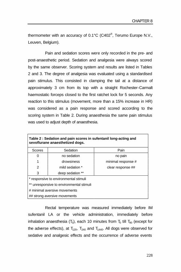
CHAPTER 8
228
thermometer with an accuracy of 0.1°C (C402®, Terumo Europe N.V.,
Leuven, Belgium).
Pain and sedation scores were only recorded in the pre- and
post-anaesthetic period. Sedation and analgesia were always scored
by the same observer. Scoring system and results are listed in Tables
2 and 3. The degree of analgesia was evaluated using a standardised
pain stimulus. This consisted in clamping the tail at a distance of
approximately 3 cm from its top with a straight Rochester-Carmalt
haemostatic forceps closed to the first ratchet lock for 5 seconds. Any
reaction to this stimulus (movement, more than a 15% increase in HR)
was considered as a pain response and scored according to the
scoring system in Table 2. During anaesthesia the same pain stimulus
was used to adjust depth of anaesthesia.
Table 2 : Sedation and pain scores in sufentanil long-acting and sevoflurane anaesthetized dogs.
Scores Sedation Pain
0 no sedation no pain
1 drowsiness minimal response #
2 mild sedation * clear response ##
3 deep sedation **
* responsive to environmental stimuli
** unresponsive to environmental stimuli
# minimal aversive movements
## strong aversive movements
Rectal temperature was measured immediately before IM
sufentanil LA or the vehicle administration, immediately before
inhalation anaesthesia (T0), each 10 minutes from T0 till T90 (except for
the adverse effects), at T120, T240 and T1440. All dogs were observed for
sedative and analgesic effects and the occurrence of adverse events

CHAPTER 8
229
(lateral recumbency, ataxia, noise-sensitivity, vomiting, defaecation,
salivation, nystagmus, excitation, …) immediately before
administration of sufentanil/vehicle, immediately before initiation of
inhalation anaesthesia (T0) and at T90, T120, T240 and T1440.
At the end of inhalation anaesthesia, oxygen was supplied
until extubation was possible. The dogs were transferred to the
recovery box. If the rectal temperature was lower than 36°C, an
infrared heating source was installed in the recovery box.
Statistical Analysis
The dogs were sorted according to decreasing body weight and
divided into 8 classes of 5 dogs each. In each class, the body weights
were comparable. Within each class the 5 dogs were randomly
allocated to the five different study groups and the 40 phases by a
computer randomisation program. The statistical tests were two-sided
and used a 0.05 type I error (α=5%).
The StatXact software (StatXact 3 For Windows (1995), Users
Manual, CYTEL Software, Cambridge, MA 02139 USA) was used for
the Kruskal-Wallis one-way analysis of variance tests, for the
Wilcoxon Mann-Whitney U tests and for the Fisher’s exact tests
(Siegel, 1977).
The treatment groups A (control suf) and B (control sevo)
were control groups. No statistics were done for these groups, since
the effects of sufentanil LA and sevoflurane were studied previously in
dogs (Bernard et al., 1990; Engelen et al., 1996a; Engelen et al.,
1996b; Engelen et al., 1996c; Short, 1996; Short and Vlaminck, 1998;
Verbeeck et al., 1998; Hoeben et al., 1999; Sterkens et al., 1999).

CHAPTER 8
230
The body weights recorded seven days before the first study
phase were compared by means of the Kruskal-Wallis one-way
analysis of variance test to check the randomisation procedure. To
evaluate the homogeneity of the several treatment groups, baseline
comparisons (T0) were also performed on rectal temperature, sedation
scores and analgesia scores of each treatment group by means of the
Kruskal-Wallis one-way analysis of variance test.
For each parameter (rectal temperature, overall analysis of
critical events, adverse events) the following tests were performed:
statistical comparisons on the mean over time (T0 to T90) was
performed between the treatment groups C0, D15 and E30 by means of
the Kruskal-Wallis one-way analysis of variance test followed by two-
by-two Wilcoxon Mann-Whitney U tests. Each of the treatment groups
C0, D15 and E30 was compared with the control groups A and B by
Wilcoxon Mann-Whitney U tests.
The individual time points after inhalation anaesthesia (T120,
T240 and T1140) were analysed in the same way as the variable mean
over time. For sedation and analgesia scores statistics were
performed on the individual values of each time point (T120, T240 and
T1140). Statistical comparisons were performed between the treatment
groups C0, D15 and E30 by means of the Kruskal-Wallis one-way
analysis of variance test followed by two-by-two Wilcoxon Mann-
Whitney U tests. Each of the treatment groups C0, D15 and E30 were
compared with the treatment groups A and B by Wilcoxon Mann-
Whitney U tests.

CHAPTER 8
231
RESULTS
Results for different measured variables are given in Table 3
and 4. No significant differences in baseline values per study group
and per parameter were observed. At T0 sedation scores were
significantly higher in groups D15 and E30 (injected 15 and 30 minutes
before T0 respectively) compared to groups A, B and C0 (p<0.01 and
p<0.001). In the post-anaesthetic period, compared to group B
(control sevo) sedation scores in sufentanil treated inhalation groups
were significantly higher at T120 in group E30 (p<0.01), at T240 in group
C0, D15 and E30 (p<0.001) and at T1440 in group E30 (p<0.05). At T120
sedation scores in group E30 were significantly higher compared to
group A (control suf) (p<0.05).
Antinociceptive effects of sufentanil LA were suggested by
differences in pain scores in the pre- and post-anaesthetic period.
Pain score at T0 was significantly lower in group E30 compared to
group B and C0 (p<0.05). Compared to group B (control sevo) pain
scores in sufentanil treated inhalation groups were significantly lower
at T120 in group C0 and E30 (p<0.01 and p<0.05), at T240 in group C0,
D15 and E30 (p<0.01; p<0.05 and p<0.01) and at T1440 in group C0 and
E30 (p<0.05). At T120 pain scores in group C0 were significantly lower
compared to group A (control suf) (p<0.05).
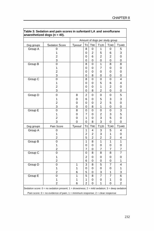
CHAPTER 8
232
Table 3: Sedation and pain scores in sufentanil LA and sevoflurane anaesthetized dogs (n = 40).
Amount of dogs per study group
Dog groups Sedation Score Tpresuf T0 T90 T120 T240 T1440 Group A 0 8 0 1 0 5
1 0 2 5 6 3 2 0 6 2 2 0 3 0 0 0 0 0
Group B 0 8 0 1 8 8 1 0 0 7 0 0 2 0 0 0 0 0 3 0 8 0 0 0
Group C 0 8 0 0 0 4 1 0 0 5 6 4 2 0 0 1 2 0 3 0 8 2 0 0
Group D 0 8 2 0 0 0 5 1 0 6 0 5 3 3 2 0 0 0 2 5 0 3 0 0 8 1 0 0
Group E 0 8 0 0 0 0 3 1 0 7 0 2 3 5 2 0 1 0 3 5 0 3 0 0 8 3 0 0
Dog groups Pain Score Tpresuf T0 T90 T120 T240 T1440 Group A 0 1 4 3 5 4
1 2 2 3 1 0 2 5 2 2 2 4
Group B 0 1 8 1 1 1 1 0 0 0 0 0 2 7 0 7 7 7
Group C 0 0 8 8 8 7 1 2 0 0 0 0 2 6 0 0 0 1
Group D 0 1 3 8 5 7 4 1 1 0 0 0 0 1 2 6 5 0 3 1 3
Group E 0 1 5 8 7 7 6 1 1 1 0 0 1 0 2 6 2 0 1 0 2
Sedation score: 0 = no sedation present; 1 = drowsiness; 2 = mild sedation; 3 = deep sedation
Pain score: 0 = no evidence of pain; 1 = minimum response; 2 = clear response

CHAPTER 8
233
The mean end-tidal sevoflurane concentration (Sevo ET%) in
group B (control sevo) over the time T0 to T90 was 2.4 ± 0.2% and
considered to be 100%. There was a significant reduction in Sevo
ET% in all sufentanil treated groups (C0, D15, E30) compared to group
B (p<0.05; p<0.01 and p<0.001). Differences between these groups
were not significant. Sevo ET% was reduced by 29.2% in group D15
(mean Sevo ET%: 1.7 ± 0.4%), by 19.8% in group C0 (mean Sevo
ET%: 1.9 ± 0.5%) and by 16.2% in group E30 (mean Sevo ET%: 2.0 ±
0.1%). The amount of thiopental administered in group B was
considered to be 100% (mean: 15.9 mg/kg ± 2.7). The amount of
thiopental needed for induction in sufentanil treated groups was
significantly reduced. This reduction was 6.8% (mean: 14.8 ± 1.4
mg/kg) in group C0 (non-significant), 30.2% (mean: 11.1 ± 1.6 mg/kg)
in group D15 (p<0.01) and 29.4% (mean: 11.2 ± 2.5 mg/kg) in group
E30 (p<0.01), all compared to group B. The reduction in administered
thiopental was significantly lower in groups D15 and E30 compared to
group C0 (p<0.01).
Body temperature decreased in all groups during the
anaesthesia period (T0 to T90). Body temperature in group D15 and E30
was significantly lower compared to group A and C0 (p<0.01 and
p<0,05). There was no significant differences between group B
(control sevo) and the sufentanil treated inhalation groups. In group A
(control suf) the initial decrease in temperature was more gradually
than in the inhalation groups. In the post-anaesthetic period body
temperature returned quickly to baseline values in group B (control
sevo). In the sufentanil treated groups C0, D15 and E30 body
temperature remained low and compared to group B the difference
was significant at every time point (T120, T240 and T1440). In the post-
anaesthetic period body temperature remained rather constant in all

CHAPTER 8
234
sufentanil groups and hypothermia was observed even after 24 hours.
Body temperature was significantly lower in group E30 compared to C0
at T120 and T240 (p<0.05). Body temperature was significantly lower
compared to group A at T120 in groups D15 and E30 (p<0.05 and
p<0.01) and at T240 in group E30 (p<0.05). Minimum body temperature
observed were in group A 36.3°C (T240), in group B 36.7°C (T80, T90),
in group C0 35.9°C (T240), in group D15 35.7°C (T90, T120) and in group
E30 35.3°C (T70, T240). When body temperature in the post-anaesthetic
period descended below 36.0°C an infrared heating source was
installed at ± 70 cm above the dogs.
In the post-anaesthetic period lateral recumbency was not
observed in group B (control sevo). The incidence of lateral
recumbency in group C0 decreased from 62.5% (T120) to 12.5% (T240)
and 0% (T1440). In group D15 incidence of lateral recumbency
decreased from 25% (T120) to12.5% (T240) and 0% (T1440). In group E30

CHAPTER 8
235
this decrease was from 75% to 62.5% and 12.5% respectively. In
group A (control suf) lateral recumbency was observed in maximally
50% of the dogs (T90), this decreased to 12.5% (T120), 25.0% (T240)
and 0% (T1440).
At T0 12.5% of the dogs in group E30 were ataxic and none in
the other groups. In the control group B (control sevo) ataxia was only
observed in 50% of the dogs at T120 . The incidence of ataxia in group
C0 was 37.5% (T120), this increased to 87.5% (T240) and decreased to
0% (T1440). In group D15 ataxia was 50% (T120), this decreased to
37.5% (T240) and 12.5% (T1440). In group E30 the incidence of ataxia
was 50% (T120), this decreased to 37.5% (T240 and T1440). In group A
(control suf) ataxia was observed in 37.5% of the dogs (T90), this
changed to 12.5% (T120), 75.0 % (T240) and 12.5% (T1440).
Incidence of increased noise sensitivity at T120 was 25%
(group B), 62.5% (group C0), 25% (group D15 and E30). At T240 the
incidence was 25% (group B), 50% (group C0 and E30), 62.5% (group
D15). Incidence of increased noise sensitivity was observed in group A
in 87.5% (T120), 100% (T240) and 75.0% (T1440).
In group B (control sevo) 12.5% of the dogs defaecated in the
period T240-T1440. Defaecation was observed in group C0 in the period
T120- T240 (12.5%). Defaecation was seen in group D15 in the period
T90-T120 (62.5%) and T120-T240 (12.5%). Defaecation was seen in group
E30 in the period T120-T240 (50.0%) and T240-T1440 (12.5%). In group A
(control suf) 12,5% of the dogs defaecated in de period T90- T120 and
T120-T240. Vomiting nor the presence of nystagmus was observed in
any dog at any time point. Salivation was seen in group A (control suf)
in 25.0% and 12.5% of the dogs at T90 and at T120 respectively. 25.0%
of the dogs in groups B, C0 and D15 showed excitation at T120. No
other adverse events were observed at any time point.
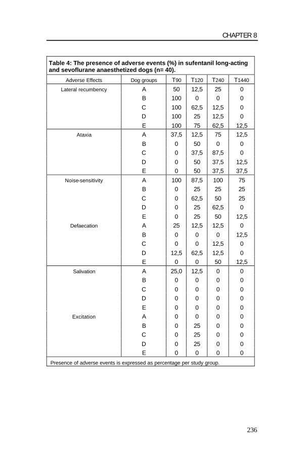
CHAPTER 8
236
Table 4: The presence of adverse events (%) in sufentanil long-acting and sevoflurane anaesthetized dogs (n= 40).
Adverse Effects Dog groups T90 T120 T240 T1440
Lateral recumbency A 50 12,5 25 0 B 100 0 0 0
C 100 62,5 12,5 0 D 100 25 12,5 0
E 100 75 62,5 12,5 Ataxia A 37,5 12,5 75 12,5
B 0 50 0 0 C 0 37,5 87,5 0
D 0 50 37,5 12,5 E 0 50 37,5 37,5
Noise-sensitivity A 100 87,5 100 75 B 0 25 25 25
C 0 62,5 50 25 D 0 25 62,5 0
E 0 25 50 12,5 Defaecation A 25 12,5 12,5 0
B 0 0 0 12,5 C 0 0 12,5 0
D 12,5 62,5 12,5 0 E 0 0 50 12,5
Salivation A 25,0 12,5 0 0 B 0 0 0 0
C 0 0 0 0 D 0 0 0 0
E 0 0 0 0 Excitation A 0 0 0 0
B 0 25 0 0 C 0 25 0 0
D 0 25 0 0 E 0 0 0 0
Presence of adverse events is expressed as percentage per study group.
CHAPTER 8
237
DISCUSSION
Haemodynamic observations during this study were
discussed in the first part (Chapter 7). Additional observations were
the degree of sedation, antinociceptive and sevoflurane-sparing
effects and the occurrence of side effects.
Early signs of sedation and analgesia occurred 15 minutes
after sufentanil LA administration and were long lasting (24 hours).
The fast decrease in pain scores in groups D15 and E30 with
respectively 3 and 5 dogs/8 with minimal pain scores at T0 indicated a
rapid systemic resorption from the IM injection site. In previous studies
sufentanil LA plasma levels very rapidly increased (0.55 ± 0.12 ng/ml
and 0.59 ± 0.34 ng/ml (mean ± SD) 15 and 30 minutes after
intramuscular administration respectively) and peak levels around
1.53 ± 0.45 ng/ml were observed around 6 hours after IM injection.
Sufentanil LA plasma levels of 0.85 ng/ml were necessary to provide
good analgesia during major surgery (Short, 1996; Short and
Vlaminck, 1998; Verbeeck et al., 1998; Hoeben et al., 1999; Sterkens
et al., 1999). Only one dog showed mild sedation at T0. The analgesic
effect was pronounced with 20/24 dogs at T120 and 22/24 dogs at T240
with minimal pain scores.
Analgesic effects and to a lesser extent, the sedative effects,
were long lasting (24 hours). At T1440 still 17/24 dogs showed minimal
pain scores compared to only one dog in group B (control sevo). In
group B (control sevo) no recumbency was observed in the recovery
period. In groups C0, D15 and E30 25% to 75% dogs were still
recumbent at T120 and 12.5%- 62.5% at T240. This illustrates the strong
sedative effects of sufentanil LA. A specific problem rose when the
CHAPTER 8
238
presence of ataxia in the dogs had to be evaluated, because many
dogs couldn’t walk, adequate scoring was not always possible.
Probably the dogs were ataxic at those time points. Nevertheless, at
T240 when no dogs were ataxic in group B ataxia was still observed in
several dogs in all sufentanil groups at T240 (37.5%-87.5%) and T1440
(12.5%-37.5%). This was indicative for the long residual sedative
effect of sufentanil LA in these dogs. Sedative effects at T1440 were
limited to drowsiness in 12/24 dogs compared to none in group B
(control sevo). The long lasting analgesic and sedative effects in all
sufentanil groups could be related to a slow decrease in plasma
sufentanil concentration. T1/2 of sufentanil was reported to be 15.8 ±
5.1 hours (Verbeeck et al., 1998; Hoeben et al., 1999; Sterkens,
1999).
Significantly more sevoflurane (100%) had to be administered
in group B (control sevo) compared to the sufentanil-sevoflurane
groups. The sparing effect of sufentanil LA after IM administration on
sevoflurane anaesthesia found (29.2%) was very similar as the
sparing effect accompanying IV administration of fentanyl and
sufentanil during enflurane and isoflurane anaesthesia in dogs (Hall et
al., 1987a; Schwieger et al., 1991; Hellyer et al., 2001). To our
knowledge sevoflurane sparing effects of opioids in dogs are not yet
described. The anesthetic sparing effect is likely caused by a
“reduction” of the MAC value of the inhalation anesthetic. The MAC-
value of potent volatile anaesthetics is reduced by increasing plasma
concentrations of opioids (Valverde et al., 1989; Sebel et al., 1992;
Brunner et al., 1994; Ilkiw et al., 1997). The degree by which MAC is
reduced may be used as a measure of opioids potency (McEwan et
al., 1993). Following intravenous application of opioids (alfentanil,
butorphanol, fentanyl, morphine, nalbuphine, remifentanil, sufentanil)

CHAPTER 8
239
during inhalation anaesthesia MAC reductions up to 73% have been
reported (Murphy and Hug, 1982a; Murphy and Hug, 1982b; Hall et
al., 1987a; Hall et al., 1987b; Schwieger et al., 1991; Michelsen et al.,
1996; Hellyer et al., 2001).
The barbiturate-sparing effect was very similar in groups D15
(30.2%) and E30 (29.4%), but, as expected, much less (6.8%) in group
C0. The barbiturate induction dosage amount is markedly less reduced
(10-20%) when tranquilizers are used as premedication.
Premedication with midazolam and diazepam, two benzodiazepines,
reduced the thiamylal (ultra-short acting barbiturate) dose required to
accomplish endotracheal intubation in dogs (Muir et al., 1991;
Tranquilli et al., 1991; Greene et al., 1993). The amount of thiopental
required to produce loss of the eyelid reflex is reduced with the
concomitant use of opiates and benzodiazepines (butorphanol and
diazepam) for premedication in humans (Sklar et al., 1989). To our
knowledge, barbiturate sparing effects of opioids were not reported in
dogs.
An important side effect was the development of hypothermia
in all sufentanil treated groups that persisted for 24 hours even in the
presence of an external heating source. Body temperature is
controlled by a complex, highly integrated system that carefully
balances heat production and heat loss. Heat is produced as a
byproduct of metabolism, and as a result of muscular work, shivering,
and chemical thermogenesis, whereas heat is lost from the body
through the channels of heat exchange: radiation, conduction,
convection and evaporation (Machon et al., 1999). In the present
study the decreased body temperature during inhalation anaesthesia
was expected since hypothermia is a common and potentially serious
complication of general anaesthesia on one hand and opioid

CHAPTER 8
240
administration on the other hand (Bissonnette, 1991). Opioids as
alfentanil, pethidine, fentanyl and sufentanil impair thermoregulatory
control by increasing the thresholds for sweating and decreasing the
thresholds for vasoconstriction and shivering (Ikeda et al., 1997;
Alfonsi, 1998). As expected, the body temperature of the dogs from
group B (control sevo) returned very fast to base line values.
Hypothermia was probably partially induced by stimulation of the
serotonine receptors and by cessation of shivering in the dogs.
Shivering is a source of metabolic heat production (Okada et al.,
1998). Body temperature deregulating effects by inhibition of shivering
are also described in humans after parenteral administration of
pethidine, sufentanil and alfentanil (Ikeda et al., 1997; Alfonsi et al.,
1998).
Noise sensitivity was evaluated with hand clapping; a clear
response of the dog was scored as present noise sensitivity. In the
post-anaesthetic period the treated sufentanil dogs could be easily
aroused from their sedation by auditory stimulation. Their reaction to
hand clapping was exaggerated, but they were sedated again soon
after cessation of hand clapping. This exaggerated response to loud
noises is also seen after fentanyl administration (Thurmon et al., 1996;
Lukasik, 1999).
None of the dogs of any study group vomited over the entire
study protocol. Initially, morphine induces vomiting by activating the
chemoreceptor trigger zone, probably through a partial dopamine
agonist effect, but appears to be the only opioid, which induces
vomiting in dogs and cats (Kromer, 1988; Zuckerman and Ferrante,
1998). Defaecation in sufentanil treated dogs tended to be more
frequent than in the control group B with up to 60% at certain time
points compared to 12%. Defaecation is also frequently seen after
CHAPTER 8
241
fentanyl injection due to the occurring anal sphincter relaxation
(Thurmon et al., 1996; Lukasik, 1999). In contrast, in human studies
opioids are associated with a generalized depressant effect on
gastrointestinal motility with reduced propulsive peristaltic activity
during prolonged opioid administration (reflex inhibition) (Zuckerman
and Ferrante, 1998). Salivation was noticed in group A at T90 and T120,
but not in other groups. Salivation was also described after morphine
administration in cats and after methadone administration in dogs
(Burroughs, 1953; Davis and Donnely, 1968).
Excitation was only seen at T120 in groups B, C0 and D15 in two
dogs per group. Morphine in clinical doses can induce excitement on
rare occasions in dogs and cats (Lukasik, 1999; Nolan, 2000).
Excitation after sufentanil administration is not mentioned in literature,
probably because the registered short acting formulation of sufentanil
is mostly used for intra-operative analgesia and not for postoperative
pain relief.
In conclusion, the present study showed that the combination
of sufentanil LA and sevoflurane anaesthesia was associated with a
significant reduction in end-tidal sevoflurane concentrations necessary
to avoid reaction to a standardized pain stimulus. This effect was most
pronounced when sufentanil LA was administered 15 minutes before
induction of anaesthesia. In the post-anaesthetic period pain scores
were lower and sedation scores higher in the sufentanil-treated
groups. In many dogs diminished pain and elevated sedation scores
persisted during 24 hours. Hypothermia was observed in all sufentanil
groups and persisted during 24 hours in spite of external heating
source. Other side effects such as lateral recumbency, ataxia, arousal
on auditory stimulation, defaecation, salivation and excitation in some
dogs were not clinically relevant.
CHAPTER 8
242
REFERENCES
Alfonsi, P., D.I. Sessler, B. Du Manoir, J.C. Levron, J.P. Le Moing, and M. Chauv, 1998 : The effects of meperidine and sufentanil on the shivering treshold in postoperative patients. Anesthesiology 89, 43-48. Bernard, J.M., P.F. Wouters, M.F. Doursout, B. Florence, J.E. Chelly, and R.G. Merin, 1990: Effects of sevoflurane and isoflurane on cardiac and coronary dynamics in chronically instrumented dogs. Anesthesiology 72, 659-662. Bissonnette, B., 1991: Body temperature and anesthesia. Anesthesiology Clinics of North America 9, 849-864. Brunner, M.D., P. Braithwaite, R. Jhaveri, A.I. McEwan, D.K. Goodman, L.R. Smith, and P.S.A. Glass, 1994: MAC reduction of isoflurane by sufentanil. British Journal of Anaesthesia 72, 42-46. Burroughs, H.E., 1953: Methadone narcosis in dogs. Journal of Small Animal Medicine 1, 301. Davis, L.E., and E.J. Donnely, 1968: Analgesic drugs in the cat. Journal of the American Veterinary Medicine Association 153, 1161. Engelen, M., A. Proost, and K. Vlaminck, 1996a: Pilot trial on the pharmacodynamic effects of sufentanil after a single intramuscular administration of a new depot formulation (0.35 mg/ml injectable solution in Miglyol) at a dose of 35 µg/kg in Beagle dogs (Protocol No. SUF-94-PREC-01). Janssen Pharmaceutica Preclinical R&D Report. Engelen, M., A. Proost, and K. Vlaminck, 1996b: Pilot trial on the pharmacodynamic effects of sufentanil after a single intramuscular administration of a new depot formulation (0.35 mg/ml injectable solution in Miglyol) at a dose of 70 µg/kg in Beagle dogs (Protocol No. SUF-95-PREC-01). Janssen Pharmaceutica Preclinical R&D Report. Engelen, M., A. Proost, and K. Vlaminck, 1996c: Pilot trial on the pharmacodynamic effects of sufentanil after a single intramuscular administration of a new depot formulation (0.50 mg/ml injectable solution in Miglyol) at a dose of 35, 50 or 70 µg/kg in Beagle dogs (Protocol No. SUF-95-PREC-02). Janssen Pharmaceutica Preclinical R&D Report. Greene, S.A., G.J. Benson, S.M. Hartsfield, 1993: Thiamylal-sparing effectnof midazolam for canine endotracheal intubation. A clinical study of 118 dogs. Veterinary Surgery 22, 69-72. Hall, R.I., M.R. Murphy, and C.C.Jr Hug, 1987a: The enflurane sparing effect of sufentanil in dogs. Anesthesiology 67, 518-525.
CHAPTER 8
243
Hall, R.I., F. Szlam, and C.C.Jr. Hug, 1987b: The enflurane-sparing effect of alfentanil in dogs. Anesthesia & Analgesia 66, 1287-1291. Hellebrekers, L.J., and R. Sap, 1991: Anesthesia in the patient with stomach dilatation-volvulus. Tijdschrift voor Diergeneeskunde 116, 130-136. Hellyer, P.W., K.R. Mama, H.L. Shafford, A.E. Wagner, and C. Kollias-Baker, 2001: Effects of diazepam and flumazenil on minimum alveolar concentrations for dogs anesthetized with isoflurane or a combination of isoflurane and fentanyl. American Journal of Veterinary Research 62, 555-560. Hoeben, D., J. Verbeeck, K. Vlaminck, R. Mostmans, and E. Anthonissens, 1999: Clinical and pharmacokinetic study with a sufentanil depot formulation: GLP tolerance trial (Protocol No. V/SUF-BEL-2). Janssen Animal Health Preclinical R&D Report, Revised Version. Ikeda, T., A. Kurz, D.I., Sessler, J. Go, M. Kurz, K. Belani, M. Larson, A.R. Bjorksten, M. Dechert, and R. Christensen, 1997: The effects of opioids on thermoregulatory responses in humans and the special antishivering action of meperidine. Annales of the New York Academy of Sciences 15, 792-798. Ilkiw, J.E., P.J. Pascoe, L.D. Fisher, 1997: Effect of alfentanil on the minimum alveolar concentration of isoflurane in cats. American Journal of Veterinary Research 58, 1274-1279. Kromer, W., 1988: Endogenous and exogenous opioids in the control of gastrointestinal motility and secretion. Pharmacological Reviews 40, 121-162. Lukasik, V.M., 1999: Premedication and sedation. In: Manual of Small Animal Anaesthesia and Analgesia, Ed. By Seymour C., Gleed R.; BSAVA, Cheltenham U.K.; 71-85. Machon, R.G., M.R. Raffe, and E.P. Robinson, 1999: Warming with a forced air blanket minimizes anesthetic-induced hypothermia in cats. Veterinary Surgery 28, 301-310. McEwan, A.I., C. Smith, O. Dyar, D. Goodman, L.R. Smith, and P.S.A. Glass, 1993: Isoflurane minimum alveolar concentration reduction by fentanyl. Anesthesiology 78, 864-869. Michelsen, L.G., M. Salmenpera, C.C Hug, F. Szlam, and D. Vander Meer, 1996: Anesthetic potency of remifentanyl in dogs. Anesthesiology 84, 865-872. Moon, P.F., J.M. Scarlett, J.W. Ludders, T.A. Conway, and S.V. Lamb, 1995: Effect of fentanyl on the medium alveolar concentration of isoflurane in swine. Anesthesiology 83, 535-542.
CHAPTER 8
244
Muir, W.W., 1998a: Anesthesia for dogs and cats with cardiovascular disease – Part I. Compendium on Continuing Education for the Practicing Veterinarian, 20, 78-87. Muir, W.W., 1998b: Anesthesia for dogs and cats with cardiovascular disease – Part II. Compendium on Continuing Education for the Practicing Veterinarian, 20, 473-484. Muir, W.W., L. Bednarski, and R. Bednarski, 1991: Thiamylal- and halothane-sparing effect of diazepam in dogs. Journal of Veterinary Pharmacology and Therapeutics 14, 46-50. Murphy, M.R., and C.C.Jr. Hug, 1982a: The anesthetic potency of fentanyl in terms of its reduction of enflurane MAC. Anesthesiology 57, 485-488. Murphy, M.R., and C.C.Jr. Hug, 1982b: The enflurane sparing effect of morphine, butorphanol, and nalbuphine. Anesthesiology 57, 489-492. Mutoh, T., R. Nishimura, H.-Y. Kim, S. Matsunaga, and N. Sasaki, 1997: Cardiopulmonary effects of sevoflurane, compared with halothane, enflurane, and isoflurane, in dogs. American Journal of Veterinary Research 58, 885-890. Nolan, A.M., 2000: Pharmacology of analgesic drugs. In: Pain Management in animals. Ed. Flecknell P. and Waterman-Pearson A. W.B. Saunders, London, U.K.; 21-52. Oden, R.V., 1989: Acute postoperative pain. Incidence, severity and the etiology of inadequate treatment. Anesthesiology Clinics of North America 7, 1-15. Okada, Y., M. Powis, A. McEwan, and A. Pierro, 1998: Fentanyl analgesia increases the incidence of postoperative hypothermia in neonates. Pediatric Surgery International 13, 508-511. Pascoe, P., 1992: Control of postoperative pain in animals receiving inhalant anesthetics. In: Short C.E., Van Poznak A. (Eds). Animal Pain. Churchill Livingstone, New York: 348-352. Polis, I., F. Gasthuys, H. Laevens, L. Van Ham, and A. De Rick, 2001: The influence of ventilation mode (spontaneous ventilation, IPPV and PEEP) on cardiopulmonary parameters in sevoflurane anaesthetized dogs. Journal of Veterinary Medicine A 48, 619-630. Rawlings, C.A., and R.J. Kolata, 1983: Cardiopulmonary effects of thiopental/lidocaine combination during anesthetic induction in the dog. American Journal of Veterinary Research 44, 144-149.

CHAPTER 8
245
Schwieger, I.M., R.I. Hall, and C.C. Hug, 1991: Less than additive antinociceptive interaction between midazolam and fentanyl in enflurane-anesthetized dogs. Anesthesiology 74, 1060-1066. Sebel, P.S., P.S.A. Glass, J.E. Fletcher, M.R. Murphy, C. Gallagher, and T. Quill, 1992: Reduction of MAC of desflurane with fentanyl. Anesthesiology 76, 52-59. Short, C.E., 1996: The evaluation of R033800 (sufentanil) as a selected Janssen Pharmaceutica opioid agonist for use in canine pain control: study report (Protocol No. SUF-94-US-01). Cornell University, USA Janssen Pharmaceutica Preclinical R&D Report. Short, C. E., and K. Vlaminck, 1998: The evaluation of R030730 as a selected Janssen Pharmaceutica opioid agonist for use in canine pain control: dose confirmation trial (Protocol No. SUF 96-US-01). Janssen Animal Health Preclinical R&D Report. Siegel, S., 1977: The Mann-Whitney U test. In: Nonparametric statistics for the behavioural sciences. McGraw – Hill Kogakusha, Ltd., 116-127. Sinatra, R.S.,1991 : Current methods of controlling postoperative pain. Yale Journal of Biology and Medicine 64, 351-374. Sklar, G.S., D.D. Sonn, and W.A. Watson, 1989: Thiopental-sparing properties of butorphanol/diazepam for induction of anesthesia in ambulatory gynecologic surgery. The Annales of Pharmacotherapy 23, 659-662. Sterkens, P., 1999: Plasma kinetics of sufentanil (R030730) in the beagle dog in an intramuscular tolerance study (V/SUF-BEL-2) of sufentanil at 50, 100 and 150 µg/kg with a miglyol depot formulation (Protocol No. R030730/FK2828). Janssen Pharmaceutica Non-clinical Pharmacokinetics Report. Thurmon, J.C., W.J. Tranquilli, and G.J. Benson,1996 : Preanesthetic and anesthetic adjuncts. In: Lumb &Jones Veterinary Anesthesia. 3rd ed., 183-209. Tranquilli, W.J., L.M. Graning, J.C. Thurmon, G.J. Benson, S.G. Moum, and E.L. Lentz, 1991: Effect of midazolam preanesthetic administration on thiamylal induction requirement in dogs. American Journal of Veterinary Research 52, 662-664. Valverde, A., D.H. Dyson, and W.N. McDonell, 1989: Epidural morphine reduces halothane MAC in the dog. Canadian Journal of Anaesthesiology 36, 629-632. Verbeeck, J., K. Vlaminck, R. Mostmans, and L. Gypen, 1998: Pharmacokinetic and pharmacodynamic study with a sufentanil long-acting formulation: GLP dose finding trial in dogs (Protocol No. SUF-97-PREC-01). Janssen Animal Health Preclinical R&D Report.

CHAPTER 8
246
Wallin, R.F., B.M. Regan, M.D. Napoli, and I.J.Stern, 1975: Sevoflurane: A new inhalational anesthetic agent. Anesthesia & Analgesia 54, 758-766. Zuckerman L.A., and F.M. Ferrante, 1998: Nonopioid and opioid analgesics. In: The management of pain. Eds.: Ashburn M.A., Rice L.J. Churchill Livingstone, Philadelphia. p.125.

GENERAL DISCUSSION
247
GENERAL DISCUSSION

GENERAL DISCUSSION
248

GENERAL DISCUSSION
249
Major progress in human and veterinary anaesthesia has been
established the last few decades. Sevoflurane was introduced into the
anaesthetic theatre in the early seventies (Wallin et al., 1975).
Sevoflurane meets entirely the new trend in volatile anaesthetic drugs
permitting more precise control of anaesthesia and perhaps most
importantly, more rapid recovery from the effects of anaesthesia
(Eger, 1994). Up to now, a lot of research has been done on
sevoflurane anaesthesia in human medicine (Katoh and Ikeda, 1987;
Lerman, 1993). However, research on sevoflurane in veterinary
anaesthesia is rather limited. Especially its clinical use in dogs and the
potential clinical applications of sevoflurane in small animal
anaesthesia are not studied widely yet. A review on the physico-
chemical and anaesthetic properties of sevoflurane in human and
small animal medicine was described in chapters 1 and 2.
In chapter 3 the influences of sevoflurane on recovery times
and haemodynamic parameters in dogs after 1 hour of anaesthesia
were evaluated in a clinical study. The emphasis was put on the
clinical aspect of the study. Was the low blood-gas solubility of
sevoflurane also accompanied by rapid recoveries in premedicated
dogs? Moreover, emergency times were compared with those after
halothane and isoflurane anaesthesia at 2 anaesthetic concentrations
(1.5 and 2 MAC). A neurolept-analgesic mixture, fentanyl and
droperidol, was chosen as premedication to simulate clinical
anaesthesia. Droperidol has a long duration of action and can
influence the recovery profile (Thurmon et al., 1996; Bissonnette et al.,
1999). Anaesthesia was induced with propofol, a short acting
induction agent, with rapid metabolisation (Shafer et al., 1988). Its low
biological half life was probably minimally influencing recovery after 1
hour of anaesthesia (Shafer, 1993; Smith et al., 1994).

GENERAL DISCUSSION
250
However, anaesthetic recovery is influenced by many other
factors. The most important factor is the blood/gas partition coefficient
of the anaesthetic agent. A low blood/gas partition coefficient allows
more rapid drug elimination and results in a shorter emergence time.
The blood/gas solubilities of sevoflurane, isoflurane and halothane in
man are respectively 0.68, 1.46 and 2.54 (Strum and Eger, 1987;
Steffey, 1996). Of minor importance for anaesthetic recovery are the
oil/gas partition coefficient, metabolisation percentage, alveolar
ventilation, cardiac output, duration of anaesthesia, percutaneous
losses, etc. (Stoelting and Eger, 1969; Carpenter et al., 1987;
Lockhart et al., 1991; Steffey, 1996).
In the present work no significant emergence times between
the 3 anaesthetic agents occurred, although the emergence time was
the longest for halothane at both anaesthetic concentrations. It could
be concluded that it was more difficult to show the kinetic advantages
of less soluble anaesthetics, as sevoflurane after short duration
anaesthetic exposures of 1 hour (Eger and Johnson, 1987). In
addition, residual effects of premedication in clinical anaesthesia
exerted a depressant effect on cognitive function nullifying any kinetic
advantage of sevoflurane over isoflurane and halothane. In
conclusion, clinically there was little difference in emergence times
between halothane, isoflurane, and sevoflurane in premedicated dogs
after 1 hour of inhalation anaesthesia.
The influence of ventilation mode on cardiopulmonary
parameters in sevoflurane anaesthetized dogs was also evaluated
during clinical anaesthesia (chapter 4). Three types of ventilation
namely spontaneous ventilation, intermittent positive pressure
ventilation and positive end expiratory pressure (5 cm H2O) were

GENERAL DISCUSSION
251
compared at 2 anaesthetic concentrations (1.5 and 2 MAC) of
sevoflurane in a clinical protocol including standard premedication and
induction. This study shows that in anaesthetized spontaneously
ventilating dogs increasing MAC values of sevoflurane from 1.5 to 2
induced a pronounced cardiopulmonary depression together with a
significant increase in HR. However, this increase was clinically not
very relevant. The increased HR could be explained by the
baroreceptor-reflex and/or by sympathetic stimulation. The
cardiopulmonary depression was characterized by decreases in SI,
CI, LVSWI and increases in PCWP and PAP. This cardiac depression
was probably related to the presence of a decreased myocardial
contractility caused by increasing the MAC value (Suga et al., 1985;
Mutoh et al., 1997). A reduced myocardial contractility is often
compensated by an increase in end diastolic pressure (Kittleson,
1988). Hence, PCWP and PAP which are good reflections of the end
diastolic pressure, increased in the present study (Brutsaert et al.,
1985). However, it should be underlined that it remains difficult to
explain the pharmacologic effects in this setting because several
drugs were used concomitantly.
Changing from SpV to IPPV or PEEP using 1.5 MAC induced
a small increase of arterial blood pressures, right cardiac pressures
and HR. Little to no influences on other cardiac parameters were
observed. On the other hand, the situation changed completely with
increasing to 2 MAC. Artificial ventilation with 2 MAC sevoflurane
induced a sever impact on the arterial pressures and cardiac related
parameters. CO, CI, SV, SI, LVSWI and RVSWI decreased
significantly. The impact was more pronounced with PEEP, but the
difference between PEEP and IPPV was not significant. Right cardiac
pressures increased, while arterial pressures decreased. The main

GENERAL DISCUSSION
252
patho-physiologic mechanism for these cardiovascular side effects of
PEEP was a decreased venous return due to the increased
intrathoracic pressure and a decreased coronary blood flow inversely
related with the PEEP level (Jacobs and Venus, 1983; Versprille,
1990).
In conclusion, sevoflurane anaesthesia at 1.5 MAC in
premedicated healthy dogs induced a relatively moderate
cardiopulmonary depression during spontaneous and controlled
ventilation (IPPV and PEEP of 5 cm H²O) and could therefore be used
safely for clinical anaesthesia. On the contrary, increasing the MAC
from 1.5 to 2 caused a marked cardiopulmonary depression.
Consequently, higher concentrations of sevoflurane must be avoided
during all ventilation modes in dogs.
The following chapters discussed some practical applications
of sevoflurane in specific anaesthesia protocols (TLV with CO2-
insufflation, the use of pre-emptive analgesia: sevoflurane in
combination with sufentanil LA). In chapter 5 the effects of
intrathoracic pressure elevation during continuous two-lung ventilation
for thoracoscopy on the cardio-respiratory parameters in sevoflurane
anaesthetized dogs were studied. An anaesthesia protocol using
standard endotracheal intubation was studied to evaluate its potential
use in veterinary practice, since alternative techniques using OLV with
bronchial blockers or double lumen tubes are technically difficult or
expensive for veterinary practice. Moreover, these techniques require
bronchoscopic confirmation of adequate tube placement (Smith et al.,
1986; Benumof, 1993). Therefore, TLV with active lung collapse was
chosen in the protocol. This technique was accompanied by a severe
hypoxemia induced by the collapse of one lung. Regions of atelectasis
GENERAL DISCUSSION
253
were clearly observed during thoracoscopy confirming the occurrence
of ventilation-perfusion mismatches due to intra-pulmonary shunting
(Cohen et al., 1988). Nevertheless, active vasoconstrictive
mechanisms in the non-ventilated lung might reduce the blood flow
and minimize the shunt. This is known as the so-called hypoxic
pulmonary vasoconstriction reflex. Recently it has been shown in dogs
and piglets that sevoflurane (up to 2 MAC) had no significant effect on
HPV (Domino et al., 1986; Okutomi and Ikeda, 1990; Lesitsky et al.,
1998; Kerbaul et al., 2000).
Three different levels of intrathoracic pressure elevation in the
left hemi-thorax (CO2-insufflation 3, 5 and 2 mm Hg) were evaluated
during IPPV in the present study. All direct cardiac parameters (blood
pressures, CO, SV, SI, LVSWI) initially decreased significantly during
ITP increase to 3, 5 and 2 mm Hg. Afterwards there was a gradual
correction of these parameters probably induced by the occurring
hypercapnia (Walley et al., 1990). Hypercapnia was observed during
CO2-insufflation most likely because of the induced capnothorax
(Peden and Prys-Roberts, 1993). The decrease in direct cardiac
parameters was probably due to decreased venous return caused by
ITP elevation and/or to a decreased myocardial contractility induced
by sevoflurane (Mutoh et al., 1997; Brock et al., 2000; Polis et al.,
2001). On the other hand, right cardiac parameters (RAP, PAP,
PCWP) increased significantly during ITP elevation at every level
compared to values before CO2-insufflation. Possible explanations
were the pulmonary tissue pressure rise and the potential hypoxic
pulmonary vasoconstriction in the collapsed pulmonary parenchyma
(Ohtsuka et al., 1999). ITP increase resulted in decreased venous
return and increased pulmonary vascular pressure compromising SI
and CO and resulting in hypotension and hypoperfusion (Lenaghan et
GENERAL DISCUSSION
254
al., 1969; Connolly, 1993). As expected, SpO2 and PaO2 decreased
significantly after CO2-insufflation, whereby the decrease was more
rapid and pronounced after consecutive ITP elevations compared to
the first pressure rise. In contrast with SpO2, PaO2 remained low at the
end of anaesthesia probably by an increased amount of blood flow in
the underlying lung, while its lung volume was compressed by the
mediastinal weight. This resulted in hypoxemia caused by existing
ventilation/perfusion mismatch and blood shunting.
In conclusion, thoracoscopic procedures in sevoflurane (1.5
MAC) anaesthetized dogs at low pressure (2 mm Hg CO2-insufflation)
into one hemithorax allowed an optimal visualisation of the
intrathoracic structures for short periods. The TLV technique with
standard intubation and CO2-insufflation using sevoflurane can be
applied in veterinary practice, although the thoracoscopy should be
accomplished in one short period of CO2-insufflation since additional
insufflation periods could lead to more rapidly occurring and more
pronounced cardiopulmonary depression. Therefore, it might be
interesting to compare this technique with OLV using bronchial
blockers in a following study, however OLV is technically more difficult
to perform and requires more equipment (bronchial blockers, double
lumen tubes, bronchoscope).
In the following chapters the combination of sufentanil LA with
sevoflurane was studied. Repetitive blood sampling and blood
pressure monitoring in unrestrained animals over a relatively long
period of time were required. The repetitive puncture of arteries and
veins or multiple consecutive peripheral catheter placement is
certainly accompanied by technical problems and stress responses,
but also by iatrogenically induced damage of the blood vessels

GENERAL DISCUSSION
255
including thrombosis and sclerosis (Mesfin et al., 1988; Bagley and
Flanders, 1990; Endres et al., 1990; Grosse-Siestrup and Lajous-
Petter, 1990). Hence, a totally implantable catheter technique with
titanium vascular access port was used in the haemodynamic study
on sufentanil LA and sevoflurane combination (Chapter 6). The
catheters were implanted in the femoral artery of the dogs, while the
vascular access ports were secured on the lumbar region. This
location was chosen to facilitate repeated port punctures. All catheters
remained patent during the study. No problems concerning wound
healing occurred. Blood sampling and blood pressure measurement
were easy to perform requiring only minimal animal restraint. Four
dogs showed an increased body temperature without signs of
lameness or local infection at the incision sites. A contamination of the
flush solution with Pseudomonas aeruginosa was the aetiology of this
finding. The problem was solved after a few days of antibiotic
treatment. Nevertheless, this problem showed the importance of
aseptic preparation and storage of the flush solution. Furthermore,
only some minor problems of little clinical importance occurred. In
conclusion the described arterial catheterisation technique with
vascular access ports was suitable and technically feasible for
experimental haemodynamic protocols.
The cardiopulmonary effects during and following clinically
conducted sevoflurane anaesthesia in sufentanil LA premedicated
dogs were evaluated (Chapter 7). Opioids are often included into the
premedication protocol because of their analgesic properties. In order
to overcome the problems associated with intermittent administration
of short acting opioids, it was postulated that a single intramuscular
administration of a potent opioid (sufentanil) in a long acting
formulation could be used as premedication, providing effective pre-

GENERAL DISCUSSION
256
emptive analgesia over an extended period of time. The goal of this
study was to evaluate potential deterioration of cardiopulmonary
influences of sufentanil LA administered at different time points in
combination with sevoflurane anaesthesia in dogs. During
anaesthesia the expected bradycardia was masked by thiopental and
sevoflurane (sympathetic and baroreceptor-reflex stimulation), while a
long lasting, but clinically not relevant bradycardia persisted for 24
hours after anaesthesia (Mutoh et al., 1997; Polis et al., 2001). In all
anaesthesia groups MAP showed a similar pattern while MAP values
were lower compared to the sufentanil group. An initial transient
period of panting occurred after sufentanil LA administration. This
panting was probably related to alterations in the thermoregulatory
centre induced by the opioid (Lukasik, 1999; Lascelles, 2000; Nolan,
2000). Respiratory depression was observed in the sufentanil-
sevoflurane groups during anaesthesia. This was probably clinically
irrelevant since high PaO2 and SaO2 values occurred during
anaesthesia. However, temporary support of ventilation with IPPV
however might be occasionally indicated. Therefore, clinical
observation and/or respiratory function monitoring with spirometry or
capnography would be helpful. Thirty minutes between sufentanil LA
premedication and induction of anaesthesia might be preferable, since
less respiratory depression occurred in this group. Obviously, in
clinically adjusted sevoflurane anaesthesia the addition of sufentanil
LA as premedication moderately enhanced the occurring
cardiopulmonary side effects during anaesthesia. No marked
differences were observed between the different sufentanil LA-
sevoflurane groups.
In the second part of this study, the antinociceptive and
sedative effects of sufentanil LA were emphasised together with other
GENERAL DISCUSSION
257
potential side effects (hypothermia, lateral recumbency, ataxia, noise-
sensitivity, defaecation, salivation, excitation,…) (Chapter 8). In
addition, the potential dosage reducing effect of sufentanil LA on
thiopental induction dosage and sevoflurane end-tidal concentration
were evaluated and an optimal time interval between the IM
administration of sufentanil LA and the beginning of sevoflurane
anaesthesia induced by thiopental was determined.
A similar sedation and analgesia pattern was observed in all
sufentanil groups. Signs of sedation and analgesia occurred 15
minutes after sufentanil LA administration and were long lasting. This
was indicative for a fast systemic resorption from sufentanil LA and a
slow decrease in plasma concentration afterwards (Verbeeck et al.,
1998; Hoeben et al., 1999). In the postanaesthetic period pain scores
were lower and sedation scores higher in the sufentanil-treated
groups. In many dogs diminished pain and elevated sedation scores
persisted during 24 hours. The present study showed that the
combination of sufentanil LA and clinically directed sevoflurane
anaesthesia was associated with a significant reduction in end-tidal
sevoflurane concentrations necessary to avoid reaction to a
standardized pain stimulus. This effect was most pronounced when
sufentanil LA was administered 15 minutes before induction of
anaesthesia. This dose dependent MAC reducing effect was
previously reported for other opioids (Valverde et al., 1989; Sebel et
al., 1992; McEwan et al., 1993; Brunner et al., 1994; Michelsen et al.,
1996; Ilkiw et al., 1997). Severe hypothermia was observed in all
sufentanil groups even after 24 hours. Hypothermia could be expected
because it is a common and potentially serious complication of
general anaesthesia and opioid administration in particular
(Bissonnette, 1991). The persistence of hypothermia was probably
due to the long acting effect of sufentanil LA causing cessation of
GENERAL DISCUSSION
258
shivering and of metabolic heat production (Okada et al., 1998).
Furthermore, some minor side effects as lateral recumbency and
ataxia were observed illustrating the sedative effects of sufentanil LA.
The presence of defaecation, excitation, salivation, and arousal on
auditory stimulation in some dogs was of little clinical importance. It
could be concluded that sufentanil LA in addition to sevoflurane
anaesthesia offered beneficial dosage reducing analgesic effects.
Because cardiopulmonary depression induced by sevoflurane was
marked at higher concentrations, its combination with an opioid is
advisable to lower anaesthetic concentration needed for surgical
interventions.
CONCLUSIONS
1/ Little difference in emergence times between halothane, isoflurane
and sevoflurane in premedicated dogs after 1 hour of inhalation
anaesthesia occurred (chapter 3).
2/ Sevoflurane anaesthesia at 1.5 MAC in premedicated healthy dogs
induced a relatively moderate cardiopulmonary depression during
spontaneous and controlled ventilation and can be used safely.
Increasing the anaesthetic concentration from 1.5 to 2 MAC caused a
marked cardiopulmonary depression. Higher concentrations of
sevoflurane are better avoided during all ventilation modes in dogs.
3/ Thoracoscopic procedures in sevoflurane (1.5 MAC) anaesthetized
dogs with low pressure (2 mm Hg) CO2-insufflation into one
hemithorax allowed an optimal visualization of the intrathoracic
structures for short periods. However, the thoracoscopic procedure
should be accomplished in one short episode since additional

GENERAL DISCUSSION
259
insufflation periods could lead to more rapidly occurring and more
pronounced cardiopulmonary depression.
4/ Femoral artery catheterisation with vascular access ports was
considered suitable and technically feasible for experimental
haemodynamic protocols in dogs.
5/ The addition of sufentanil LA as premedication before clinically
adjusted sevoflurane anaesthesia moderately enhanced the occurring
cardiopulmonary depression.
6/ Sufentanil LA in addition to sevoflurane anaesthesia offered
beneficial dosage reducing effects. Long lasting hypothermia was a
major side effect. An optimal time period of 15 minutes should be
respected between sufentanil LA administration and induction of
sevoflurane anaesthesia to benefit from the reduction in sevoflurane.
GENERAL DISCUSSION
260
REFERENCES
Bagley, R.S., J.A. Flanders, 1990: The use of totally implantable vascular access systems. Compendium on Continuing Education for Practising Veterinarians 12, 22-27. Benumof, J.L., 1993: The position of a double-lumen tube should be routinely determined by fibreoptic bronchoscopy. Journal of Cardiothoracic and Vascular Anesthesia 7, 513-514. Bissonnette, B.,1991: Body temperature and anesthesia. Anesthesiology Clinics of North America 9, 849-864. Bissonnette, B., H. Swan, P. Ravussin, and V. Un, 1999: Neuroleptanesthesia: current status. Canadian Journal of Anaesthesiology 46, 154-168. Brock, H., R. Rieger, C. Gabriel, W. Pölz, W. Moosbauer, and S. Necek, 2000: Haemodynamic changes during thoracoscopic surgery. The effects of one-lung ventilation compared with carbon dioxide insufflation. Anaesthesia 55, 10-16. Brunner, M.D., P. Braithwaite, R. Jhaveri, A.I. McEwan, D.K. Goodman, L.R. Smith, and P.S.A. Glass, 1994: MAC reduction of isoflurane by sufentanil. British Journal of Anaesthesia 72, 42-46. Brutsaert, D.L., F.E. Rademakers, S.U. Sys, T.C. Gillebert, and P.R. Housmans,1985: Analysis of relaxation in the evaluation of the ventricular function of the heart. Progress in Cardiovascular Disease 28, 143-163. Carpenter, R.L., E.I.II Eger, B.H. Johnson, J.D. Unadkat, and L.B. Sheiner, 1987: Does the duration of anesthetic administration affect the pharmacokinetics or metabolism of inhaled anesthetics in humans? Anesthesia & Analgesia 66, 1-8. Cohen, E., J.B. Eisenkraft, D.M. Thys, P.A. Kirschner, and J.A. Kaplan, 1988: Oxygenation and hemodynamic changes during one-lung ventilation: effects of CPAP10, PEEP10, and CPAP10/PEEP10. Journal of Cardiothoracic
Anesthesia 2, 34-40. Connolly, J.P., 1993: Hemodynamic measurements during a tension pneumothorax. Critical Care Medicine 21, 294-296. Domino, K.B., L. Borowec, C.M. Alexander, J.J. Williams, L. Chen, C. Marshall, and B.E. Marshall, 1986: Influence of isoflurane on hypoxic pulmonary vasoconstriction in dogs. Anesthesiology 64, 423-429.
GENERAL DISCUSSION
261
Endres, D.R., R. Akimoto, M. Lavelle-Jones, and H.E. Wahlstrom, 1990: A simple method of maintaining chronic vascular access in the dog. Journal of Investigational Surgery 3, 267-278. Eger, E.I. II, 1994: New inhaled anesthetics. Anesthesiology 80, 906-922. Eger, E.I.II, and B.H. Johnson, 1987: Rates of awakening from anesthesia with I-653, halothane, isoflurane, and sevoflurane: A test of the effect of anesthetic concentration and duration in rats. Anesthesia & Analgesia 66, 977-982. Evans, K.L., D.D. Smeak, C.G. Couto, A.S. Hammer, and J.S. Gaynor, 1994: Comparison of two indwelling central venous access catheters in dogs undergoing fractionated radiotherapy. Veterinary Surgery 23, 135-142. Grosse-Siestrup, C., and A.M. Lajous-Petter, 1990: Totally implantable catheter system in the dog. Journal of Investigational Surgery 3, 373-385. Hoeben, D., J. Verbeeck, K. Vlaminck, R. Mostmans, and E. Anthonissens, 1999: Clinical and pharmacokinetic study with a sufentanil depot formulation: GLP tolerance trial (Protocol No. V/SUF-BEL-2). Janssen Animal Health Preclinical R&D Report, Revised Version. Ilkiw, J.E., P.J. Pascoe, and L.D. Fisher, 1997: Effect of alfentanil on the minimum alveolar concentration of isoflurane in cats. American Journal of Veterinary Research 58, 1274-1279. Jacobs, H.K., and B. Venus, 1983: Left ventricular regional myocardial blood flows during controlled positive pressure ventilation and PEEP in dogs. Critical Care Medicine 11, 872-875.
Katoh, T., and K. Ikeda, 1987: The minimum alveolar concentration (MAC) of sevoflurane in humans. Anesthesiology 66, 301-303. Kerbaul, F., M. Bellezza, C. Guidon, L. Roussel, M. Imbert, J.P. Carpentier, and J.P. Auffray, 2000: Effects of sevoflurane on hypoxic pulmonary vasoconstriction in anaesthetized piglets. British Journal of Anaesthesia 85, 440-445. Kittleson, M.D., 1988: Management of heart failure: Concepts, Therapeutic Strategies, and Drug Pharmacology. In: Fox P.R. (ed.), Canine and Feline Cardiology. Churchill Livingstone, New York, pp. 171-204. Lascelles, B.D.X., 2000 : Clinical pharmacology of analgesic agents. In: Animal Pain. A practice-oriented approach to an effective pain control in animals. Ed.: Hellebrekers L.J., Van der Wees Uitgeverij, Utrecht, The Netherlands; 85-116.
GENERAL DISCUSSION
262
Lenaghan, R., Y.J. Silva, and A.J. Walt, 1969: Hemodynamic alterations associated with expansion rupture of the lung. Archives of Surgery 99, 339-343. Lerman, J., 1993: Sevoflurane and desflurane in paediatric patients. Current Opinion on Anaesthesiology 6, 527-531. Lesitsky, M.A., S. Davis, and P.A. Murray, 1998: Preservation of hypoxic pulmonary vasoconstriction during sevoflurane and desflurane anesthesia compared to the conscious state in chronically instrumented dogs. Anesthesiology 89, 1501-1508. Lockhart, S.L., N. Yasuda, N. Peterson, M.J. Laster, S. Taheri, R.B. Weiskopf, and E.I.II Eger, 1991: Comparison of percutaneous losses of sevoflurane and isoflurane in humans. Anesthesia & Analgesia 72, 212-215. Lukasik, V.M., 1999: Premedication and sedation. In: Manual of Small Animal Anaesthesia and Analgesia, Ed. By Seymour C., Gleed R.; BSAVA, Cheltenham U.K.; 71-85. McEwan, A.I., C. Smith, O. Dyar, D. Goodman, L.R. Smith, and P.S.A. Glass, 1993: Isoflurane minimum alveolar concentration reduction by fentanyl. Anesthesiology 78, 864-869. Mesfin, G.M., M.J. Higgins, W.P. Brown, and D. Rosnick, 1988: Cardiovascular complications of chronic catheterization of the jugular vein in the dog. Veterinary Pathology 25, 492-502. Michelsen, L.G., M. Salmenpera, C.C. Hug, F. Szlam, and D. Vander Meer, 1996: Anesthetic potency of remifentanyl in dogs. Anesthesiology 84, 865-872. Mutoh, T., R. Nishimura, H.-Y. Kim, S. Matsunaga, and N. Sasaki, 1997: Cardiopulmonary effects of sevoflurane, compared with halothane, enflurane and isoflurane, in dogs. American Journal of Veterinary Research 58, 885-890. Nolan, A.M., 2000: Pharmacology of analgesic drugs. In: Pain Management in animals. Ed. Flecknell P. and Waterman-Pearson A. W.B. Saunders, London, U.K.; 21-52. Ohtsuka, T., K. Imanaka, M. Endoh, T. Kohno, J. Nakajima, Y. Kotsuka, and S. Takamoto, 1999: Hemodynamic effects of carbon dioxide insufflation under single-lung ventilation during thoracoscopy. Annales of Thoracic Surgery 68, 29-33. Okada, Y., M. Powis, A. McEwan, and A. Pierro, 1998: Fentanyl analgesia increases the incidence of postoperative hypothermia in neonates. Pediatric Surgery International 13, 508-511.

GENERAL DISCUSSION
263
Okutomi, T., and K. Ikeda, 1990: Sevoflurane has no inhibitory effect on hypoxic pulmonary vasoconstriction (HPV) in dogs. Journal of Anesthesiology 4, 123-130. Peden, C.J., and C. Prys-Roberts, 1993: Capnothorax: implications for the anaesthetist. Anaesthesia 48, 664-666. Polis, I., F. Gasthuys, H. Laevens, L. Van Ham, and A. De Rick, 2001: The influence of ventilation mode (spontaneous ventilation, IPPV and PEEP) on cardiopulmonary parameters in sevoflurane anaesthetized dogs. Journal of Veterinary Medicine A 48, 619-630. Sebel, P.S., P.S.A. Glass, J.E. Fletcher, M.R. Murphy, C. Gallagher, and T. Quill, 1992: Reduction of MAC of desflurane with fentanyl. Anesthesiology 76, 52-59. Shafer, A.,V.A. Doze, S.L. Shafer, and P.F. White, 1988: Pharmacokinetics and pharmacodynamics of propofol infusions during general anesthesia. Anesthesiology 69, 348-356. Shafer, S.L., 1993: Advances in propofol pharmacokinetics and pharmacodynamics. Journal of Clinical Anesthesia 5(suppl. 1), 14-21. Smith, G.B., N.P. Hirsch, and J. Ehrenwerth, 1986: Placement of double-lumen endobronchial tubes. British Journal of Anaesthesia 58, 1317-1320. Smith, I., P.F. White, M. Nathanson, and R. Gouldson, 1994: Propofol. An update on its clinical use. Anesthesiology 81, 1005-1043. Steffey, E.P., 1996: Inhalation Anesthetics. In: Thurmon, J.C., W.J. Tranquilli, G.J. Benson (eds), Lumb & Jones’ Veterinary Anesthesia. 3rd edn. pp. 297-329. Williams & Wilkins, Baltimore.
Stoelting, R.K., and E.I.II Eger, 1969: The effects of ventilation and anesthetic solubility on recovery from anesthesia: An in vivo and analog analysis before and after equilibration. Anesthesiology 30, 290-296. Strum, D.P., and E.I.II Eger, 1987: Partition coefficients for sevoflurane in human blood, saline, and olive oil. Anesthesia & Analgesia 66, 654-656. Suga, H., Y. Igarashi, O. Yamada, and Y. Goto, 1985: Mechanical efficiency of the left ventricle as a function of preload, afterload, and contractility. Heart and Vessels 1, 3-8. Thurmon, J.C., W.J. Tranquilli, G. Benson (eds), 1996: Preanesthetics and anesthetic adjuncts. In: Lumb & Jones’ Veterinary Anesthesia. 3rd edn, pp. 186. Williams & Wilkins, Baltimore.
GENERAL DISCUSSION
264
Valverde, A., D.H. Dyson, and W.N. McDonell, 1989: Epidural morphine reduces halothane MAC in the dog. Canadian Journal of Anaesthesiology 36, 629-632. Verbeeck, J., K. Vlaminck, R. Mostmans, and L. Gypen, 1998: Pharmacokinetic and pharmacodynamic study with a sufentanil long-acting formulation: GLP dose finding trial in dogs (Protocol No. SUF-97-PREC-01). Janssen Animal Health Preclinical R&D Report. Versprille, A., 1990: The pulmonary circulation during mechanical ventilation. Acta Anaesthaesiologica Scandinavica 34, S94, 51-62. Walley, K., T. Lewis, and L. Wood, 1990: Acute respiratory acidosis decreases left ventricular contractility but increases cardiac output in dogs. Circulation Research 67, 628-635. Wallin, R.F., B.M. Regan, M.D. Napoli, and I.J. Stern, 1975: Sevoflurane: a new inhalational anesthetic agent. Anesthesia & Analgesia 54, 758.

SUMMARY
265
SUMMARY

SUMMARY
266
SUMMARY
267
The use of inhalation anaesthesia in veterinary small animal
practice is quickly gaining ground under the impulse of recent
developments in human anaesthesia. Until now, halothane and to a
lesser extent isoflurane are commonly used as inhalant anaesthetic
agents for veterinary anaesthesia. Recently, sevoflurane has been
developed and is nowadays routinely used in human anaesthesia.
Physico-chemical characteristics of sevoflurane, its influences on
body systems and economic considerations of sevoflurane in clinical
anaesthesia practice are described and summarised in chapters 1 and
2 of this work. The major objectives of this thesis were to examine the
clinical use of sevoflurane in veterinary anaesthesia and the potential
application of sevoflurane in some specific anaesthesia protocols.
In the first part of this work the influences of sevoflurane on
recovery times and on cardiopulmonary parameters with different
ventilation patterns and at 2 MAC multiples (1.5 and 2 MAC) were
determined (chapter 3 and 4). The influence of 3 volatile agents (Halo,
Iso and Sevo) at 2 concentrations (1.5 and 2 MAC) on non-invasive
cardio-respiratory parameters and recovery times (first eyelid reflex,
emergence time) following clinical anaesthesia was studied. After
premedication with fentanyl-droperidol (5 µg/kg and 0.25 mg/kg IM)
and induction with propofol (5 mg/kg IV) six dogs were randomly
anaesthetised for one hour for a standard neurologic stimulation test.
A wide individual variation in respiration rate (induced by an initial
hyperpnea) was observed in the 1.5 MAC protocols. Heart rate was
significantly lower during 1.5 and 2 MAC halothane when compared to
isoflurane and sevoflurane. An increase from 1.5 to 2 MAC induced
significant decreases in diastolic and mean arterial blood pressure in
all groups without significant changes in the systolic arterial pressures.
Time for a first eyelid reflex was significantly longer after 2 MAC

SUMMARY
268
compared to the 1.5 MAC protocol. There was no significant
difference between the 3 anaesthetic agents. Although emergence
time was longest for halothane at both anaesthetic concentrations, no
significant difference in emergence time was observed for the 3
volatile agents. Clinically there was little difference in emergence
times between halothane, isoflurane, and sevoflurane in premedicated
dogs after 1 hour of inhalation anaesthesia.
Furthermore, the effects of sevoflurane on cardiopulmonary
parameters were emphasized. Three types of ventilation (SpV, IPPV
and PEEP) were compared at 2 anaesthetic concentrations (1.5 and 2
MAC). Increasing the MAC value during sevoflurane anaesthesia with
spontaneous ventilation induced a marked cardiopulmonary
depression; on the other hand, HR increased significantly, but this
was clinically not relevant. The influences of artificial respiration on
cardiopulmonary parameters during 1.5 MAC sevoflurane anaesthesia
were moderate and clinically acceptable. In contrast, PEEP ventilation
during 2 MAC concentration had very pronounced depressant
influences, especially on right cardiac parameters. In conclusion, at
1.5 MAC, a surgical anaesthesia level, sevoflurane could be used
safely in healthy dogs during spontaneous and controlled ventilation
(IPPV and PEEP of 5 cm H²O). Higher concentrations of sevoflurane
should better be avoided during all ventilation modes in dogs,
because of marked cardiopulmonary depression.
In the second part of the thesis, the application of sevoflurane
in different anaesthesia protocols was studied. First, the
cardiopulmonary influences of different levels of carbon dioxide
insufflation (3, 5 and 2 mm Hg) during two-lung ventilation were
studied in 6 sevoflurane (1.5 MAC) anaesthetized dogs for left sided
SUMMARY
269
thoracoscopy (chapter 5). Although carbon dioxide insufflation into the
left hemithorax with an intrapleural pressure of 2 to 5 mm Hg
compromised cardiac functioning in 1.5 MAC sevoflurane
anaesthetized dogs, it could be an efficacious adjunct for
thoracoscopic procedures. Intrathoracic view was satisfactory with an
intrapleural pressure of 2 mm Hg. Therefore, the intrathoracic
pressure rise during thoracoscopy with two-lung ventilation should be
kept as low as possible. Additional insufflation periods should be
avoided, since a more rapid and more severe cardiopulmonary
depression would occur.
Secondly, the cardiopulmonary effects of sufentanil LA in
sevoflurane anaesthetized dogs was evaluated together with the
occurrence of antinociceptive and sedative effects and other opioid
side effects. An optimal time interval between the administration of
sufentanil LA and the induction of sevoflurane anaesthesia was
examined in addition to the possible dosage reducing effects of
sufentanil LA on thiopental and sevoflurane (chapter 7 and 8). The
combination of sufentanil LA followed by clinically adjusted
sevoflurane anaesthesia induced a moderate cardiopulmonary
depression. The combination of sufentanil LA and clinically directed
sevoflurane anaesthesia was associated with a significant reduction in
the sevoflurane end-tidal concentration necessary to avoid reaction to
a standardized pain stimulus. This effect was most pronounced when
sufentanil LA was administered 15 minutes before induction of
anaesthesia. In the post-anaesthetic period pain scores were lower
and sedation scores higher in the sufentanil-treated groups. In many
dogs diminished pain and elevated sedation scores persisted during
24 hours. In conclusion, sufentanil LA in addition to sevoflurane
anaesthesia offered beneficial dosage reducing analgesic effects;
SUMMARY
270
although some minor side effects (hypothermia, lateral recumbency,
ataxia, arousal on auditory stimulation, defaecation, salivation and
excitation occurred. To achieve this advantageous dosage reducing
effect 15 minutes should be respected between sufentanil LA
administration and induction of sevoflurane anaesthesia.
For this hemodynamic study repetitive arterial blood samples
and blood pressure measurement were required. Therefore, a method
using coated polyurethane catheters and titanium vascular access
ports (VAP) with a silicone membrane providing arterial access for a
longer period was described in forty dogs (chapter 6). This technique
allowed repeated arterial blood pressure measurement and blood
sampling in unrestrained conscious and anaesthetised dogs. Catheter
extraction caused by the dogs did not occur. On the other hand,
infection with Pseudomonas aeruginosa due to a contaminated
heparinised flush solution was diagnosed in 4 dogs. The dogs healed
rapidly after an appropriate antibiotic therapy. It could be concluded
that the described arterial catheterisation technique with vascular
access port over a two weeks period was suitable and technically
feasible for experimental protocols in dogs.
271
SAMENVATTING
Onder invloed van recente ontwikkelingen in de humane
anesthesie neemt het gebruik van inhalatie anesthesie in de kleine
huisdierenpraktijk snel toe. Tot op heden werd als inhalatie
anestheticum meestal gebruik gemaakt van halothaan of in iets
mindere mate van isofluraan. Sevofluraan dat recent op de markt
kwam, wordt tegenwoordig al veel aangewend in de humane
anesthesie. In hoofdstuk 1 en 2 van dit proefschrift wordt als inleiding
eerst ingegaan op de fysisch-chemische eigenschappen, de invloed
op de verschillende orgaansystemen en enkele economische
aspecten van sevofluraan gebruik tijdens de klinische anesthesie
voornamelijk bij de mens. Hoofdstukken 3 - 5 en 7 – 8 zijn gewijd aan
onderzoek over het gebruik van sevofluraan bij de hond. Evaluatie
van de klinische aanwending van sevofluraan in de kleine
huisdierenpraktijk enerzijds en evaluatie van enkele mogelijke
toepassingen in specifieke anesthesie protocols vormden de
belangrijkste doelstellingen van dit proefschrift.
In het eerste deel van het proefschrift werd de invloed van een
eind-expiratorische concentratie van 1.5 en 2 MAC sevofluraan,
isofluraan en halothaan op de ontwaaktijd (ooglid reflex en
extubatietijd) en op enkele hemodynamische parameters tijdens een
klinische anesthesie bij de hond onderzocht (hoofdstuk 3). De honden
werden gepremediceerd met fentanyl-droperidol (5 µg/kg en 0.25
mg/kg IM) en vervolgens werd de anesthesie geïnduceerd met
propofol (5 mg/kg IV). Tijdens de één uur durende anesthesie werd

SAMENVATTING
272
een standaard neurologische stimulatietest uitgevoerd. De
hartfrequentie was significant lager gedurende halothaan anesthesie
dan tijdens isofluraan en sevofluraan anesthesie en dit bij beide MAC
waarden. Bij concentratie toename van 1.5 naar 2 MAC ontstond een
significante daling van de diastolische en gemiddelde arteriële
bloeddruk in alle anesthesiegroepen. Dit ging echter niet gepaard met
significante veranderingen in de systolische arteriële bloeddruk. Een
positieve ooglid reflex was significant vlugger aanwezig na toepassen
van het 1.5 MAC protocol in vergelijking met het 2 MAC protocol. Er
was echter geen significant verschil in terugkeren van de ooglidreflex
tussen de 3 inhalatie anesthetica onderling. De extubatietijd was het
langst na halothaan anesthesie voor beide anesthesie concentraties,
maar toch waren er geen significante verschillen merkbaar tussen de
3 anesthetica. Er werd besloten dat er na een klinische anesthesie
van 1 uur bij gepremediceerde honden geen verschil was in ontwaak
tijden tussen halothaan, isofluraan en sevofluraan.
Vervolgens werd de mogelijke invloed van sevofluraan op
verschillende cardiopulmonaire parameters bij de hond onderzocht.
Drie ventilatie technieken (spontane ventilatie, intermittent positive
pressure ventilation (IPPV) en positive end expiratory pressure
ventilation (PEEP)) werden vergeleken bij gebruik van 2
anesthetische concentraties (1.5 en 2 MAC). Verhogen van de eind
expiratorische concentratie tot 2 MAC tijdens spontane ventilatie
veroorzaakte een duidelijke cardiopulmonaire depressie, en
anderzijds een significante, maar klinisch irrelevante stijging van de
hartfrequentie. Tijdens IPPV waren de cardiorespiratoire invloeden
met gebruik van 1.5 MAC eerder gematigd en zeker klinisch
aanvaardbaar. Bij 2 MAC daarentegen waren de cardiopulmonaire
invloeden van PEEP uitgesproken nefast voornamelijk op rechter hart
SAMENVATTING
273
parameters. Er werd besloten dat een concentratie van 1.5 MAC
sevofluraan (meestal gepaard gaande met een chirurgisch anesthesie
niveau), veilig kon gebruikt worden bij de gezonde hond en dit zowel
bij spontane als artificiële respiratie (IPPV en PEEP van 5 cm H2O).
Hogere sevofluraan concentraties daarentegen zouden beter
vermeden worden voor anesthesie van de hond ongeacht de ventilatie
methode.
In het tweede deel van het proefschrift werd de mogelijke
toepassing van sevofluraan bij verschillende anesthesie protocols bij
de hond onderzocht. Een eerste toepassing was de anesthesie voor
thoracoscopie waar Two Lung Ventilation, TLV, met CO2-insufflatie
gebruikt wordt. Een tweede toepassing was de combinatie van een
sevofluraan anesthesie met een premedicatie met een langwerkend
opiaat, sufentanil LA.
Bij de thoracoscopie werden in eerste instantie
cardiopulmonaire invloeden van CO2-insufflatie (3, 5 en 2 mm Hg)
gedurende TLV tijdens sevofluraan anesthesie bestudeerd (hoofdstuk
5). CO2-insufflatie in de linker thorax helft met een intrapleurale druk
van 2 tot 5 mm Hg onderdrukte de hartwerking tijdens 1.5 MAC
sevofluraan anesthesie. Desondanks werd deze techniek als een
efficiënte hulpmiddel beschouwd voor thoracoscopische ingrepen.
Gezien de cardiopulmonaire onderdrukking wordt de intrathoracale
druktoename tijdens thoracoscopie met TLV best zo laag mogelijk
gehouden. Een intrapleurale druk van slechts 2 mm Hg gaf reeds een
goede zichtbaarheid in de thorax. Hierbij is het wel aan te bevelen dat
het thoracoscopisch onderzoek tijdens één enkele insufflatieperiode
kan uitgevoerd worden, daar een sneller optredende en meer
uitgesproken cardiopulmonaire depressie optreedt bij herinsufflatie.
SAMENVATTING
274
Om de kombinatie van een sevofluraan anesthesie met
sufentanil LA te evalueren werden de hemodynamische invloeden van
een premedicatie met sufentanil LA tijdens en na sevofluraan
anesthesie bij de hond onderzocht. Daarnaast werden eveneens de
sedatieve, antinociceptieve en eventuele andere opiaat effecten onder
de loep genomen. Het dosis reducerend effect van sufentanil LA op
thiopental inductie en sevofluraan anesthesie werd bestudeerd. Er
werd een optimaal tijdsinterval tussen de toediening van sufentanil LA
en de inductie van sevofluraan anesthesie bepaald (hoofdstuk 7 en 8).
De combinatie van sufentanil LA premedicatie en sevofluraan
anesthesie induceerde een matige cardiopulmonaire onderdrukking.
Door sufentanil LA premedicatie was er een lagere eind expiratoire
concentratie van sevofluraan nodig om een vergelijkbare analgesie/
anesthesie te bekomen. Dit effect was meest uitgesproken wanneer
sufentanil LA 15 minuten voor de inductie werd toegediend. Na de
anesthesie bleven de pijnscores lager en de sedatiescores hoger in
de sufentanil LA groepen. Dit analgetisch en sedatief effect van
sufentanil LA bleef bij vele honden aanwezig gedurende 24 uur. Een
gedurende 24 uur persisterende hypothermie was een klinisch
belangrijke nevenwerking opgemerkt na sufentanil LA premedicatie.
Voor de beoordeling van de gasuitwisseling en het meten van
de bloeddruk tijdens en na de sufentanil LA/ sevofluraan anesthesie
was herhaalde arteriële bloedmonstername voor bloed-gas analyse
en een arteriële toegang voor bloeddrukmeting noodzakelijk. De
arteriële katheterisatie techniek die bij deze honden (n=40) gebruikt
werd wordt beschreven (hoofdstuk 6). Deze techniek maakt gebruik
van polyurethaan katheders en een titanium “vascular access port”.
Herhaalde arteriële bloeddrukmetingen en bloedafnames waren
SAMENVATTING
275
hierdoor mogelijk bij geänesthesieerde en niet geänesthesieerde
honden. De honden ondervonden weinig hinder van deze techniek en
deden geen pogingen om de katheter te verwijderen. Bij 4 honden
werd er wel een infectie (koorts en arthritis) vastgesteld. Dit bleek te
wijten aan het gebruik van gecontamineerde heparine spoelvloeistof
(Pseudomonas aeruginosa). Verwijderen van het implantatiemateriaal
en een aangepaste antibioticatherapie brachten volledige en snelle
genezing.

DANKWOORD
276

277
DANKWOORD
Graag had ik een aantal mensen willen bedanken voor hun
grote hulp en onvoorwaardelijke steun in de realisatie van dit
proefschrift.
Mijn grote liefde voor de anesthesie bij gezelschapsdieren heb
ik ten dele geërfd van mijn promotor en “anesthesiebaas” Prof. Dr. F.
Gasthuys. Zijn overweldigend enthousiasme voor veterinaire
anesthesie werkte zodanig aanstekelijk, dat mijn eerste schuchtere
stappen in dit wereldje al snel zijn uitgegroeid tot een grote interesse
voor de anesthesie bij kleine huisdieren in al zijn aspecten. Steeds
kon ik bij hem terecht voor alle problemen en vragen bij het schrijven
van dit proefschrift. Met een engelen geduld (en zowaar dit meen ik!)
heeft hij vele bladzijden tekst kritisch gelezen. Ondertussen heeft mijn
onwennigheid van de eerste maanden bij jouw aanwezigheid plaats
gemaakt voor een groot respect en vriendschap. Frank, bedankt voor
alles.
Aan Prof. Dr. Y. Moens heb ik de impuls te danken die zorgde
voor de voltooiing van het doctoraatsonderzoek. Jij hebt de vakgroep
doen inzien dat het hoog tijd was om in de hoogste versnelling verder
te werken om alles nog binnen de vooropgestelde termijn te kunnen
voltooien. Vele e-mails tussen Bern en Merelbeke hielden me op het
rechte pad, wanneer het schrijven sommige momenten minder goed
vooruitging. Beste Yves, ik zal jouw dagelijkse aanwezigheid hier in
de kliniek altijd blijven missen en ik hoop dat we onze wekelijkse mails
ook zonder dit doctoraat blijven verderzetten.
DANKWOORD
278
De start van mijn doctoraatsonderzoek kwam dan weer tot
stand door toedoen van Prof. Dr. L. Van Ham. Hij heeft me mijn eerste
stapjes in het experimenteel werk bijgebracht. Luc, bedankt voor de
vele uren die we samen doorgebracht hebben naast een slapende en
vooral “recoverende” hond.
Dr. Vlaminck K. en Dr. Hoeben D. Van Janssen Animal Health
ben ik erg dankbaar omdat zij me de mogelijkheid geboden hebben
aan hun zeer interessant onderzoeksproject mee te werken. Op die
manier heb ik kennis mogen maken met de wereld van het
wetenschappelijk onderzoek en ik heb er absoluut geen kater aan
overgehouden. Dagmar, bedankt voor alle nuttige tips en snelle
antwoorden op mijn vele vragen.
Dr. Tshamala M. verdient een pluim voor de vele “vascular
access port” implantaties en extracties bij onze beagletjes. Uren
hebben we samen in onderzoeksruimte 3 doorgebracht. Een strijd
tegen de klok, met af en toe wat tegenslag, een lach en een traan,
maar het is ons uiteindelijk toch gelukt. Clément, Yves, Marije,
Mariano en ik vormden een hecht operatieteam. Bedankt allemaal.
Ondanks de enorme tijdsdruk, heb ik er toch van genoten. Mariano,
ginder ver in Costa Rica, ik heb nu al een tijdje niets meer van je
gehoord, maar het ga je goed met je studies diergeneeskunde. Die
vele operaties brachten ook veel te wassen en te steriliseren
chirurgisch materiaal met zich mee. Bedankt, Elke, Hubert en
Marleen.
Dr. Laevens H. en Anthonissens E. zou ik willen bedanken
voor de statistische verwerking van alle resultaten. Zonder jullie had ik
dat zelf nooit tot een goed einde kunnen brengen.
DANKWOORD
279
Ik zou ook graag mijn jongere collega’s in de bloemetjes willen
zetten omdat ze het dagelijkse “anesthesiewerk” in onze kliniek
overeind hebben gehouden, wanneer ik weer eens achter mijn PC
zat. Yves, Iris en Isabel, jullie mogen terecht fier zijn op jullie prestatie,
jullie hebben mooi werk geleverd.
Mijn beagletjes werden steeds verzorgd als koningen, bij het
minste dat er iets met ze scheelde wisten Geneviève en Stefaan me
alle details te melden. Bedankt voor de goede zorgen. Tijdens
experimenteel onderzoek met dieren is hun goede gezondheid
immers van primordiaal belang.
Iedereen bij ons in de vakgroep weet wel dat ik een haat-
liefde verhouding heb met mijn PC: ik kan er niet zonder, maar ik zal
het ding waarschijnlijk nooit helemaal goed begrijpen. Daarom wil ik
Filip en Willy bedanken voor hun oeverloos geduld bi de hulp om mijn
proefschrift in een mooi jasje te gieten.
Mijn “privé-maatschappelijk medewerkers” hier in de vakgroep
zijn zeker Valérie, Piet en Sylvie geweest. Jullie hebben dat misschien
niet zozeer beseft, maar onze gesprekken -vaak over koetjes en
kalfjes- hebben me enorm geholpen de moed erin te houden. Vele
uren achter de PC doen je immers vergeten dat er beneden in de
kliniek ook nog mensen rondlopen en interessante dingen gebeuren.
Bedankt, voor alles.
De overige leden van de begeleidingscommissie, Prof. Dr. De
Rick A. en Prof. Dr. Mortier E. wil ik ook bedanken voor hun
waardevolle opmerkingen bij de presentatie van het manuscript. Prof.
DANKWOORD
280
Dr. De Schepper J. en Prof. Dr. De rick A., mijn vakgroepvoorzitters,
wil ik hierbij extra bedanken omdat ze me de laatste jaren van mijn
assistentenmandaat de mogelijkheid gegeven hebben extra tijd in het
schrijven van mijn proefschrift te steken. Bovendien vertoonden ze
steeds een grote interesse voor de gemaakte vorderingen.
Vervolgens wil ik ook een woordje van dank richten aan mijn
familie. Mama en papa, bedankt voor alle steun en vooral ook voor de
opvang van “boem-boem” Brammeke op momenten dat Geert en ik
weer eens vastzaten met deadlines en weekenddiensten.
Tenslotte wil ik nog “mijne Geert” bedanken, bij jou sta ik
waarschijnlijk het meest van al in het krijt. Je hebt me al die tijd
geholpen en moed ingesproken, ook al was het zeker niet altijd even
gemakkelijk. Nu is het jouw beurt om een proefschrift bij mekaar te
schrijven en aan mij om begrip op te brengen voor dat vele werk. Ik
zal ook proberen de komende jaren een betere mama te zijn voor jou,
Brammeke, mijn lieve schat. Dat heb je wel verdiend. Straks ben je
groot zonder dat ik het beseft heb, maar nu is het eindelijk aan jou om
terug op nummer 1 te staan!
Ingeborgh

CURRICULUM VITAE
281
CURRICULUM VITAE
Ingeborgh Polis werd geboren op 21 mei 1969 te Fraipont. Na
het beëindigen van het secundair onderwijs, afdeling Latijn-Grieks,
aan het Onbevlekte Ontvangenis Instituut te Tongeren, is zij in 1987
gestart met de studies Diergeneeskunde aan de Universiteit Gent. Zij
behaalde in 1994 het diploma van dierenarts met grote
onderscheiding. Zij is in 1996 getrouwd met Geert Hoflack en in 1998
moeder geworden van Bram.
In juli 1994 trad zij in dienst bij de vakgroep Geneeskunde en
Klinische Biologie van de Kleine Huisdieren, eerst als deeltijds vrij
assistent. De overige 50 % was ze werkzaam in Dierenkliniek
Kerberos te Leuven in samenwerking met Dr. L. Brants en Dr. P. Van
Aerschot. In oktober 1995 werd ze aangesteld als voltijds assistent
onder leiding van professoren De Schepper, De Rick, Van Ham,
Moens en Gasthuys bij de vakgroep Geneeskunde en Klinische
Biologie van de Kleine Huisdieren. Zij genoot een algemene opleiding
in de geneeskunde van kleine huisdieren, maar heeft zich vooral
toegelegd op de anesthesie bij kleine huisdieren in al zijn aspecten.
Zij heeft het vakgebied van de anesthesie volledig uitgebouwd in de
vakgroep Geneeskunde en Klinische Biologie van de Kleine
Huisdieren in opdracht van Prof. Dr. Gasthuys en Prof. Dr. Moens. Zij
staat in voor de praktische organisatie en coördinatie van de
anesthesie in de Cel Chirurgie van de Kleine Huisdieren.
Verschillende onderzoeken op het vlak van de anesthesie en de
analgesie bij de kleine huisdieren in opdracht van de farmaceutische
industrie werden door haar mede opgesteld en volledig uitgevoerd.

CURRICULUM VITAE
282
Daarnaast is zij verantwoordelijk voor het klinisch onderricht in de
anesthesie van de kleine huisdieren aan de studenten van het laatste
jaar. Zij heeft verschillende lessen over anesthesie en intensieve
zorgen voor het Post Universitair Onderwijs en voor de cursus
Vakdierenarts Kleine Huisdieren gegeven. Sinds januari 2000 volgt ze
een alternatief programma van het “European College of Veterinary
Anaesthesia” en zal zij na voltooiing van het programma deelnemen
aan het Europees examen voor “Diplomate”.
Haar interesse voor het gebruik van sevoflurane bij de hond
ontstond vanuit de dagelijkse ervaring met inhalatie anesthesie bij de
hond. Het eigenlijke onderzoek werd gestart in 1999. In september
2001 behaalde zij het getuigschrift van de doctoraatsopleiding in de
diergeneeskundige wetenschappen.

PUBLICATIONS
283
PUBLICATIONS * Van Ham L.M.L., Thoonen H., Barber J.S., Trees E.J., Polis I., De Cock H., Hoorens J.K. (1996). Neospora caninum infection in the dog: typical and atypical cases. Vlaams Diergeneeskundig tijdschrift 65, 326-335. * Van Ham L., Tshamala M., Polis I. (1996). Succesvolle verwijdering van een intracranieel meningioma van grote omvang bij de kat. VDV-magazine 46, 26. * Barber J.S., Van Ham L., Polis I., Trees A.J. (1997). Seroprevalence of antibodies to Neospora caninum in Belgian dogs. Journal of Small Animal Practice 38, 15-16. * Polis I., Gasthuys F., Van Ham L. (1999). Sevofluraan: een nieuw inhalatie-anestheticum voor hond en kat. Deel I. Vlaams Diergeneeskundig Tijdschrift 68 (6), 261-266. * Polis I., Gasthuys F., Van Ham L. (1999). Sevofluraan: een nieuw inhalatie-anestheticum voor hond en kat. Deel II. Vlaams Diergeneeskundig Tijdschrift 68 (6), 267-272. * Bhatti S., Van Ham L., Putcuyps I., De Bosschere H., Polis I., Van Goethem B. (2001). Atlantoaxial cartilaginous exostosis causing spinal cord compression in a mature Bernese mountain dog. Journal of Small Animal Practice 42, 79-81. * Polis I., Gasthuys F., Van Ham L., Laevens H. (2001). Recovery times and evaluation of clinical haemodynamic parameters of sevoflurane, isoflurane and halothane anaesthesia in mongrel dogs. Journal of Veterinary Medicine A series 48 (7), 401-411. * De Rycke L., Gielen I., Polis I., Van Ryssen B., van Bree H., Simoens P. (2001). Thoracoscopic anatomy of dogs positioned in lateral recumbency. Journal of the American Hospital Association 37, 543-548. * Polis I., Gasthuys F., Laevens H., Van Ham L., De Rick A. (2001). The influence of ventilation mode (spontaneous ventilation, IPPV and PEEP) on cardiopulmonary parameters in sevoflurane anaesthetized dogs. Journal of Veterinary Medicine A series 48 (10), 619-630.
PUBLICATIONS
284
* Polis I., Gasthuys F., Gielen I., Van Ryssen B., van Bree H., Laevens H., De Rycke L. (2001). The effects of intrathoracic pressure elevation on cardio-respiratory parameters during sevoflurane anaesthesia with continuous two lung ventilation for thoracoscopy in dogs. Journal of Veterinary Medicine A series, (In press).

PUBLICATIONS
285

PUBLICATIONS
286
Recommended