Sede Amministrativa: Università degli Studi di Padova
Dipartimento Di Scienze Del Farmaco
SCUOLA DI DOTTORATO DI RICERCA IN: Biologia e Medicina della
Rigenerazione
INDIRIZZO: Scienze Epatologiche e Gastroenterologiche
CICLO: XXV
SITE-SPECIFIC RISK FACTORS FOR PORTAL VEIN THROMBOSIS AND EVALUATION OF ANTICOAGULATION EFFICACY IN PATIENTS WITH
CIRRHOSIS
Direttore della Scuola: Ch.ma Prof.ssa Maria Teresa Conconi
Coordinatore d’indirizzo: Ch.mo Prof. Giacomo Carlo Sturniolo
Supervisori: Dott.ssa Patrizia Burra e Dott. Marco Senzolo
Dottorando: Kryssia Isabel Rodríguez Castro

1
ABBREVIATIONS .................................................................... 5
SUMMARY ............................................................................... 7
RIASSUNTO .......................................................................... 10
INTRODUCTION .................................................................... 13
PROCOAGULANT FACTORS ...................................................... 13
ANTICOAGULANT FACTORS ..................................................... 15
OVERVIEW OF HEMOSTASIS ALTERATIONS IN CIRRHOSIS ............. 16
THROMBIN GENERATION .......................................................... 19
CLINICAL EVIDENCE OF PRO-THROMBOTIC COMPLICATIONS IN
CIRRHOSIS ............................................................................. 23
Venous thromboembolism ................................................ 23
Portal vein thrombosis ...................................................... 24
RISK FACTORS FOR THE DEVELOPMENT OF PORTAL VEIN
THROMBOSIS IN CIRRHOSIS ...................................................... 25
ENDOTHELIAL INJURY AND DEVELOPMENT OF PORTAL VEIN
THROMBOSIS ......................................................................... 28
CLINICAL CONSEQUENCES OF PORTAL VEIN THROMBOSIS IN
CIRRHOSIS AND RATIONALE FOR TREATMENT ............................. 30
AIMS ...................................................................................... 35
I. ENDOTHELIAL DYSFUNCTION IN CIRRHOSIS:
CHARACTERIZATION OF STRUCTURAL AND FUNCTIONAL
ASPECTS OF THE PORTAL VEIN WHICH LEAD TO IN SITU
THROMBOSIS ....................................................................... 37
MATERIALS AND METHODS ...................................................... 37
Venous samples ............................................................... 37
Sample obtainment ........................................................... 38

2
Sample retrieval and conservation .................................... 38
Immunohistochemical evaluation of FVIII .......................... 39
Immonofluorescence evaluation of TM .............................. 39
RESULTS ............................................................................... 41
Characteristics of patients and controls ............................. 41
Immunohistochemistry ...................................................... 43
Immunofluorescence ......................................................... 44
II. PREDICTORS OF RESPONSE TO ANTICOAGULANT
THERAPY IN CIRRHOSIS PATIENTS WITH PORTAL VEIN
THROMBOSIS. ...................................................................... 45
MATERIALS AND METHODS ...................................................... 45
Patients ............................................................................. 45
Definition of extent and age of thrombosis ........................ 45
Thrombophilia screening ................................................... 46
Anticoagulation protocol .................................................... 47
Follow-up and imaging ...................................................... 48
Analyzed variables ............................................................ 48
Endpoints .......................................................................... 49
Statistical analysis ............................................................. 50
RESULTS ............................................................................... 51
Patient and thrombus characteristics ................................ 51
Recanalization under anticoagulation protocol .................. 52
Factors associated with response to anticoagulation ........ 55
III. ANTICOAGULANT RESPONSE TO LOW-MOLECULAR-
WEIGHT HEPARIN IN PLASMA FROM PATIENTS WITH
ADVANCED CIRRHOSIS. ..................................................... 59
MATERIALS AND METHODS ...................................................... 59
Patients ............................................................................. 59
Blood collection and plasma preparation ........................... 60

3
Thrombophilia screening and evaluation of coagulation
factors ............................................................................... 60
Addition of Enoxaparin to plasma samples ....................... 61
Anti-Xa activity measurement ........................................... 61
Thrombin Generation ........................................................ 62
Statistical Analysis ............................................................ 63
RESULTS ............................................................................... 64
Patient characteristics and coagulation profile .................. 64
......................................................................................... 66
Determination of anti-Xa activity in plasma ....................... 66
Thrombin generation ......................................................... 67
DISCUSSION ......................................................................... 75
CONCLUSIONS ..................................................................... 95
BIBLIOGRAPHY .................................................................... 97

4
5
aPTT Activated Partial Thromboplastin Time
AT Antithrombin
CAT Calibrated Automated Thrombogram
CT Computerized Tomography Scan
DVT Deep Vein Thrombosis
ELISA Enzyme-Linked Immunosorbent Assay
ETP Endogenous Thrombin Potential
FII Factor II
FITC Fluorescein isothiocyanate
FV Factor V
FVIII Factor VIII
FXa Activated Factor X
HIT Heparin-induced thrombocytopenia
IgG Immunoglobulin G
IL Interleukin
INR International Normalized Ratio
LMWH Low Molecular Weight Heparin
MRI Magnetic Resonance Imaging
RNA Ribo-Nucleic Acid
OCT Optimal Cutting Temperature Compound
PBS Phosphate buffered saline
PC Protein C
PE Pulmonary Embolism
PIVKA Proteins Induced by Vitamin K Absence
PPP Platelet-Poor Plasma
PRP Platelet-Rich Plasma
PVT Portal Vein Thrombosis
TG Thrombin Generation

6
TM Thrombomodulin
TNF Tumor Necrosis Factor
VKA Vitamin K Antagonists
VTE Venous Thromboembolism
vWF von Willebrand Factor
7
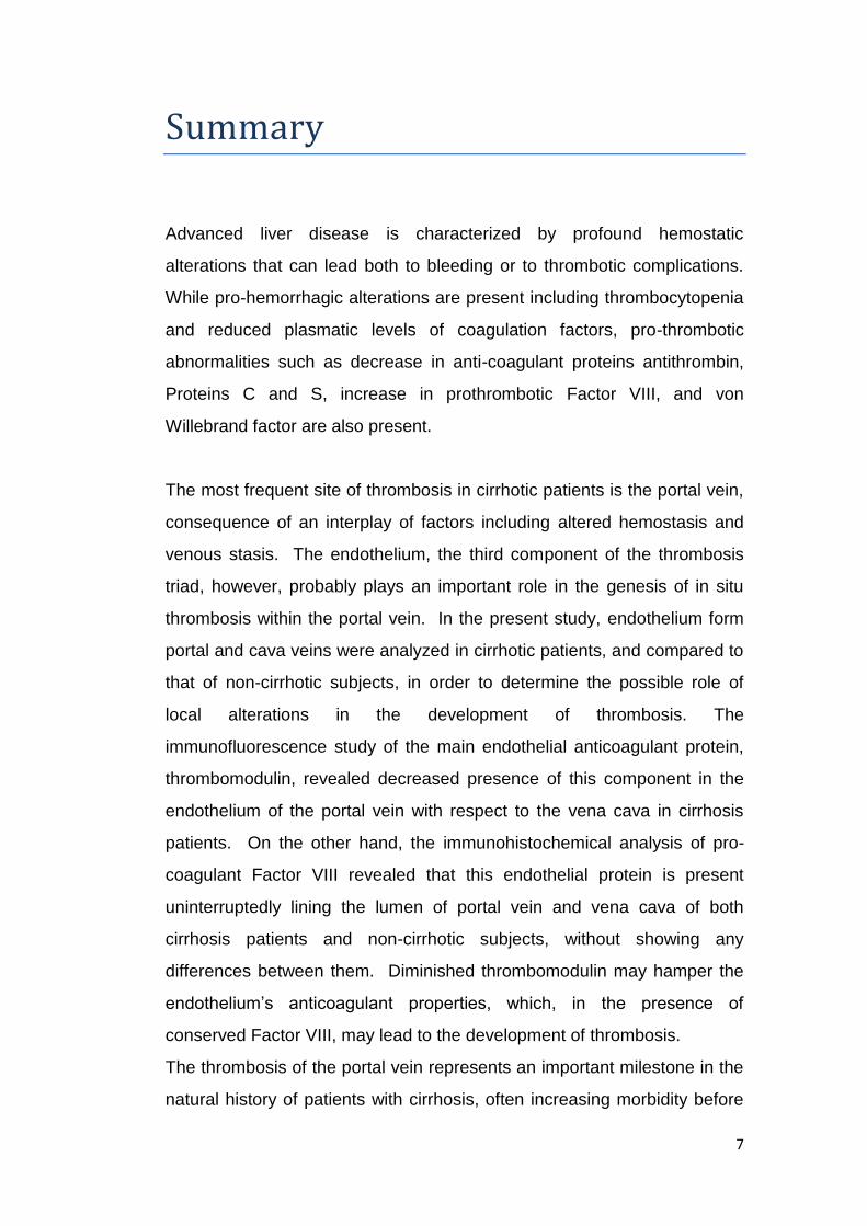
Advanced liver disease is characterized by profound hemostatic
alterations that can lead both to bleeding or to thrombotic complications.
While pro-hemorrhagic alterations are present including thrombocytopenia
and reduced plasmatic levels of coagulation factors, pro-thrombotic
abnormalities such as decrease in anti-coagulant proteins antithrombin,
Proteins C and S, increase in prothrombotic Factor VIII, and von
Willebrand factor are also present.
The most frequent site of thrombosis in cirrhotic patients is the portal vein,
consequence of an interplay of factors including altered hemostasis and
venous stasis. The endothelium, the third component of the thrombosis
triad, however, probably plays an important role in the genesis of in situ
thrombosis within the portal vein. In the present study, endothelium form
portal and cava veins were analyzed in cirrhotic patients, and compared to
that of non-cirrhotic subjects, in order to determine the possible role of
local alterations in the development of thrombosis. The
immunofluorescence study of the main endothelial anticoagulant protein,
thrombomodulin, revealed decreased presence of this component in the
endothelium of the portal vein with respect to the vena cava in cirrhosis
patients. On the other hand, the immunohistochemical analysis of pro-
coagulant Factor VIII revealed that this endothelial protein is present
uninterruptedly lining the lumen of portal vein and vena cava of both
cirrhosis patients and non-cirrhotic subjects, without showing any
differences between them. Diminished thrombomodulin may hamper the
endothelium’s anticoagulant properties, which, in the presence of
conserved Factor VIII, may lead to the development of thrombosis.
The thrombosis of the portal vein represents an important milestone in the
natural history of patients with cirrhosis, often increasing morbidity before

8
and mortality after liver transplantation. Obtainment of recanalization
through anticoagulation is therefore paramount, and in the present study,
an analysis was performed regarding factors that may have an impact on
efficacy of anticoagulation with low molecular weight heparin in cirrhotic
patients with this complication. Anticoagulation with low molecular weight
heparin was demonstrated to be a valid strategy for achieving portal vein
recanalization, with a response rate of 65.2%, including complete
recanalization in 24 of the 46 treated patients, after a mean of 4.5 months
(±3.1 months) of anticoagulation. Whereas the hemostatic status of
patients did not correlate with the response to anticoagulation, the interval
between thrombus onset and start of therapy was the only predictive factor
of therapeutic efficacy. Specifically, thrombus age at diagnosis (1.9 ± 1.2
months vs 6.3 ± 4.5 months in the recanalization group and in the non-
recanalization group, respectively, p<.001), and the interval between
thrombus onset and start of anticoagulation (3.2 ± 1.7 months vs 7.78 ±
4.5 months in the recanalization group and in the non-recanalization
group, respectively, p<.002) were the principal determinants of therapeutic
efficacy. This underlines the importance of prompt diagnosis and start of
therapy to increase the probability of successful anticoagulant therapy.
Although in cirrhosis the low levels of antithrombin, which is necessary for
the action of heparins, could theoretically hamper the anticoagulant effect,
the clinical efficacy of anticoagulant therapy with low molecular weight
heparin has been herein demonstrated.
The anticoagulant effect of low molecular weight heparin was then tested
in vitro using the thrombin generation assay, and concentrations within the
therapeutic range achieved reduction of endogenous thrombin potential
notwithstanding the marked reduction in antithrombin levels that were
present in plasma from cirrhotic patients and the low plasma anti-Xa
activity determined in vitro. In particular, patients with Child Pugh C

9
cirrhosis were characterized by antithrombin levels which were as low as
those of subjects with the prothrombotic condition of genetic antithrombin
deficit (42±14% versus 52±4%, respectively, p=.06). At low molecular
weight heparin 0.35 UI/mL concentration in vitro, anti-Xa activity was
significantly lower in Child Pugh B and Child Pugh C patients as compared
to controls (p<0.001), as well as in patients with congenital AT defect as
compared to controls (p<0.001). Despite low levels of antithrombin and
anti-Xa activity, patients with cirrhosis showed a greater anticoagulant
effect of low molecular weight heparin, with a mean endogenous thrombin
potential reduction of 72.6±11% (p=0.02 versus controls). This increased
susceptibility of cirrhosis patients with advanced stages of the disease
may therefore actually warrant dose reduction of anticoagulation.
10
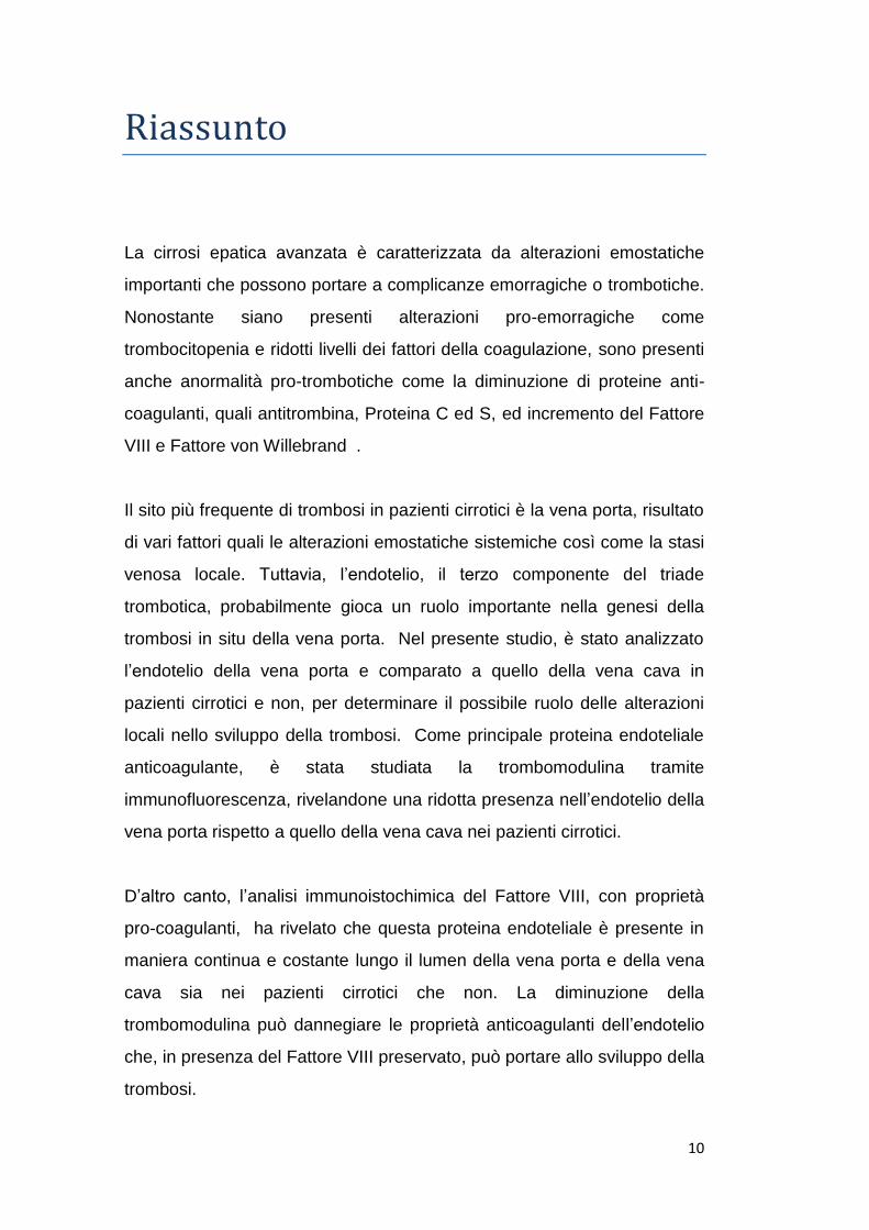
La cirrosi epatica avanzata è caratterizzata da alterazioni emostatiche
importanti che possono portare a complicanze emorragiche o trombotiche.
Nonostante siano presenti alterazioni pro-emorragiche come
trombocitopenia e ridotti livelli dei fattori della coagulazione, sono presenti
anche anormalità pro-trombotiche come la diminuzione di proteine anti-
coagulanti, quali antitrombina, Proteina C ed S, ed incremento del Fattore
VIII e Fattore von Willebrand .
Il sito più frequente di trombosi in pazienti cirrotici è la vena porta, risultato
di vari fattori quali le alterazioni emostatiche sistemiche così come la stasi
venosa locale. Tuttavia, l’endotelio, il terzo componente del triade
trombotica, probabilmente gioca un ruolo importante nella genesi della
trombosi in situ della vena porta. Nel presente studio, è stato analizzato
l’endotelio della vena porta e comparato a quello della vena cava in
pazienti cirrotici e non, per determinare il possibile ruolo delle alterazioni
locali nello sviluppo della trombosi. Come principale proteina endoteliale
anticoagulante, è stata studiata la trombomodulina tramite
immunofluorescenza, rivelandone una ridotta presenza nell’endotelio della
vena porta rispetto a quello della vena cava nei pazienti cirrotici.
D’altro canto, l’analisi immunoistochimica del Fattore VIII, con proprietà
pro-coagulanti, ha rivelato che questa proteina endoteliale è presente in
maniera continua e costante lungo il lumen della vena porta e della vena
cava sia nei pazienti cirrotici che non. La diminuzione della
trombomodulina può dannegiare le proprietà anticoagulanti dell’endotelio
che, in presenza del Fattore VIII preservato, può portare allo sviluppo della
trombosi.
11
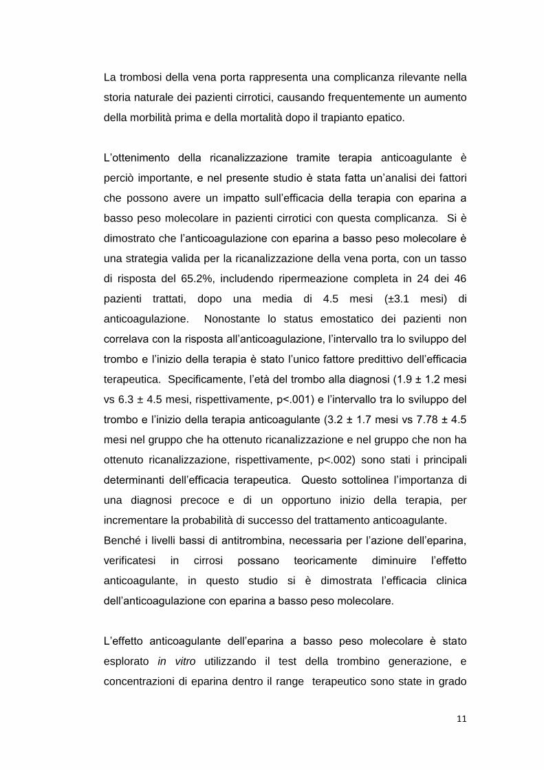
La trombosi della vena porta rappresenta una complicanza rilevante nella
storia naturale dei pazienti cirrotici, causando frequentemente un aumento
della morbilità prima e della mortalità dopo il trapianto epatico.
L’ottenimento della ricanalizzazione tramite terapia anticoagulante è
perciò importante, e nel presente studio è stata fatta un’analisi dei fattori
che possono avere un impatto sull’efficacia della terapia con eparina a
basso peso molecolare in pazienti cirrotici con questa complicanza. Si è
dimostrato che l’anticoagulazione con eparina a basso peso molecolare è
una strategia valida per la ricanalizzazione della vena porta, con un tasso
di risposta del 65.2%, includendo ripermeazione completa in 24 dei 46
pazienti trattati, dopo una media di 4.5 mesi (±3.1 mesi) di
anticoagulazione. Nonostante lo status emostatico dei pazienti non
correlava con la risposta all’anticoagulazione, l’intervallo tra lo sviluppo del
trombo e l’inizio della terapia è stato l’unico fattore predittivo dell’efficacia
terapeutica. Specificamente, l’età del trombo alla diagnosi (1.9 ± 1.2 mesi
vs 6.3 ± 4.5 mesi, rispettivamente, p<.001) e l’intervallo tra lo sviluppo del
trombo e l’inizio della terapia anticoagulante (3.2 ± 1.7 mesi vs 7.78 ± 4.5
mesi nel gruppo che ha ottenuto ricanalizzazione e nel gruppo che non ha
ottenuto ricanalizzazione, rispettivamente, p<.002) sono stati i principali
determinanti dell’efficacia terapeutica. Questo sottolinea l’importanza di
una diagnosi precoce e di un opportuno inizio della terapia, per
incrementare la probabilità di successo del trattamento anticoagulante.
Benché i livelli bassi di antitrombina, necessaria per l’azione dell’eparina,
verificatesi in cirrosi possano teoricamente diminuire l’effetto
anticoagulante, in questo studio si è dimostrata l’efficacia clinica
dell’anticoagulazione con eparina a basso peso molecolare.
L’effetto anticoagulante dell’eparina a basso peso molecolare è stato
esplorato in vitro utilizzando il test della trombino generazione, e
concentrazioni di eparina dentro il range terapeutico sono state in grado

12
di ridurre la generazione della trombina, nonostante la spiccata riduzione
nei livelli plasmatici di antitrombina e i bassi livelli di anti-Xa determinati in
vitro. In particolare, i pazienti in classe C di Child Pugh si sono
caratterizzati da livelli di antitrombina bassi quanto quelli presenti in
pazienti con la condizione protrombotica di deficit genetico di questa
proteina (42±14% vs 52±4%, rispettivamente, p=.06). Alla concentrazione
in vitro di 0.35 UI/mL di eparina a basso peso molecolare, l’attività anti-Xa
è stata significativamente più bassa in pazienti in classi di Child Pugh B e
C rispetto ai controlli (p<.001), così come in pazienti con difetto genetico
dell’antitrombina rispetto ai controlli (p<.001). Nonostante i ridotti livelli di
attività anti-Xa, i pazienti cirrotici hanno dimostrato un maggiore effetto
anticoagulante dell’eparina a basso peso molecolare, con una riduzione
del potenziale endogeno di trombina di 72.6±11% (p=0.02 vs i controlli).
Data l’incrementata suscettibilità dei pazienti cirrotici in stadi avanzati della
malattia epatica, potrebbe essere necessaria la riduzione della dose di
anticoagulazione.

13
Multiple changes occur in the hemostatic system as a result of deranged
liver function. Being the liver the primary site of synthesis of most
coagulation factors, as well as of proteins which keep these mechanisms
in check, severe derangement of its function has long been known to
result in bleeding complications such as epistaxis, gastrointestinal
bleeding from esophago-gastric varices, and gum bleeding, but also in
thrombotic complications.
Routinely performed coagulation profile is abnormal in the majority of
these patients, and clinical consequences of alterations in this complex
interplay may lead to bleeding or to thrombosis. In patients with severe
liver disease, hemostasis is affected due to diminished synthesis of factors
II, V, VI, IX, X, XI, XIII, fibrinogen, protein C, protein S, oftentimes
coexisting with Vitamin K deficiency due to malabsorption or malnutrition.
Dysfibrinogenemia, enhanced fibrinolysis, impaired clearance of activated
clotting factors, plasminogen activators, and fibrinogen degradation
products all contribute to altered hemostasis in cirrhosis. Coagulation in
patients with decompensated liver cirrhosis can also be affected by other
factors like infections, endogenous heparinoids, renal failure, and
endothelial dysfunction(1-3).
Procoagulant factors
While von Willebrand factor is synthesized by the endothelium (4) and
Factor VIII is primarily synthesized by hepatic sinusoidal cells(5;6), the
liver is the site of synthesis of fibrinogen and factors II, V, VII, IX, X, XI and
XII (7). Thus the plasma concentration of factor VIII is not decreased with
14
liver disease, and may be even increased, as many chronic liver diseases
are associated with chronic inflammation (8). This probably obeys to an
increase in endothelial synthesis, a reduced clearance via low-density
lipoprotein receptor-related protein (4), and increased levels of von
Willebrand factor. However, the biological activity of the synthetized
molecule is lower than the plasmatic concentration (9). Furthermore,
Factor VIII is elevated in fulminant hepatic failure but decreased in
disseminated intravascular coagulation (DIC) (10).
Vitamin K is an essential cofactor for the production of biologically active
forms of the coagulation factors II, VII, IX and X, enhancing hepatic post-
ribosomal conversion of certain glutamic acid residues in the protein
precursors, to -carboxyglutamic acid (Gla). These active forms of the
clotting factors chelate calcium at the Gla site, resulting in effective
hemostatic function. When -carboxylation is impaired due to deficiency or
antagonism of vitamin K, inert precursors are synthetized, (known as
Proteins Induced by Vitamin K Absence [PIVKA]) and released into the
blood stream (11). The clinical significance of these precursors is not
clear. In the case of prothrombin, a specific and sensitive immunoassay
has been developed which is able to detect small decreases in Gla
content of this incomplete PIVKA prothrombin before any changes occur in
conventional coagulation tests (12). In cholestasis, reduction of vitamin K
absorption from the small intestine due to decreased bile salt production
can be compensated with parenteral administration of vitamin K 10mg
daily for 24-48 hours, but in parenchymal liver disease decreased levels of
coagulation factors are dependent on a decreased synthesis, so that there
is no improvement with vitamin K administration (13). Nevertheless 25% of
patients with acute liver injury have a subclinical deficit of vitamin K which
may benefit from parenteral administration, with corresponding
improvement of the INR (14).
15
Anticoagulant Factors
Antithrombin III (ATIII) is a non-vitamin K-dependent glycoprotein
synthesised by the liver but also by the endothelium (15). It has low
concentration in patients with liver disease, probably due to reduced
synthesis and/or increased consumption due to hyperfibrinolysis (16).
ATIII replacement does not correct hyperfibrinolysis in patients with liver
cirrhosis. Usually the ATIII deficit is mild and thrombosis as a complication
is very rare, reported only sporadically (17).
Proteins C and S are vitamin K dependent glycoproteins synthesised
mainly by hepatocytes (18). Therefore during acute or chronic liver
disease, their concentrations can be decreased concomitantly with the
other coagulation factors, but usually not below 20% of normal values
(19). Genetic deficiency of protein C is rare in the general population, but
found in 20% patients with Budd-Chiari syndrome (BCS). In patients with
liver disease who also have genetic deficiency, plasma concentration is
often lower than 20%. When there is severe liver disease, it can be difficult
to exclude coexistent genetic deficiency as levels may be very low, due to
very depressed synthesis (20). In this situation a concomitant finding of a
normal level of factor II and protein C/factor VII ratio, can help to confirm a
coexistent genetic deficit (21). In acquired deficiency of vitamin K, a
defective protein C lacking -carboxyl (PIVKA) is produced (22). Protein C
deficiency is not associated with extrahepatic portal vein thrombosis (23).
Genetic deficiency of protein S is extremely rare, yet accounts up to 7% of
patients with BCS or portal vein thrombosis (PVT), especially in series
from Asia (24).
16
Overview of hemostasis alterations in cirrhosis
The fact that a rebalanced hemostatic status, which can be easily tipped
towards bleeding or towards thrombotic complications not only is evident
from laboratory studies, but also from the clinical point of view. It is ever
clearer that patients with cirrhosis are not “auto-anticoagulated”, as
previously thought, but actually have a greater risk than non-cirrhotic
counterparts for developing thrombotic complications as well. Clinically,
evidence is accumulating regarding a relative hypercoagulable state which
is present in patients with cirrhosis. (Table 1).
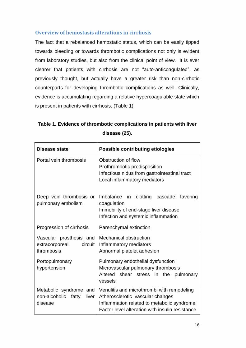
Table 1. Evidence of thrombotic complications in patients with liver
disease (25).
Disease state Possible contributing etiologies
Portal vein thrombosis Obstruction of flow
Prothrombotic predisposition
Infectious nidus from gastrointestinal tract
Local inflammatory mediators
Deep vein thrombosis or
pulmonary embolism
Imbalance in clotting cascade favoring
coagulation
Immobility of end-stage liver disease
Infection and systemic inflammation
Progression of cirrhosis Parenchymal extinction
Vascular prosthesis and
extracorporeal circuit
thrombosis
Mechanical obstruction
Inflammatory mediators
Abnormal platelet adhesion
Portopulmonary
hypertension
Pulmonary endothelial dysfunction
Microvascular pulmonary thrombosis
Altered shear stress in the pulmonary
vessels
Metabolic syndrome and
non-alcoholic fatty liver
disease
Venulitis and microthrombi with remodeling
Atherosclerotic vascular changes
Inflammation related to metabolic syndrome
Factor level alteration with insulin resistance

17
From the point of view of laboratory parameters, patients with chronic or
acute liver failure show profound abnormalities in their haemostatic and
coagulation system(2;26).
Despite routine coagulation tests may be compatible with a bleeding
tendency, both pro and anti-coagulation factors are affected, the latter of
which are not well reflected in these tests (27). Reduced levels of FII, FIX,
FXI, FXII are characteristic of the coagulation profile of a typical patient
with cirrhosis, and levels are correlated to the severity of liver disease.
However, anticoagulant factors are also decreased in cirrhosis, and
therefore a new thrombotic-hemostatic balance is reached(2;28).
In fact, more global tests such as thrombin generation test (TG), which are
able to detect the overall effect of deficient anticoagulant mechanisms in
plasma of cirrhosis patients, have shown that endogenous thrombin
potential in cirrhosis patients is not significantly different from that of
healthy subjects(29). Specifically, when the thrombin generation test is
performed in the presence of thrombomodulin, an endothelial receptor
which catalyzes the thrombin-mediated conversion of Protein C into its
active form activated Protein C, the amount of thrombin generated is
similar between plasma from cirrhotic and from healthy subjects(30;31).
As mentioned above, aside from acquired coagulation-hemostatic defects,
it has been demonstrated that genetic thrombophilias are more frequent in
cirrhosis patients with PVT when compared to cirrhosis patients without
PVT. In a study by Amitrano et al, the frequencies of Factor V Leiden and
of Prothrombin A20210 polymorphism were reportedly 13% and 34.8% in
cirrhotic patients with PVT, whereas frequencies were 7.5% and 2.5% in
cirrhotic patients without PVT (32).
18
Regarding primary hemostasis, chronic liver disease is characterized by a
variable degree of thrombocytopenia due to increased platelet destruction,
increased splenic and/or hepatic sequestration, or to reduced levels of
thrombopoietin and by altered platelet function due to defective
thromboxane A2 synthesis, storage pool deficiency and abnormalities of
the platelet glycoprotein Ib (33-39).
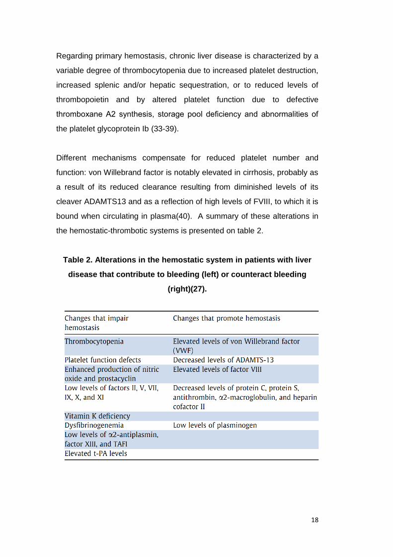
Different mechanisms compensate for reduced platelet number and
function: von Willebrand factor is notably elevated in cirrhosis, probably as
a result of its reduced clearance resulting from diminished levels of its
cleaver ADAMTS13 and as a reflection of high levels of FVIII, to which it is
bound when circulating in plasma(40). A summary of these alterations in
the hemostatic-thrombotic systems is presented on table 2.
Table 2. Alterations in the hemostatic system in patients with liver
disease that contribute to bleeding (left) or counteract bleeding
(right)(27).

19
Thrombin generation
Thrombin is often seen as the end-product of the coagulation cascade and
as such responsible for the final step, the conversion of the soluble protein
fibrinogen into the insoluble fibrin clot. However, the coagulation cascade
is actually composed of many feedback loops and inhibitory pathways that
are all directly or indirectly influenced by the developing thrombin
concentration, which renders thrombin pivotal throughout the entire
coagulation-anticoagulation process.
The thrombin generation assay in plasma is a test which assesses
coagulation globally, and is able to detect alterations both in coagulation
and anticoagulation mechanisms (41) (Figure 1). In contrast to
determination of thrombin generation in vivo, which detects byproducts of
an ongoing thrombus formation such as prothrombin fragments 1-2 and d-
dimer, the thrombin generation assay measures ex vivo the potential
capacity of a certain sample to generate thrombin, triggered by tissue
factor. The sample analyzed may be platelet-poor plasma (PPP), or
platelet-rich plasma (PRP), and the interplay between all coagulation
components is analyzed concomitantly, with the exception of the vessel
wall. To this end, thrombomodulin and tissue factor are added to the
assay, rendering the test a very close approximation to what occurs inside
the vessel. Phospholipids are added as platelet substitutes when PPP is
analyzed, providing a pro-coagulant surface. This method can monitor the
initiation, propagation and decay phases of thrombin generation. In
addition, this assay also measures the endogenous thrombin potential
(ETP), which represents the activity of thrombin multiplied by the time for
which it remains active in plasma(42).
Thrombin generated from the plasma sample after addition of a suitable
trigger (tissue factor, recalcification of thawed plasma), is measured using
20
a fluorogenic substrate. The amount of generated thrombin is plotted
against time to construct a thrombin generation curve (or thrombogram)
(Figure 2). A computer program developed by Hemker and collaborators
calculates the parameters which characterize the thrombogram i.e., the
lag time, the peak height, the time to peak and the endogenous thrombin
potential (ETP) (Figure 3) (43). The latter is the area under the thrombin
generation curve and represents the total amount of thrombin formed over
time, after exclusion of the contribution to amidolysis of the
alpha2macroglobulin-thrombin complex that is unable to convert fibrinogen
into fibrin, but retains amidolytic activity towards the thrombin-specific
synthetic substrates. The fluorescent signal has the drawback of not being
linear with product concentration(44). To compensate for this and for the
effects of substrate consumption, the calibrated automated thrombogram
(CAT) method has been developed, which continuously compares the
signal from the experimental sample to that of a fixed known thrombin
activity (45). This method allows visualizing the thrombin concentration in
clotting PPP or PRP in 24 parallel experiments. In the present study, the
Thrombinoscope™ software (Thrombinoscope BV) was used, together
with the Thrombinograph™, a 96-well plate fluorimeter (Thermo Scientific).
A thrombin calibrator was used for every sample, to correct for the inner
filter effect, donor-to-donor variability in color of plasma, substrate
depletion and instrumental differences.
The study of thrombin generation – performed either with clotting-based
assays, or with chromogenic substrates – is an established tool in blood
coagulation research –(43;46-49). It has been shown that the thrombin
generation assay is able to detect thrombophilic phenotypes in subjects at
risk of VTE (50-52). When this test is performed in the presence of
thrombomodulin or activated protein C, alterations in the pathway of the
natural anticoagulant protein C are unmasked.
21
In population-based studies Lutsey et al. demonstrated an increased risk
for VTE (primarily idiopathic) for elevated peak values of TG (53).
Furthermore, Tripodi and colleagues demonstrated a correlation between
TG values and clinical risk assessment of VTE (low, medium, high risk of
VTE), when thrombin generation assay was performed in the presence of
thrombomodulin(54). In cirrhosis patients, studies performed using the
thrombin generation assay have demonstrated an imbalance between pro-
and anti-coagulant factors which tends towards hypercoagulability and is
unveiled by the addition of thrombomodulin(55). Finally, the thrombin
generation test has also been used to study the anticoagulant effect of
multiple anticoagulant drugs, showing adequate sensibility to detecting
their action (56).
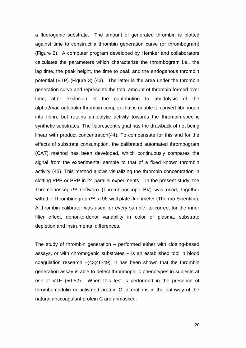
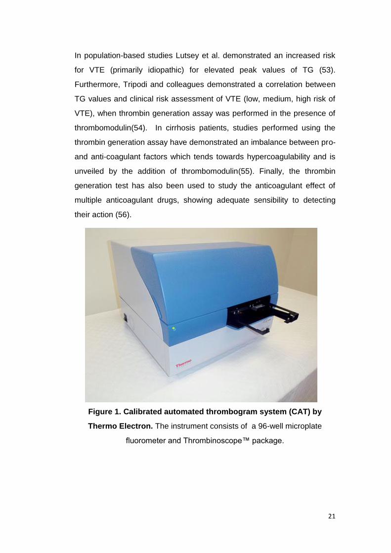
Figure 1. Calibrated automated thrombogram system (CAT) by
Thermo Electron. The instrument consists of a 96-well microplate
fluorometer and Thrombinoscope™ package.

22
Figure 2. Typical thrombin generation curve (thrombogram).
The endogenous thrombin potential (ETP) corresponds to the area under
the curve.
Thrombin Generation curve
0 10 20 300
100
200
300
400
500Cmax: peak of thrombin
ETP: area under the curve
Lag Time: time to reaction start
Time (min)
FII
(n
M)
Figure 3. Thrombin generation curve and parameters.
Thrombin generated and subsequently degraded is plotted against time.

23
Clinical evidence of pro-thrombotic complications in cirrhosis
Venous thromboembolism
Patients with cirrhosis appear to have a higher incidence of unprovoked
deep vein thrombosis and pulmonary embolism (DVT/PE) compared with
the general population. Although there are no prospective studies that
report on the incidence of venous thromboembolism (VTE) in patients with
cirrhosis, a nationwide population-based study undertaken in Denmark
evaluating more than 99000 patients with thromboembolism showed that
cirrhosis and liver disease carry a greater risk of VTE (OR of 2.10, and of
3.58 if age < 55 years)(57). In a prospective cohort study with case-control
analysis of 6550 patients with VTE in the United Kingdom, Huerta et al.
found that in patients with chronic liver diseases, the odds ratio for PE was
1.75 (CI 0.91–3.36), and the odds ratio for DVT/PE combined was 1.65 (CI
0.97–2.82), demonstrating not only that these patients are not protected
from thromboembolic events, but rather that cirrhosis is a predisposing
condition(58).
Eight articles, of which two are case-control studies and six are
retrospective studies, have been published up to date specifically aimed at
investigating the incidence of DVT and PE in patients with cirrhosis (59-
66). (Table 3)

24
Table 3. Prevalence of venous thromboembolism in patients with
cirrhosis admitted for hospitalization (59-66).
Reference Type of study Study population (n)
Incidence of DVT/PE (%)
DVT number (%)
PE number (%)
DVT+PE number (%)
Northup et al. (2006) Case control 21000 113 (0.5%) 74 (65.5%) 22 (19.5%) 17 (15%)
Garcia Fuster et al. (2008) Retrospective 2074 17 (0.8%) 10 (59%) 6 (35%) 1 (6%)
Gulley et al. (2008) Case control 963 18 (1.87%) NA NA NA
Lesmana et al. (2010) Retrospective 256 12 (4.7%) 12 (100) 0 (0%) 0 (0%)
Ali et al.(2010) Retrospective 449798 8231 (1.8%) 4335 (0.9%) 3688 (0.8%) 208 (0,8%)
Dabbagh et al.(2010) Retrospective 190 12 (6.3%) NA NA NA
Wu et al. (2010) Retrospective 649879 52881 (8.1) NA NA NA
Aldawood et al. (2011) Retrospective 226 6 (2.7%) 6 (100%) 0 (0%) 0 (0%)
DVT: Deep vein thrombosis; NA: data not available; PE: pulmonary embolism. Adapted from(67).
Portal vein thrombosis
The portal vein is certainly the most common site for thrombus formation in
patients with advanced liver disease. Moreover, non-neoplastic portal vein
thrombosis (PVT) is more frequent in cirrhosis than in the general
population, with a prevalence of 0.6 to 26% in patients without
hepatocellular carcinoma, although the wide range in reported prevalence
is owed to the different groups of cirrhotic patients studied and the
different diagnostic method (68-74).
25
Risk factors for the development of portal vein thrombosis in
cirrhosis
The occurrence of pathological thrombosis is determined by an alteration
in the physiological equilibrium that regulates coagulation and
anticoagulation dynamics, and shifts regarding any one of the components
of Virchow’s triad (venous stasis, endothelial injury and
hypercoagulatiblity) may result in thrombosis. Both local and systemic
factors can result in the development of site-specific thrombosis,
particularly PVT.
In patients with end-stage liver disease, venous stasis results from
splanchnic vasodilatation and architectural derangement, which primarily
involves the cirrhotic liver. In one prospective study regarding a cohort of
73 patients with cirrhosis, reduced portal flow velocity below 15 cm/s was
the only independent variable that correlated with the risk of developing
PVT at 1 year of follow-up(75). However, the prospective study by
Francoz et al. on 251 patients with cirrhosis listed for liver transplantation
failed to find a correlation between portal flow direction (hepatofugal or
hepatopetal) and risk of development of PVT(70). Recent in vivo evidence
shows that the ratio of the most important pro and anticoagulant factors,
factor VIII and protein C, respectively, demonstrates a strong imbalance in
favor of factor VIII in cirrhotics, indicating a hypercoagulable state(55).
In several series, elevated levels of factor VIII have been shown to be a
risk factor associated with either primary or cirrhosis-associated
PVT(76;77). This has been confirmed in a larger cohort that evaluated 58
non-cirrhotic patients with PVT, 27 cirrhosis patients with PVT and 200
with DVT, in whom a strong association between elevated levels of FVIII
and the risk of PVT was demonstrated (odds ratio for thrombosis and fVIII
levels above 129 UI/dl being 6.0 for cirrhosis)(78).
26
The possible role of inherited thrombophilic abnormalities has been
advocated in several cross-sectional studies reporting a thrombophilic
genotype in up to 9% of patients with cirrhosis and PVT. It has also been
demonstrated that polymorphisms TT677 of methylene-trahydrofolate
reductase, and G20210A in the prothrombin gene are significantly more
frequent in this group than in controls(32;68;71). Although not
demonstrated in all studies regarding the liver transplant population(79), in
other studies, the risk of PVT has been shown to be independently
associated with the severity of cirrhosis, being paradoxically more
frequent in those patients with worsening indices of coagulation and those
with worsening portal hypertension (i.e. ascites and
encephalopathy)(73;80).
Furthermore, PVT is more likely to occur in late stages of cirrhosis, and the
combination of sclerotherapy and history of abdominal surgery are
associated with an increased risk of PVT, which is probably not the case
of a causal relationship, but rather reflecting a more advanced disease
stage (68). The presence of previous treatments for portal hypertension
(sclerotherapy, TIPS, shunt surgery, previous splenectomy) and severity of
liver disease classified as Child Pugh C are two factors which have been
found to be significantly associated with an increased risk of PVT (73), and
the former was also demonstrated in other studies analyzing risk factors
for PVT in liver transplantation series(74;81).
Table 4 summarizes the most important reported risk factors for the
development of PVT in patients who undergo liver transplantation
(70;73;74;79;80;82-89).
27
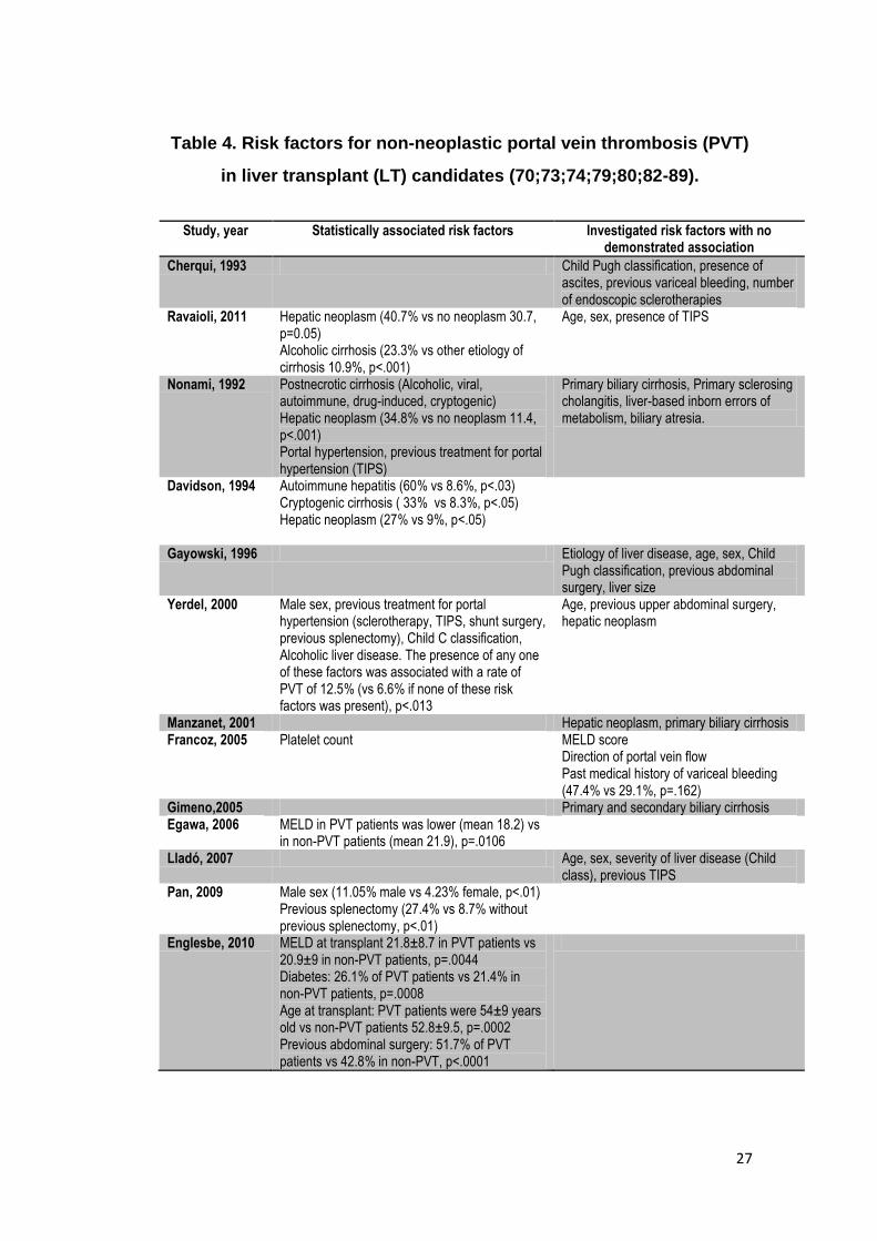
Table 4. Risk factors for non-neoplastic portal vein thrombosis (PVT)
in liver transplant (LT) candidates (70;73;74;79;80;82-89).
Study, year Statistically associated risk factors Investigated risk factors with no demonstrated association
Cherqui, 1993 Child Pugh classification, presence of ascites, previous variceal bleeding, number of endoscopic sclerotherapies
Ravaioli, 2011 Hepatic neoplasm (40.7% vs no neoplasm 30.7, p=0.05) Alcoholic cirrhosis (23.3% vs other etiology of cirrhosis 10.9%, p<.001)
Age, sex, presence of TIPS
Nonami, 1992 Postnecrotic cirrhosis (Alcoholic, viral, autoimmune, drug-induced, cryptogenic) Hepatic neoplasm (34.8% vs no neoplasm 11.4, p<.001) Portal hypertension, previous treatment for portal hypertension (TIPS)
Primary biliary cirrhosis, Primary sclerosing cholangitis, liver-based inborn errors of metabolism, biliary atresia.
Davidson, 1994 Autoimmune hepatitis (60% vs 8.6%, p<.03) Cryptogenic cirrhosis ( 33% vs 8.3%, p<.05) Hepatic neoplasm (27% vs 9%, p<.05)
Gayowski, 1996 Etiology of liver disease, age, sex, Child Pugh classification, previous abdominal surgery, liver size
Yerdel, 2000 Male sex, previous treatment for portal hypertension (sclerotherapy, TIPS, shunt surgery, previous splenectomy), Child C classification, Alcoholic liver disease. The presence of any one of these factors was associated with a rate of PVT of 12.5% (vs 6.6% if none of these risk factors was present), p<.013
Age, previous upper abdominal surgery, hepatic neoplasm
Manzanet, 2001 Hepatic neoplasm, primary biliary cirrhosis Francoz, 2005 Platelet count MELD score
Direction of portal vein flow Past medical history of variceal bleeding (47.4% vs 29.1%, p=.162)
Gimeno,2005 Primary and secondary biliary cirrhosis Egawa, 2006 MELD in PVT patients was lower (mean 18.2) vs
in non-PVT patients (mean 21.9), p=.0106
Lladó, 2007 Age, sex, severity of liver disease (Child class), previous TIPS
Pan, 2009 Male sex (11.05% male vs 4.23% female, p<.01) Previous splenectomy (27.4% vs 8.7% without previous splenectomy, p<.01)
Englesbe, 2010 MELD at transplant 21.8±8.7 in PVT patients vs 20.9±9 in non-PVT patients, p=.0044 Diabetes: 26.1% of PVT patients vs 21.4% in non-PVT patients, p=.0008 Age at transplant: PVT patients were 54±9 years old vs non-PVT patients 52.8±9.5, p=.0002 Previous abdominal surgery: 51.7% of PVT patients vs 42.8% in non-PVT, p<.0001

28
Endothelial injury and development of portal vein thrombosis
Endothelial alterations of the portal vein in cirrhosis patients which can
compromise natural antithrombotic mechanisms, together with the
hemostatic plasmatic alterations as well as venous stasis, may play a key
role in development of site-specific thrombosis of the portal vein.
Cirrhosis is being increasingly recognized as a condition characterized by
vascular damage, which can result in significant impairment of the
physiologic mechanisms that ensure vessel permeability and the myriad of
its homeostatic functions (90). A generalized inflammatory state is
perpetuated by altered substance hepatic metabolism, shear stress due to
disturbed hemodynamics and volume distribution, and the presence of
circulating bacterial products caused by bacterial translocation from the
gut.
Evidence of altered vascular permeability has been demonstrated in
animal models of cirrhosis and more recently in cirrhosis patients (91).
Moreover, proteins and glycoproteins associated with endothelial injury
and inflammation, such as heparin-like substances, are notably elevated in
cirrhosis. There is evidence that cirrhosis -and its possible concomitant
events such as bacterial translocation, portal hypertension, and ascites -
determines a particular set of conditions that challenge the endothelium’s
physiologic anti-thrombotic and anticoagulant capacity.
Furthermore, there is evidence to support the concept of greater
inflammation and endothelial injury within the portal vein with respect to
other venous beds in cirrhosis patients(91). D-dimer, an established
marker of inflammation, which is also associated with abnormal activation
of the coagulation cascade, has been demonstrated to reach significantly
higher levels in blood samples drawn from the portal vein when compared
to levels in blood samples from the jugular vein of cirrhosis patients (92). A

29
similar pattern has also been demonstrated for levels of prothrombin F1+2,
demonstrating significantly higher levels in the portal vein vs the jugular
vein, which represents systemic venous blood (92). Moreover, elevated
levels of glycosaminoglycans, constituents of the glycocalyx, are elevated
in patients undergoing liver transplant for advanced cirrhosis, and probably
reflect shedding from an injured endothelium (3).
Regarding the endothelial anticoagulant mechanisms, a fundamental role
is played by thrombomodulin. This cell surface-expressed transmembrane
glycoprotein, predominantly synthesized by vascular endothelial cells, is a
critical cofactor for thrombin-mediated activation of protein C, an event
further amplified by the endothelial protein C receptor, as part of the
natural anticoagulant mechanisms. This protein, which is present in both
large vessels and capillary endothelium, and has a crucial role in
anticoagulation, has also been implicated in inflammation (93). In fact, this
protein has also been studied with regard to endothelial injury, linking both
inflammation and coagulation, conditions that present concomitantly in
cirrhosis. This endothelial cell surface glycoprotein forms a 1:1 complex
with thrombin, and this binding activates protein C approximately 1000x
faster than thrombin alone. Activated protein C, in turn, together with
Protein S, inactivates Factor Va and VIIIa. TM also modulates
inflammation independently of activated protein C, by activating anti-
inflammatory pathways, negatively regulating complement, and by
facilitating the activation of tissue activatable fibrinolysis inhibitor (TAFI),
which modulates both fibrinolysis and complement-mediated inflammation
(94-97).
30
Clinical consequences of portal vein thrombosis in cirrhosis and
rationale for treatment
Clinical manifestations of PVT vary from asymptomatic disease to a life
threatening complication. In a study on 79 cirrhotic patients with newly
diagnosed PVT, 39% presented with gastrointestinal bleeding (from
varices or portal hypertensive gastropathy) and 18% had abdominal pain,
amongst which 70% had intestinal infarction due to the extension of the
thrombus into the mesenteric vein (71).
PVT may worsen portal hypertension and may lead to variceal bleeding,
development or worsening of preexisting ascites, and increases mortality
and morbidity after liver transplantation. As emerged from a recently
published systematic review, the presence of non-neoplastic PVT at LT
entails a greater 30-day mortality after surgery when compared to patients
without PVT (10.5% vs 7.7%, respectively (p=.01)(98). One-year mortality
is also increased in patients who undergo LT in the presence of PVT in
contrast with those with patent portal vein at transplant (18.8% vs 15.3%,
respectively (p<.001). Furthermore, the presence of complete or Grade IV
PVT according to Yerdel adversely affects outcome after liver
transplantation, as shown in table 5.
31
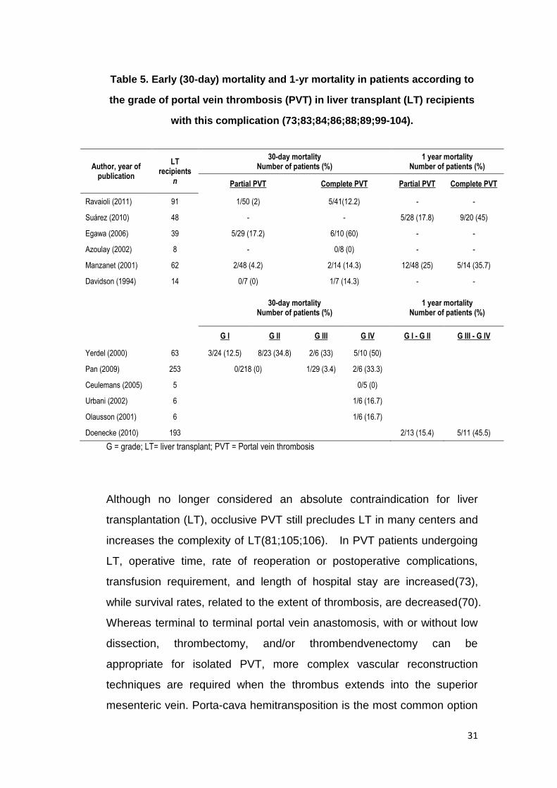
Table 5. Early (30-day) mortality and 1-yr mortality in patients according to
the grade of portal vein thrombosis (PVT) in liver transplant (LT) recipients
with this complication (73;83;84;86;88;89;99-104).
Author, year of publication
LT recipients
n
30-day mortality Number of patients (%)
1 year mortality Number of patients (%)
Partial PVT Complete PVT Partial PVT Complete PVT
Ravaioli (2011) 91 1/50 (2) 5/41(12.2) - -
Suárez (2010) 48 - - 5/28 (17.8) 9/20 (45)
Egawa (2006) 39 5/29 (17.2) 6/10 (60) - -
Azoulay (2002) 8 - 0/8 (0) - -
Manzanet (2001) 62 2/48 (4.2) 2/14 (14.3) 12/48 (25) 5/14 (35.7)
Davidson (1994) 14 0/7 (0) 1/7 (14.3) - -
30-day mortality Number of patients (%)
1 year mortality Number of patients (%)
G I G II G III G IV G I - G II G III - G IV
Yerdel (2000) 63 3/24 (12.5) 8/23 (34.8) 2/6 (33) 5/10 (50)
Pan (2009) 253 0/218 (0) 1/29 (3.4) 2/6 (33.3)
Ceulemans (2005) 5
0/5 (0)
Urbani (2002) 6 1/6 (16.7)
Olausson (2001) 6 1/6 (16.7)
Doenecke (2010) 193 2/13 (15.4) 5/11 (45.5)
G = grade; LT= liver transplant; PVT = Portal vein thrombosis
Although no longer considered an absolute contraindication for liver
transplantation (LT), occlusive PVT still precludes LT in many centers and
increases the complexity of LT(81;105;106). In PVT patients undergoing
LT, operative time, rate of reoperation or postoperative complications,
transfusion requirement, and length of hospital stay are increased(73),
while survival rates, related to the extent of thrombosis, are decreased(70).
Whereas terminal to terminal portal vein anastomosis, with or without low
dissection, thrombectomy, and/or thrombendvenectomy can be
appropriate for isolated PVT, more complex vascular reconstruction
techniques are required when the thrombus extends into the superior
mesenteric vein. Porta-cava hemitransposition is the most common option
32
during transplantation with PVT extending into the superior mesenteric
vein(107), but is characterized by survival of 60% and 38% at 1 and 3
years, respectively, and entails a 30% bleeding complications related to
residual portal hypertension after transplantation(108).
These recent studies underscore the need for a clear protocol for the
treatment of PVT in patients with cirrhosis, regardless of whether LT
might be a future prospect or not.
Hence, the aims of PVT management in patients with cirrhosis listed for
LT are the achievement of complete or partial recanalization and the
prevention of thrombosis progression. These goals can be achieved
either through the placement of, a transjugular intrahepatic portosystemic
shunt (TIPS), with or without local thrombolysis or thrombectomy(109;110)
or employing anticoagulation(111).
The rationale for the use of anticoagulation to treat PVT in patients with
underlying liver disease derives from larger experiences published in
patients with PVT and no liver disease, and the rare event of spontaneous
recanalization (112;113). In patients with non-cirrhotic PVT, recanalization
rate is achieved in 40% of patients who receive early
anticoagulation(112;114;115). Since recanalization has been shown to
occur only within 6 months from the start of anticoagulation, the duration of
anticoagulation is currently recommended for at least 3 and preferably for
up to 6 months(112;114). Evidence for the use of anticoagulation to treat
PVT in patients with cirrhosis is more scarce.
The development of thrombotic complications in cirrhosis patients warrants
a therapeutic approach that is similar to the non-cirrhosis setting.
However, the anticoagulant of choice is still not known and a suboptimal
utilization of prophylactic fractionated heparin in cirrhotic patients has been

33
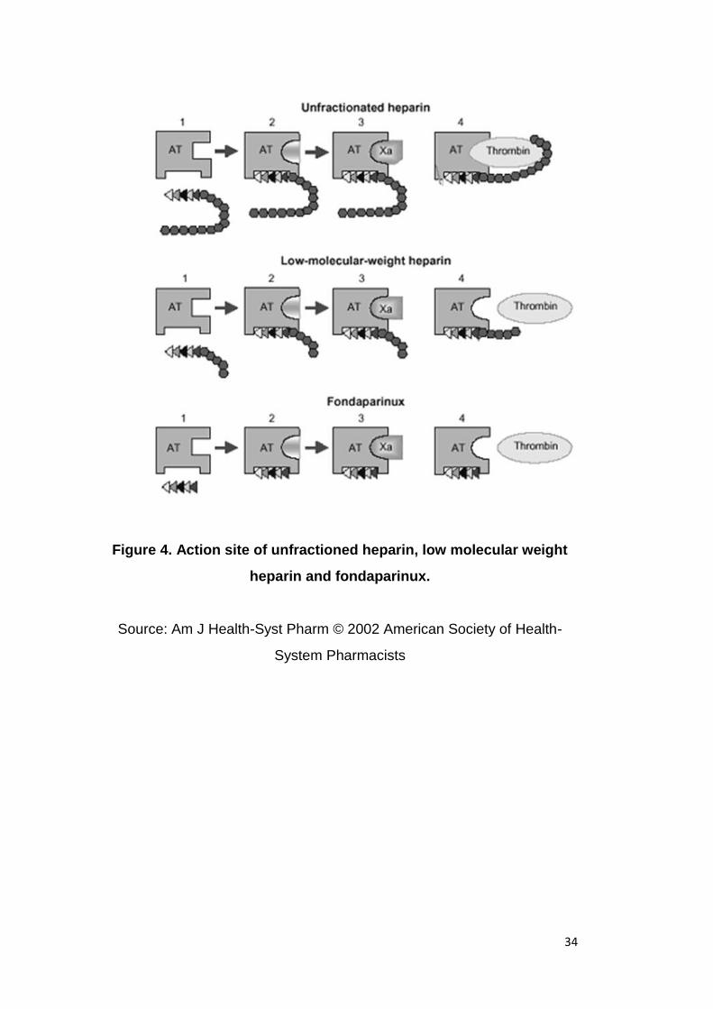
recently described(65). Several low-molecular-weight heparins (LMWHs)
are currently available as anticoagulant drugs for the prophylaxis and
treatment of thrombosis in non-cirrhotic patients. LWMHs act by
stimulating antithrombin (AT)-mediated inhibition of factor Xa; those with
saccharide chains above a critical length also exert an inhibitory effect on
thrombin. Due to their pharmacokinetic properties, LMWHs have a much
more predictable anticoagulant effect than unfractionated heparin. (Figure
4) This allows for monitoring of clotting times to be avoided and makes it
possible to use weight-adjusted or fixed doses of LWMHs depending on
the clinical setting. Since LMWHs exert their anticoagulant effect by
means of AT, the reduction of plasmatic levels of this protein seen in
patients with advanced liver disease could theoretically hamper its
anticoagulant effect.
Recently, Bechmann et al have demonstrated that after administration of
LWMH, cirrhotic patients reach lower levels of anti-Xa activity than
controls, which correlate to the severity of liver disease(116). Although
this finding could also suggest the need to increase the dose of LWMH in
cirrhotics, Lisman and colleagues showed in vitro that the anti-Xa assay
underestimates LWMH plasma levels in patients with cirrhosis(117;118).
Low molecular weight heparin (LMWH) has been shown to be effective for
the treatment of PVT in 45-75% of patients; however it is still not clear
which thrombus and patient characteristics, including hemostatic
coagulation balance, are predictive of the response to anticoagulation
therapy.
34
Figure 4. Action site of unfractioned heparin, low molecular weight
heparin and fondaparinux.
Source: Am J Health-Syst Pharm © 2002 American Society of Health-
System Pharmacists

35
Project I: Endothelial dysfunction in cirrhosis:
characterization of structural and functional aspects of the
portal vein which lead to in situ thrombosis.
1. To analyze the integrity of the endothelial lining in cirrhotic patients
using specific markers for endothelial cells, such as FVIII, and to
compare it to that of other systemic venous territories, as
exemplified by the vena cava, and to compare these findings to
those of the portal vein and vena cava of non-cirrhotic subjects.
2. To study the anticoagulant properties of the endothelium as
represented by the distribution of thrombomodulin, the main
endothelial anticoagulant protein, in the portal vein and vena cava
of cirrhotic subjects and compare it to that of non-cirrhotic subjects.
Project II: Predictors of response to anticoagulant therapy in
cirrhosis patients with portal vein thrombosis.
3. To assess hemostatic status in terms of pro- and anti-coagulant
factors, as well as clinical characteristics of the thrombus and
patients, as predictors of therapeutic efficacy of anticoagulation with
low molecular weight heparin to treat portal vein thrombosis in
patients with cirrhosis.
36
Project III: Anticoagulant response to low molecular weight
heparin in plasma from patients with advanced cirrhosis.
4. To evaluate the effect of low molecular weight heparin on
endogenous thrombin potential in plasma from patients with
cirrhosis.
5. To correlate the anticoagulant efficacy of low molecular weight
through thrombin generation assay with levels of anti-Factor Xa
(activated Factor X) in plasma from patients with cirrhosis.
6. To correlate the anticoagulant effect of low molecular weight
heparin in vitro with the severity of liver disease.
7. To correlate the efficacy of low molecular weight heparin in vitro
with the determined plasmatic levels of coagulation factors and
antithrombin in plasma from patients with cirrhosis.

37
Materials and Methods
Venous samples
Venous samples from vena cava and portal vein were from obtained from
adult subjects at transplant surgery in the case of cirrhosis patients, and
during organ retrieval in non-cirrhosis donors (controls). All procedures
were performed according to the Helsinki declaration. Criteria for exclusion
of patients were: hepatocellular carcinoma (HCC), fulminant or acute
hepatic failure, extrahepatic neoplasms, known genetic or acquired
thrombophilia or overt thrombotic complications (DVT, PVT, or VTE)
subsequently confirmed by determination of prothrombotic mutations
(Factor V Leiden, prothrombin polymorphism G20210A) and pediatric LT
recipients (<18 years of age). Criteria for exclusion of controls were
similar to criteria used to exclude potential donors (neoplasm, abdominal
trauma). As part of the protocol for organ retrieval, visual assessment by
the surgeon and histological intraoperative analysis exclude the presence
of significant liver disease.

38
Sample obtainment
Upon notice from the on-call transplant coordinator, the equipment
necessary for sample transportation and preservation was prepared.
Vena cava and portal vein samples were obtained by sterile surgical
excision of a 1 cm-wide circumferential specimen immediately after the
liver explant in the case of cirrhotic patients, before immersion in the
preserving UW (University of Wisconsin) cold solution, and after infusion of
cold preserving UW solution in non-cirrhotic subjects, as part of the
protocol in explant surgery. Specimens were received on sterile gauze.
Sample retrieval and conservation
Samples thus obtained were immediately prepared for conservation using
a sterile technique. According to the position of the lumen, all samples
were neatly and sharply sectioned transversally (perpendicularly to the
luminal surface) into approximately 0.4 cm-diameter fragments for future
analyses, obtaining fragments of the full-depth vessel wall. One fragment
of each venous sample (vena cava and vena porta) was immediately
conserved in buffered formalin, for immunohistochemical analysis. One
fragment of each venous sample (vena cava and vena porta) was
immediately embedded in optimal cutting temperature compound (OCT)
frozen by isopentane, which was previously cooled using liquid nitrogen.
The OCT-embedded samples were then placed into criovials and stored in
liquid nitrogen until surgery was terminated and the team returned to the
base hospital. Subsequently, frozen samples were immediately stored at -
80°C and formalin-fixed samples were stored at room temperature until
processing and analysis. All samples were processed within six months of
procurement.
39
Immunohistochemical evaluation of FVIII
Formalin-fixed samples were processed for routine paraffin embedding.
Serial 2 µm-thick slices were cut using a microtome, mounted onto clean
slides and stored at room temperature. Thus obtained slices were then
dewaxed in xylene (twice for 5 min each) and rehydrated through serial
acohols (100%, 95%, 70%; two changes of 3 min each) to distilled water
(twice for 5 min each), and air-dried for thirty minutes.
Immunohistochemical staining for factor VIII-related antigen (FVIII-RAG),
as a marker for endothelial cells (119) was performed with rabbit anti-
human Factor VIII conjugated with horseradish peroxidase antibody (Dako
Cytomation, Denmark) diluted 1:100 in buffer containing 20 mM Tris-HCL
pH 7.4 and 150 mM NaCl for two hours at room temperature. The
antibodies were developed with 3, 3’-diaminobenzidine (DAB, Fluka,
Milan, Italy), slides were then rinsed in phosphate buffered saline (PBS),
and slides were mounted using coverslips with Vector ® mounting
medium. Examination of the samples was performed with a light
microscope Leica DMS 5000 (Leica, BM Medical, Padua, Italy). For this
analysis all images were viewed and captured at 20 x and 40x
magnification.
Immonofluorescence evaluation of TM
Cryostat sections were prepared from OCT-embedded frozen samples by
cutting 4 µm-slices at -15° from frozen tissue bound to a chuck, and
mounted on clean slides using static force. Mounted tissues were stored at
-20°.
40
Before use, slides were air-dried for 30 minutes. For immunofluorescence
analysis, tissue samples were fixed in 2% paraformaldehyde (PF) in PBS
for 20 minutes at room temperature, treated with 50 mM NH4Cl, and
permeabilized with 0.5% Triton X-100 in PBS for 15 minutes. Tissues
were then incubated for one hour with mouse anti-human TM (Diagnostica
Stago, Asnières, France) at a concentration of 1:50, as the primary
antibody. Following two rinsing procedures with PBS, tissues were
subsequently incubated for one hour with goat anti-mouse IgG Fluorescein
isothiocyanate (FITC)-conjugated antibody (Diagnostica Stago, Asnières,
France) at a concentration of 1:400, as the secondary antibody.
Examination of samples was performed with a fluorescent microscope
Leica IMDM 6000 (Leica, BM Medical, Padua, Italy). FITC fluorescence
was visualized by excitation at 475-490 and emission at 530 nm. Cell
nuclei staining was performed with Höechst 1 µg/mL for 8 minutes at
room temperature in the dark and fluorescence of nuclei was visualized by
excitation at 330-385 nm with a 450 nm barrier filter. All samples were
analyzed by differential interference contrast (DIC) objective. For this
analysis, all images were viewed and captured at 20x and 40x
magnification.

41
Results
I: Endothelial dysfunction in cirrhosis: characterization of
structural and functional aspects of the portal vein which
lead to in situ thrombosis
Four portal vein and four vena cava samples from adult patients with
cirrhosis were obtained at liver transplantation, and four portal vein and
four vena cava samples from non-cirrhosis adult controls were obtained at
surgery for organ donation.
Characteristics of patients and controls
Mean age was 50.25±15.1 in cirrhotic patients, vs 64.75±14.6 in non-
cirrhotic subjects. The etiology of liver disease was HCV in two patients,
while one patient had autoimmune hepatitis/primary sclerosing cholangitis
overlap syndrome, and the remaining patient had alcohol-related liver
disease. MELD scores were 17, 38, 15, and 20 at liver transplant, and all
four patients had evidence of portal hypertension, including refractory
ascites in two, moderate ascites in the other two patients, and esophageal
varices in three patients. Causes of death in organ donors were: cerebral
anoxia in two, intracerebral hemorrhage in one, and non-abdominal
trauma in the remaining patient. (Table 6).
42
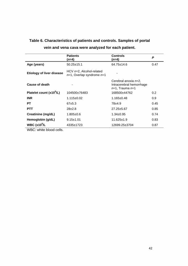
Table 6. Characteristics of patients and controls. Samples of portal
vein and vena cava were analyzed for each patient.
Patients (n=4)
Controls (n=4)
P
Age (years) 50.25±15.1 64.75±14.6 0.47
Etiology of liver disease HCV n=2, Alcohol-related n=1, Overlap syndrome n=1
-
Cause of death - Cerebral anoxia n=2, Intracerebral hemorrhage n=1, Trauma n=1
Platelet count (x109/L) 104500±76483 168500±44762 0.2
INR 1.115±0.02 1.165±0.48 0.9
PT 67±5.3 78±4.9 0.45
PTT 28±2.8 27.25±5.67 0.85
Creatinine (mg/dL) 1.805±0.6 1.34±0.95 0.74
Hemoglobin (g/dL) 9.15±1.01 11.625±1.9 0.83
WBC (x109/L 4335±1723 12699.25±3704 0.87
WBC: white blood cells.

43
Immunohistochemistry
Sixteen samples (portal vein samples and vena cava samples from four
cirrhosis patients and from four controls) were analyzed. FVIII was
ubiquitously, consistently, and homogeneously present in all vessels
studied, delineating the one-cell thick endothelial lining of the portal vein
and vena cava samples in all patients, without any significant differences
between patients and controls, nor between vena cava and the portal vein.
(Figure 5).
Figure 5. Immunohistochemical peroxidase staining for FVIII of portal
vein and vena cava of a patient with cirrhosis and a non-cirrhotic
control. A) Section of vena cava from control subject. B) Section of vena
cava from cirrhotic patient. C) Section of portal vein from control subject.
D) Section of portal vein from cirrhotic patient. *Vessel lumen. **Vessel
wall, composed of collagen fibers and smooth muscle. Arrow: Endothelial
lining. Objective 20x and 40x.

44
Immunofluorescence
TM was present in the endothelial lining of the portal vein in non-cirrhotic
subjects. In contrast, the TM fluorescence signal was less intense in the
analyzed portal vein sample of a cirrhotic patient. These results are
preliminary, and represent the portal vein of three control cases and three
cases of cirrhotic patients. (Figure 6).
Figure 6. Immunofluorescence staining with FITC thrombomodulin
(TM) on sections of portal vein from a control subject (left panel) and
from a cirrhotic patient (right panel).
A;C) interference contrast. B;D) FITC staining of TM. *Vessel lumen.
**Vessel wall. Arrow: Endothelial lining which stains for TM).
Objective 40x magnification.

45
Materials and Methods
Patients
A retrospective analysis of cirrhotic patients prospectively and
consecutively evaluated and treated for thrombosis of the portal vein
according to the local anticoagulation protocol at the Multivisceral
Transplant Unit, Department of Surgical, Oncological, and
Gastroenterological Sciences of the Padua University Hospital from
January 2007 to October 2012 was performed. Exclusion criteria were:
absence of underlying liver disease, present or recent (during the two
weeks prior to evaluation) use of anti-platelet agents, and the presence of
hepatocellular carcinoma. Patients with isolated thrombosis of the
superior mesenteric vein or splenic vein, without involvement of the portal
vein, were not included in the analysis. Upon initial evaluation, and as part
of the local treatment protocol, every patient underwent clinical evaluation
and blood sampling.
Definition of extent and age of thrombosis
At the first evaluation, all patients underwent computer tomography (CT)
scanning or magnetic resonance imaging (MRI) to define the grade of
46
occlusion of the vessel(s) and determine the extension of thrombosis into
the splenic vein and/or the superior mesenteric vein. Thrombosis was
defined as partial or total, when thrombotic material occupied <90%, or
≥90% of the vessel lumen, respectively. Thrombus age was estimated
based on past medical history, analysis of previous radiological studies,
and radiological characteristics of the thrombus at diagnosis. Thrombus
was arbitrarily defined as new if there was a recent episode of abdominal
pain associated with radiological image of PVT compatible with fresh
thrombus, with no evidence of collateral circulation at hepatic hilum on
cross-sectional imaging. The thrombus was defined as recent (≤ 6 months)
when imaging in the previous 6 months demonstrated no thrombosis and
there was no established cavernous transformation of the portal vein.
When signs of long standing thrombus were present (i.e. established
cavernous transformation, defined as multiple small collaterals in and
around the recanalizing or occluded main portal vein), the time interval
was determined using previous radiological imaging demonstrating
absence of thrombosis, and clinical history of previous diagnosis of PVT.
Based on the thus obtained information, thrombus age was recorded as a
discrete variable and also classified as ≤6 months or >6 months.
Thrombophilia screening
Upon initial evaluation (and before the start of anticoagulation), all patients
underwent blood sampling for determination of: platelet count, PT, PTT,
INR, Lupus Anticoagulant, anticardiolipin and anti-b2 glycoprotein I
antibodies, levels of antithrombin, Protein C and S antigen and activity,
activated protein C resistance, Factor VIII, Factor IX, Factor XI,
Fibrinogen, and FVIII:PC Ratio. DNA analysis for Factor V Leiden (FVL)
and Prothrombin G20210A mutations was also performed.
47
Prothrombin time (PT), INR and activated partial thromboplastin time
(aPTT) were assessed with commercially available methods. Factor II, V,
VII, VIII, IX, XI activities and fibrinogen levels were measured using
commercially available reagents on BCT (Siemens, Germany) (120). AT
activity was detected by a chromogenic method (Antithrombin III, Roche
Diagnostic, Milan, Italy). Protein C anticoagulant and chromogenic
activities were assessed using the Protein C and the Berichrom PC kits,
respectively (Siemens, Germany) on BCT (Siemens, Germany), as
previously reported (121). Protein S activity was measured using a
coagulometric method (ProS IL, Milan, Italy) on ACL 9000 (IL, Milan, Italy)
(122). Activated protein C resistance was measured using a “home-made”
method on ACL 3000 (IL, Italy) as previously described (123). DNA
analysis for Factor V Leiden (FVL) and Prothrombin G20210A mutations
was performed as previously described (124). Lupus anticoagulant (LAC)
was detected by PTT-LA (Diagnostica Stago, Asnieres, France) and
DRVVT (Siemens, Marburg, Germany) on BCT (Siemens, Germany). The
presence of LAC was established according to the guidelines of the
“Scientific and Standardization Committee of the International Society on
Thrombosis and Haemostasis: Subcommittee of the Lupus An
ticoagulant/Phospholipid Dependent Antibodies” (125). Anticardiolipin and
anti-β2-glicoprotein-I antibodies both IgG and IgM were detected by
commercially available ELISA kits (Orgentec, Mainz, Germany).
Anticoagulation protocol
According to the local protocol, before starting anticoagulation, all patients
underwent full blood count, routine laboratory tests to evaluate coagulation
and renal function, and endoscopic screening for esophageal varices.
Patients with previous variceal bleeding and those with Grade II
esophageal varices with red signs and grade III varices with or without red

48
signs completed variceal eradication by endoscopic band ligation at least
15 days prior to being started on anticoagulation. The study was
conducted according to Declaration of Helsinki and patients enrolled in the
treatment group gave their consent before starting the protocol.
The anticoagulation protocol consisted of administration of LMWH 1.5
mg/kg/day, and a 40% dose reduction was applied to patients with platelet
count <50x109/L. If serum creatinine was >150 umol/L or Creatinine
Clearance <50 mL/min monitoring of anticoagulation was performed by
assaying dosage of anti-Xa activity at 6 h after low weight molecular
heparin (LWMH) administration. Anticoagulation was continued until
recanalization or for a period of 12 months. In cases in which
recanalization was not achieved after a year of therapy, anticoagulation
was maintained at a prophylactic dose in order to avoid thrombus
extension.
Follow-up and imaging
All patients were evaluated with abdominal Doppler ultrasound every two
months during the first six months of therapy, and with CT scan/MRI and
abdominal ultrasound every six months. Patency of the portal vein was
assessed at 6 and 12 months from the start of anticoagulation therapy.
Efficacy of anticoagulation therapy was defined as complete or >50%
recanalization of the portal vein and its main branches.
Analyzed variables
The following patient variables were analyzed: age, sex, etiology of liver
disease, severity of liver disease according to Child-Pugh class and MELD

49
score, INR, aPTT, PT, platelet count, patient status at timepoints 6 and 12
months (dead, alive, or transplanted), months of follow-up, plasmatic
levels of FVIII, FIX, FXI, fibrinogen, antithrombin, protein S, protein C,
calculus of FVIII:Protein C ratio, and the presence of FV Leiden mutation
or Prothrombin G20210A polymorphism. The following variables
associated to the thrombosis of the portal vein and the anticoagulation
therapy were analyzed: grade of occlusion of the portal vein, extension to
superior mesenteric vein and/or splenic vein and/or to intrahepatic
branches, presence of portal cavernoma, estimated thrombus age at time
of diagnosis, time interval between thrombus onset and start of
anticoagulation therapy, achievement of recanalization or failure to
recanalize, time interval between start of anticoagulation therapy and
recanalization of the portal vein, progression or extension of thrombosis
into splanchnic vessels, continuation of anticoagulation at a prophylaxis
dose or lack of prophylactic anticoagulation after recanalization, the event
of liver transplant, patency of portal vein at liver transplant, adverse events
such as bleeding from esophageal varices or other sites, heparin-induced
thrombocytopenia (HIT), and the need to discontinue anticoagulation
therapy.
Endpoints
The endpoints were (i) complete or > 50% patency of previously
thrombosed portal vein trunk or main branches; (ii) maintained patency of
superior mesenteric vein and splenic veins; (iii) progression of thrombosis
into the portal vein or extension into splenic or mesenteric veins; (iv)
bleeding, intestinal infarction, liver transplantation or death.

50
Statistical analysis
Quantitative variables are expressed as the mean ± SD and qualitative
variables as absolute and relative frequencies. Comparisons between
groups of quantitative and qualitative variables were made by the
Wilcoxon and Chi square tests respectively. Multivariate analysis of
variables associated with recanalization was performed by discriminant
analysis. Time to recanalization rates were assessed using Cox models.
Comparisons of recanalization rates with risk factors were made by the log
rank test. All tests were two-sided, and P <0.05 was considered
significant. Data handling and analysis were performed with SPSS version
12.0 software (SPSS Inc., Chicago, IL, USA).

51
Results
II. Predictors of response to anticoagulant therapy in
cirrhosis patients with portal vein thrombosis
Medical records for all patients treated with anticoagulation for PVT and
who were evaluated at the Portal Hypertension Clinic of the Multivisceral
Transplant Unit of the Padua University Hospital from January 2007 to
October 2012 were reviewed. Forty-six patients were included in the
study, and were followed for a mean of 25.2 months (range 3-68 months)
from the time of initial evaluation for PVT at our center and until the
closure of the study (October 2012).
Patient and thrombus characteristics
The characteristics of the study population and of the thrombosed vessels
are shown in table 7. Mean age was 59 years (range 41-80), patients
were predominantly of male sex (34/46), and mean MELD score was 12.8
(±4.2). Estimated thrombus age at diagnosis was ≤6 months in 39/46
(84.8%) of cases. Regarding the grade of portal vein occlusion, PVT was
partial in 36/46 (78%) of cases. Twenty-one patients (45.7%) had portal
cavernoma, and PVT was extended into the superior mesenteric vein or
splenic vein in 14/46 patients, and to the intrahepatic branches in 11
patients.
Thrombophilic mutations were found in 4 patients (heterozygosity for FV
Leiden n=2, heterozygosity for Prothrombin G20210A n=1, and the
presence of both mutations in heterozygosity in one single patient).

52
Recanalization under anticoagulation protocol
Fifteen patients underwent banding of esophageal varices before starting
anticoagulation (mean number of sessions to eradication was 2 (range: 1–
3)). Thirty of the 46 treated patients (65.2%) responded to anticoagulation
after a mean of 4.5 months (±3.1 months) of anticoagulation (Figure 7); 26
during the first 6 months of therapy and 4 in the following 6 months of
therapy. Only 2/11 patients in whom the interval between thrombus onset
and start of anticoagulation therapy was greater than 6 months presented
recanalization. Complete recanalization was achieved in a total of 24
patients, while in 6 patients recanalization was partial. (Figure 8).

53
Table 7. Characteristics of cirrhotic patients with portal vein
thrombosis (PVT) treated with anticoagulation and PVT
characteristics.
Studied variable P
Mean age ± SD 59 (41-80)
Sex (Male/Female) 34/12
Etiology of liver disease Alcohol 15/46 HBV/HCV 22/46 Cholestatic/ Cryptogenic 9/46
Child-Pugh Class A/B/C 21/19/6
Thrombus age at diagnosis (mean ± SD) 3.46 ± 3.58 months
Thrombus age at diagnosis ≤6 months >6 months
39 7
Interval between thrombus onset and start of anticoagulation 4.76 ± 3.89 months
Interval between thrombus onset and start of anticoagulation ≤6 months >6 months
35 11
Extension to splanchnic vessels Superior Mesenteric Vein Splenic Vein
11 (23.9%) 4 (8.7%)
Extension to intrahepatic branches (yes/no) 11 (23.9%)
Occlusion grade (total/partial) 10/36
Presence of cavernoma 21 (45.6%)
Full-dose therapy 24 (52.2%)
Reduced-dose therapy 22 (47.8%)
PT 67.7 ± 15.8
INR 1.3 ±0.19
aPTT 36.02 ±7.67
Factor VIII 164.6±57.9
Factor IX 82.3±28.1
Factor XI 66.5±31.1
Fibrinogen 249.2±146.4
Antithrombin 65.5±22.8
Protein S 80 ± 21.1
Protein C 48.3 ± 19.5
FVIII:PC Ratio 3.65 ± 1.46
Platelet count (x109/L) 68.2 ± 30.8

54
Figure 7. Frequency of recanalization of portal vein at different
months after the start of anticoagulation treatment.
Mean = 4.5 months; standard deviation = 3.1; N = 30.
Adverse effects of anticoagulation therapy were observed in 8 patients:
bleeding from esophageal varices n = 2, hematuria n = 1, epistaxis n = 1
(did not require suspension of anticoagulant therapy); cerebral
hemorrhage n = 1 and HIT (heparin-induced thrombocytopenia) n = 3, in
all of whom anticoagulation therapy with heparin was suspended.
0
1
2
3
4
5
6
7
8
9
1 2 3 4 5 6 7 8 9 10 11 12
Pa
tie
nts
in
wh
om
re
ca
na
liza
tio
n w
as a
ch
ieve
d
(N
)
Interval to recanalization (months)
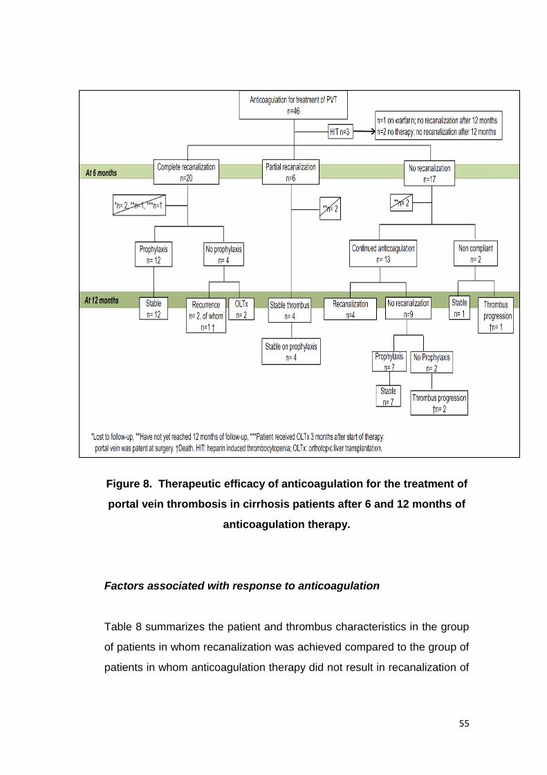
55
Figure 8. Therapeutic efficacy of anticoagulation for the treatment of
portal vein thrombosis in cirrhosis patients after 6 and 12 months of
anticoagulation therapy.
Factors associated with response to anticoagulation
Table 8 summarizes the patient and thrombus characteristics in the group
of patients in whom recanalization was achieved compared to the group of
patients in whom anticoagulation therapy did not result in recanalization of
56
the portal vein, and analyzed variables are compared between the two
groups.
Thrombus age at diagnosis (1.9 ± 1.2 months vs 6.3 ± 4.5 months in the
recanalization group and in the non-recanalization group, respectively,
p<.001), the interval between thrombus onset and start of anticoagulation
(3.2 ± 1.7 months vs 7.78 ± 4.5 months in the recanalization group and in
the non-recanalization group, respectively, p<.002), and the presence of
intrahepatic branch extension of the thrombosis (p=.025) were the only
variables that correlated with response to anticoagulation therapy.
At logistic regression analysis, the interval between thrombus onset and
start of anticoagulation therapy (≤6 months vs > 6 months in responders
vs non- responders) correlated with the probability of recanalization
(p=.0051). A longer interval between thrombus onset and start of
anticoagulation therapy was associated with therapeutic failure (HR 0.734
(risk describes the probability of recanalization)).
At survival analysis using Kaplan Meier curves, the recanalization
portal vein showed no significant impact on transplant-free survival.
(Figure 9) However, mortality was higher in patients who did not present
recanalization (p=.03); of five deaths in all the study population, four were
related to PVT: one patient had thrombosis recurrence after recanalization
had been achieved, but no prophylactic anticoagulation had ensued; one
death occurred in a patient who never achieved recanalization due to non-
compliance to therapy, and two other deaths occurred in patients in whom
no recanalization had been achieved, while one death occurred in a
patient who had adequately responded to therapy, but died on the waiting
list for LT of hepatic insufficiency.

57
Table 8. Clinical characteristics of patients and portal vein
thrombosis in patients in whom recanalization was achieved and in
patients in whom recanalization was not achieved with
anticoagulation.
Studied variable Recanalized PVT n = 30
Not-Recanalized PVT n = 16
P
Mean age ± SD 59.5 ± 11.2 58.1 ± 9.8 NS
Sex (Male/Female) 22/8 12/4 NS
Etiology of liver disease Alcohol 11/30 HBV/HCV 15/30 Cholestatic/ Cryptogenic 4/30
Alcohol 4/16 HBV/HCV 7/16 Cholestatic/ Cryptogenic 5/16
NS
Child-Pugh Class A/B/C 15/14/1 6/5/5 NS
Thrombus age at diagnosis (mean ± SD) 1.9 ± 1.2 months 6.3 ± 4.5 months .001
Thrombus age at diagnosis ≤6 months >6 months
30 0
10 6
.001
Interval between thrombus onset and start of anticoagulation
3.2 ± 1.7 months 7.78 ± 4.5 months .002
Interval between thrombus onset and start of anticoagulation
≤6 months >6 months
28 2
7 9
.001
Extension to splanchnic vessels Superior Mesenteric Vein (yes/no) Splenic Vein (yes/no)
9/21 4/26
2/14 0/16
NS
Extension to intrahepatic branches (yes/no) 4/26 7/9 .025
Occlusion grade (total/partial) 6/24 4/12 NS
Presence of cavernoma 12 9 NS
Full-dose therapy 13 11 NS
Reduced-dose therapy 17 5 NS
PT 69.2 ± 14.2 66.7±18.4 NS
INR 1.3 ± 0.1 1.4±0.2 NS
aPTT 35.6 ± 7.6 36.3±7.5 NS
Factor VIII 158.4 ± 39.0 177.2 ± 90.4 NS
Factor IX 83.5 ± 25.9 82.5 ± 35.2 NS
Factor XI 67.4 ± 26.0 68 ± 41.9 NS
Fibrinogen 233.1 ± 89.0 263.1 ± 227.9 NS
Antithrombin 63.9 ± 18.8 69.8 ± 30.2 NS
Protein S 79.7 ± 19.3 84.2 ± 23.7 NS
Protein C 51.7 ± 16.5 44.2 ± 24.3 NS
FVIII:PC Ratio 3.3 ± 1.2 4.2 ± 1.7 NS
Platelet count (x109/L) 84.5 ± 38.2 58.1 ± 23.4 NS

58
Figure 9. Transplant-free survival in cirrhosis patients stratified
according to response to anticoagulation therapy for the treatment of
portal vein thrombosis (PVT).

59
--
Materials and Methods
Patients
Thirty patients admitted to the Multivisceral Transplant Unit, Department of
Surgical, Oncological and Gastroenterological Sciences of the Padua
University Hospital, who had been previously diagnosed with cirrhosis
were included in the study, and were prospectively and consecutively
enrolled for each class of Child Pugh until equal distribution of severity of
liver disease was attained. Exclusion criteria were: use of antiplatelet
agents, previous or ongoing anticoagulation treatment, presence of PVT
and active hepatocellular carcinoma or extrahepatic neoplasm. Two
cohorts of patients, ten with heterozygous inherited type 1 AT defect and
ten healthy subjects were used as control groups. Patients with type 1 AT
deficiency presented approximately 50% reduction of both AT antigen and
activity and belonged to thrombophilic families that have been previously
studied (126;127). None of the patients with inherited AT deficit presented
other associated thrombophilic defects. Exclusion criteria for the control
groups were: age younger than 18 years, pregnancy, presence of active
cancer, ongoing anticoagulant or hormonal treatments, and history of
previous thrombotic events. Informed consent according to the Helsinki
Declaration was obtained from each patient before inclusion in the study.

60
Blood collection and plasma preparation
Fasting whole blood samples (15 mL) from each patient and control were
drawn by clean venipuncture and collected into vacuum tubes containing
109 mmol/L trisodium citrate as an anticoagulant, at a blood to
anticoagulant ratio of 9:1, using the BD-Vacutainer® system (Plymouth,
UK). Platelet poor plasma was prepared by double centrifugation at 2000
g for 10 min. Plasma was aliquoted in plastic tubes, snap-frozen and
stored at -80 °C until use.
Thrombophilia screening and evaluation of coagulation factors
Prothrombin time (PT), INR and activated partial thromboplastin time
(aPTT) were assessed with commercially available methods. Factor II, V,
VII, VIII, IX, XI activities and fibrinogen levels were measured using
commercially available reagents on BCT (Siemens, Germany) (120). AT
activity was detected by a chromogenic method (Antithrombin III, Roche
Diagnostic, Milan, Italy). Protein C anticoagulant and chromogenic
activities were assessed using the Protein C and the Berichrom PC kits,
respectively (Siemens, Germany) on BCT (Siemens, Germany), as
previously reported (121).Protein S activity was measured using a
coagulometric method (ProS IL, Milan, Italy) on ACL 9000 (IL, Milan, Italy)
(122). Plasminogen activity was evaluated by a chromogenic method
(Berichrom Plasminogen, Siemens, Germany). Activated protein C
resistance was measured using a “home-made” method on ACL 3000 (IL,
Italy) as previously described(123). DNA analysis for Factor V Leiden
(FVL) and Prothrombin G20210A mutations was performed as previously
described (124). Lupus anticoagulant (LAC) was detected by PTT-LA
(Diagnostica Stago, Asnieres, France) and DRVVT (Siemens, Marburg,
Germany) on BCT (Siemens, Germany). The presence of LAC was

61
established according to the guidelines of the “Scientific and
Standardization Committee of the International Society on Thrombosis and
Haemostasis: Subcommittee of the Lupus An ticoagulant/Phospholipid
Dependent Antibodies” (125) Anticardiolipin and anti-β2-glicoprotein-I
antibodies both IgG and IgM were detected by commercially available
ELISA kits (Orgentec, Mainz, Germany).
Addition of Enoxaparin to plasma samples
Enoxaparin (Clexane® (Sanofi-Aventis) at 0.5 UI/mL or 0.7 UI/mL final
anti-Xa concentration was added to plasma samples of every cirrhotic
patient, patients with genetic AT deficit, and controls.
Anti-Xa activity measurement
Anti Xa activity was measured at basal conditions and after addition of
enoxaparin at a final concentration of 0.35UI/mL and 0.7 UI/mL of anti-Xa
activity through an amidolytic method according to the manufacturer
instructions, with the BCT (Siemens, Germany). A fixed concentration of
bovine activated factor X (FXa) and a chromogenic substrate are added to
the plasma sample. Hydrolyzation of the substrate mediated by FXa
depends on the inhibition of FXa acted by the complex heparin-
antithrombin present in the tested plasma sample. The hydrolysis of the
substrate is inversely proportional to the LMWH concentration present in
the plasma sample.

62
Thrombin Generation
Thrombin generation test (TGT) was performed on platelet-poor plasma
(PPP) with the fluorimetric method described by Hemker, Calibrated
Automated Thrombogram® (CAT)(44;45) and expressed as Endogenous
thrombin potential (ETP). In this test, coagulation is activated on PPP,
using the commercial trigger composed of recombinant tissue factor (TF)
at a concentration of 5 pM and phospholipids at a concentration of 4μMol.
Thrombin generation curves were calibrated with a thrombin Calibrator
(Thrombinoscope BV, Maastricht, The Netherlands) at each cycle of the
test.
A fluorogenic substrate (Z-Gly-Gly-Arg-AMC, BACHEM AG, Bubendorf,
Switzerland) was dispensed in each well at a final concentration of 300 μM
to allow a continuous registration of thrombin generation, fluorescence
produced was read in real time by a fluorometer, Fluoroskan Ascent®
(Thermo Labsystems, Helsinki, Finland). A software (Thrombinoscope BV,
Maastricht, The Netherlands)® recorded fluorescence generated and
created a curve of thrombin generation. The area under the curve is
expressed as ETP, while the peak of the curve corresponds to the peak of
thrombin generated (CMax.)
TG with determination of ETP was performed at basal conditions and after
addition of enoxaparin at a final concentration of 0.35UI/mL and 0.7 UI/mL
of anti-Xa activity. The anticoagulant effect of enoxaparin was expressed
as the ratio between ETP after addition of anticoagulant (0.35 ETP ratio
and 0.7 ETP ratio) and ETP obtained at basal conditions in each group of
patients and controls.
TG tests were performed in the same way in another aliquot of plasma
with the addition of soluble (TM) (Recombinant human thrombomodulin

63
(CD141), American diagnostica inc., Stamford, USA), at a final well
concentration of 6nM.
Statistical Analysis
Continuous variables were expressed as mean ± standard deviation (SD)
or as medians and ranges. The nonparametric Mann-Whitney U and
Kruskal-Wallis H test were used to test for differences between median
values when appropriate. Spearman’s correlation coefficient was used to
assess correlation between continuous different variables and Pearson
Chi-square or Fisher’s exact test with Pearson Correlation Coefficient for
dicothomus variables. P values of .05 or less were considered statistically
significant. All analyses were performed with SPSS version 11.5 software
(Chicago, IL).

64
Results
III. Anticoagulant response to low-molecular-weight heparin
in plasma from patients with advanced cirrhosis.
Patient characteristics and coagulation profile
The main characteristics of the study population are reported in Table 9.
Thirty patients with cirrhosis, (24 males and 6 females), were included,
and they were equally distributed according to the severity of liver disease
as expressed by the Child Pugh classes (10 Child Pugh A, 10 Child Pugh
B, and 10 Child Pugh C patients). Ten patients with heterozygous type 1
AT defect and 10 healthy subjects were included as controls.
Table 9. Demographic and clinical characteristics of cirrhosis
patients, subjects with genetic antithrombin defect, and healthy
controls.

65
Plasma activity of coagulation factors and inhibitors, platelet count, aPTT,
INR and PT are shown in Table 10. Patients with AT deficiency had lower
levels of AT, and patients with liver cirrhosis showed a statistically
significant prolongation of aPTT and INR, a decrease in PT and all
measured coagulation proteins, except for factor VIII, as compared to
controls. The reduction in levels of plasmatic factors was consistent with
the severity of liver disease. In particular , patients with Child Pugh C
cirrhosis were characterized by lower AT activity levels than patients with
AT inherited defect (42±14% versus 52±4%, respectively, p=.06) (Figure
10)
No patient or control had antiphospholipid antibodies and none was carrier
of factor V Leiden and/or prothrombin G201210A mutation.
Table 10. Coagulation parameters in controls, cirrhotic patients, and
patients with antithrombin defect type 1.

66
Figure 10. Plasmatic coagulometric antithrombin activity in healthy
controls, cirrhotic patients distributed according to Child Pugh class,
and in subjects with genetic antithrombin defect type 1.
Determination of anti-Xa activity in plasma
After the addition of LMWH to plasma samples at an estimated final
concentration of LMWH 0.35 UI/mL, measured values of anti-Xa activity
were 0.36 ± 0.04 UI/mL in healthy subjects, 0.34 ± 0.05 UI/mL, 0.27 ± 0.6
UI/mL, 0.21 ± 0.09 UI/mL in Child Pugh A, Child Pugh B, and Child Pugh C

67
class cirrhotic patients, respectively, and 0.34 ± 0.01 UI/mL in patients with
congenital AT defect. At LMWH 0.35 UI/mL concentration, anti-Xa activity
was significantly lower in Child Pugh B and Child Pugh C patients as
compared to controls (p<0.001), as well as in patients with congenital AT
defect as compared to controls (p<0.001). Likewise, at LMWH 0.7 UI/mL,
anti-Xa activity was significantly lower in Child Pugh B and Child Pugh C
cirrhotics (0.59±0.03 and 0.51±0.08 UI/mL) and in patients with AT defect
(0.64 ± 0.04 UI/mL) than in controls (0.72 ± 0.009 UI/mL) (p<0.001), and in
Child Pugh C cirrhotics than in patients with AT defect (p<0.001).
Thrombin generation
When thrombin generation was performed without addition of enoxaparin,
patients with cirrhosis showed decreased endogenous thrombin potential,
expressed as ETP (nMol*min), as compared to controls and to patients
with congenital AT defect, independently of the severity of liver disease
(Figure 11).

68
Figure 11. Native endogenous thrombin potential (ETP) in healthy
controls, cirrhotic patients according to Child class, and subjects
with genetic antithrombin defect type 1.
After the addition of enoxaparin at a final concentration of 0.35 IU/mL,
there was a significantly greater decrease in thrombin generation in
controls than in patients with congenital AT defect, with a mean ETP
reduction of 51.6±14% and 30.8±10%, respectively (p=.003). Despite low
levels of AT and anti-Xa activity, patients with cirrhosis showed a greater
anticoagulant effect of LMWH, with a mean ETP reduction of 72.6±11%
(p=0.02 versus controls).

69
The decrease in thrombin generation with LMWH in cirrhotic patients
mirrored the severity of liver disease. Although Child Pugh class C patients
had AT levels similar to patients with heterozygous AT defect, they
exhibited the greatest reduction in ETP, with a mean reduction of 82.6 ±
10% . On the contrary, patients with AT defect were characterized by an
apparent low response to LMWH with a mean reduction of ETP by
30.8±10% with respect to basal conditions.
In cirrhotic patients, the anticoagulant response to LMWH at an estimated
concentration equal to 0.35 IU/mL of anti-Xa activity expressed as ETP
ratio correlated with all coagulation parameters tested, except for
INR.(Table 11).
Table 11. Correlations between coagulation parameters and ETP ratio
0.35 in cirrhotic patients.
70
Interestingly, the plasma AT activity level was the parameter that most
strongly correlated with the response to LWMH expressed as ETP ratio
(r=.64, p<0.001) (Figure 12).
No correlation was found between ETP ratio 0.35 and severity of liver
disease expressed as Child Pugh score, or serum bilirubin, albumin or
creatinine levels.
When thrombin generation was performed after addition of enoxaparin at a
final concentration of 0.7 UI/ml, ETP was not measurable in 9 (4 in Child
Pugh B and 5 in Child Pugh C) out of 20 patients with more advanced
cirrhosis because of complete abolition of thrombin generation, as
compared to 1 out of 10 controls. In the remaining samples, in which
generated thrombin reached a measurable threshold, the anticoagulation
was almost complete in all groups, with ETP ratio 0.7 of 0.26±0.08,
0.19±0.06, 0.10±0.08 in Child Pugh A, B, and C cirrhotic patients
respectively), compared to 0.15±0.15 in controls. Patients with
heterozygous AT defect showed “resistance” to LMWH, with an ETP ratio
of 0.56±0.47.

71
Figure 12. Correlation between endogenous thrombin potential (ETP)
ratio at 0.35 UI/mL and plasmatic antithrombin levels.
In native plasma, amongst patients with cirrhosis, only those in Child Pugh
class C showed a significant increase in the ETP ratio with and without
(TM), compared with controls and patients with AT genetic defect
(0.97±0.03 vs 0.85±0.05, and 0.87±0.06, respectively, P<.05).(Figure 13).
After the addition of enoxaparin at a final concentration of 0.35 IU/mL, the
ETP ratio with and without TM mirrored the results obtained in the native
samples (Figure 14). Moreover, when thrombin generation was performed
after the addition of enoxaparin at a concentration of 0.7 IU/ml in samples
with the addition of TM, ETP was not measurable in 7 cirrhotic patients
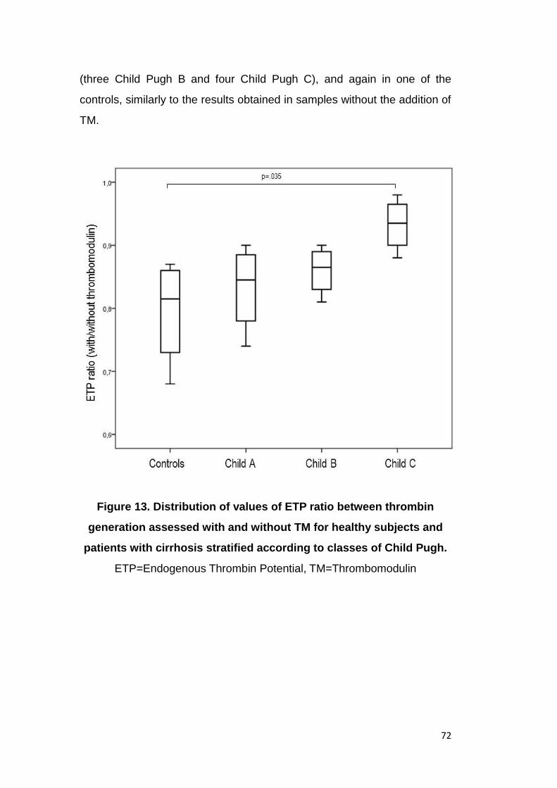
72
(three Child Pugh B and four Child Pugh C), and again in one of the
controls, similarly to the results obtained in samples without the addition of
TM.
Figure 13. Distribution of values of ETP ratio between thrombin
generation assessed with and without TM for healthy subjects and
patients with cirrhosis stratified according to classes of Child Pugh.
ETP=Endogenous Thrombin Potential, TM=Thrombomodulin

73
Figure 14. The ETP ratio of plasma with 0.35 UI mL enoxaparin to
native plasma in samples with 6 nM TM in healthy controls, cirrhotic
patients according to Child Pugh classes and subjects with a genetic
antithrombin (AT) defect type 1.
ETP=Endogenous Thrombin Potential, TM=Thrombomodulin

74

75
According to Virchow’s triad, the three key role players in the formation of
thrombosis are: blood stasis, alterations of the coagulation cascade, and
endothelial dysfunction. In cirrhosis patients, the most common site of
thrombosis is the portal vein, probably reflecting the consequence of all
three mechanisms. Venous stasis present in portal hypertension due to
the architectural derangement of the cirrhotic liver, likely combines with an
endothelium that has lost, partially or completely, its anticoagulant
properties. The characteristic altered coagulation status of advanced liver
disease, characterized by diminished levels of pro- but also anti-coagulant
circulating factors, probably also contributes to the establishment of portal
vein thrombosis.
The arm-to-tongue circulation time of the blood (∼30 seconds) is short
compared with a whole blood clotting time, so thrombin formed in flowing
blood in vivo is rapidly diluted and inactivated before clotting can occur.
Thrombin only builds up in unstirred boundary layers at cell surfaces and
in the stagnant plasma caught in a clot or an aggregate, and transport by
diffusion will therefore tend to govern reaction rates (42). Within the
portal vein of cirrhosis patients, flow is severely hampered, with inversion
of flow direction being a relatively common finding (128). In the study by
Zocco and collaborators, where portal flow velocity was determined using
Doppler ultrasound in cirrhotic patients who were subsequently followed
for the development of PVT, patients who developed PVT had a mean
basal value of portal flow velocity of 11.8 ± 2.6 cm/s, and a velocity below
15 cm/s was the only independent predictor of occurrence of PVT(75).
Notably, however, low flow in the portal vein (19.6 ± 5.7 cm/s) was also
found in all cirrhotics, also in those who had not developed PVT one year
after initial Doppler evaluation. Venous stasis can favor the initiation of the
76
coagulation cascade, which, in the presence of an endothelium that has
lost its anticoagulant properties, may result in thrombus formation.
Actual development of thrombosis has been demonstrated to injure the
venous wall, causing a reaction that has been described as similar to
wound healing, associated with increased procollagen, total collagen, and
metalloproteinases (129).
Not only existing thrombosis, however, but also hypertension, has been
experimentally shown to induce measurable changes. Hayashi et al. used
a rabbit model in which left femoral venous pressure was elevated by
constriction of the external iliac vein (130). Compared with the
contralateral normal vein, the femoral vein, which experienced the higher
intravascular pressure (16 vs 7 mmHg), displayed increased wall thickness
and elevated vascular tone.
Regarding the integrity of the endothelium’s anticoagulant functions, TM
plays a fundamental role and was therefore studied. TM is an integral
membrane glycoprotein that has a major role in the regulation of
intravascular coagulation(131). This glycoprotein can change the function
of thrombin, the main procoagulant enzyme, to an anticoagulant through
activation of the protein C pathway(132). In turn, activated protein C in the
presence of protein S inactivates factor VIIIa and factor Va, and thereby
inhibits further formation of thrombin.
High levels of circulating TM have been demonstrated after induction of
liver damage in animal models and other diseases with systemic
inflammation(93;133). Circulating TM has been found to be either normal
or elevated in patients with liver disease, and to be correlated with
endothelial damage, without showing, however, a direct relationship with
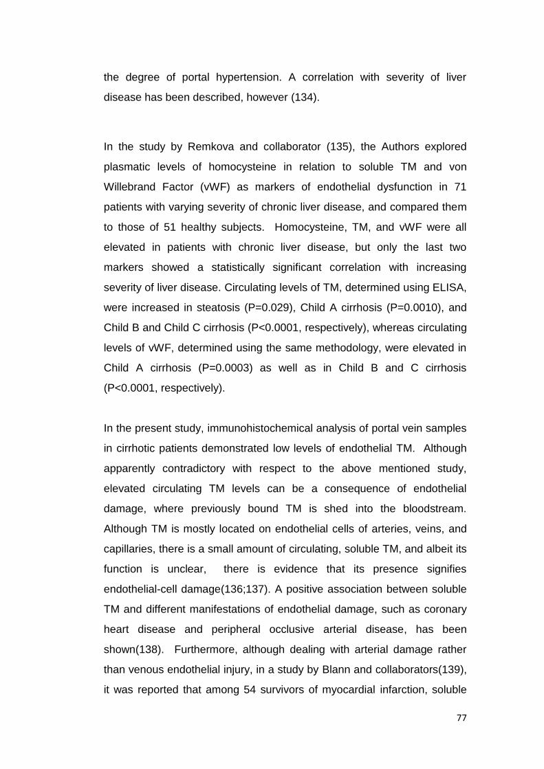
77
the degree of portal hypertension. A correlation with severity of liver
disease has been described, however (134).
In the study by Remkova and collaborator (135), the Authors explored
plasmatic levels of homocysteine in relation to soluble TM and von
Willebrand Factor (vWF) as markers of endothelial dysfunction in 71
patients with varying severity of chronic liver disease, and compared them
to those of 51 healthy subjects. Homocysteine, TM, and vWF were all
elevated in patients with chronic liver disease, but only the last two
markers showed a statistically significant correlation with increasing
severity of liver disease. Circulating levels of TM, determined using ELISA,
were increased in steatosis (P=0.029), Child A cirrhosis (P=0.0010), and
Child B and Child C cirrhosis (P<0.0001, respectively), whereas circulating
levels of vWF, determined using the same methodology, were elevated in
Child A cirrhosis (P=0.0003) as well as in Child B and C cirrhosis
(P<0.0001, respectively).
In the present study, immunohistochemical analysis of portal vein samples
in cirrhotic patients demonstrated low levels of endothelial TM. Although
apparently contradictory with respect to the above mentioned study,
elevated circulating TM levels can be a consequence of endothelial
damage, where previously bound TM is shed into the bloodstream.
Although TM is mostly located on endothelial cells of arteries, veins, and
capillaries, there is a small amount of circulating, soluble TM, and albeit its
function is unclear, there is evidence that its presence signifies
endothelial-cell damage(136;137). A positive association between soluble
TM and different manifestations of endothelial damage, such as coronary
heart disease and peripheral occlusive arterial disease, has been
shown(138). Furthermore, although dealing with arterial damage rather
than venous endothelial injury, in a study by Blann and collaborators(139),
it was reported that among 54 survivors of myocardial infarction, soluble
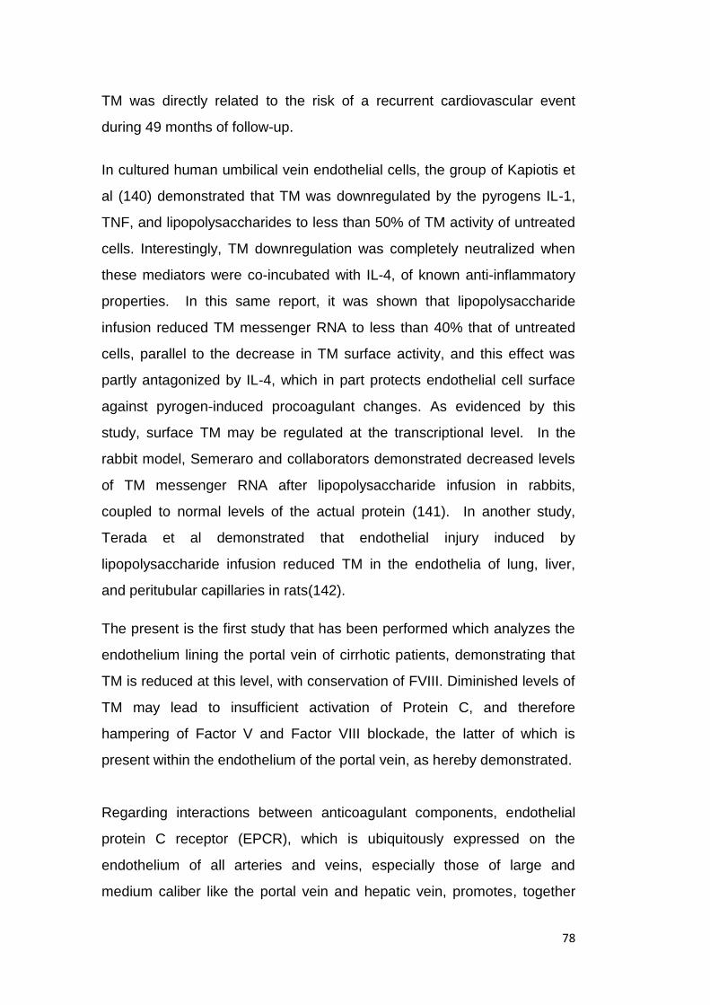
78
TM was directly related to the risk of a recurrent cardiovascular event
during 49 months of follow-up.
In cultured human umbilical vein endothelial cells, the group of Kapiotis et
al (140) demonstrated that TM was downregulated by the pyrogens IL-1,
TNF, and lipopolysaccharides to less than 50% of TM activity of untreated
cells. Interestingly, TM downregulation was completely neutralized when
these mediators were co-incubated with IL-4, of known anti-inflammatory
properties. In this same report, it was shown that lipopolysaccharide
infusion reduced TM messenger RNA to less than 40% that of untreated
cells, parallel to the decrease in TM surface activity, and this effect was
partly antagonized by IL-4, which in part protects endothelial cell surface
against pyrogen-induced procoagulant changes. As evidenced by this
study, surface TM may be regulated at the transcriptional level. In the
rabbit model, Semeraro and collaborators demonstrated decreased levels
of TM messenger RNA after lipopolysaccharide infusion in rabbits,
coupled to normal levels of the actual protein (141). In another study,
Terada et al demonstrated that endothelial injury induced by
lipopolysaccharide infusion reduced TM in the endothelia of lung, liver,
and peritubular capillaries in rats(142).
The present is the first study that has been performed which analyzes the
endothelium lining the portal vein of cirrhotic patients, demonstrating that
TM is reduced at this level, with conservation of FVIII. Diminished levels of
TM may lead to insufficient activation of Protein C, and therefore
hampering of Factor V and Factor VIII blockade, the latter of which is
present within the endothelium of the portal vein, as hereby demonstrated.
Regarding interactions between anticoagulant components, endothelial
protein C receptor (EPCR), which is ubiquitously expressed on the
endothelium of all arteries and veins, especially those of large and
medium caliber like the portal vein and hepatic vein, promotes, together

79
with TM, protein C activation by increasing at least by 5-fold the catalytic
efficiency of the thrombin-thrombomodulin complex. Although it has been
demonstrated that soluble levels of EPCR are increased in chronic liver
disease (143), this may be not sufficient to compensate for the low levels
of TM that have been found in the present study. It is possible, moreover,
that endothelial damage diminishes not only TM, but also EPCR, further
compromising the anticoagulant mechanisms of the endothelium. This
issue has not yet been explored, and future studies in this direction could
help clarify the interactions of these proteins with the other anticoagulant
components within the endothelium of the portal vein and other venous
territories in cirrhosis patients.
Factor VIII (FVIII), on the other hand, is a pro-coagulant factor which is
ubiquitously expressed in the endothelium. It is an essential cofactor for
FIXa-mediated catalysis of factor X to Xa. In its soluble form, FVIII is
notably increased in cirrhosis, and the most widely accepted mechanism is
the increase of von Willebrand factor, to which it binds when in circulation.
An established marker of endothelial dysfunction, the elevation in FVIII
has been shown to parallel the increase in severity of liver disease (144-
146).
As demonstrated in the present study, portal vein endothelium in cirrhosis
patients, as well as systemic venous territories as represented by the vena
cava, do not show differences with regard to the presence of FVIII, which
appears consistently conserved. The coupling between diminished TM
and conserved FVIII may represent hampering of a protective
anticoagulant mechanism by the first, and the maintenance of a pro-
coagulant mechanism by the latter, which possibly favor the development
of PVT.
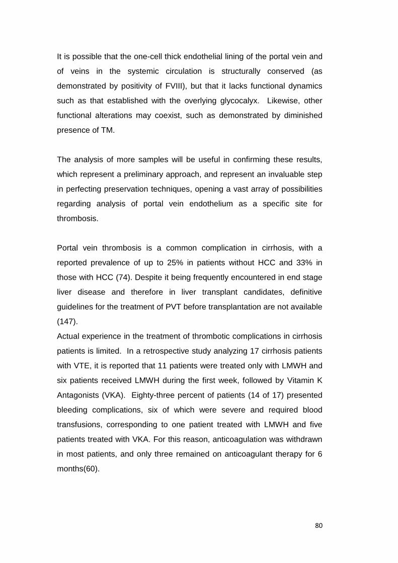
80
It is possible that the one-cell thick endothelial lining of the portal vein and
of veins in the systemic circulation is structurally conserved (as
demonstrated by positivity of FVIII), but that it lacks functional dynamics
such as that established with the overlying glycocalyx. Likewise, other
functional alterations may coexist, such as demonstrated by diminished
presence of TM.
The analysis of more samples will be useful in confirming these results,
which represent a preliminary approach, and represent an invaluable step
in perfecting preservation techniques, opening a vast array of possibilities
regarding analysis of portal vein endothelium as a specific site for
thrombosis.
Portal vein thrombosis is a common complication in cirrhosis, with a
reported prevalence of up to 25% in patients without HCC and 33% in
those with HCC (74). Despite it being frequently encountered in end stage
liver disease and therefore in liver transplant candidates, definitive
guidelines for the treatment of PVT before transplantation are not available
(147).
Actual experience in the treatment of thrombotic complications in cirrhosis
patients is limited. In a retrospective study analyzing 17 cirrhosis patients
with VTE, it is reported that 11 patients were treated only with LMWH and
six patients received LMWH during the first week, followed by Vitamin K
Antagonists (VKA). Eighty-three percent of patients (14 of 17) presented
bleeding complications, six of which were severe and required blood
transfusions, corresponding to one patient treated with LMWH and five
patients treated with VKA. For this reason, anticoagulation was withdrawn
in most patients, and only three remained on anticoagulant therapy for 6
months(60).

81
Notwithstanding existing evidence in favor of avoiding thrombosis
progression, current guidelines do not comprise treatment strategies for
management of patients in the waiting list for liver transplantation. To date,
135 cirrhotic patients with PVT in 4 published cohorts have been treated
with anticoagulation, resulting in a portal vein recanalization rate ranging
from 46% to 85% (70;148-150).
In the prospective study by Francoz et al (70), 19 cirrhosis patients
evaluated for liver transplantation, 18 with partial and one with complete
PVT, were treated with LMWH followed by VKA, and were compared
against a historical control group of 10 patients who received no
anticoagulation. All but two anticoagulated patients in Child class B or C,
and fourteen had had previous variceal bleeding. The rate of complete
recanalization was 42.1% at the end of follow up, but few details were
provided about the time interval between the diagnosis of thrombosis and
anticoagulation, and the time interval between the start of anticoagulation
and recanalization. It is noteworthy that patients with complete PVT at the
time of liver transplantation had significantly lower survival after transplant.
This further confirms the fact that at least in patients with cirrhosis and
PVT listed for liver transplantation, anticoagulation should be used.
In the second study by Amitrano et al. therapeutic dose (200UI/KG/die) of
LWMH was used in 29 patients with cirrhosis and PVT. Recanalization
was attained in 33.3% of cases within 6 months of treatment, but reached
a 75% rate in those patients who continued anticoagulation treatment
beyond 6 months, with time to recanalization which ranged from 1 to 17
months(148). This percentage is exceptionally high in comparison with the
efficacy of anticoagulation in a non-cirrhotic population with PVT (27%-
45.4%) and cannot be explained either by the time interval to
anticoagulation, nor by the presence/absence of underlying thrombophilic
conditions, which were not investigated by the authors.

82
In the retrospective study by Delgado et al, analyzing data from 55
cirrhosis patients with PVT who received anticoagulant therapy for acute
or subacute thrombosis (n=31), or due to progression of previously known
PVT (n=24), partial or complete recanalization was achieved in 33
patients, and early initiation of anticoagulation was the only factor which
was significantly associated with anticoagulant response. After stopping
anticoagulation, 5 of the 13 patients who had had recanalization on
anticoagulant therapy presented rethrombosis of the portal vein a median
of 1.3 months after cessation of anticoagulation. The Authors of this study
recommend the lifelong maintenance of anticoagulant therapy after
thrombus disappearance (151).
Senzolo and collaborators recently published a prospective study
proposing an algorithm for the treatment of PVT in cirrhosis including the
use of anticoagulation and transjugular intrahepatic portosystemic, and
report on several patients which are comprehended in the present study
population, but also on the group of patients from Royal Free Hospital that
constituted the untreated arm (152). In that study, 35 patients were
included in the protocol and 33 were anticoagulated with the standard
therapeutic dose of nadroparin (95 anti-Xa U/Kg body weight td), with a
40% dose reduction with platelet count below 50000x109/L. A complete
recanalization rate of 36% was reported, and partial response to
anticoagulation therapy was seen in 27% of patients. Mean interval
between starting of anticoagulation and recanalization was 5.5±2.6 months
(range 1-10).
The present study, which aside from broadening the study population,
explores the coagulation profile as a possible predictor of response to
anticoagulation, largely confirms these results, with a response rate of
65%. Complete recanalization rates are higher (52%), at the expense of a

83
smaller percent representation of partial recanalization rates (13%). This
can be explained by the fact that the present study includes recently
evaluated and treated patients, who presented for evaluation with a recent
thrombus onset, due to the ever-increasing awareness of the feasibility
and importance of anticoagulation treatment in these patients, which is
reflected on increased referral to tertiary centers. The present study also
confirms that the time interval between diagnosis of PVT and
anticoagulation is the strongest predictor of response to anticoagulant
treatment.
These data are similar to those obtained in the non-cirrhotic population
with PVT reported by Plessier et al (112), in which the one year
recanalization rate was 38%, with no patient obtaining recanalization
when anticoagulation was started beyond 12 months from the diagnosis of
PVT. Interestingly, in this series evaluating non-cirrhotic patients with PVT,
the presence of ascites and thrombotic splenic vein involvement were
independently associated with failure to obtain recanalization.
This probably confirms that chronicity of the thrombus (clinically
associated with the appearance of portal hypertension in non-cirrhotic
patients) is associated with the lack of efficacy of anticoagulation. In
contrast with the study by Amitrano and collaborators, anticoagulation was
continued for another 6 months in the case of those patients who had not
achieved recanalization after the first 6 months(148).
Importantly, an aspect that could contribute to the lack of response to
anticoagulation in some patients can be owed to the fact that a worse
stage of liver disease can preclude a prolonged anticoagulation treatment.
However, lack of studies do not allow for clarification of this bias; present
evidence does not indicate that patients followed have had to abandon
anticoagulant therapy prematurely due to increasing severity of liver

84
disease, or have perished while on anticoagulant therapy, thus reducing
the available time for thrombus resolution. Arguably, liver disease is less
severe in patients who are able to tolerate anticoagulation for several
months before undergoing liver transplant, and this could represent an
inherent bias. However, in the few studies published, patients with severe
liver disease have been included (Child C cirrhosis), and this has not
hampered the efficacy of anticoagulation, and comparability of groups
anticoagulated vs non-anticoagulated have been corroborated, with the
exception of variceal bleeding being less frequent in groups undergoing
anticoagulation(70;148).
In the present study, thrombosis progression was observed in one of four
patients who did not complete 12 months of anticoagulation, within 2
months of anticoagulation cessation. Of 9 non-responders who completed
12 months of anticoagulation, two had thrombosis progression after 3 and
4 months of anticoagulation cessation, respectively, and in the absence of
prophylaxis. Two other patients presented thrombosis recurrence after 1
and 4 months of anticoagulation cessation after obtainment of complete
recanalization, in the absence of prophylaxis. In contrast, no patient on
prophylaxis presented thrombus recurrence, 16 after complete or partial
response to therapy, and 7 after a course of 12 months of anticoagulation
without obtainment of recanalization.
These data are similar to those reported by Amitrano et al (148), where
thrombus progression was reported in 2/5 of non-responders, and in 1/10
of non-responders in the study by Francoz et al(70). In patients who were
not anticoagulated, thrombus extension was observed in 6/10 patients.
In the present study, albeit 86.7% of patients in whom recanalization was
achieved this event occurred within the first 6 months of therapy, in 4/30
patients this event was attained in the subsequent 6 months of

85
anticoagulation, which supports the notion that it is worthwhile to continue
anticoagulation therapy beyond the initial 6 months, for a similar period of
time.
The present study underlines the two important steps which must be met
in the management of this complication in cirrhosis: early diagnosis and
early treatment. Early start of anticoagulation therapy after thrombus
formation was the most important predictor of efficacy of anticoagulation
for the treatment of PVT in cirrhosis patients, and from this derives the
importance of not delaying diagnosis. An old, organized thrombus is less
likely to disappear on anticoagulation therapy with respect to a fresh
thrombus.
This is in line with what reported by Delgado et al (153), in that the only
factor that was statistically associated with the therapeutic efficacy of
anticoagulation was the delay in start of treatment (15±27 days in
responders vs 55±86 days in non-responders, p=.042). This group also
confirmed the lack of association between efficacy of anticoagulation
therapy and studied factors such as age, gender, history of previous
variceal bleeding, history of encephalopathy, previous episodes of portal
hypertension decompensation, Child-Pugh score, platelet count (x109/L),
renal function, presence of an underlying thrombophilia, presence of
symptoms at diagnosis, extension of PVT into the splachnic vasculature,
and presence of ascites.
Similarly, the presence of thrombophilia, as well as the coagulation status,
as described by the determination of coagulation factors does not seem to
influence response to anticoagulation. In the present study, in fact,
response to anticoagulation therapy neither thrombus progression nor
recurrence were associated with coagulation factors nor thrombophilic
defects. However, it is noteworthy that these tests offer only an

86
approximation of the actual hemostasis-coagulation dynamics taking place
inside the portal vein. On the other hand, Amitrano and collaborators did
not find any differences in clinical features or extension of portal
thrombosis between early or late responders and those that were non-
responders to anticoagulation (148).
In the present series, aside from a long-standing thrombus, the extension
of thrombosis into intrahepatic branches was another negative predictors
of response. The latter was associated with increasing severity of liver
disease, although the correlation did not reach statistical significance, but
this is probably due at least in part to the small number of patients with this
particular condition.
Interestingly, response to anticoagulation was independent of the stage of
cirrhosis and of the use of a full dose or a reduced dose of anticoagulation,
which is dictated by platelet number and therefore correlates with severity
of liver disease. These data support the notion that even patients with
advanced cirrhosis may benefit from anticoagulation, and that if a full dose
is not possible, recanalization can still be attempted with dose reduction.
In the present study, neither occlusion grade nor extension to other
splanchnic vessels were associated with the efficacy of anticoagulation.
This probably reflects the fact that acute thrombosis can debut as an
extensive thrombus, and thrombus extension is not necessarily a temporal
becoming of a chronic thrombosis, and that acutely formed, fresh thrombi
are more susceptible to the action of anticoagulants. In the study by
Francoz and collaborators (70), factors that were associated with
response to anticoagulation were not specifically sought for; however, the
only case of complete PVT was found in the group of non-responders.
However, only one of 19 treated patients had complete occlusion, and
therefore no conclusions can be drawn from this data.
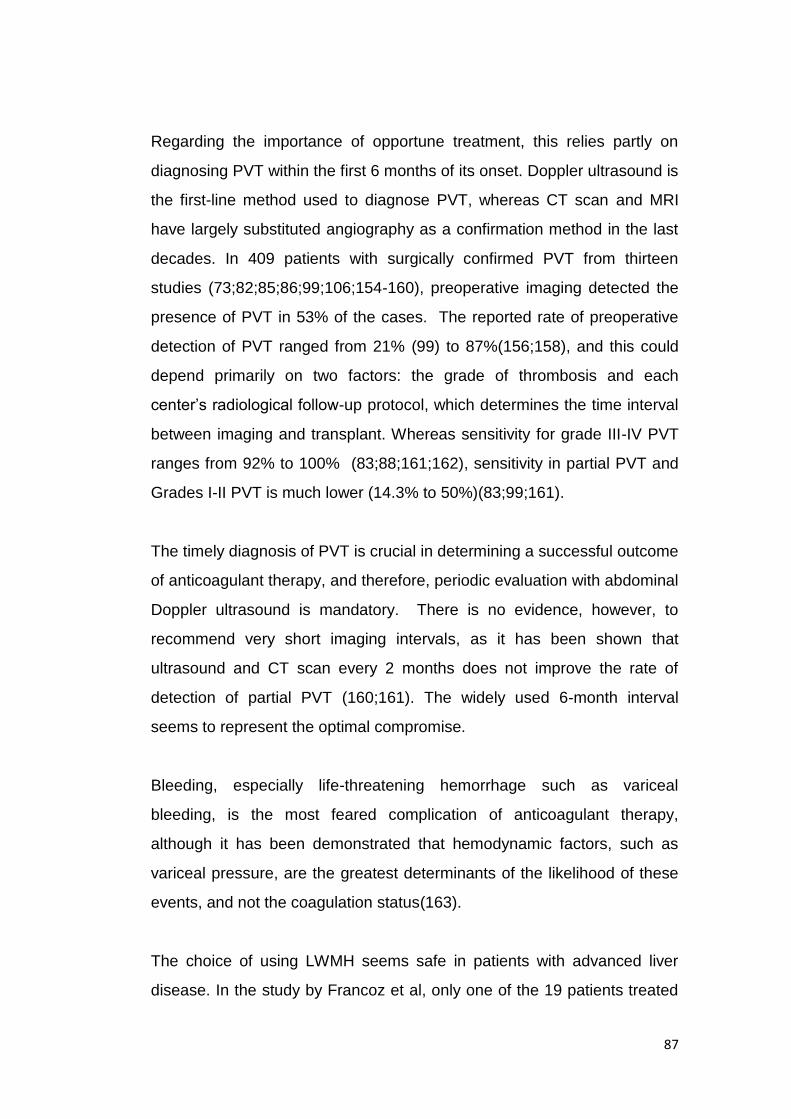
87
Regarding the importance of opportune treatment, this relies partly on
diagnosing PVT within the first 6 months of its onset. Doppler ultrasound is
the first-line method used to diagnose PVT, whereas CT scan and MRI
have largely substituted angiography as a confirmation method in the last
decades. In 409 patients with surgically confirmed PVT from thirteen
studies (73;82;85;86;99;106;154-160), preoperative imaging detected the
presence of PVT in 53% of the cases. The reported rate of preoperative
detection of PVT ranged from 21% (99) to 87%(156;158), and this could
depend primarily on two factors: the grade of thrombosis and each
center’s radiological follow-up protocol, which determines the time interval
between imaging and transplant. Whereas sensitivity for grade III-IV PVT
ranges from 92% to 100% (83;88;161;162), sensitivity in partial PVT and
Grades I-II PVT is much lower (14.3% to 50%)(83;99;161).
The timely diagnosis of PVT is crucial in determining a successful outcome
of anticoagulant therapy, and therefore, periodic evaluation with abdominal
Doppler ultrasound is mandatory. There is no evidence, however, to
recommend very short imaging intervals, as it has been shown that
ultrasound and CT scan every 2 months does not improve the rate of
detection of partial PVT (160;161). The widely used 6-month interval
seems to represent the optimal compromise.
Bleeding, especially life-threatening hemorrhage such as variceal
bleeding, is the most feared complication of anticoagulant therapy,
although it has been demonstrated that hemodynamic factors, such as
variceal pressure, are the greatest determinants of the likelihood of these
events, and not the coagulation status(163).
The choice of using LWMH seems safe in patients with advanced liver
disease. In the study by Francoz et al, only one of the 19 patients treated
88
with anticoagulation presented upper gastrointestinal bleeding, but in this
case the bleeding episode was caused by a post ligation ulcer, and
anticoagulation did not have to be discontinued in any patient (70). In the
study by Amitrano and collaborators, of 28 patients treated with
anticoagulation, none presented side effects such to consider interruption
of therapy; mild anemia due to portal hypertensive gastropathy was seen
in two patients, improving after iron supplementation (148). Delgado et al
report bleeding related to anticoagulation (variceal bleeding in all) in 5 of
55 treated patients, and platelet count below 50x109/L was the only factor
significantly associated with a higher risk of a hemorrhagic complication
(153). At a prophylactic dose in the setting of primary prevention of PVT,
LMWH was proven safe in a recently published prospective study in which
enoxaparin 4000 UI/day was administered to 34 Child B and C cirrhosis
patients for 12 months. No hemorrhagic events were reported, and
suspension of therapy was necessary in only one patient because of
thrombocytopenia (164).
In contrast, Garcia Fuster and collaborators reported bleeding
complications in 85% of patients (35% of them requiring transfusions) in a
series of 17 cirrhosis patients treated with vitamin K antagonists for DVT
(60). Moreover, an early cohort study evaluating 29,000 INR
measurements during a period of 6 months, demonstrated that underlying
liver disease or alcohol abuse were independently correlated with risk of
excessive anticoagulation (INR≥6) (165). Landefeld and collaborators
retrospectively evaluated predictors of major bleeding in hospitalized
patients receiving initial long term warfarin therapy. Among 411 patients
evaluated for different risk factors, bleeding episodes significantly
correlated with the presence of worsening liver function (166).
In the present study, one serious adverse event (a non-fatal cerebral
haemorrhage) occurred which was possibly related to anticoagulation.

89
Risk factors correlated with the underlying liver disease were absent (PLT
count was 110 x 109/L while INR and creatinine were within the normal
range).
Heparin induced thrombocytopenia occurred in three patients, and a
similar high frequency has been reported with the use of LMWH in
splanchnic vein thrombosis (167). LWMH was suspended in all three
patients: one was switched to warfarin, and two patients did not receive
any anticoagulation treatment. In all three patients the thrombosis
remained stable (there was no thrombus progression and no
recanalization was observed).
Attainment and maintenance of portal vein patency did not have an impact
on transplant-free survival, but this is in part due to the fact that PVT is
more frequent in patients with more advanced cirrhosis, who have a
greater probability of being transplanted. If only the event death as such is
considered, rethrombosis and thrombus progression entailed a mortality of
80%, and only one of the five deaths in the present series occurred in a
patient who had adequately responded to therapy. However, this study
was not powered to show survival differences, and prolonged follow-up
studies are needed to assess survival.
Regarding secondary prophylaxis after achieving recanalization, 12
patients continued prophylactic anticoagulation, and remained free of
rethrombosis at 6 months’ follow-up. In contrast, of four patients who had
achieved complete recanalization but did not continue anticoagulation
prophylaxis, two had recurrence of the thrombosis, and in one case this
led to death. Anticoagulation prophylaxis was also continued in 6 other
patients after achievement of partial recanalization, and of the four
patients evaluated six months later, none had presented thrombus
recurrence. Of 9 patiens in whom recanalization was not achieved even
after 12 months of therapy, seven continued anticoagulation prophylaxis,
90
and remain stable after 4, and 7 months of follow-up, respectively. This is
in contrast with the two patients in whom recanalization was not achieved
after 12 months of therapy, and in whom prophylactic anticoagulation was
not continued; these two patients presented progression of the thrombus
into splanchnic vasculture and one into the vena cava, and led to death in
both cases.
Although the recommendation to continue anticoagulation in patients with
PVT who do not obtain recanalization cannot be made based on current
evidence, the efficacy of anticoagulation in preventing progression of
thrombosis to a total splanchnic vein thrombosis should be taken into
consideration, especially in patients who are potential or future candidates
for liver transplantation.
Patients with cirrhosis are prone to develop thrombotic complications
including PVT and DVT. Despite these complications may warrant
anticoagulant therapy, current treatment guidelines do not provide specific
indications for anticoagulation in patients with liver disease and
thrombosis.
Furthermore, low levels of antithrombin, which is necessary for the action
of low molecular weight heparin, could theoretically hamper the
anticoagulant effect of these drugs(116), and data on their use in patients
with cirrhosis are still limited. Clinically, however, low molecular weight
heparin is effective in the treatment of thrombotic complications in these
patients, notwithstanding low levels of antithrombin and low levels of anti-
Xa activity. Moreover, in a small in vitro study by Lisman and
collaborators(117), it was demonstrated that the anti-Xa assay
substantially underestimates the LMWH mass present in plasma from
patients with cirrhosis, and actually a fixed amount of LMWH is as effective
in cirrhotic plasma as it is in plasma from normal pool plasma.

91
Thrombin generation has been recently used to investigate haemostatic
balance in patients with liver disease and can be useful to evaluate the
efficacy of LWMH in this setting of patients.
The present study represents an evaluation of the in vitro effect of LMWH
on thrombin generation in cirrhotic patients, in controls and in a group of
AT-deficient patients.
Child Pugh C patients demonstrated the greatest response to LMWH, as
compared to controls. On the contrary, patients with heterozygous AT
deficiency showed an in vitro resistance to the effect of LMWH, despite
similar plasma levels of AT as compared to Child Pugh C cirrhotics. This
increased effect of LWMH was also evident when a therapeutic dose of
LMWH was used (0.7 U anti-Xa/mL), which completely inhibited thrombin
generation in approximately about one-third of cirrhotic patients and
resulted in a thrombin generation similar to controls in the remaining
cirrhotic samples.
When the experiments were repeated in samples with the addition of TM
in order to unveil the acquired protein C deficiency in patients with
cirrhosis, the response to the addition of LMWH did not change. Moreover,
the proportion of plasma samples from patients with more advanced
cirrhosis (Child C) whose thrombin generation was completely abated
after the addition of LMWH at 0.7 IU/ml was not different between
samples with and without TM. This confirms the notion that, in cirrhotic
patients, the newly rebalanced hemostatic equilibrium due to the
concomitant decrease in pro-coagulant and anticoagulant factors can be
easily perturbed.
92
Contrary to what is expected, in cirrhotics the effect of LWMH inversely
correlated with AT levels. Thus, the lower the level of AT, the higher the
anticoagulant response to LWHM. This may be due to the concomitant
reduction of all procoagulant factors, in particular of factor Xa which is the
target of LMWH inhibition. On the contrary, subjects with inherited AT
deficiency had normal plasma levels of all procoagulant factors. As a
consequence, the reduced effect of LMWH caused by low AT levels, was
not counterbalanced by the reduction of other procoagulant factors and
possibly results in the in vitro LMWH resistance.
In agreement with Bechman et al, the present study shows that plasma
from patients with chronic liver disease present with lower anti-Xa activity
when a similar dose of LMWH is added, as compared to control plasma.
However, we also found that in the presence of a lower anti-Xa activity,
plasma from patients with end stage liver disease exhibited a more
pronounced response to anticoagulation as compared to control plasma.
Therefore, patients in Child Pugh C class, despite very low plasma anti-
Xa activity, demonstrated the highest anticoagulation effect, as opposed to
what can be seen in patients with isolated AT defect.
This apparently conflicting data can be explained by considering the
characteristic of the method used to measure anti-Xa activity in the plasma
sample.
As the test is performed by adding fixed amounts (in excess) of FXa to the
sample, the result of the reaction depends only on LMWH-AT complex
generation and concentration. Adding a fixed amount of LMWH in all
samples, the differences in the levels of anti-Xa activity depend merely on
AT plasma levels. Patients with AT deficiency showed similar anti-Xa
levels to those of Child A cirrhotic patients, and not significantly different
from that of controls; this could be due to the difference in the levels of

93
coagulation factors in plasma samples. Theoretically, the test mechanism
implies saturation of Xa, and holds true for plasma of patients with
normal AT level. The test’s results could be influenced by decreased
levels of endogenous FXa, as well as by the reduced levels of other
coagulation factors.
Therefore, anti-Xa determination does not seem to be a suitable test for
monitoring anticoagulation in cirrhotic patients, since low anti-Xa levels are
not indicative of a reduction in response to LMWH. On the contrary, the
present findings show that thrombin generation assay can be useful in the
evaluation of anticoagulant effect of heparin in cirrhotic patients with
impaired clotting system.
However, these findings need to be further confirmed in vivo. As the
matter of fact, an additional influence on pharmacokynetics and
distribution of LWMH in cirrhotic patients with ascites may derive from
altered water content and body mass index, thus leading to unpredictable
LWMH concentrations.
It is also important to consider that different LWMHs might not have the
same effect in patients with cirrhosis, due to the different ratio of inhibition
of factor Xa and thrombin. This ratio can vary from 2.2 to 4.1 among
commercially available LWMH. For example, enoxaparin, which is
characterized by an anti- Xa/anti- factor IIa ratio of 3.8, might exert an
different effect than dalteparin, which has a ratio of 2.7.

94
95
Aside from the reduction of flow characteristic of portal hypertension and
cirrhosis, there seems to be an apparent imbalance between pro- and anti-
coagulant factors that could, together with other local factors, contribute to
the development of PVT.
The structure of the endothelial lining of the portal vein in cirrhosis patients
seems to be conserved, and its aspect regarding the endothelial marker
FVIII is similar to that of other venous territories, exemplified by the vena
cava, as well as to that of portal vein and vena cava endothelial lining of
non-cirrhotic subjects. That is, the endothelium of the portal vein in
cirrhosis patients seems to be characterized by a normal representation of
FVIII, which is one of the most important drivers of TF-independent
coagulation.
On the contrary, the anticoagulation mechanism mediated by TM seems to
be underrepresented in the portal vein endothelium of cirrhotic patients
when compared to that of non-cirrhotic subjects. This imbalance, together
with the mechanical alterations of portal vein flow, may be responsible for
the elevated incidence of PVT in cirrhosis patients. Further studies are
needed regarding other protective mechanisms such as
glycosaminoglycans and endothelial Protein C receptor.
When PVT develops in cirrhosis patients, anticoagulant therapy with
LMWH is effective in 65.2% of cases, with achievement of complete portal
vein patency in 52.2% of patients. Furthermore, recanalization is achieved
during the first 6 months of anticoagulation therapy in most patients, but as
recanalization is reached in yet another, albeit small group of patients who
continue anticoagulant therapy beyond 6 months, it is worthwhile to
continue anticoagulation for 12 months.
96
In particular, the hemostatic systemic status does not seem to have an
impact on the response to anticoagulant therapy, but rather, the interval
between PVT onset and start of anticoagulation therapy. The continuation
of anticoagulation at a prophylactic dose for 6 months after recanalization
seems to be effective in preventing recurrence of PVT after suspension of
anticoagulation at a therapeutic dose.
Cirrhosis patients therefore demonstrate an adequate clinical response to
anticoagulant therapy, notwithstanding their low antithrombin levels,
actually showing a greater susceptibility to increasing concentrations of
LMWH in advanced cirrhosis, apparent from sharply abated thrombin
generation curves and decreasing endogenous thrombin potential.
Henceforth, patients with cirrhosis may have an enhanced anticoagulant
response to LWMH, which increases with the severity of liver disease.
Anti-X activity is not a good marker of response to LWMH in patients with
liver disease and should not be used in clinical practice to monitor the
anticoagulant effect.
Finally, dose adjustment of LWMHs might be warranted according to the
Child Pugh class in order to avoid excessive anticoagulation and possible
bleeding complications. Clinical studies on the in vivo effect of LMWH in
cirrhotic patients with thrombosis are needed to further validate this
hypothesis.

97
(1) Caldwell SH, Hoffman M, Lisman T, Macik BG, Northup PG,
Reddy KR, et al. Coagulation disorders and hemostasis in liver
disease: pathophysiology and critical assessment of current
management. Hepatology 2006 Oct;44(4):1039-46.
(2) Lisman T, Caldwell SH, Burroughs AK, Northup PG, Senzolo M,
Stravitz RT, et al. Hemostasis and thrombosis in patients with liver
disease: the ups and downs. J Hepatol 2010 Aug;53(2):362-71.
(3) Senzolo M, Cholongitas E, Thalheimer U, Riddell A, Agarwal S,
Mallett S, et al. Heparin-like effect in liver disease and liver
transplantation. Clin Liver Dis 2009 Feb;13(1):43-53.
(4) Ferro D, Quintarelli C, Lattuada A, Leo R, Alessandroni M,
Mannucci PM, et al. High plasma levels of von Willebrand factor
as a marker of endothelial perturbation in cirrhosis: relationship to
endotoxemia. Hepatology 1996 Jun;23(6):1377-83.
(5) Wion KL, Kelly D, Summerfield JA, Tuddenham EG, Lawn RM.
Distribution of factor VIII mRNA and antigen in human liver and
other tissues. Nature 1985 Oct 24;317(6039):726-9.
(6) Hollestelle MJ, Geertzen HG, Straatsburg IH, van Gulik TM, van
Mourik JA. Factor VIII expression in liver disease. Thromb
Haemost 2004 Feb;91(2):267-75.
(7) Kelly DA, Summerfield JA. Hemostasis in liver disease. Semin
Liver Dis 1987 Aug;7(3):182-91.
(8) Kerr R. New insights into haemostasis in liver failure. Blood
Coagul Fibrinolysis 2003 Jun;14 Suppl 1:S43-S45.

98
(9) Maisonneuve P, Sultan Y. Modification of factor VIII complex
properties in patients with liver disease. J Clin Pathol 1977
Mar;30(3):221-7.
(10) Mueller MM, Bomke B, Seifried E. Fresh frozen plasma in patients
with disseminated intravascular coagulation or in patients with
liver diseases. Thromb Res 2002 Oct 31;107 Suppl 1:S9-17.
(11) Blanchard RA, Furie BC, Jorgensen M, Kruger SF, Furie B.
Acquired vitamin K-dependent carboxylation deficiency in liver
disease. N Engl J Med 1981 Jul 30;305(5):242-8.
(12) Belle M, Brebant R, Guinet R, Leclercq M. Production of a new
monoclonal antibody specific to human des-gamma-
carboxyprothrombin in the presence of calcium ions. Application
to the development of a sensitive ELISA-test. J Immunoassay
1995 May;16(2):213-29.
(13) Sherlock S, Dooley J. The haematololgy of liver disease. In:
Blackwell Publishing, editor. Diseases of the liver and biliary
system. 11 ed. Oxford: 2002. p. 47-64.
(14) Pereira SP, Rowbotham D, Fitt S, Shearer MJ, Wendon J,
Williams R. Pharmacokinetics and efficacy of oral versus
intravenous mixed-micellar phylloquinone (vitamin K1) in severe
acute liver disease. J Hepatol 2005 Mar;42(3):365-70.
(15) Schipper HG, ten Cate JW. Antithrombin III transfusion in patients
with hepatic cirrhosis. Br J Haematol 1982 Sep;52(1):25-33.
(16) Liebman HA, McGehee WG, Patch MJ, Feinstein DI. Severe
depression of antithrombin III associated with disseminated
intravascular coagulation in women with fatty liver of pregnancy.
Ann Intern Med 1983 Mar;98(3):330-3.
99
(17) Carmassi F, Morale M, De Negri F, Carrai M. Modulation of
hemostatic balance with antithrombin III replacement therapy in a
case of liver cirrhosis associated with recurrent venous
thrombosis. J Mol Med 1995 Feb;73(2):89-93.
(18) Fair DS, Marlar RA. Biosynthesis and secretion of factor VII,
protein C, protein S, and the Protein C inhibitor from a human
hepatoma cell line. Blood 1986 Jan;67(1):64-70.
(19) Mannucci PM, Vigano S. Deficiencies of protein C, an inhibitor of
blood coagulation. Lancet 1982 Aug 28;2(8296):463-7.
(20) Senzolo M, Cholongitas CE, Patch D, Burroughs AK. Update on
the classification, assessment of prognosis and therapy of Budd-
Chiari syndrome. Nat Clin Pract Gastroenterol Hepatol 2[4], 182-
190. 2005.
(21) Minnema MC, Janssen HL, Niermeijer P, de Man RA. Budd-Chiari
syndrome: combination of genetic defects and the use of oral
contraceptives leading to hypercoagulability. J Hepatol 2000
Sep;33(3):509-12.
(22) Lisman T, Leebeek FW, de Groot PG. Haemostatic abnormalities
in patients with liver disease. J Hepatol 2002 Aug;37(2):280-7.
(23) Primignani M, Martinelli I, Bucciarelli P, Battaglioli T, Reati R,
Fabris F, et al. Risk factors for thrombophilia in extrahepatic portal
vein obstruction. Hepatology 2005 Mar;41(3):603-8.
(24) Bhattacharyya M, Makharia G, Kannan M, Ahmed RP, Gupta PK,
Saxena R. Inherited prothrombotic defects in Budd-Chiari
syndrome and portal vein thrombosis: a study from North India.
Am J Clin Pathol 2004 Jun;121(6):844-7.

100
(25) Northup PG, Sundaram V, Fallon MB, Reddy KR, Balogun RA,
Sanyal AJ, et al. Hypercoagulation and thrombophilia in liver
disease. J Thromb Haemost 2008 Jan;6(1):2-9.
(26) Senzolo M, Burroughs AK. Hemostasis alterations in liver disease
and liver transplantation. In: Kitchens G, Alving B, Kessler G, eds.
Consultative Hemostasis and Thrombosis. Philadelphia, PA:
Saunders; 490-502. 2007.
(27) Lisman T, Leebeek FW, de Groot PG. Haemostatic abnormalities
in patients with liver disease. J Hepatol 2002 Aug;37(2):280-7.
(28) Lisman T, Leebeek FW. Hemostatic alterations in liver disease: a
review on pathophysiology, clinical consequences, and treatment.
Dig Surg 2007;24(4):250-8.
(29) Lisman T, Bakhtiari K, Pereboom IT, Hendriks HG, Meijers JC,
Porte RJ. Normal to increased thrombin generation in patients
undergoing liver transplantation despite prolonged conventional
coagulation tests. J Hepatol 2010 Mar;52(3):355-61.
(30) Tripodi A, Salerno F, Chantarangkul V, Clerici M, Cazzaniga M,
Primignani M, et al. Evidence of normal thrombin generation in
cirrhosis despite abnormal conventional coagulation tests.
Hepatology 2005 Mar;41(3):553-8.
(31) Dahlback B. Progress in the understanding of the protein C
anticoagulant pathway. Int J Hematol 2004 Feb;79(2):109-16.
(32) Amitrano L, Brancaccio V, Guardascione MA, Margaglione M,
Iannaccone L, D'Andrea G, et al. Inherited coagulation disorders
in cirrhotic patients with portal vein thrombosis. Hepatology 2000
Feb;31(2):345-8.
101
(33) Peck-Radosavljevic M. Thrombocytopenia in liver disease. Can J
Gastroenterol 2000 Nov;14 Suppl D:60D-6D.
(34) Goulis J, Chau TN, Jordan S, Mehta AB, Watkinson A, Rolles K,
et al. Thrombopoietin concentrations are low in patients with
cirrhosis and thrombocytopenia and are restored after orthotopic
liver transplantation. Gut 1999 May;44(5):754-8.
(35) Rubin MH, Weston MJ, Langley PG, White Y, Williams R. Platelet
function in chronic liver disease: relationship to disease severity.
Dig Dis Sci 1979 Mar;24(3):197-202.
(36) Ordinas A, Escolar G, Cirera I, Vinas M, Cobo F, Bosch J, et al.
Existence of a platelet-adhesion defect in patients with cirrhosis
independent of hematocrit: studies under flow conditions.
Hepatology 1996 Nov;24(5):1137-42.
(37) Laffi G, Marra F, Gresele P, Romagnoli P, Palermo A, Bartolini O,
et al. Evidence for a storage pool defect in platelets from cirrhotic
patients with defective aggregation. Gastroenterology 1992
Aug;103(2):641-6.
(38) Violi F, Leo R, Vezza E, Basili S, Cordova C, Balsano F. Bleeding
time in patients with cirrhosis: relation with degree of liver failure
and clotting abnormalities. C.A.L.C. Group. Coagulation
Abnormalities in Cirrhosis Study Group. J Hepatol 1994
Apr;20(4):531-6.
(39) Ordinas A, Maragall S, Castillo R, Nurden AT. A glycoprotein I
defect in the platelets of three patients with severe cirrhosis of the
liver. Thromb Res 1978 Aug;13(2):297-302.
(40) Lisman T, Bongers TN, Adelmeijer J, Janssen HL, de Maat MP,
de Groot PG, et al. Elevated levels of von Willebrand Factor in
102
cirrhosis support platelet adhesion despite reduced functional
capacity. Hepatology 2006 Jul;44(1):53-61.
(41) Hemker HC, Giesen P, AlDieri R, Regnault V, de SE,
Wagenvoord R, et al. The calibrated automated thrombogram
(CAT): a universal routine test for hyper- and hypocoagulability.
Pathophysiol Haemost Thromb 2002 Sep;32(5-6):249-53.
(42) Hemker HC, al DR, Beguin S. Thrombin generation assays:
accruing clinical relevance. Curr Opin Hematol 2004
May;11(3):170-5.
(43) Hemker HC, Willems GM, Beguin S. A computer assisted method
to obtain the prothrombin activation velocity in whole plasma
independent of thrombin decay processes. Thromb Haemost
1986 Aug 20;56(1):9-17.
(44) Chantarangkul V, Clerici M, Bressi C, Giesen PL, Tripodi A.
Thrombin generation assessed as endogenous thrombin potential
in patients with hyper- or hypo-coagulability. Haematologica 2003
May;88(5):547-54.
(45) Hemker HC, Giesen P, al DR, Regnault V, de SE, Wagenvoord R,
et al. Calibrated automated thrombin generation measurement in
clotting plasma. Pathophysiol Haemost Thromb 2003;33(1):4-15.
(46) MACFARLANE RG, BIGGS R. A thrombin generation test; the
application in haemophilia and thrombocytopenia. J Clin Pathol
1953 Feb;6(1):3-8.
(47) PITNEY WR, DACIE JV. A simple method of studying the
generation of thrombin in recalcified plasma; application in the
investigation of haemophilia. J Clin Pathol 1953 Feb;6(1):9-14.
103
(48) Seegers WH. A personal perspective on hemostasis and
thrombosis (1937-1981). Semin Thromb Hemost 1981 Dec;7(3-
4):177-307.
(49) Houbouyan L, Padilla A, Gray E, Longstaff C, Barrowcliffe TW.
Inhibition of thrombin generation by heparin and LMW heparins: a
comparison of chromogenic and clotting methods. Blood Coagul
Fibrinolysis 1996 Jan;7(1):24-30.
(50) van Veen JJ, Gatt A, Makris M. Thrombin generation testing in
routine clinical practice: are we there yet? Br J Haematol 2008
Sep;142(6):889-903.
(51) Castoldi E, Rosing J. Thrombin generation tests. Thromb Res
2011 Feb;127 Suppl 3:S21-S25.
(52) Segers O, van OR, ten CH, Rosing J, Castoldi E. Thrombin
generation as an intermediate phenotype for venous thrombosis.
Thromb Haemost 2010 Jan;103(1):114-22.
(53) Lutsey PL, Folsom AR, Heckbert SR, Cushman M. Peak thrombin
generation and subsequent venous thromboembolism: the
Longitudinal Investigation of Thromboembolism Etiology (LITE)
study. J Thromb Haemost 2009 Oct;7(10):1639-48.
(54) Tripodi A, Martinelli I, Chantarangkul V, Battaglioli T, Clerici M,
Mannucci PM. The endogenous thrombin potential and the risk of
venous thromboembolism. Thromb Res 2007;121(3):353-9.
(55) Tripodi A, Primignani M, Chantarangkul V, Dell'Era A, Clerici M,
de FR, et al. An imbalance of pro- vs anti-coagulation factors in
plasma from patients with cirrhosis. Gastroenterology 2009
Dec;137(6):2105-11.

104
(56) al DR, Alban S, Beguin S, Hemker HC. Thrombin generation for
the control of heparin treatment, comparison with the activated
partial thromboplastin time. J Thromb Haemost 2004
Aug;2(8):1395-401.
(57) Sogaard KK, Horvath-Puho E, Gronbaek H, Jepsen P, Vilstrup H,
Sorensen HT. Risk of venous thromboembolism in patients with
liver disease: a nationwide population-based case-control study.
Am J Gastroenterol 2009 Jan;104(1):96-101.
(58) Huerta C, Johansson S, Wallander MA, Garcia Rodriguez LA.
Risk factors and short-term mortality of venous thromboembolism
diagnosed in the primary care setting in the United Kingdom. Arch
Intern Med 2007 May 14;167(9):935-43.
(59) Northup PG, McMahon MM, Ruhl AP, Altschuler SE, Volk-
Bednarz A, Caldwell SH, et al. Coagulopathy does not fully
protect hospitalized cirrhosis patients from peripheral venous
thromboembolism. Am J Gastroenterol 2006 Jul;101(7):1524-8.
(60) Garcia-Fuster MJ, Abdilla N, Fabia MJ, Fernandez C, Oliver V,
Forner MJ. [Venous thromboembolism and liver cirrhosis]. Rev
Esp Enferm Dig 2008 May;100(5):259-62.
(61) Lesmana CR, Inggriani S, Cahyadinata L, Lesmana LA. Deep
vein thrombosis in patients with advanced liver cirrhosis: a rare
condition? Hepatol Int 2010;4(1):433-8.
(62) Gulley D, Teal E, Suvannasankha A, Chalasani N, Liangpunsakul
S. Deep vein thrombosis and pulmonary embolism in cirrhosis
patients. Dig Dis Sci 2008 Nov;53(11):3012-7.
(63) Dabbagh O, Oza A, Prakash S, Sunna R, Saettele TM.
Coagulopathy does not protect against venous thromboembolism
105
in hospitalized patients with chronic liver disease. Chest 2010
May;137(5):1145-9.
(64) Ali M, Ananthakrishnan AN, McGinley EL, Saeian K. Deep vein
thrombosis and pulmonary embolism in hospitalized patients with
cirrhosis: a nationwide analysis. Dig Dis Sci 2011 Jul;56(7):2152-
9.
(65) Aldawood A, Arabi Y, Aljumah A, Alsaadi A, Rishu A, Aldorzi H, et
al. The incidence of venous thromboembolism and practice of
deep venous thrombosis prophylaxis in hospitalized cirrhotic
patients. Thromb J 2011;9(1):1.
(66) Wu H, Nguyen GC. Liver cirrhosis is associated with venous
thromboembolism among hospitalized patients in a nationwide US
study. Clin Gastroenterol Hepatol 2010 Sep;8(9):800-5.
(67) Rodriguez-Castro KI, Simioni P, Burra P, Senzolo M.
Anticoagulation for the treatment of thrombotic complications in
patients with cirrhosis. Liver Int 2012 Nov;32(10):1465-76.
(68) Mangia A, Villani MR, Cappucci G, Santoro R, Ricciardi R,
Facciorusso D, et al. Causes of portal venous thrombosis in
cirrhotic patients: the role of genetic and acquired factors. Eur J
Gastroenterol Hepatol 2005 Jul;17(7):745-51.
(69) Erkan O, Bozdayi AM, Disibeyaz S, Oguz D, Ozcan M, Bahar K,
et al. Thrombophilic gene mutations in cirrhotic patients with portal
vein thrombosis. Eur J Gastroenterol Hepatol 2005
Mar;17(3):339-43.
(70) Francoz C, Belghiti J, Vilgrain V, Sommacale D, Paradis V,
Condat B, et al. Splanchnic vein thrombosis in candidates for liver
106
transplantation: usefulness of screening and anticoagulation. Gut
2005 May;54(5):691-7.
(71) Amitrano L, Guardascione MA, Brancaccio V, Margaglione M,
Manguso F, Iannaccone L, et al. Risk factors and clinical
presentation of portal vein thrombosis in patients with liver
cirrhosis. J Hepatol 2004 May;40(5):736-41.
(72) Amitrano L, Brancaccio V, Guardascione MA, Margaglione M,
Sacco M, Martino R, et al. Portal vein thrombosis after variceal
endoscopic sclerotherapy in cirrhotic patients: role of genetic
thrombophilia. Endoscopy 2002 Jul;34(7):535-8.
(73) Yerdel MA, Gunson B, Mirza D, Karayalcin K, Olliff S, Buckels J,
et al. Portal vein thrombosis in adults undergoing liver
transplantation: risk factors, screening, management, and
outcome. Transplantation 2000 May 15;69(9):1873-81.
(74) Nonami T, Yokoyama I, Iwatsuki S, Starzl TE. The incidence of
portal vein thrombosis at liver transplantation. Hepatology 1992
Nov;16(5):1195-8.
(75) Zocco MA, Di SE, De CR, Novi M, Ainora ME, Ponziani F, et al.
Thrombotic risk factors in patients with liver cirrhosis: correlation
with MELD scoring system and portal vein thrombosis
development. J Hepatol 2009 Oct;51(4):682-9.
(76) Fimognari FL, De SA, Piccheri C, Moscatelli R, Gigliotti F, Vestri
A, et al. Evaluation of D-dimer and factor VIII in cirrhotic patients
with asymptomatic portal venous thrombosis. J Lab Clin Med
2005 Oct;146(4):238-43.
107
(77) Martinelli I. von Willebrand factor and factor VIII as risk factors for
arterial and venous thrombosis. Semin Hematol 2005
Jan;42(1):49-55.
(78) Martinelli I, Primignani M, Aghemo A, Reati R, Bucciarelli P,
Fabris F, et al. High levels of factor VIII and risk of extra-hepatic
portal vein obstruction. J Hepatol 2009 May;50(5):916-22.
(79) Llado L, Fabregat J, Castellote J, Ramos E, Torras J, Jorba R, et
al. Management of portal vein thrombosis in liver transplantation:
influence on morbidity and mortality. Clin Transplant 2007
Nov;21(6):716-21.
(80) Englesbe MJ, Schaubel DE, Cai S, Guidinger MK, Merion RM.
Portal vein thrombosis and liver transplant survival benefit. Liver
Transpl 2010 Aug;16(8):999-1005.
(81) Englesbe MJ, Kubus J, Muhammad W, Sonnenday CJ, Welling T,
Punch JD, et al. Portal vein thrombosis and survival in patients
with cirrhosis. Liver Transpl 2010 Jan;16(1):83-90.
(82) Cherqui D, Duvoux C, Rahmouni A, Rotman N, Dhumeaux D,
Julien M, et al. Orthotopic liver transplantation in the presence of
partial or total portal vein thrombosis: problems in diagnosis and
management. World J Surg 1993 Sep;17(5):669-74.
(83) Ravaioli M, Zanello M, Grazi GL, Ercolani G, Cescon M, Del GM,
et al. Portal vein thrombosis and liver transplantation: evolution
during 10 years of experience at the University of Bologna. Ann
Surg 2011 Feb;253(2):378-84.
(84) Davidson BR, Gibson M, Dick R, Burroughs A, Rolles K.
Incidence, risk factors, management, and outcome of portal vein

108
abnormalities at orthotopic liver transplantation. Transplantation
1994 Apr 27;57(8):1174-7.
(85) Gayowski TJ, Marino IR, Doyle HR, Echeverri L, Mieles L, Todo
S, et al. A high incidence of native portal vein thrombosis in
veterans undergoing liver transplantation. J Surg Res 1996 Feb
1;60(2):333-8.
(86) Manzanet G, Sanjuan F, Orbis P, Lopez R, Moya A, Juan M, et al.
Liver transplantation in patients with portal vein thrombosis. Liver
Transpl 2001 Feb;7(2):125-31.
(87) Gimeno FA, Calvo J, Loinaz C, Meneu JC, Perez B, Gomez R, et
al. Comparative analysis of the results of orthotopic liver
transplantation in patients with and without portal vein thrombosis.
Transplant Proc 2005 Nov;37(9):3899-903.
(88) Egawa H, Tanaka K, Kasahara M, Takada Y, Oike F, Ogawa K, et
al. Single center experience of 39 patients with preoperative portal
vein thrombosis among 404 adult living donor liver
transplantations. Liver Transpl 2006 Oct;12(10):1512-8.
(89) Pan C, Shi Y, Zhang JJ, Deng YL, Zheng H, Zhu ZJ, et al. Single-
center experience of 253 portal vein thrombosis patients
undergoing liver transplantation in China. Transplant Proc 2009
Nov;41(9):3761-5.
(90) Wiest R, Groszmann RJ. The paradox of nitric oxide in cirrhosis
and portal hypertension: too much, not enough. Hepatology 2002
Feb;35(2):478-91.
(91) Iwakiri Y, Groszmann RJ. Vascular endothelial dysfunction in
cirrhosis. J Hepatol 2007 May;46(5):927-34.
109
(92) Delahousse B, Labat-Debelleix V, Decalonne L, d'Alteroche L,
Perarnau JM, Gruel Y. Comparative study of coagulation and
thrombin generation in the portal and jugular plasma of patients
with cirrhosis. Thromb Haemost 2010 Oct;104(4):741-9.
(93) Morser J. Thrombomodulin links coagulation to inflammation and
immunity. Curr Drug Targets 2012 Mar;13(3):421-31.
(94) Abeyama K, Stern DM, Ito Y, Kawahara K, Yoshimoto Y, Tanaka
M, et al. The N-terminal domain of thrombomodulin sequesters
high-mobility group-B1 protein, a novel antiinflammatory
mechanism. J Clin Invest 2005 May;115(5):1267-74.
(95) Van de Wouwer M, Conway EM. Novel functions of
thrombomodulin in inflammation. Crit Care Med 2004 May;32(5
Suppl):S254-S261.
(96) Conway EM, Van de Wouwer M, Pollefeyt S, Jurk K, Van AH, De
VA, et al. The lectin-like domain of thrombomodulin confers
protection from neutrophil-mediated tissue damage by
suppressing adhesion molecule expression via nuclear factor
kappaB and mitogen-activated protein kinase pathways. J Exp
Med 2002 Sep 2;196(5):565-77.
(97) Shi CS, Shi GY, Hsiao SM, Kao YC, Kuo KL, Ma CY, et al. Lectin-
like domain of thrombomodulin binds to its specific ligand Lewis Y
antigen and neutralizes lipopolysaccharide-induced inflammatory
response. Blood 2008 Nov 1;112(9):3661-70.
(98) Rodriguez-Castro KI, Porte RJ, Nadal E, Germani G, Burra P,
Senzolo M. Management of Nonneoplastic Portal Vein
Thrombosis in the Setting of Liver Transplantation: A Systematic
Review. Transplantation 2012 Dec 15;94(11):1145-53.

110
(99) Suarez AG, Barrera PL, Alamo Martinez JM, Serrano Diez-
Canedo J, Bernal BC, Marin Gomez LM, et al. Outcomes of liver
transplantation in candidates with portal vein thrombosis.
Transplant Proc 2010 Oct;42(8):3156-8.
(100) Azoulay D, Adam R, Castaing D, Muresan S, Essomba A, Vibert
E, et al. [Liver transplantation with cavoportal or renoportal
anastomosis: a solution in cases of diffuse portal thrombosis].
Gastroenterol Clin Biol 2002 Apr;26(4):325-30.
(101) Ceulemans B, Aerts R, Monbaliu D, Coosemans W, Verslype C,
Van SW, et al. Liver transplantation using cavoportal
transposition: an effective treatment in patients with complete
splanchnic venous thrombosis. Transplant Proc 2005
Mar;37(2):1112-4.
(102) Urbani L, Cioni R, Catalano G, Iaria G, Bindi L, Biancofiore G, et
al. Cavoportal hemitransposition: patient selection criteria and
outcome. Transplant Proc 2002 Dec;34(8):3331-3.
(103) Doenecke A, Tsui TY, Zuelke C, Scherer MN, Schnitzbauer AA,
Schlitt HJ, et al. Pre-existent portal vein thrombosis in liver
transplantation: influence of pre-operative disease severity. Clin
Transplant 2010 Jan;24(1):48-55.
(104) Olausson M, Norrby J, Mjornstedt L, Liden H, Friman S. Liver
transplantation using cavoportal hemitransposition- a life-saving
procedure in the presence of extensive portal vein thrombosis.
Transplant Proc 2001 Feb;33(1-2):1327-8.
(105) Vianna R, Giovanardi RO, Fridell JA, Tector AJ. Multivisceral
transplantation for diffuse portomesenteric thrombosis in a patient
with life-threatening esophagogastroduodenal bleeding.
Transplantation 2005 Aug 27;80(4):534-5.
111
(106) Lendoire J, Raffin G, Cejas N, Duek F, Barros SP, Trigo P, et al.
Liver transplantation in adult patients with portal vein thrombosis:
risk factors, management and outcome. HPB (Oxford)
2007;9(5):352-6.
(107) Borchert DH. Cavoportal hemitransposition for the simultaneous
thrombosis of the caval and portal systems - a review of the
literature. Ann Hepatol 2008 Jul;7(3):200-11.
(108) Selvaggi G, Weppler D, Nishida S, Moon J, Levi D, Kato T, et al.
Ten-year experience in porto-caval hemitransposition for liver
transplantation in the presence of portal vein thrombosis. Am J
Transplant 2007 Feb;7(2):454-60.
(109) Senzolo M, Tibbals J, Cholongitas E, Triantos CK, Burroughs AK,
Patch D. Transjugular intrahepatic portosystemic shunt for portal
vein thrombosis with and without cavernous transformation.
Aliment Pharmacol Ther 2006 Mar 15;23(6):767-75.
(110) Senzolo M, Patch D, Miotto D, Ferronato C, Cholongitas E,
Burroughs AK. Interventional treatment should be incorporated in
the algorithm for the management of patients with portal vein
thrombosis. Hepatology 2008 Oct;48(4):1352-3.
(111) Senzolo M, Ferronato C, Burra P, Sartori MT. Anticoagulation for
portal vein thrombosis in cirrhotic patients should be always
considered. Intern Emerg Med 2009 Apr;4(2):161-2.
(112) Plessier A, Darwish-Murad S, Hernandez-Guerra M, Consigny Y,
Fabris F, Trebicka J, et al. Acute portal vein thrombosis unrelated
to cirrhosis: a prospective multicenter follow-up study. Hepatology
2010 Jan;51(1):210-8.
112
(113) Sarin SK, Agarwal SR. Extrahepatic portal vein obstruction.
Semin Liver Dis 2002 Feb;22(1):43-58.
(114) Valla DC. Thrombosis and anticoagulation in liver disease.
Hepatology 2008 Apr;47(4):1384-93.
(115) Amitrano L, Guardascione MA, Scaglione M, Pezzullo L,
Sangiuliano N, Armellino MF, et al. Prognostic factors in
noncirrhotic patients with splanchnic vein thromboses. Am J
Gastroenterol 2007 Nov;102(11):2464-70.
(116) Bechmann LP, Sichau M, Wichert M, Gerken G, Kroger K, Hilgard
P. Low-molecular-weight heparin in patients with advanced
cirrhosis. Liver Int 2011 Jan;31(1):75-82.
(117) Lisman T, Porte RJ. Towards a rational use of low-molecular-
weight heparin in patients with cirrhosis. Liver Int 2011
Aug;31(7):1063.
(118) Tripodi A, Primignani M, Lemma L, Chantarangkul V, Dell'Era A,
Iannuzzi F, et al. Detection of the imbalance of procoagulant
versus anticoagulant factors in cirrhosis by a simple laboratory
method. Hepatology 2010 Jul;52(1):249-55.
(119) Burgdorf WH, Mukai K, Rosai J. Immunohistochemical
identification of factor VIII-related antigen in endothelial cells of
cutaneous lesions of alleged vascular nature. Am J Clin Pathol
1981 Feb;75(2):167-71.
(120) Simioni P, Tormene D, Tognin G, Gavasso S, Bulato C, Iacobelli
NP, et al. X-linked thrombophilia with a mutant factor IX (factor IX
Padua). N Engl J Med 2009 Oct 22;361(17):1671-5.
(121) Simioni P, Kalafatis M, Tormene D, Luni S, Zerbinati P, Barzon L,
et al. Abnormal propeptide processing resulting in the presence of

113
two abnormal species of protein C in plasma: characterization of
the dysfunctional protein C Padua3 (protein C(R-1L/propeptide)).
Thromb Haemost 2001 Oct;86(4):1017-22.
(122) Tormene D, Fortuna S, Tognin G, Gavasso S, Pagnan A,
Prandoni P, et al. The incidence of venous thromboembolism in
carriers of antithrombin, protein C or protein S deficiency
associated with the HR2 haplotype of factor V: a family cohort
study. J Thromb Haemost 2005 Jul;3(7):1414-20.
(123) Sartori MT, Simioni P, Patrassi GM, Theodoridis P, Tormene D,
Girolami A. Combined heterozygous plasminogen deficiency and
factor V Leiden defect in the same kindred. Clin Appl Thromb
Hemost 2000 Jan;6(1):36-40.
(124) Tormene D, De S, V, Grandone E, Za T, Perlati M, Rossi E, et al.
The G20210A prothrombin variant and the risk of venous
thromboembolism or fetal loss in pregnant women: a family study.
J Thromb Haemost 2007 Nov;5(11):2193-6.
(125) Pengo V, Tripodi A, Reber G, Rand JH, Ortel TL, Galli M, et al.
Update of the guidelines for lupus anticoagulant detection.
Subcommittee on Lupus Anticoagulant/Antiphospholipid Antibody
of the Scientific and Standardisation Committee of the
International Society on Thrombosis and Haemostasis. J Thromb
Haemost 2009 Oct;7(10):1737-40.
(126) Sanson BJ, Simioni P, Tormene D, Moia M, Friederich PW,
Huisman MV, et al. The incidence of venous thromboembolism in
asymptomatic carriers of a deficiency of antithrombin, protein C,
or protein S: a prospective cohort study. Blood 1999 Dec
1;94(11):3702-6.

114
(127) Simioni P, Sanson BJ, Prandoni P, Tormene D, Friederich PW,
Girolami B, et al. Incidence of venous thromboembolism in
families with inherited thrombophilia. Thromb Haemost 1999
Feb;81(2):198-202.
(128) von HA, Frieling T, Haussinger D. Color Doppler sonographic
evaluation of spontaneous portosystemic shunts and inversion of
portal venous flow in patients with cirrhosis. J Clin Ultrasound
2000 Sep;28(7):332-9.
(129) Deatrick KB, Eliason JL, Lynch EM, Moore AJ, Dewyer NA,
Varma MR, et al. Vein wall remodeling after deep vein thrombosis
involves matrix metalloproteinases and late fibrosis in a mouse
model. J Vasc Surg 2005 Jul;42(1):140-8.
(130) Hayashi K, Mori K, Miyazaki H. Biomechanical response of
femoral vein to chronic elevation of blood pressure in rabbits. Am
J Physiol Heart Circ Physiol 2003 Feb;284(2):H511-H518.
(131) Sadler JE. Thrombomodulin structure and function. Thromb
Haemost 1997 Jul;78(1):392-5.
(132) Esmon CT. Thrombomodulin as a model of molecular
mechanisms that modulate protease specificity and function at the
vessel surface. FASEB J 1995 Jul;9(10):946-55.
(133) Okamoto T, Tanigami H, Suzuki K, Shimaoka M.
Thrombomodulin: a bifunctional modulator of inflammation and
coagulation in sepsis. Crit Care Res Pract 2012;2012:614545.
(134) Tacke F, Schoffski P, Trautwein C, Manns MP, Ganser A, von
DM. Tissue factor and thrombomodulin levels are correlated with
stage of cirrhosis in patients with liver disease. Blood Coagul
Fibrinolysis 2001 Oct;12(7):539-45.

115
(135) Remkova A, Remko M. Homocysteine and endothelial markers
are increased in patients with chronic liver diseases. Eur J Intern
Med 2009 Sep;20(5):482-6.
(136) Ishii H, Majerus PW. Thrombomodulin is present in human
plasma and urine. J Clin Invest 1985 Dec;76(6):2178-81.
(137) Ishii H, Uchiyama H, Kazama M. Soluble thrombomodulin antigen
in conditioned medium is increased by damage of endothelial
cells. Thromb Haemost 1991 May 6;65(5):618-23.
(138) Blann AD, Seigneur M, Steiner M, Boisseau MR, McCollum CN.
Circulating endothelial cell markers in peripheral vascular disease:
relationship to the location and extent of atherosclerotic disease.
Eur J Clin Invest 1997 Nov;27(11):916-21.
(139) Blann AD, Amiral J, McCollum CN. Prognostic value of increased
soluble thrombomodulin and increased soluble E-selectin in
ischaemic heart disease. Eur J Haematol 1997 Aug;59(2):115-20.
(140) Kapiotis S, Besemer J, Bevec D, Valent P, Bettelheim P, Lechner
K, et al. Interleukin-4 counteracts pyrogen-induced
downregulation of thrombomodulin in cultured human vascular
endothelial cells. Blood 1991 Jul 15;78(2):410-5.
(141) Semeraro N, Triggiani R, Montemurro P, Cavallo LG, Colucci M.
Enhanced endothelial tissue factor but normal thrombomodulin in
endotoxin-treated rabbits. Thromb Res 1993 Sep 15;71(6):479-
86.
(142) Terada Y, Eguchi Y, Nosaka S, Toba T, Nakamura T, Shimizu Y.
Capillary endothelial thrombomodulin expression and fibrin
deposition in rats with continuous and bolus lipopolysaccharide
administration. Lab Invest 2003 Aug;83(8):1165-73.

116
(143) Biguzzi E, Franchi F, Bucciarelli P, Colombo M, Romeo R.
Endothelial protein C receptor plasma levels increase in chronic
liver disease, while thrombomodulin plasma levels increase only
in hepatocellular carcinoma. Thromb Res 2007;120(2):289-93.
(144) McComb RD, Jones TR, Pizzo SV, Bigner DD. Specificity and
sensitivity of immunohistochemical detection of factor VIII/von
Willebrand factor antigen in formalin-fixed paraffin-embedded
tissue. J Histochem Cytochem 1982 Apr;30(4):371-7.
(145) Hollestelle MJ, Geertzen HG, Straatsburg IH, van Gulik TM, van
Mourik JA. Factor VIII expression in liver disease. Thromb
Haemost 2004 Feb;91(2):267-75.
(146) Baele G, Matthijs E, Barbier F. Antihaemophilic factor A activity, F
VIII-related antigen and von Wilebrand factor in hepatic cirrhosis.
Acta Haematol 1977;57(5):290-7.
(147) Tsochatzis EA, Senzolo M, Germani G, Gatt A, Burroughs AK.
Systematic review: portal vein thrombosis in cirrhosis. Aliment
Pharmacol Ther 2010 Feb 1;31(3):366-74.
(148) Amitrano L, Guardascione MA, Menchise A, Martino R, Scaglione
M, Giovine S, et al. Safety and efficacy of anticoagulation therapy
with low molecular weight heparin for portal vein thrombosis in
patients with liver cirrhosis. J Clin Gastroenterol 2010
Jul;44(6):448-51.
(149) Delgado MG, Seijo S, Yepes I, Achecar L, Catalina MV, Garcia-
Criado A, et al. Efficacy and safety of anticoagulation on patients
with cirrhosis and portal vein thrombosis. Clin Gastroenterol
Hepatol 2012 Jul;10(7):776-83.
117
(150) Senzolo M, Sartori M, Rossetto V, Burra P, Cillo U, Boccagni P, et
al. Prospective evaluation of anticoagulation and transjugular
intrahepatic portosistemic shunt for the management of portal vein
thrombosis in cirrhosis. Liver Int 2012 Jul;32(6):919-27.
(151) Delgado MG, Seijo S, Yepes I, Achecar L, Catalina MV, Garcia-
Criado A, et al. Efficacy and Safety of Anticoagulation on Patients
With Cirrhosis and Portal Vein Thrombosis. Clin Gastroenterol
Hepatol 2012 Jan 28.
(152) Senzolo M, Sartori M, Rossetto V, Burra P, Cillo U, Boccagni P, et
al. Prospective evaluation of anticoagulation and transjugular
intrahepatic portosystemic shunt for the management of portal
vein thrombosis in cirrhosis. Liver Int 2012 Jul;32(6):919-27.
(153) Delgado MG, Seijo S, Yepes I, Achecar L, Catalina MV, Garcia-
Criado A, et al. Efficacy and safety of anticoagulation on patients
with cirrhosis and portal vein thrombosis. Clin Gastroenterol
Hepatol 2012 Jul;10(7):776-83.
(154) Shi LW, Verran D, Chang D, Drenckhahn C, Fisher J, Stewart G,
et al. Primary liver transplantation with preexisting portal vein
thrombosis. Transplant Proc 2003 Feb;35(1):354-5.
(155) Figueras J, Torras J, Rafecas A, Fabregat J, Ramos E, Moreno
G, et al. Extra-anatomic venous graft for portal vein thrombosis in
liver transplantation. Transpl Int 1997;10(5):407-8.
(156) Robles R, Fernandez JA, Hernandez Q, Marin C, Ramirez P,
Sanchez-Bueno F, et al. Eversion thromboendovenectomy in
organized portal vein thrombosis during liver transplantation. Clin
Transplant 2004 Feb;18(1):79-84.
118
(157) Orlando G, De LL, Toti L, Zazza S, Angelico M, Casciani CU, et
al. Liver transplantation in the presence of portal vein thrombosis:
report from a single center. Transplant Proc 2004 Jan;36(1):199-
202.
(158) Brancatelli G, Federle MP, Pealer K, Geller DA. Portal venous
thrombosis or sclerosis in liver transplantation candidates:
preoperative CT findings and correlation with surgical procedure.
Radiology 2001 Aug;220(2):321-8.
(159) Shi Z, Yan L, Zhao J, Li B, Wen T, Xu M, et al. Prevention and
treatment of rethrombosis after liver transplantation with an
implantable pump of the portal vein. Liver Transpl 2010
Mar;16(3):324-31.
(160) Dumortier J, Bizollon T, Scoazec JY, Chevallier M, Bancel B,
Berger F, et al. Orthotopic liver transplantation for idiopathic portal
hypertension: indications and outcome. Scand J Gastroenterol
2001 Apr;36(4):417-22.
(161) Cho JY, Suh KS, Shin WY, Lee HW, Yi NJ, Lee KU. Thrombosis
confined to the portal vein is not a contraindication for living donor
liver transplantation. World J Surg 2008 Aug;32(8):1731-7.
(162) Ramos AP, Reigada CP, Ataide EC, Almeida JR, Cardoso AR,
Caruy CA, et al. Portal vein thrombosis and liver transplantation:
long term. Transplant Proc 2010 Mar;42(2):498-501.
(163) Nevens F, Bustami R, Scheys I, Lesaffre E, Fevery J. Variceal
pressure is a factor predicting the risk of a first variceal bleeding:
a prospective cohort study in cirrhotic patients. Hepatology 1998
Jan;27(1):15-9.

119
(164) Villa E, Camma C, Marietta M, Luongo M, Critelli R, Colopi S, et
al. Enoxaparin prevents portal vein thrombosis and liver
decompensation in patients with advanced cirrhosis.
Gastroenterology 2012 Nov;143(5):1253-60.
(165) Brigden ML, Kay C, Le A, Graydon C, McLeod B. Audit of the
frequency and clinical response to excessive oral anticoagulation
in an out-patient population. Am J Hematol 1998 Sep;59(1):22-7.
(166) Landefeld CS, Cook EF, Flatley M, Weisberg M, Goldman L.
Identification and preliminary validation of predictors of major
bleeding in hospitalized patients starting anticoagulant therapy.
Am J Med 1987 Apr;82(4):703-13.
(167) Primignani M, Era AD, Fabris F, et al. High incidence of heparin
induced thrombocytopenia (HIT) in splanchnic vein thrombosis
treated with low weight molecular heparin (LWMH). 48(suppl. 2):
p. S113. Am J Transplant 2008.
Recommended