SLAC–PUB–6327November 1993(T/E)
SLAC MEASUREMENTS OF THE
NEUTRON SPIN-STRUCTURE FUNCTION?
Gerassimos G. Petratos
representing the SLAC E142 Collaboration
Stanford Linear Accelerator Center
Stanford University, Stanford, California 94309
ABSTRACT
Results from a measurement of the neutron spin-dependent structure function
gn1 (x) over a range in x from 0.03 to 0.6 and with Q2 > 1 (GeV/c)2 are presented.
The experiment consisted of scattering a longitudinally polarized electron beam
from the Stanford Linear Accelerator off a polarized 3He target and detecting
scattered electrons in two magnetic spectrometers. The results are interpreted in
the quark-parton model and used to test the Bjorken sum rule.
Presented at Hadron 93: Biennial Conference on Hadron Spectroscopy
Como, Italy, June 21–25, 1993.
? Work supported by Department of Energy contract DE–AC03–76SF00515.
The results of a measurement of the neutron spin structure function gn1 in
the Bjorken x range from 0.03 to 0.6 are presented. The experiment used a
longitudinally polarized beam from the Stanford Linear Accelerator and a polarized
3He target. The new measurement is complimentary to previous measurements of
the proton spin structure function gp1 at SLAC[1,2]
and CERN.[3]
Interpretation of
the latter measurements in the context of the quark-parton model of the nucleon
suggested that the quarks carry a small fraction of the proton spin and that the
strange sea polarization is large and negative, a rather surprizing result.
The spin structure functions G1 and G2 of the nucleons are determined by
measuring the cross section asymmetries[4,5]
A‖ =σ(↑⇑)− σ(↑⇓)
σ(↑⇑) + σ(↑⇓)=
1− ε(1 + εR)W1
[(E + E′ cos θ)MG1 −Q2G2
], (1)
and
A⊥ =σ(↑⇒)− σ(↑⇐)
σ(↑⇒) + σ(↑⇐)=
(1− ε)E′(1 + εR)W1
[(MG1 + 2EG2) sin θ] (2)
in deep inelastic scattering of polarized electrons from polarized nucleons. Here ↑and ↓ denote the longitudinal spin of the incoming electron (along or opposite the
direction of its momentum), and ⇑ and ⇓ or ⇐ and⇒ denote the longitudinal or
transverse spin of the target nucleon. The asymmetries are functions of the nucleon
mass M , the incident electron energy E, the scattered electron energy E′ and angle
θ, the degree of the longitudinal polarization of the virtual photon exchanged in
the scattering ε = [1 + 2(1 + ν2/Q2) tan2(θ/2)]−1, and the spin-averaged structure
functions W1 and W2 which are related via R = [(1 + ν2/Q2)W2/W1 − 1].
In the Bjorken scaling limit of large four-momentum Q2 = 4EE′ sin2(θ/2) and
energy ν = E −E′ transfers, the spin structure functions are predicted to become
functions only of the Bjorken scaling variable x = Q2/2Mν, which is the fraction
of the nucleon momentum carried by the struck quark in the scattering
Mν2G1(x,Q2)→ g1(x), (3)
2
Mν2G2(x,Q2)→ g2(x). (4)
In the quark-parton model, the nucleon spin structure function g1(x) is related
to the quark plus anti-quark momentum distributions qi(x):
g1(x) =∑i
z2i [q↑i (x)− q↓i (x)], (5)
where ↑ (↓) represents spins parallel (anti-parallel) to the nucleon spin, and the
sum is over all quark flavors of charge zi. This structure function is also related to
the asymmetry A1 defined by (neglecting g2(x))
A1(x) ∼=A‖D∼= g1(x)
MW1(x), (6)
where D = (1−εE′/E)/(1+εR) is the fraction of the incident electron polarization
carried by the virtual photon. The A1 asymmetry is a measure of the probability
that the quark spins are aligned with the nucleon spin.
The spin structure functions gp1 and gn1 of the proton and neutron are
related by the Bjorken sum rule[6]
including first-order perturbative quantum
chromodynamics (QCD) corrections[7]
1∫0
[gp1(x)− gn1 (x)
]dx =
1
6
∣∣∣∣gAgV∣∣∣∣ [1− αs(Q
2)
π
], (7)
where gA and gV are the weak couplings from nucleon beta decay, and αs(Q2) is
the QCD coupling constant. This sum rule, first derived from current algebra and
based on isospin symmetry, is a rigorous prediction of perturbative QCD.
3
Separate sum rules for the proton and neutron were derived by Ellis and Jaffe[8]
assuming SU(3) symmetry and an unpolarized strange sea:
1∫0
gp1(x)dx ' 1
18(9F −D)
[1− αs(Q2)
π
], and (8)
1∫0
gn1 (x)dx ' 1
18(6F − 4D)
[1− αs(Q2)
π
], (9)
where F and D are SU(3) invariant matrix elements of the axial vector current.
The integrals over the spin structure functions have a simple interpretation in
the quark-parton model:
1∫0
gp1(x)dx ' 1
2(4
9∆u+
1
9∆d+
1
9∆s)
[1− αs(Q2)
π
]and (10)
1∫0
gn1 (x)dx ' 1
2(1
9∆u+
4
9∆d+
1
9∆s)
[1− αs(Q2)
π
], (11)
where the ∆q↑i =∫ 1
0 [q↑i (x) − q↓i (x)]dx represent the integrals over the momentum
distributions of the up, down, and strange quarks of the nucleon. A measurement
of the integrals of the spin structure functions in conjunction with the neutron
beta decay relationship ∆u − ∆d = F + D and the hyperon decay relationship
∆d − ∆s = F − D can be used to solve for the ∆u,∆d, and ∆s quark spin
distributions.
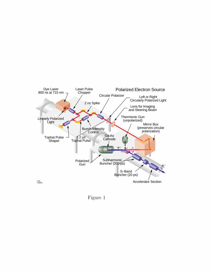
Polarized electrons with energies of 19 to 26 GeV and 2 µA intensity were
produced by a laser optically pumped AlGaAs source (see Fig. 1). The helicity
of the beam was reversed randomly on a pulse-to-pulse basis by reversing the
circular polarization of the excitation photons. The beam polarization was
4
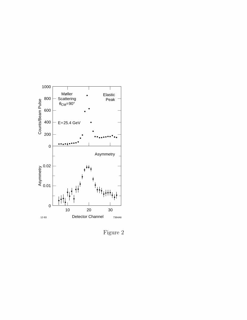
monitored during the experiment by measuring the cross section asymmetry in
Møller scattering off polarized electrons in a magnetized ferromagnetic foil (see
Fig. 2). Electrons scattered at 90◦ in the center-of-mass frame were detected in a
magnetic spectrometer consisting of a dipole magnet and an array of proportional
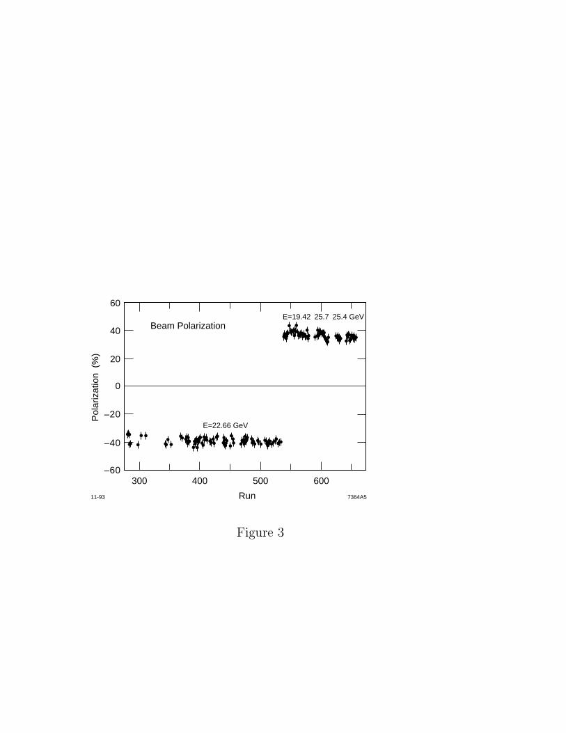
tubes. The beam polarization was found to be stable over the duration of the
experiment with an average value of (39± 2)% (see Fig. 3).
The experiment used a polarized 3He target. The nucleon spin structure of a
polarized 3He target is the same as a polarized free neutron target to the extent
that the 3He nucleus is in a space-symmetric S state. In an S state, the two
proton spins are aligned antiparallel due to the Pauli exclusion principle, implying
that scattering from a polarized 3He nucleus represents scattering from a polarized
neutron. The presence of some S′ and D state admixtures in the 3He ground
state complicates the above picture by introducing a polarized proton component
opposite to that of the neutron. Theoretical calculations[9−11]
have shown that these
admixtures have a small effect in the cross section asymmetry measurements and
that the theoretical uncertainty in extracting the spin structure function gn1 (x) is
small (see Fig. 4).
The target is based on the technique of 3He polarization by spin exchange
collisions with Rb vapor.[12]
The Rb atoms are polarized via laser optical pumping
by absorbing circularly polarized photons at a wavelength of 795 nm. The spin
exchange from Rb to 3He occurs due to the hyperfine interaction between the
polarized valence electron of Rb and the 3He nucleus.
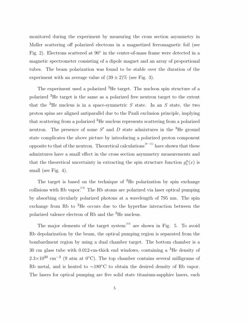
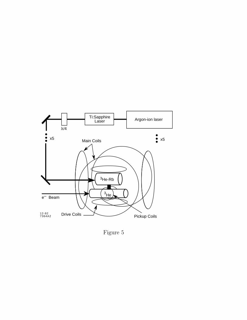
The major elements of the target system[13]
are shown in Fig. 5. To avoid
Rb depolarization by the beam, the optical pumping region is separated from the
bombardment region by using a dual chamber target. The bottom chamber is a
30 cm glass tube with 0.012-cm-thick end windows, containing a 3He density of
2.3×1020 cm−3 (9 atm at 0◦C). The top chamber contains several milligrams of
Rb metal, and is heated to ∼180◦C to obtain the desired density of Rb vapor.
The lasers for optical pumping are five solid state titanium-sapphire lasers, each
5
pumped by an argon-ion laser and producing greater than 20 watts of power. The
axis of quantization for polarization is established by the magnetic field produced
by the two main Helmholtz coil sets. The drive and pickup coils are used for the
3He polarization measurements.
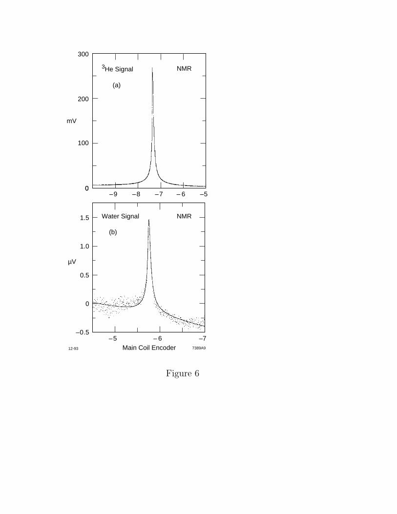
The 3He nuclear polarization was measured by means of nuclear magnetic
resonance (NMR) adiabatic fast passage.[12]
The 3He NMR signals (see Fig. 6) were
calibrated to the NMR signals of the proton polarization at thermal equilibrium in
water (see Fig. 6). The water sample was contained in a cell identical to the 3He
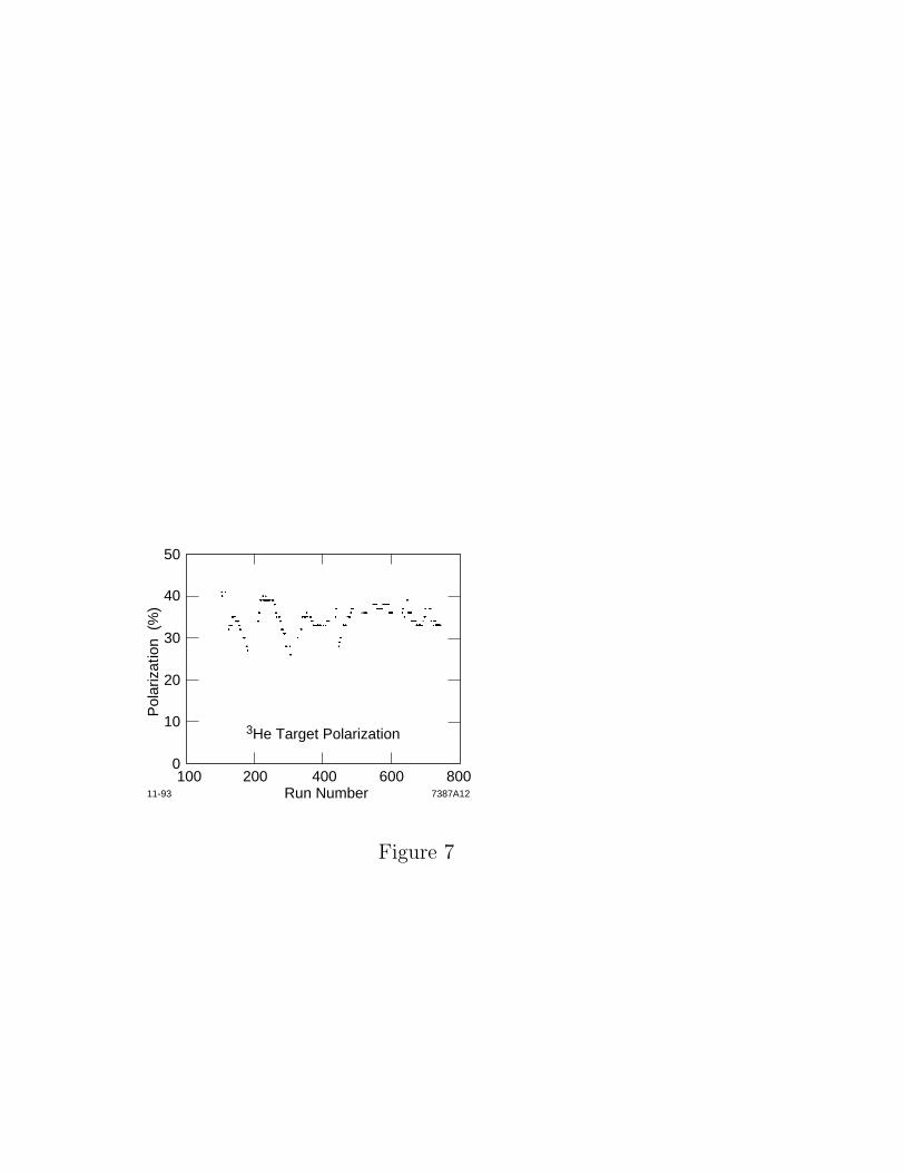
bombardment cell. The average 3He polarization was ∼ 35%, as can be seen in
Fig. 7. The fractional uncertainty in the 3He polarization measurement was ±7%,
dominated by uncertainties in the water calibration.
The neutron asymmetries were extracted from the 3He measured asymmetries
assuming[9]
that the polarization of the neutron in 3He is ∼ 87%. A correction for
the polarization of the two protons in 3He (∼ −2.7% per proton) was applied. The
latter correction[9]
used the proton asymmetry results from the CERN experiment.[3]
No other corrections were made for the fact that the polarized neutron is embedded
in the 3He nucleus.
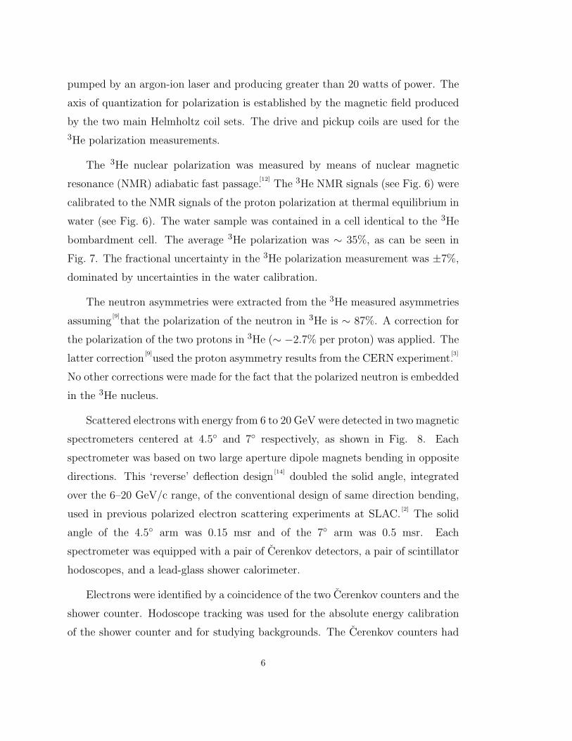
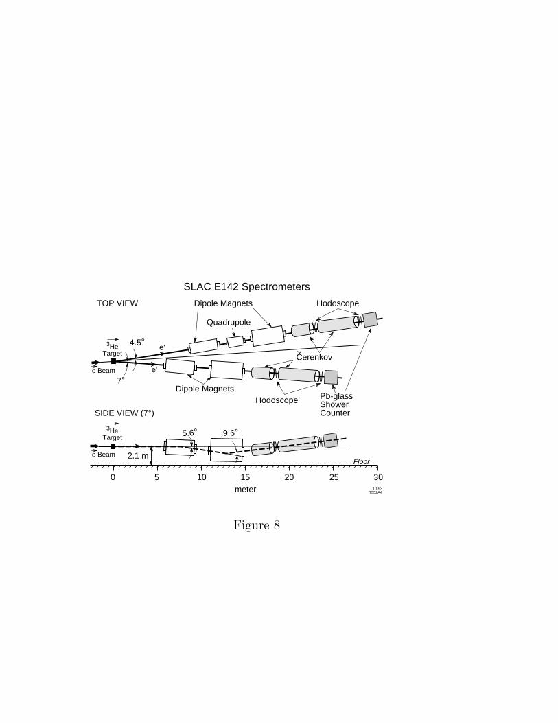
Scattered electrons with energy from 6 to 20 GeV were detected in two magnetic
spectrometers centered at 4.5◦ and 7◦ respectively, as shown in Fig. 8. Each
spectrometer was based on two large aperture dipole magnets bending in opposite
directions. This ‘reverse’ deflection design[14]
doubled the solid angle, integrated
over the 6–20 GeV/c range, of the conventional design of same direction bending,
used in previous polarized electron scattering experiments at SLAC.[2]
The solid
angle of the 4.5◦ arm was 0.15 msr and of the 7◦ arm was 0.5 msr. Each
spectrometer was equipped with a pair of Cerenkov detectors, a pair of scintillator
hodoscopes, and a lead-glass shower calorimeter.
Electrons were identified by a coincidence of the two Cerenkov counters and the
shower counter. Hodoscope tracking was used for the absolute energy calibration
of the shower counter and for studying backgrounds. The Cerenkov counters had
6
an efficiency of over 99% (∼ 7 photoelectrons per incident electron). A typical
Cerenkov counter pulse-height spectrum is shown in Fig. 9. The shower counters
were ∼98% efficient with a resolution (rms) of 15%/√E′.
The true asymmetries A‖ and A⊥ were derived from the measured raw
asymmetries (A‖)raw and (A⊥)raw:
A‖ = (A‖)rawPePtf and A⊥ = (A⊥)rawPePtf, (12)
where Pe and Pt are the beam and target polarizations, respectively, and f is
the fraction of events originating from polarized neutrons in the target. False
asymmetries were found to be consistent with zero by comparing data with target
spins in opposite directions (see Fig. 10).
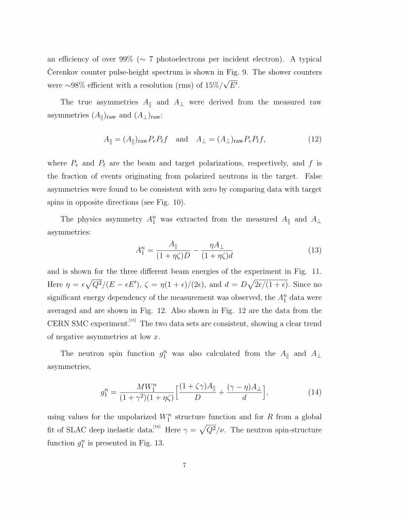
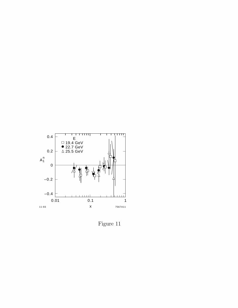
The physics asymmetry An1 was extracted from the measured A‖ and A⊥
asymmetries:
An1 =A‖
(1 + ηζ)D− ηA⊥
(1 + ηζ)d(13)
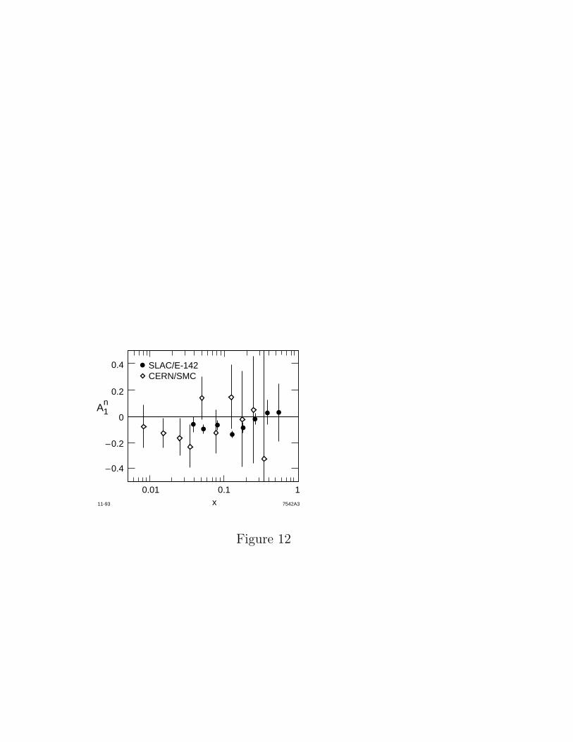
and is shown for the three different beam energies of the experiment in Fig. 11.
Here η = ε√Q2/(E − εE′), ζ = η(1 + ε)/(2ε), and d = D
√2ε/(1 + ε). Since no
significant energy dependency of the measurement was observed, the An1 data were
averaged and are shown in Fig. 12. Also shown in Fig. 12 are the data from the
CERN SMC experiment.[15]
The two data sets are consistent, showing a clear trend
of negative asymmetries at low x.
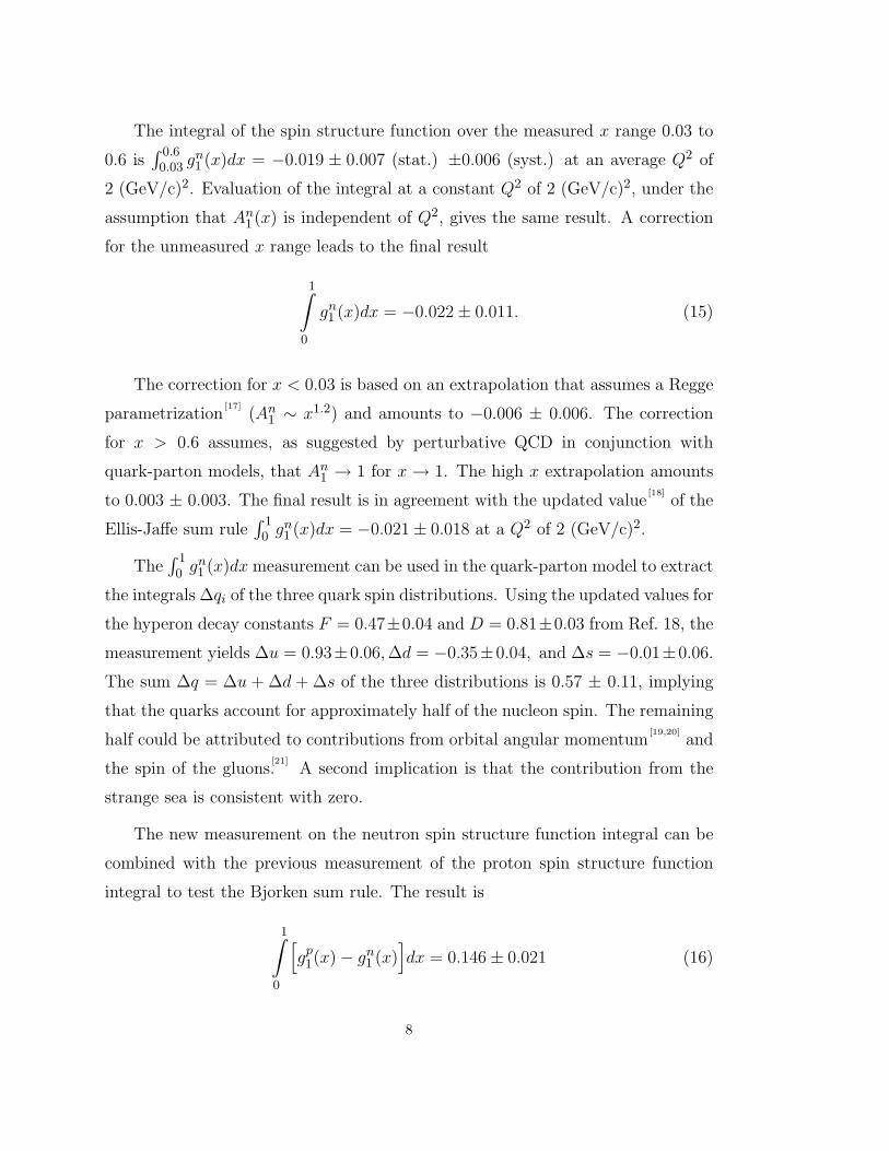
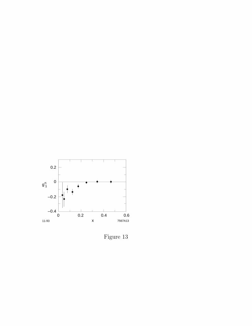
The neutron spin function gn1 was also calculated from the A‖ and A⊥
asymmetries,
gn1 =MWn
1
(1 + γ2)(1 + ηζ)
[(1 + ζγ)A‖D
+(γ − η)A⊥
d
], (14)
using values for the unpolarized Wn1 structure function and for R from a global
fit of SLAC deep inelastic data.[16]
Here γ =√Q2/ν. The neutron spin-structure
function gn1 is presented in Fig. 13.
7
The integral of the spin structure function over the measured x range 0.03 to
0.6 is∫ 0.6
0.03 gn1 (x)dx = −0.019 ± 0.007 (stat.) ±0.006 (syst.) at an average Q2 of
2 (GeV/c)2. Evaluation of the integral at a constant Q2 of 2 (GeV/c)2, under the
assumption that An1 (x) is independent of Q2, gives the same result. A correction
for the unmeasured x range leads to the final result
1∫0
gn1 (x)dx = −0.022± 0.011. (15)
The correction for x < 0.03 is based on an extrapolation that assumes a Regge
parametrization[17]
(An1 ∼ x1.2) and amounts to −0.006 ± 0.006. The correction
for x > 0.6 assumes, as suggested by perturbative QCD in conjunction with
quark-parton models, that An1 → 1 for x→ 1. The high x extrapolation amounts
to 0.003 ± 0.003. The final result is in agreement with the updated value[18]
of the
Ellis-Jaffe sum rule∫ 1
0 gn1 (x)dx = −0.021± 0.018 at a Q2 of 2 (GeV/c)2.
The∫ 1
0 gn1 (x)dx measurement can be used in the quark-parton model to extract
the integrals ∆qi of the three quark spin distributions. Using the updated values for
the hyperon decay constants F = 0.47±0.04 and D = 0.81±0.03 from Ref. 18, the
measurement yields ∆u = 0.93±0.06,∆d = −0.35±0.04, and ∆s = −0.01±0.06.
The sum ∆q = ∆u + ∆d + ∆s of the three distributions is 0.57 ± 0.11, implying
that the quarks account for approximately half of the nucleon spin. The remaining
half could be attributed to contributions from orbital angular momentum[19,20]
and
the spin of the gluons.[21]
A second implication is that the contribution from the
strange sea is consistent with zero.
The new measurement on the neutron spin structure function integral can be
combined with the previous measurement of the proton spin structure function
integral to test the Bjorken sum rule. The result is
1∫0
[gp1(x)− gn1 (x)
]dx = 0.146± 0.021 (16)
8
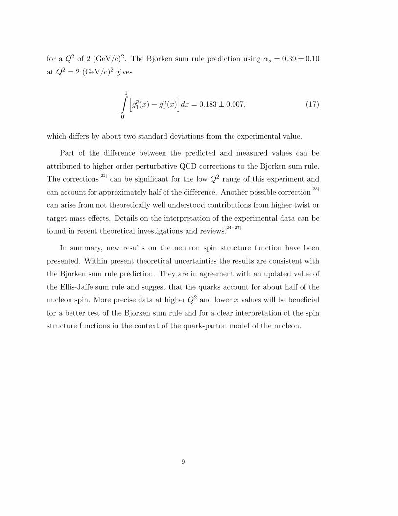
for a Q2 of 2 (GeV/c)2. The Bjorken sum rule prediction using αs = 0.39 ± 0.10
at Q2 = 2 (GeV/c)2 gives
1∫0
[gp1(x)− gn1 (x)
]dx = 0.183± 0.007, (17)
which differs by about two standard deviations from the experimental value.
Part of the difference between the predicted and measured values can be
attributed to higher-order perturbative QCD corrections to the Bjorken sum rule.
The corrections[22]
can be significant for the low Q2 range of this experiment and
can account for approximately half of the difference. Another possible correction[23]
can arise from not theoretically well understood contributions from higher twist or
target mass effects. Details on the interpretation of the experimental data can be
found in recent theoretical investigations and reviews.[24−27]
In summary, new results on the neutron spin structure function have been
presented. Within present theoretical uncertainties the results are consistent with
the Bjorken sum rule prediction. They are in agreement with an updated value of
the Ellis-Jaffe sum rule and suggest that the quarks account for about half of the
nucleon spin. More precise data at higher Q2 and lower x values will be beneficial
for a better test of the Bjorken sum rule and for a clear interpretation of the spin
structure functions in the context of the quark-parton model of the nucleon.
9
REFERENCES
1. M. J. Alguard et al., Phys. Rev. Lett. 37, 1258 (1976); 37, 1261 (1976).
2. G. Baum et al., Phys. Rev. Lett. 51, 1135 (1983).
3. J. Ashman et al., Nucl. Phys. B238, 1 (1989).
4. C. E. Carlson and W.-K. Tung, Phys. Rev. D5, 721 (1972).
5. A. J. G. Hey and J. E. Mandula, Phys. Rev. D5, 2610 (1972).
6. J. D. Bjorken, Phys. Rev. 148, 1467 (1966); Phys. Rev. D1, 1376 (1970).
7. J. Kodaira et. al., Phys. Rev. D20, 627 (1979); Nucl. Phys. B159, 99 (1979).
8. J. Ellis and R. Jaffe, Phys. Rev. D9, 1444 (1974).
9. C. Ciofi degli Atti et al., Phys. Rev. C48, 968 (1993).
10. R. Shulze and P. Sauer, Phys. Rev. C48, 38 (1993).
11. R. M. Woloshyn, Nucl. Phys. A496, 749 (1989).
12. T. E. Chupp et al., Phys. Rev. C36, 2244 (1987); C45, 915 (1992).
13. H. Middleton et al., in Proceedings of Workshop on Polarized Gas Targets,
Madison, WI, May 1993.
14. G. G. Petratos et al., SLAC–PUB–5678 (1991).
15. B. Adeva et.al., Phys. Lett. B362, 553 (1993).
16. L. W. Whitlow et al., Phys. Lett. B250, 193 (1990); B282, 475 (1992).
17. A. Schafer, Phys. Lett. B208, 175 (1988).
18. R. L. Jaffe and A. V. Manohar, Nucl. Phys. B337, 509 (1990).
19. L.M. Sehgal, Phys. Rev. D10, 1663 (1974).
20. J. Ellis and M. Karliner, Phys. Lett. B213, 73 (1988).
21. G. Altarelli and G. G. Ross, Phys. Lett. B212, 391 (1988)
10
22. S. A. Larin and J. A. M. Vermaseren, Phys. Lett. B259, 345 (1991).
23. I. I. Balitsky et al., Phys. Lett. B242, 245 (1990).
24. F. E. Close, Rutherford prepring RAL–93–040 (1993).
25. G. Preparata and P. Ratcliffe, Milano preprint MITH–93/15 (1993).
26. J. Ellis and M. Karliner, CERN preprint CERN–TH–7022/93 (1993).
27. X. Ji and P. Unrau, MIT preprint MIT–CTP–2232 (1993).
11
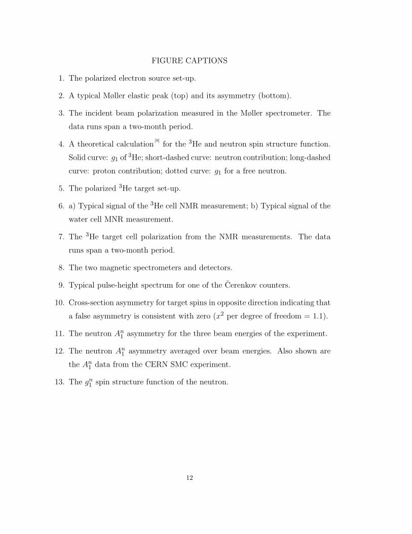
FIGURE CAPTIONS
1. The polarized electron source set-up.
2. A typical Møller elastic peak (top) and its asymmetry (bottom).
3. The incident beam polarization measured in the Møller spectrometer. The
data runs span a two-month period.
4. A theoretical calculation[9]
for the 3He and neutron spin structure function.
Solid curve: g1 of 3He; short-dashed curve: neutron contribution; long-dashed
curve: proton contribution; dotted curve: g1 for a free neutron.
5. The polarized 3He target set-up.
6. a) Typical signal of the 3He cell NMR measurement; b) Typical signal of the
water cell MNR measurement.
7. The 3He target cell polarization from the NMR measurements. The data
runs span a two-month period.
8. The two magnetic spectrometers and detectors.
9. Typical pulse-height spectrum for one of the Cerenkov counters.
10. Cross-section asymmetry for target spins in opposite direction indicating that
a false asymmetry is consistent with zero (x2 per degree of freedom = 1.1).
11. The neutron An1 asymmetry for the three beam energies of the experiment.
12. The neutron An1 asymmetry averaged over beam energies. Also shown are
the An1 data from the CERN SMC experiment.
13. The gn1 spin structure function of the neutron.
12
Dye Laser800 ns at 715 nm
Bunch IntensityControl
Tophat PulseShaper
PolarizedGun
Ga AsCathode
e–
Left or RightCircularly Polarized Light
Lens for Imagingand Steering Beam
Thermionic Gun(unpolarized)
SubharmonicBuncher (200 ps)
S–BandBuncher (20 ps)
Accelerator Section
Mirror Box(preserves circular
polarization)
e–
4-937368A1
Polarized Electron SourceLaser PulseChopper
Circular Polarizer
2 ns Spike
1.2 µsTophat Pulse
Linearly PolarizedLight
Figure 1
1000
800
600
400
200
0
0.02
0.01
0
12-93
10 20 30
Asymmetry
ElasticPeak
Cou
nts/
Bea
m P
ulse
Asy
mm
etry
Detector Channel 7364A6
MøllerScatteringθCM=90°
E=25.4 GeV
Figure 2
E=19.42 25.7 25.4 GeV
E=22.66 GeV
600500400300
60
40
20
0
–20
–40
–60
Pol
ariz
atio
n (
%)
Run
Beam Polarization
7364A511-93
Figure 3

0
–0.2
–0.4
–0.60.01 0.1 1
x
g1
11-93 7567A2
Figure 4
Pickup Coils
e– Beam
12-927364A2
Main Coils
Drive Coils
3He-Rb
3He3He
λ/4
x5
Argon-ion laser
x5
Ti:SapphireLaser
Figure 5
mV
µV
Main Coil Encoder
3He Signal
(a)
300
200
100
00
1.5
1.0
0.5
0
–0.5
–9
–5 – 6 –7
–8 –7 – 6 –5
12-93 7389A9
Water Signal
(b)
NMR
NMR
Figure 6
50
40
30
20
10
0200 400 600 800
Pol
ariz
atio
n (
%)
Run Number
3He Target Polarization
11-93 7387A12
100
Figure 7
����������
TOP VIEW
4.5°
7°
Cerenkov
Dipole Magnets
Dipole MagnetsPb-glassShowerCounter
5.6° 9.6°
0
2.1 m
10 15 20
meter
Floor
SIDE VIEW (7°)
3HeTarget
10-937552A4
Hodoscope
Hodoscope
25 30
e Beam
e Beam
3HeTarget
e'
e'
5
Quadrupole
SLAC E142 Spectrometers
Figure 8
0.4
0.2
0
–0.2
–0.4
E 19.4 GeV22.7 GeV25.5 GeV
0.1 10.01
x
A1 n
7567A1111-93
Figure 11
0.01 0.1 1
–0.4
–0.2
0
0.2
0.4 SLAC/E-142CERN/SMC
x 7542A311-93
A1n
Figure 12
7567A13x
0
0.2
–0.2
–0.4
g 1n
0 0.2 0.4 0.611-93
Figure 13
Recommended