Stanford Synchrotron Radiation Lightsource
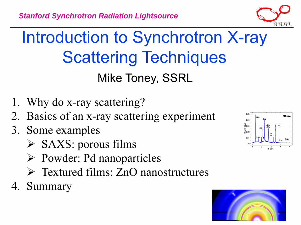
Introduction to Synchrotron X-ray Scattering Techniques
Mike Toney, SSRL
1. Why do x-ray scattering?2. Basics of an x-ray scattering experiment3. Some examples
SAXS: porous filmsPowder: Pd nanoparticlesTextured films: ZnO nanostructures
4. Summary
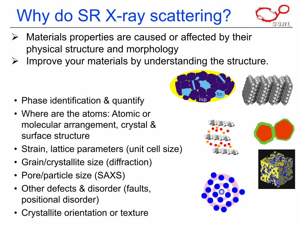
Why do SR X-ray scattering?Materials properties are caused or affected by their physical structure and morphologyImprove your materials by understanding the structure.
• Phase identification & quantify• Where are the atoms: Atomic or
molecular arrangement, crystal & surface structure
• Strain, lattice parameters (unit cell size)• Grain/crystallite size (diffraction)• Pore/particle size (SAXS)• Other defects & disorder (faults,
positional disorder)• Crystallite orientation or texture
hcpfcc
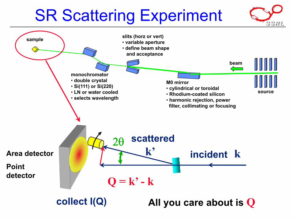
SR Scattering Experiment
monochromator• double crystal• Si(111) or Si(220)• LN or water cooled• selects wavelength
source
beam
M0 mirror• cylindrical or toroidal• Rhodium-coated silicon• harmonic rejection, power
filter, collimating or focusing
slits (horz or vert)• variable aperture• define beam shape
and acceptance
sample
k2θ
incidentscattered
k’
Q = k’ - k
All you care about is Qcollect I(Q)
Area detector
Point detector
SR Scattering Experiment
k2θ
incidentscattered
k’
Q = k’ - k
All you care about is Q
Area detector
Point detector
At SSRL:• Area detector: 11-3• Point detectors: 2-1, 7-2, 10-2• SAXS: 1-4
collect I(Q)
0 1 2 3 410-2
10-1
100
101
102
103
104
105
1 2 3 4Q (Å)
010-2
10-1
100
101
102
103
104
105
I(Q)
(arb
. uni
ts)
Diffraction:peaks
Scattering:the rest
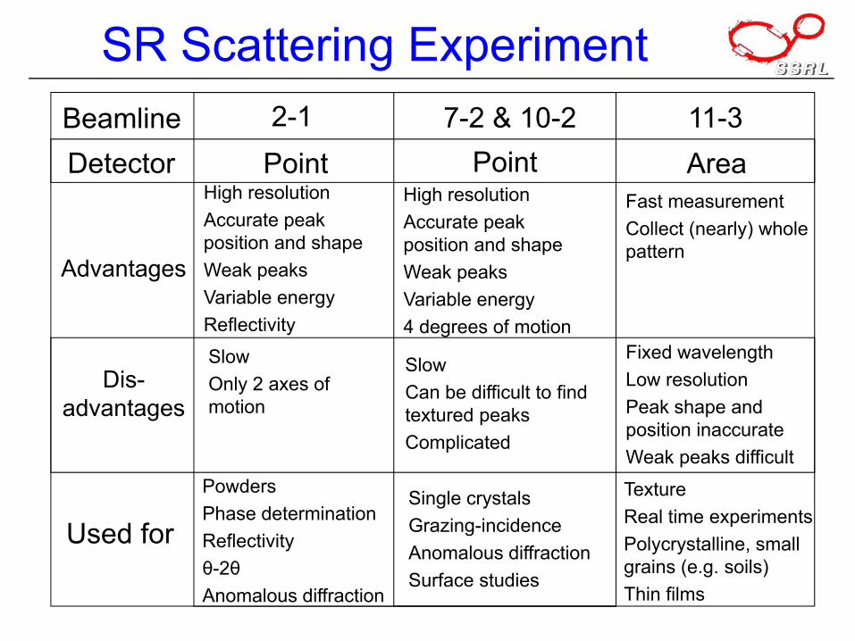
SR Scattering ExperimentBeamline 2-1 7-2 & 10-2 11-3Detector Point Point Area
Advantages
Dis-advantages
Used for
High resolutionAccurate peak position and shapeWeak peaksVariable energyReflectivity
High resolutionAccurate peak position and shapeWeak peaksVariable energy4 degrees of motion
Fast measurementCollect (nearly) whole pattern
SlowOnly 2 axes of motion
SlowCan be difficult to find textured peaksComplicated
Fixed wavelengthLow resolutionPeak shape and position inaccurateWeak peaks difficult
PowdersPhase determinationReflectivityθ-2θAnomalous diffraction
Single crystalsGrazing-incidenceAnomalous diffractionSurface studies
TextureReal time experimentsPolycrystalline, small grains (e.g. soils)Thin films
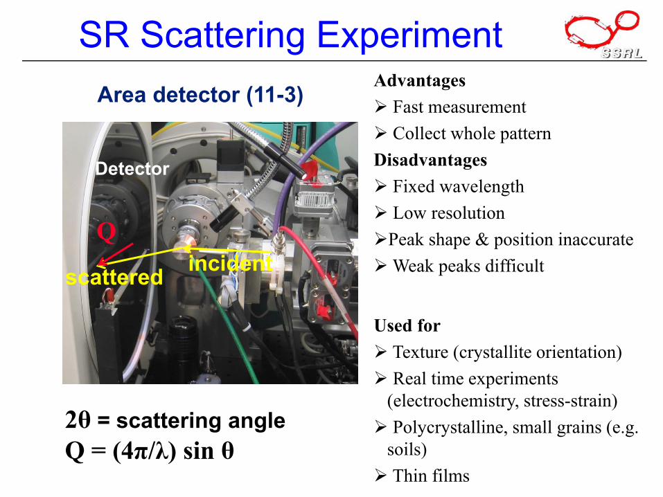
SR Scattering Experiment
2θ = scattering angleQ = (4π/λ) sin θ
incidentscattered
Detector
Q
Area detector (11-3)Advantages
Fast measurementCollect whole pattern
DisadvantagesFixed wavelengthLow resolutionPeak shape & position inaccurateWeak peaks difficult
Used forTexture (crystallite orientation)Real time experiments
(electrochemistry, stress-strain)Polycrystalline, small grains (e.g.
soils)Thin films
SR Scattering Experiment
incident x-rays
diffracted x-rays
sample
2θθ
θ
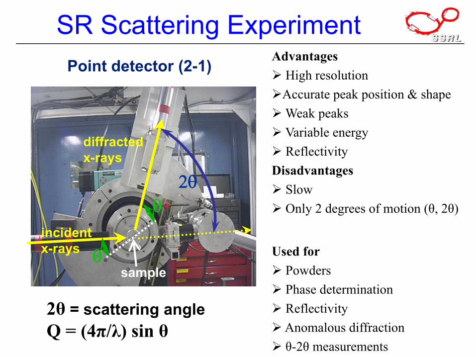
Point detector (2-1)
2θ = scattering angleQ = (4π/λ) sin θ
AdvantagesHigh resolutionAccurate peak position & shapeWeak peaksVariable energyReflectivity
DisadvantagesSlowOnly 2 degrees of motion (θ, 2θ)
Used forPowdersPhase determinationReflectivityAnomalous diffractionθ-2θ measurements
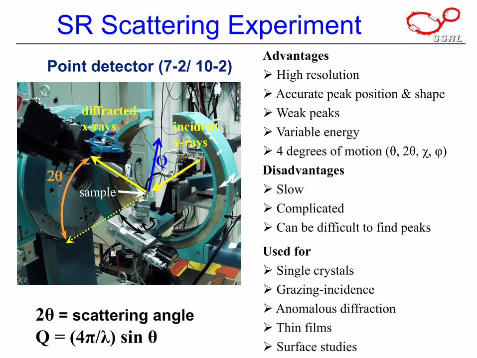
SR Scattering ExperimentPoint detector (7-2/ 10-2)
2θ = scattering angleQ = (4π/λ) sin θ
AdvantagesHigh resolutionAccurate peak position & shapeWeak peaksVariable energy4 degrees of motion (θ, 2θ, χ, φ)
DisadvantagesSlowComplicated Can be difficult to find peaks
Used forSingle crystalsGrazing-incidenceAnomalous diffractionThin filmsSurface studies
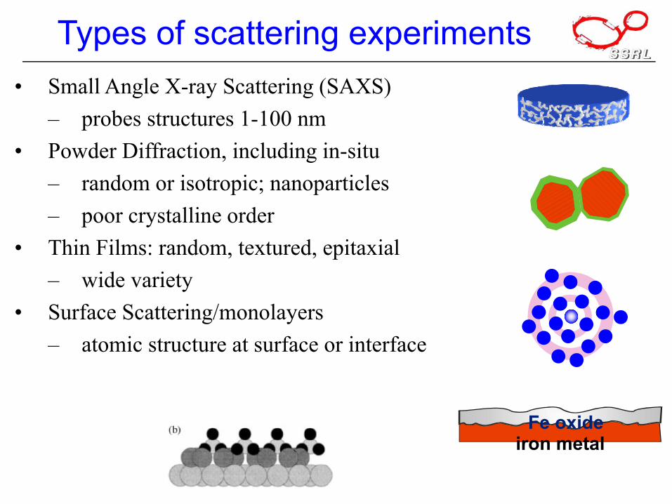
Types of scattering experiments• Small Angle X-ray Scattering (SAXS)
– probes structures 1-100 nm• Powder Diffraction, including in-situ
– random or isotropic; nanoparticles– poor crystalline order
• Thin Films: random, textured, epitaxial– wide variety
• Surface Scattering/monolayers– atomic structure at surface or interface
iron metalFe oxide
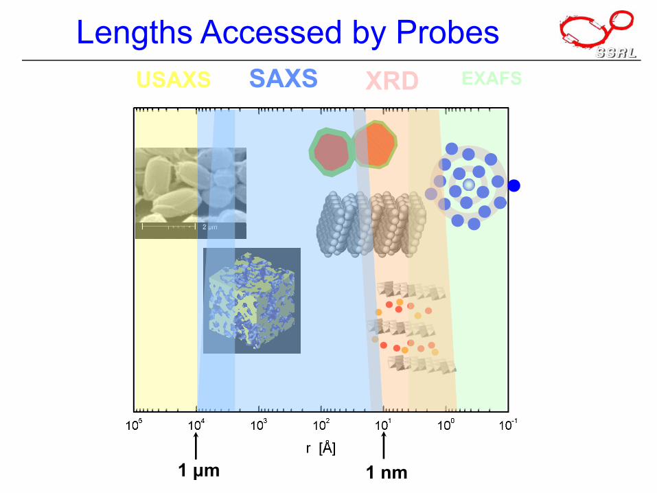
Lengths Accessed by Probes
1 nm1 μm
EXAFSXRDSAXSUSAXS
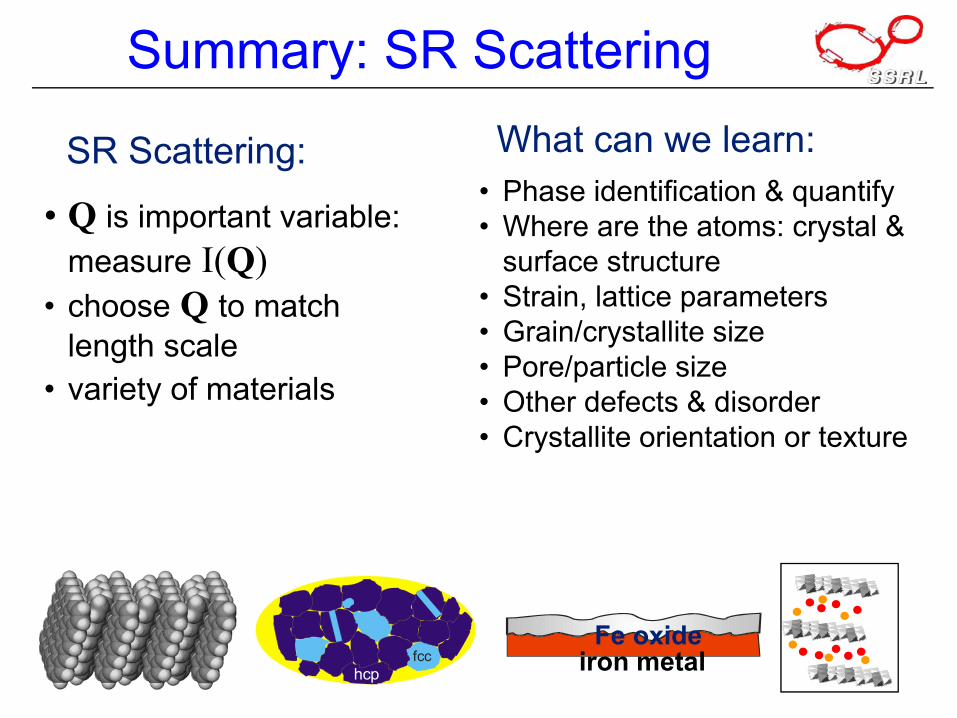
Summary: SR Scattering
• Q is important variable: measure I(Q)
• choose Q to match length scale
• variety of materials
hcpfcc
• Phase identification & quantify• Where are the atoms: crystal &
surface structure• Strain, lattice parameters• Grain/crystallite size• Pore/particle size• Other defects & disorder• Crystallite orientation or texture
What can we learn:SR Scattering:
iron metalFe oxide
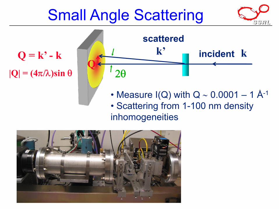
Small Angle Scattering
|Q| = (4π/λ)sin θ
• Measure I(Q) with Q ∼ 0.0001 – 1 Å-1
• Scattering from 1-100 nm density inhomogeneities
k
2θ
incidentscattered
k’Q = k’ - kQ
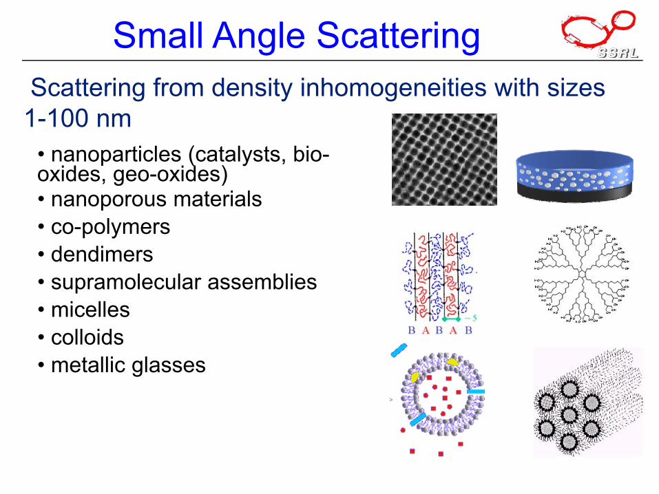
Small Angle ScatteringScattering from density inhomogeneities with sizes 1-100 nm
• nanoparticles (catalysts, bio-oxides, geo-oxides)• nanoporous materials• co-polymers• dendimers• supramolecular assemblies• micelles• colloids• metallic glasses
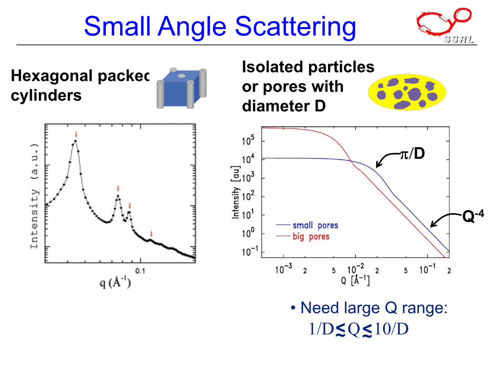
Small Angle Scattering
• Need large Q range:1/D Q 10/D<~ <~
Hexagonal packedcylinders
Isolated particlesor pores with diameter D
π/D
Q-4
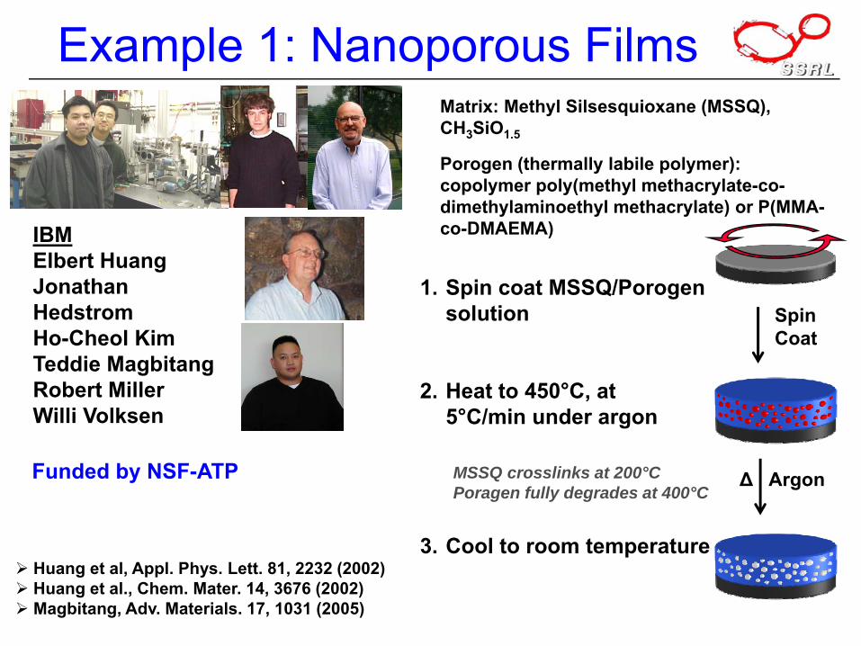
Example 1: Nanoporous Films
Spin coat MSSQ/Porogensolution
Heat to 450°C, at5°C/min under argon
Cool to room temperature
1.
2.
3.
Matrix: Methyl Silsesquioxane (MSSQ), CH3SiO1.5
Porogen (thermally labile polymer): copolymer poly(methyl methacrylate-co-dimethylaminoethyl methacrylate) or P(MMA-co-DMAEMA)
Δ Argon
Spin Coat
MSSQ crosslinks at 200°CPoragen fully degrades at 400°C
Huang et al, Appl. Phys. Lett. 81, 2232 (2002)Huang et al., Chem. Mater. 14, 3676 (2002)Magbitang, Adv. Materials. 17, 1031 (2005)
IBMElbert HuangJonathan HedstromHo-Cheol KimTeddie MagbitangRobert MillerWilli Volksen
Funded by NSF-ATP
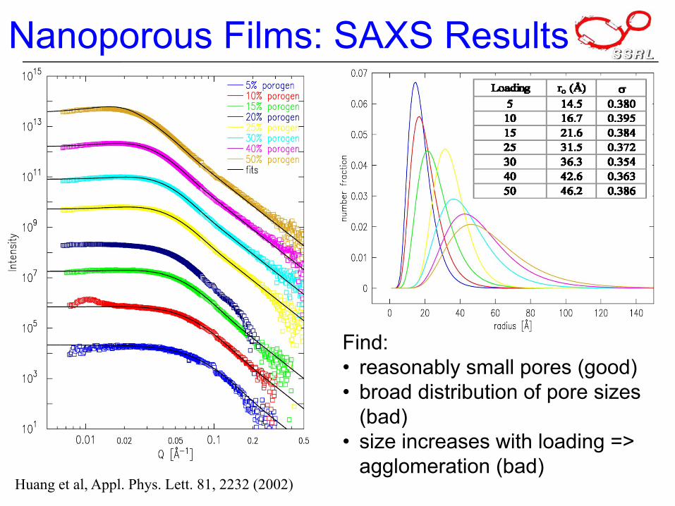
Huang et al, Appl. Phys. Lett. 81, 2232 (2002)
Nanoporous Films: SAXS Results
Find:• reasonably small pores (good)• broad distribution of pore sizes
(bad)• size increases with loading =>
agglomeration (bad)
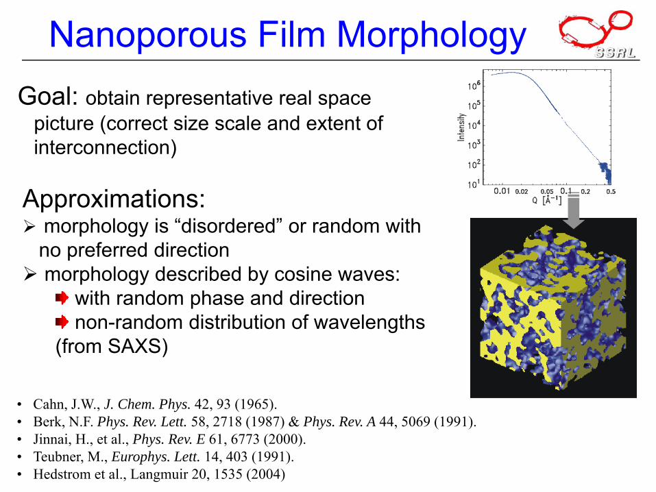
Goal: obtain representative real space picture (correct size scale and extent of interconnection)
• Cahn, J.W., J. Chem. Phys. 42, 93 (1965).• Berk, N.F. Phys. Rev. Lett. 58, 2718 (1987) & Phys. Rev. A 44, 5069 (1991).• Jinnai, H., et al., Phys. Rev. E 61, 6773 (2000).• Teubner, M., Europhys. Lett. 14, 403 (1991).• Hedstrom et al., Langmuir 20, 1535 (2004)
Approximations:morphology is “disordered” or random with no preferred directionmorphology described by cosine waves:
with random phase and directionnon-random distribution of wavelengths
(from SAXS)
Nanoporous Film Morphology
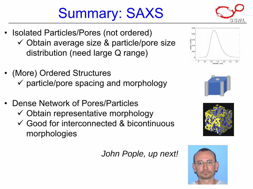
Summary: SAXS• Isolated Particles/Pores (not ordered)
Obtain average size & particle/pore size distribution (need large Q range)
• (More) Ordered Structuresparticle/pore spacing and morphology
• Dense Network of Pores/ParticlesObtain representative morphologyGood for interconnected & bicontinuousmorphologies
John Pople, up next!
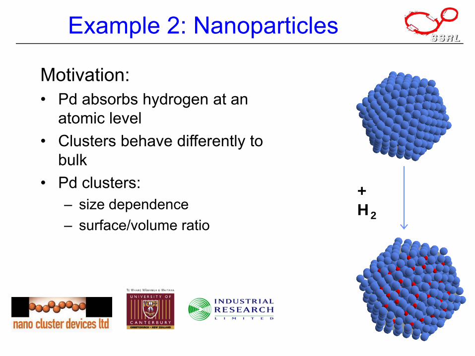
Example 2: Nanoparticles
+ H2
Motivation:• Pd absorbs hydrogen at an
atomic level• Clusters behave differently to
bulk• Pd clusters:
– size dependence– surface/volume ratio
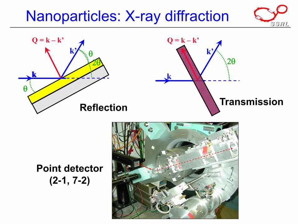
Nanoparticles: X-ray diffraction
k’Q = k – k’
k
θ
θ
k2θ
k’Q = k – k’
k
2θ
Reflection Transmission
Point detector(2-1, 7-2)
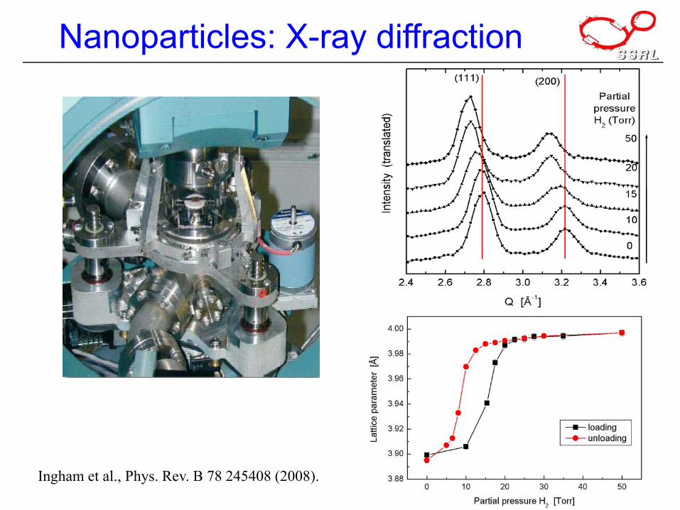
Nanoparticles: X-ray diffraction
Ingham et al., Phys. Rev. B 78 245408 (2008).
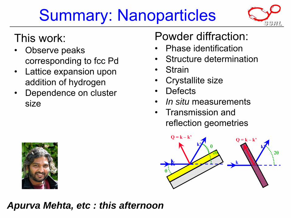
Summary: Nanoparticles
Apurva Mehta, etc : this afternoon
This work:• Observe peaks
corresponding to fcc Pd• Lattice expansion upon
addition of hydrogen• Dependence on cluster
size
Powder diffraction:• Phase identification• Structure determination• Strain• Crystallite size• Defects• In situ measurements• Transmission and
reflection geometries
k’Q = k – k’
k
θ
θ
k
2θk’
Q = k – k’
k
2θ
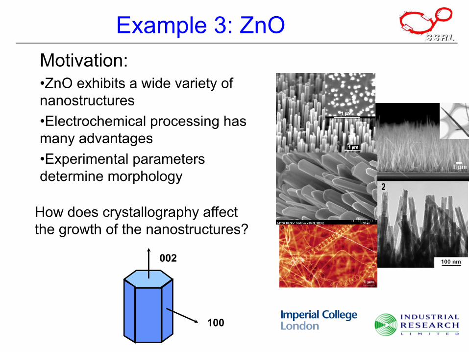
Example 3: ZnOMotivation:•ZnO exhibits a wide variety of nanostructures•Electrochemical processing has many advantages•Experimental parameters determine morphology
100
002
How does crystallography affect the growth of the nanostructures?
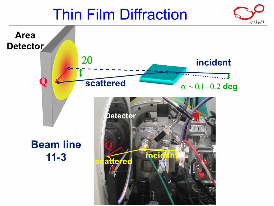
Thin Film DiffractionArea
Detector2θ incident
α ~ 0.1−0.2 degQ scattered
Beam line11-3 incidentscattered
Detector
Q
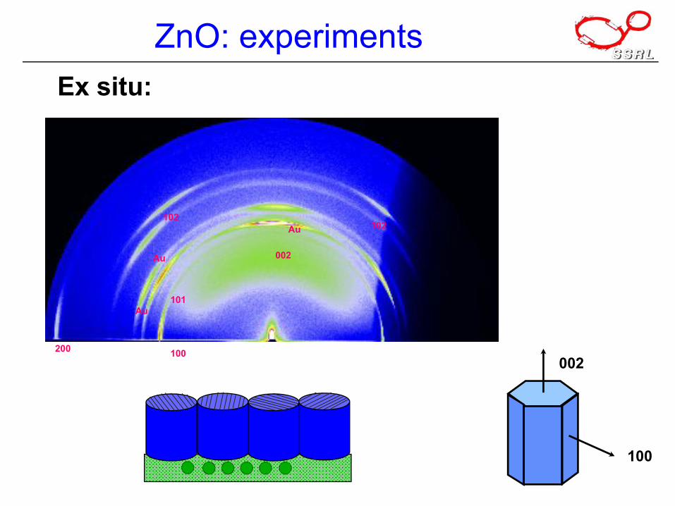
ZnO: experimentsEx situ:
100
002
002
100
101
Au
Au
Au
102102
200
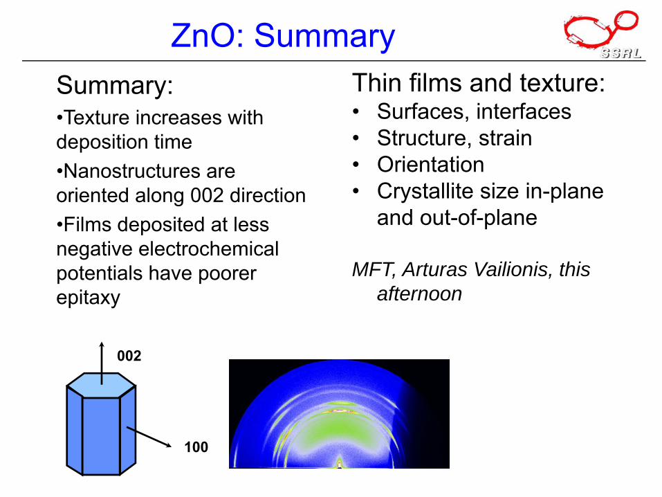
ZnO: SummarySummary:•Texture increases with deposition time•Nanostructures are oriented along 002 direction•Films deposited at less negative electrochemical potentials have poorer epitaxy
100
002
Thin films and texture:• Surfaces, interfaces• Structure, strain• Orientation• Crystallite size in-plane
and out-of-plane
MFT, Arturas Vailionis, this afternoon
Summary• Typical SR x-ray scattering experiment & some examples:
porous films, nanoparticles, textured films
• To be covered in this workshop: - SAXS- Powder- Poorly ordered - Films: random, textured, epitaxial- Monolayers

Bibliography
• B Warren, “X-ray Diffraction”, Dover (1990): $13.16 & eligible for FREE Super Saver Shipping on
• BD Cullity & SR Stock, “X-ray Diffraction”, Prentice Hall (2001): $118.40.
• J Als-Nielsen & D McMorrow, “Elements of Modern X-ray Physics”, Wiley (2001): $84.10.
More Bibliography• M. Tolan, “X-ray Scattering from Soft-Matter: Materials Science and Basic
Research”, Springer (1998).• RL Snyder, K. Fiala & HJ Bunge, Eds., “Defect and Microstructure Analysis by
Diffraction”, Oxford (1999).• V Holy, U Pietsch, T Baumbach, “High-Resolution X-Ray Scattering: From
Thin Films to Lateral Nanostructures”, Springer-Verlag, (2004)• O Glatter, “Small Angle X-Ray Scattering”, Academic (1982).• “Modern Aspects of Small Angle X-Ray Scattering”, H. Brumberger (Editor),
Springer (1994).• Int. Union of Crystallography: Links to everything crystallographic.
www.iucr.org . See : www.iucr.org/cww-top/edu.index.html and also Teaching and Education in Crystallography: www.minerals.csiro.au/mirror/w3vlc/edu.index.html.
• Structural data for thousands of minerals: database.iem.ac.ru/mincryst/ • Lawrence Berkeley: X-ray interactions with matter, data & calculations www-
cxro.lbl.gov/optical_constants/ • International Centre for Diffraction Data - purveyors of the Powder Diffraction
File (PDF) www.icdd.com
Recommended