Study of the Preparation of Mesoporous Magnetic Microspheres
and Their Applications
M å r t e n E r i c s o n
Master of Science ThesisStockholm 2009

Mårten Ericson
STUDY OF THE PREPARATION OF MESOPOROUS MAGNETIC MICROSPHERES
AND THEIR APPLICATIONS
PRESENTED AT
INDUSTRIAL ECOLOGY ROYAL INSTITUTE OF TECHNOLOGY
www.ima.kth.se
Master of Science Thesis
STOCHOLM 2009
Examiner: Per Olof Persson, Industrial Ecology

TRITA-IM 2009:25 ISSN 1402-7615 Industrial Ecology, Royal Institute of Technology www.ima.kth.se
Royal Institute of Technology
III
Abstract Treatment of wastewater using magnetic technology is a rising field. In this thesis, the latest
research on the subject is reviewed and several adsorbents with different coatings, which
impart them unique properties, are discussed. Separation of particles from aqueous solution
using magnetic technology is more convenient compared to conventional techniques, such as
filtration and centrifugation. The adsorbents described in this thesis are effective for
adsorption of several types of contaminants, such as heavy metals and different types of dyes.
Magnetic microspheres were synthesised using porous polystyrene microspheres as
template. The microspheres were first sulfonated using chlorosulfonic acid followed by
stirring in the presence of ferrous chloride which then was oxidised and magnetic
nanoparticles were formed on the surface.
The sulfonated microspheres had a surface area of 420 m2/g and the magnetic 175 m2/g,
indicative of Fe3O4 nanoparticles were successfully formed in the pores. The weight fraction
of the Fe3O4 nanoparticles in the magnetic microspheres was 33 %.
Adsorption and desorption studies of the cationic dye, methylene blue, using mesoporous
magnetic microspheres were performed. The results show that the mesoporous magnetic
microspheres have good ability to adsorb methylene blue at low concentrations. In a cycle
study the adsorption efficiency were nearly 100 % throughout the study. Using a 6/4
EtOH/H2O with saturated KCl solution the desorption efficiency in the cycle study were
about 95 %.
The microspheres were used as carriers for TiO2 in order to overcome the problem with the
separation of TiO2 from solution. The TGA results show that the microspheres contained
about 12 % of TiO2. The TiO2 coated microspheres were used for the photocatalytic
degradation of phenol. However, the TiO2 microspheres did not work. This was a result from
that the phenol had too little contact with the TiO2. A possible way of solving this problem
could be to decrease the size of the microspheres, thus increase the surface area.
Lysozyme was adsorbed and separated using the porous microspheres. The lysozyme
adsorption worked best at pH 9.6, which is the pI for lysozyme. The lysozyme could be
extracted from the microspheres by using a pH 13 buffer. Also, by using MeOH/H2O and
EtOH/H2O solutions with saturated KCl the lysozyme could be desorbed. An adsorption and
desorption mechanism was also presented.
Keywords: Adsorption, bioseparation, magnetic adsorbents, magnetic microspheres,
magnetic separation, photocatalyst, wastewater
Royal Institute of Technology
IV
Sammanfattning
Vattenrening med magnetisk teknologi är en ny och alltmer uppmärksammad teknik.
Magnetisk separation är ett enkelt och snabbt sätt att separera något från en lösning.
Magnetisk separation är mer lätthanterligt jämfört med traditionell separationsteknik såsom
centrifugering och filtrering.
Med porösa polystyren mikrosfärer som mall, syntetiserades magnetiska mikrosfärer. Först
så sulfonerades mikrosfärerna med klorosulfonisk syra, följt av att de rördes om i en
järnkloridlösning. Magnetiska nanopartiklar bildades i porerna och på ytan av mikrosfärerna.
Sulfonerade mikrosfärerna hade en specifik ytarea på 420 m2/g och de magnetiska 175 m2/g, detta indikerar att Fe3O4-nanopartiklar bildades på ytan och i porerna. Massfraktionen
av Fe3O4 var 33 %.
Adsorption- och desorptionsstudier på de magnetiska mikrosfärerna utfördes. Färgämnet
metylblått användes i studien. Resultaten visade att magnetiska mikrosfärerna hade en bra
adsorptionsförmåga vid låga koncentrationer av metylblått. Cykelstudier visade att
adsorptionsverkningsgraden var nära 100 % under flera adsorptionscykler. Desorptionsförsök
med olika lösningsmedel visade att en mättad KCl 6/4 EtOH/H2O lösning gav en desorptions-
verkningsgrad på ca 95 %.
Mikrosfärerna användes som mall och kärna för att syntetisera en TiO2-fotokatalysator, detta
för att överkomma problemet som finns med separation av rent TiO2 pulver från lösning. TGA
resultaten visade att mikrosfärerna innehöll ca 12 % TiO2. De syntetiserade TiO2-
mikrosfärerna användes till att bryta ner fenol fotokatalytiskt. Dock fungerade inte detta
experiment. En anledning var att fenolen hade för lite kontakt med TiO2. En lösning på detta
problem är att använda mikrosfärer med högre specifik ytarea.
Proteinet lysozym användes som modellprotein för försök att separera proteiner från lösning
genom att använda porösa mikrosfärer. Resultatet visade att lysozym kunde adsorberas vid
pH 9.6. Med en pH 13 buffer kunde lysozymet sedan extraheras från mikrosfärerna. En
mekanism för adsorptionen och desorptionen på mikrosfärerna presenterades.
Nyckelord: Adsorption, magnetiska adsorbenter, bioseparation, magnetiska mikrosfärer, fotokatalys
Royal Institute of Technology
VI
Table of Contents Chapter 1: Introduction .............................................................................................................. 1
1.1 Aims and Objectives ........................................................................................................ 2 1.2 Methods ............................................................................................................................ 2 1.3 Limitations ....................................................................................................................... 2
Chapter 2: Background ............................................................................................................... 3
2.1 Updated Research Progress in Magnetic Adsorbents ...................................................... 3 2.1.1 Mesoporous Magnetic Adsorbents ............................................................................ 3 2.1.2 Coated Magnetic Nano-Adsorbents .......................................................................... 6 2.1.3 Magnetic Carbon Adsorbents .................................................................................. 10 2.1.4 Other Magnetic Adsorbents .................................................................................... 11 2.1.5 Conclusions ............................................................................................................. 12
2.2 Updated Research Progress in Magnetic Photoocatalyst ............................................... 14 2.2.1 Different Preparation Techniques and Magnetic Carriers ....................................... 14 2.2.2 Degradation of Organic Pollutants by Magnetic Photocatalysts ............................. 16 2.2.3 Conclusions ............................................................................................................. 18
2.3 Updated Research Progress in Bioseparation Using Magnetic Technology .................. 19 2.3.1 Bioseparation ........................................................................................................... 19 2.3.2 Conclusions ............................................................................................................. 20
Chapter 3: Magnetic Microspheres for Removal of Methylene Blue From Wastewater ......... 21
3.1 Experimental .................................................................................................................. 21 3.1.1 Materials .................................................................................................................. 21 3.1.2 Preparation .............................................................................................................. 21 3.1.3 Characterisation ....................................................................................................... 22 3.1.4 Adsorption and Desorption Optimisation ............................................................... 22
3.2 Results and Discussion ................................................................................................... 23 3.2.1 Preparation of Magnetic Microspheres ................................................................... 23 3.2.2 Characterisation ....................................................................................................... 24 3.2.3 Adsorption and Desorption Study of Methylene Blue ............................................ 25 3.2.4 Adsorption and Desorption Mechanism .................................................................. 27
3.3 Conclusions .................................................................................................................... 30 Chapter 4: Mesoporous Microspheres with Photocatalytic Ability ......................................... 33
4.1 Experimental .................................................................................................................. 33 4.1.1 Materials .................................................................................................................. 33 4.1.2 Preparation of Photocatalytic Microspheres ........................................................... 33 4.1.3 Characterisation ....................................................................................................... 33 4.1.4 Photocatalytic Degradation of Phenol ..................................................................... 33
4.2 Results & Discussion ..................................................................................................... 34 4.2.1 Preparation of Photocatalytic Microspheres ........................................................... 34 4.2.2 Characterisation ....................................................................................................... 34 4.2.3 Photocatalytic Degradation of Phenol ..................................................................... 35
4.3 Conclusions .................................................................................................................... 37
Royal Institute of Technology
VII
Chapter 5: Mesoporous Microspheres for Bioseparation ......................................................... 39
5.1 Experimental .................................................................................................................. 39 5.1.1 Material ................................................................................................................... 39 5.1.2 Separation of Lysozyme .......................................................................................... 39
5.2 Results & Discussion ..................................................................................................... 40 5.2.1 Separation of Lysozyme .......................................................................................... 40 5.2.2 Adsorption and Desorption Mechanism .................................................................. 42
5.3 Conclusions .................................................................................................................... 43 Chapter 6: Conclusions ............................................................................................................ 45
References ................................................................................................................................ 47
Chapter 1 .............................................................................................................................. 47 Chapter 2 .............................................................................................................................. 48 Chapter 3 .............................................................................................................................. 55 Chapter 4 .............................................................................................................................. 56 Chapter 5 .............................................................................................................................. 56 Chapter 6 .............................................................................................................................. 56
Publications .............................................................................................................................. 57
Acknowledgment ..................................................................................................................... 57
End Note ................................................................................................................................... 57

1
CHAPTER 1: INTRODUCTION It is important to develop new techniques for the treatment of wastewater but also to
improve the existing ones. In most of all industry there are non-environmentally friendly
chemicals in the effluents. There is a need to remove these substances but also to recycle and
reuse them, which is beneficial from an economical point of view.
Today there are several techniques available for removal of pollutants from effluents. A few
worth to mention are chemical oxidation, biological treatment, nanofiltration, and adsorption
[1.1-1.4]. There has been a lot of research in the field of adsorption. Prominent among
recently studied adsorbents are zeolites, hydrogels, nanotubes, waste materials, and active
carbon [1.5-1.11]. Active carbon is the most commonly used adsorbent because it adsorbs a
variety of substances and possesses a great adsorption capacity [1.12]. However, active
carbon is generally difficult to separate from aqueous solutions. One approach to solve this
problem has been to incorporate magnetic particles [1.13], a technique which has drawn much
attention lately.
One of the main applications for photocatalysts is the degradation of pollutants from waste
water. This technique is becoming increasingly important, especially for removing low trace
contaminants but also for bacteria and viruses [1.14, 1.15]. Some of the advantages with
photocatalytic decomposition are, low costs, complete mineralisation thus, no waste disposal
problem [1.16]. The photocatalysts have the same problems as many adsorbents, due to the
size they are difficult to separate from aqueous solutions. Also within this field magnetic
particles have been incorporated with the photocatalysts, thus making the separation easy and
convenient [1.17].
Bioseparation is a growing field and with the increasing research proteomics, new and better
separation techniques is required in order to meet the demands of high selectivity, efficiency
etc. [1.18]. One technique that has recently got much attention is to use magnetically
separable particles with tailored coatings, which can adsorb different types of proteins [1.19].
This technique fulfils the high demands that are required for the separation of proteins.
The applications for the porous polymer microspheres are numerous, for example, ion
exchange, liquid chromatography, adsorption, purification, and catalyst [1.20-1.24]. Some of
the reasons for their extensive applications are that the microspheres show high physical
endurances, temperature durability, inertness, and low degradability. Hence, integrating
magnetic particles in the microspheres opens for applications in new areas, such as
environmental remediation.
In this thesis, we focus on the latest research progress of magnetic adsorbents for
wastewater treatment. Different coatings of the magnetic particles and their applications,
including their adsorption abilities are discussed. Further, adsorption, desorption, and
separation studies using porous magnetic microspheres were performed. Then, TiO2 coated
2
microspheres were synthesised and used for the photocatalytic degradation of phenol. Finally,
the porous microspheres were used for separation of protein.
This was the final degree project work in the education of Master of Science in Chemical
Engineering at The Royal Institute of Technology (KTH). The work has been executed at the
State Key Laboratory of Chemical Engineering, Department of Chemical Engineering and
Biological Engineering, Zhejiang University, China.
1.1 AIMS AND OBJECTIVES
The aims of this thesis were to review and increase the knowledge about the recent studies
in wastewater treatment using magnetic technology. Following, implement the knowledge
gained from the literature study in similar cases and develop new ideas for future advances in
the waste water treatment. The objectives were to study the methods in the treatment of
wastewater using magnetic technology and to improve/develop strategies for separation and
removal of pollutants from wastewater. Further, to explore the possibility of using the
microspheres as carriers for photocatalysts. Finally, by using the porous microspheres perform
a bioseparation.
1.2 METHODS
The thesis started with an extensive literature study in the recent research progress on the
applications of magnetic adsorbents in the treatment of wastewater, which resulted in an
article later to be published in a scientific journal.
Weekly seminars where the research group presented recent published articles in contiguous
fields. This was to keep everyone updated on the recent research in various fields and to gain
new ideas to implement in the research.
The experimental work was done at the State Key Laboratory of Chemical Engineering,
Zhejiang University. The work included: repeating previous work done by others, this to
familiarise with the methods and techniques. Then to design, develop, improve, and evaluate
applications for magnetic microspheres based on the previous work.
UV/Vis-spectrophotometry, XRD, TGA, and N2-sorption have been used to the evaluate
results from different experiments that were carried out.
1.3 LIMITATIONS
The research was performed under a short period of time, therefore the limitations were the
time frame. However, new inspiring ideas have been formed during this work which could be
fundamental for further research.

3
CHAPTER 2: BACKGROUND
2.1 UPDATED RESEARCH PROGRESS IN MAGNETIC ADSORBENTS
The areas of application for magnetic particles are numerous, for example data-storage,
cancer treatment, drug release, nuclear waste, and magnetic resonance imaging [2.1-2.5].
Adsorbents with incorporated magnetic particles have proved to be useful for removal of
different contaminants, such as heavy metals and dyes [2.6, 2.7] and the magnetic adsorbents
show great ability for regeneration and reuse [2.8].
Separating and removing particles using magnetic technology have shown to be more
efficient and selective compared to other methods such as centrifugation and filtration.
Magnetic adsorbents have also shown to be efficient for rapid separation of large volume
samples and in suspended systems [2.9-2.11].
There are some differences between nano- and micro-sized magnetic adsorbents. For one,
the nano-sized adsorbents have no internal diffusion resistance. The internal diffusion
resistance may cause slow adsorption and desorption rates. Also, the nano-sized adsorbents
have greater specific surface area compared to the micro-sized. This reduces the amount of
adsorbent needed to achieve satisfactory results [2.12, 2.13]. The magnetic force acting on
particles in a field gradient is direct proportional to the particle volume. If the magnetic
adsorbent is too small no separation will occur when applying a magnetic force. This is due to
the force will not be strong enough to overcome the Brownian motion [2.10].
Fe3O4 is the most commonly used material in the field for synthesising magnetic adsorbents.
The nano-sized Fe3O4-particles show superparamagnetic properties in room temperature and
can be attracted by a magnet. However, the particles do not retain the magnetism after the
magnet is removed. This is due to their superparamagnetic properties. Also, the magnetic
nano-particles do not cluster and an easy separation using an external magnetic field can be
achieved [2.6].
2.1.1 MESOPOROUS MAGNETIC ADSORBENTS
Mesoporous materials have applications in many areas, such as separation, catalysis, and
adsorption [2.14]. Incorporating Fe3O4-particles in mesoporous materials is of great interest
for the development of high quality adsorbents combined with convenient separation using an
external magnetic field. However, poor magnetic response in the magnetic adsorbents has
been a problem. The underlying cause has been found to be structure irregularities and low
Fe3O4 mass fraction [2.15-2.17].
Recently Liu et al. [2.6] have prepared mesoporous magnetic microspheres using
macroporous polydivinylbenzene (PDVB) microspheres with a diameter of 350-450 µm as a
template. The PDVB microspheres were first sulfonated with chlorosulfonic acid.
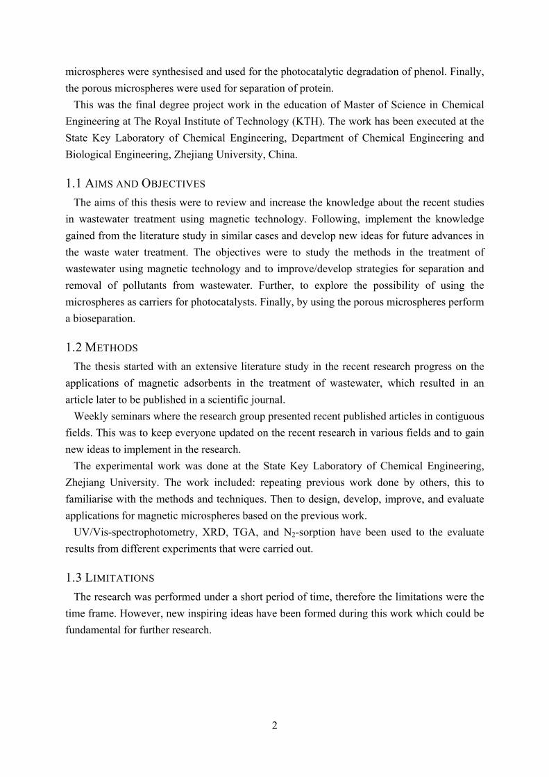
Subsequently, the sulfonated microspheres were stirred in the presence of ferrous chloride and
4
magnetic Fe3O4 nano-particles (MNPs) were formed and distributed all over the pore wall. A
scheme of the process is given in Figure 2.1.
: Magnetic particle
SO3-
Na+ -O3S
SO3-
-O3S
+Na
Na+
+Na
Magneticmicrosphere
Polystyremicrosphere
CH2Cl2
SO3H
HO3S
SO3H
HO3S
Sulfonatedmicrosphere
Fe2+SO3
-
-O3SSO3
-
-O3S
Fe2+
NaOH
H2O2
FeCl2SO3HCl
Figure 2.1 Scheme for preparing mesoporous magnetic microspheres [2.6]
The synthesised mesoporous magnetic microspheres were used for removal of the dyes
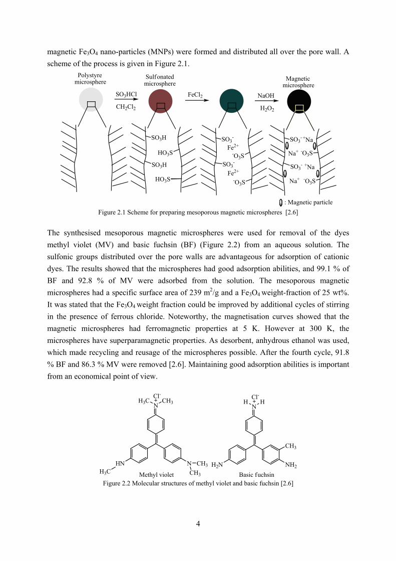
methyl violet (MV) and basic fuchsin (BF) (Figure 2.2) from an aqueous solution. The
sulfonic groups distributed over the pore walls are advantageous for adsorption of cationic
dyes. The results showed that the microspheres had good adsorption abilities, and 99.1 % of
BF and 92.8 % of MV were adsorbed from the solution. The mesoporous magnetic
microspheres had a specific surface area of 239 m2/g and a Fe3O4 weight-fraction of 25 wt%.
It was stated that the Fe3O4 weight fraction could be improved by additional cycles of stirring
in the presence of ferrous chloride. Noteworthy, the magnetisation curves showed that the
magnetic microspheres had ferromagnetic properties at 5 K. However at 300 K, the
microspheres have superparamagnetic properties. As desorbent, anhydrous ethanol was used,
which made recycling and reusage of the microspheres possible. After the fourth cycle, 91.8
% BF and 86.3 % MV were removed [2.6]. Maintaining good adsorption abilities is important
from an economical point of view.
NCH3H3C
NHN CH3
CH3H3C
NHH
NH2H2N
CH3
Methyl violet Basic fuchsin
Cl- Cl-
Figure 2.2 Molecular structures of methyl violet and basic fuchsin [2.6]
5
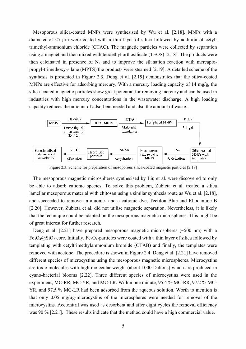
Mesoporous silica-coated MNPs were synthesised by Wu et al. [2.18]. MNPs with a
diameter of <5 µm were coated with a thin layer of silica followed by addition of cetyl-
trimethyl-ammonium chloride (CTAC). The magnetic particles were collected by separation
using a magnet and then mixed with tetraethyl orthosilicate (TEOS) [2.18]. The products were
then calcinated in presence of N2 and to improve the silanation reaction with mercapto-
propyl-trimethoxy-silane (MPTS) the products were steamed [2.19]. A detailed scheme of the
synthesis is presented in Figure 2.3. Dong et al. [2.19] demonstrates that the silica-coated
MNPs are effective for adsorbing mercury. With a mercury loading capacity of 14 mg/g, the
silica-coated magnetic particles show great potential for removing mercury and can be used in
industries with high mercury concentrations in the wastewater discharge. A high loading
capacity reduces the amount of adsorbent needed and also the amount of waste.
Figure 2.3. Scheme for preparation of mesoporous silica-coated magnetic particles [2.19]
The mesoporous magnetic microspheres synthesised by Liu et al. were discovered to only
be able to adsorb cationic species. To solve this problem, Zubieta et al. treated a silica
lamellar mesoporous material with chitosan using a similar synthesis route as Wu et al. [2.18],
and succeeded to remove an anionic- and a cationic dye, Tectilon Blue and Rhodamine B
[2.20]. However, Zubieta et al. did not utilise magnetic separation. Nevertheless, it is likely
that the technique could be adapted on the mesoporous magnetic microspheres. This might be
of great interest for further research.
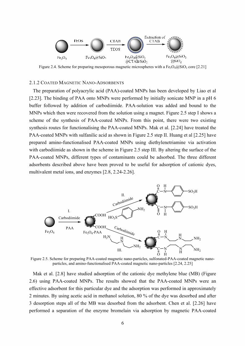
Deng et al. [2.21] have prepared mesoporous magnetic microspheres (~500 nm) with a
Fe3O4@SiO2 core. Initially, Fe3O4-particles were coated with a thin layer of silica followed by
templating with cetyltrimethylammonium bromide (CTAB) and finally, the templates were
removed with acetone. The procedure is shown in Figure 2.4. Deng et al. [2.21] have removed
different species of microcystins using the mesoporous magnetic microspheres. Microcystins
are toxic molecules with high molecular weight (about 1000 Daltons) which are produced in
cyano-bacterial blooms [2.22]. Three different species of microcystins were used in the
experiment; MC-RR, MC-YR, and MC-LR. Within one minute, 95.4 % MC-RR, 97.2 % MC-
YR, and 97.5 % MC-LR had been adsorbed from the aqueous solution. Worth to mention is
that only 0.05 mg/µg-microcystins of the microspheres were needed for removal of the
microcystins. Acetonitril was used as desorbent and after eight cycles the removal efficiency
was 90 % [2.21]. These results indicate that the method could have a high commercial value.
6
Figure 2.4. Scheme for preparing mesoporous magnetic microspheres with a Fe3O4@SiO2 core [2.21]
2.1.2 COATED MAGNETIC NANO-ADSORBENTS
The preparation of polyacrylic acid (PAA)-coated MNPs has been developed by Liao et al
[2.23]. The binding of PAA onto MNPs were performed by initially sonicate MNP in a pH 6
buffer followed by addition of carbodiimide. PAA-solution was added and bound to the
MNPs which then were recovered from the solution using a magnet. Figure 2.5 step I shows a
scheme of the synthesis of PAA-coated MNPs. From this point, there were two existing
synthesis routes for functionalising the PAA-coated MNPs. Mak et al. [2.24] have treated the
PAA-coated MNPs with sulfanilic acid as shown in Figure 2.5 step II. Huang et al [2.25] have
prepared amino-functionalised PAA-coated MNPs using diethylenetriamine via activation
with carbodiimide as shown in the scheme in Figure 2.5 step III. By altering the surface of the
PAA-coated MNPs, different types of contaminants could be adsorbed. The three different
adsorbents described above have been proved to be useful for adsorption of cationic dyes,
multivalent metal ions, and enzymes [2.8, 2.24-2.26].
PAA
Carbodiimide
HN
NH2
H2N C
C
O
O
N
N NH
HN
H
H
NH2
NH2
Fe3O4
C
O
N
H
SO3H
C
O
N
H
SO3HNH2
HO3S
Carbodiimide
CarbodiimideFe3O4-PAA
COOH
COOH
II.
III.
I.
Figure 2.5. Scheme for preparing PAA-coated magnetic nano-particles, sulfonated-PAA-coated magnetic nano-
particles, and amino-functionalised PAA-coated magnetic nano-particles [2.24, 2.25] Mak et al. [2.8] have studied adsorption of the cationic dye methylene blue (MB) (Figure
2.6) using PAA-coated MNPs. The results showed that the PAA-coated MNPs were an
effective adsorbent for this particular dye and the adsorption was performed in approximately
2 minutes. By using acetic acid in methanol solution, 80 % of the dye was desorbed and after
3 desorption steps all of the MB was desorbed from the adsorbent. Chen et al. [2.26] have
performed a separation of the enzyme bromelain via adsorption by magnetic PAA-coated

7
MNPs. Also here the adsorption process was rapid. In approximately 1 minute the bromelain
present in the solution was adsorbed. The adsorptions were quick due to the absence of
internal diffusion. [2.8, 2.26]
Figure 2.6. Molecular structure of methylene blue
As demonstrated by Mak et al., [2.8] PAA-coated MNPs can be very useful for removal of
cationic dyes. Unfortunately, it is not as easy for anionic dyes to be adsorbed onto PAA-
coated MNPs and they also show a low ability for adsorption of heavy metal ions [2.27]. Mak
et al. [2.24] have used the sulfonated PAA-coated MNPs for adsorption of multivalent cation
metal ions from aqueous solutions. The result and a comparison of adsorption of multivalent
cation metal ions from aqueous solutions are shown in Table 2.1. The results were found to be
satisfying. The sulfonated PAA-coated MNPs adsorbed nearly 100 % of the multivalent metal
ions, in comparison with the PAA-coated MNPs which only adsorbed approximately 30-40 %
of the metal ions in the study [2.24].
Table 2.1. Comparison of recovery of metal cations using either sulfonated magnetic nano-particles or PAA-
coated magnetic nano-particles, with initial concentration of 100 mg/L for each metal specie [2.24]
Recovery (%)
Cation Sulfonated magnetic nano-particles PAA-coated magnetic nano-particle
Co2+ ~100 30.5
Al3+ ~100 34.5
Ni2+ ~100 37.1
Cu2+ 97.2 38.3
Ag+ ~100 78.5
Sulfonated PAA-coated MNPs do not adsorb anionic metals or substances. To circumvent
this problem and to get a magnetic nano-adsorbent for both anionic and cationic species,
Huang et al. [2.25] have used the amino-functionalised PAA-coated MNPs. The results in
Table 2.2 confirm that amino-functionalised MNPs have good adsorption abilities, both for
cationic and anionic species. The recovery was nearly 100 % for all the different metal ions.
This demonstrates the high feasibility of the method and also its superiority compared to
sulfonated PAA-coated MNPs and PAA-coated MNPs. The adsorption rate was fast, and
equilibrium was achieved in only a few minutes [2.25].
Humic acid (HA) coated MNPs have recently been synthesised by Liu et al. [2.28]. Ferric
chloride and ferrous sulphate were dissolved in water followed by the addition of ammonium
hydroxide and HA-sodium salt. The precipitate was the HA-coated MNPs and without further

8
treatment they could be used for removal of Cd(II), Cu(II), Hg(II), and Pb(II) from the
aqueous solution. Both HA and Fe3O4 are naturally abundant [2.29, 2.30], so they possess a
minimal treat for the environment. Also, for the preparation of HA-coated MNPs
environmentally friendly salts and solvents were used [2.28]. The feasibility of the HA-coated
MNPs for removal of heavy metals from water is shown in Table 2.3.
Table 2.2. Comparison of recovery of metal ions using either amino-functionalised magnetic nano-particles, or PAA-coated magnetic nano-particles, with initial concentration of 100 mg/L [2.25]
Recovery (%)
Metal ions Amino-functionalised magnetic
nano-particles
PAA-coated magnetic
nano-particles
Fe3+ 100 82.3
Ag+ 100 41.0
Cu2+ ~100 34.7
Co2+ ~100 32.3
Fe2+ 98.7 30.1
Ni2+ 100 18.6
Cd2+ 100 8.5
AuCl4- ~100 -
PdCl4- 100 -
HCrO4- 96 -
The removal efficiency is pH-dependent, and as a result the adsorption efficiency can differ
up to 20 % for Cu and about 7 % for Cd in unfavourable conditions. The study showed that
there was no interference in adsorption of the four metal species related to adsorption of other
natural abundant metals in water, such as calcium. Further, the influence of dissolved organic
matter on adsorption of heavy metal onto HA-coated MNPs was minimal. This concludes that
the HA-coated MNP s selectively adsorb heavy metals.
Table 2.3. Comparison of removal (%) of heavy metals from water from different sources, with initial concentration C in mg/L [2.28]
Hg Pb Cd Cu
Matrix pH C Removal C Removal C Removal C Removal
Tap water 7.8 1.011 99.8 1.006 99.4 1.001 96.8 1.077 86.0
Ground water 6.4 1.012 99.8 1.005 99.5 1.002 92.1 1.005 99.7
River water 7.6 1.011 99.9 1.005 99.4 1.001 92.3 1.008 86.0
Lake water 7.2 1.011 99.2 1.006 99.4 1.001 91.7 1.011 82.4
Sea water 7.7 1.013 99.9 1.20 99.0 1.009 99.0 1.040 79.3
Chang et al. [2.27] have coated MNPs with carboxymethyl chitosan, via activation by
carbodiimide followed by addition of a carboxymethyl chitosan solution. Chitosan have many

9
amino-groups that can serve as chelation sites, and this makes it prominent to adsorb several
metal cations, [2.31-2.33] fluoride and numerous types of dyes [2.27, 2.34-2.36]. Chitosan is a
natural polysaccharide which is a non-toxic and biodegradable polymer product produced by
deacetylation of chitin. There are many applications for chitosan in various fields, such as
biomedicine, drug delivery, and paper industry [2.37-2.39]. In a study by Chang et al. [2.34] Co(II) was adsorbed from aqueous solution using the chitosan MNPs. The adsorption rate
proved to be fast, and equilibrium was reached in approximately 1 minute. In previous
reports, Chang et al. have used the chitosan MNPs for removal of Cu(II) ions. The results
showed that the maximum adsorption capacity was 21.5 mg/g and that the adsorption rate was
fast. Also here the equilibrium was achieved in approximately 1 minute [2.35]. Recovering
metals from effluents is not only important from an environmental aspect, but also from an
economical point of view, due to high raw material costs.
Two acid dyes, acid green 25 and crocein orange g (Figure 2.7) have been used in an
adsorption study by Chang et al. [2.27]. The adsorption abilities were high for both of the
dyes, 1883 mg/g for crocein orange g and 1471 mg/g for acid green 25. This was due to the
large specific surface area of the MNPs. However, acid dyes have a strong affinity to chitosan
which causes problems in the desorption step. The maximum desorption achieved for crocein
orange g was 83.6 % and for acid green 66.1 %. Desorption efficiency is as important as
adsorption efficiency.
Figure 2.7. Molecular structures of acid green 25 and crocein orange g [2.27]
With low desorption percentage the economy of the method becomes lower. It has been
suggested that multiple desorption steps could improve desorption percentage, but no further
research has been done. Figure 2.8 shows the adsorption mechanism of the acid dyes onto the
amino-groups of chitosan [2.27]. Chitosan MNPs can be used not merely for adsorption of
cationic species, but also for anionic species as shown in this case.
Figure 2.8. The mechanism of adsorption of acid dyes where R’(SO3
-)2: acid green 25 and R’’SO3-: crocein orange g [2.27]

10
Low concentrations of fluoride in drinking water have beneficial effects on dental health,
however higher concentrations have negative effects on human health [2.40, 2.41]. Several
methods for removing fluoride from water have been developed, such as reverse osmosis, and
nanofiltration [2.42, 2.43]. The drawbacks of the methods have often been high operational
costs. Wei et al. [2.36] have studied the adsorption of fluoride on chitosan MNPs, and also the
surface structure. Groups of NH, NH2, NH3, and Fe-O, create an amorphous surface on the
chitosan MNPs. As mentioned earlier these groups work as chelation sites, on which the
fluoride ions can be disposed. The results showed that the initial adsorption rate was 2.08
mg/g min. The high adsorption efficiency of fluoride using chitosan MNPs gives the method
great commercial value.
2.1.3 MAGNETIC CARBON ADSORBENTS
Active carbon is a good adsorbent with a high specific surface area. Due to the good
adsorption ability, active carbon has often been used over the years for removal of all types of
pollutants [2.44-2.47]. It has been reported by Long et al. [2.48] that carbon nanotubes
(CNTs) are more efficient in adsorbing dioxins in comparison with active carbon. Active
carbon is generally very difficult to separate from aqueous solution [1.13]. Incorporating the
CNTs with MNPs makes it easy to separate adsorbent from solution by utilising the magnetic
properties. Hence, overcoming the problem caused by their size which occurs when using
conventional separation techniques, such as filtration or centrifugation for the separation of
nano-sized particles [2.34].
Peng et al. [2.49] have prepared the CNTs by catalytic pyrolysis of a mixture containing
propylene and hydrogen with Ni as catalyst. The MNPs were incorporated by initially
dissolving the catalyst particles in nitric acid and hydrofluoric acid. Finally, ferric chloride
and ferrous sulphate were mixed with the CNTs under nitrogen, and NaOH was added. The
CNTs with incorporated MNPs can be used for adsorption of heavy metals and dyes from
aqueous solution [2.49-2.51]. By chemical oxidation, the CNTs improve their metal
adsorption due to progression of the number of oxygen-containing groups on the surface. In
the study by Peng et al., [2.49] the adsorption of Pb(II) and Cu(II) using the magnetic CNTs
were nearly 100 %, with a CNT recovery rate of approximately 98 %. Attempts of removing
dyes from water using magnetic CNTs have been executed by Qu et al. [2.50] and Gong et al.
[2.51]. Three different dyes were examined in these studies, MB, neutral red (NR), and
brilliant cresyl blue (BCB) (Figure 2.6, 2.9). The results showed that the CNTs possess high
adsorption abilities for these specific dyes, 99.2% MB, 98.3 % NR, and 98.8% BCB were
adsorbed [2.51].
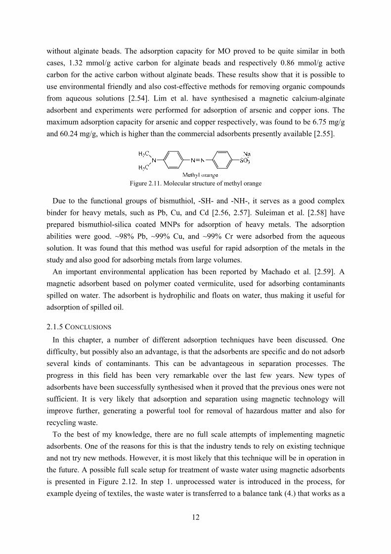
From rice husk based activated carbon (RHC) Yang et al. [1.13] have synthesised
mesoporous MNPs. The MNPs were used for adsorption of MB from aqueous solution. A
scheme of the preparation and adsorption of the MNPs is shown in Figure 2.10. The RHC-
11
Fe3O4 particles have a specific surface area of 770 m2/g and an adsorption capacity of 321
mg/g for MB in aqueous solution. Yang et al. show an environmentally friendly way of
removing contaminants.
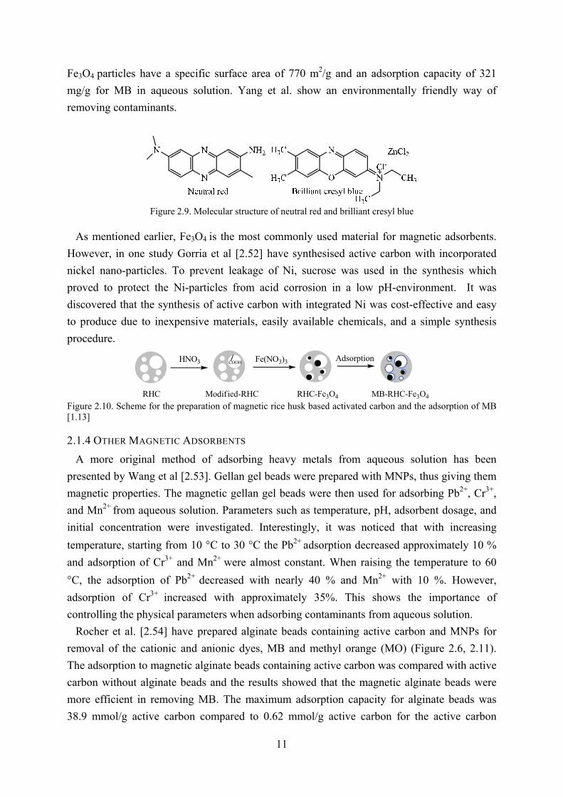
Figure 2.9. Molecular structure of neutral red and brilliant cresyl blue
As mentioned earlier, Fe3O4 is the most commonly used material for magnetic adsorbents.
However, in one study Gorria et al [2.52] have synthesised active carbon with incorporated
nickel nano-particles. To prevent leakage of Ni, sucrose was used in the synthesis which
proved to protect the Ni-particles from acid corrosion in a low pH-environment. It was
discovered that the synthesis of active carbon with integrated Ni was cost-effective and easy
to produce due to inexpensive materials, easily available chemicals, and a simple synthesis
procedure.
RHC Modif ied-RHC RHC-Fe3O4 MB-RHC-Fe3O4
HNO3 COOH AdsorptionCOOH Fe(NO3)3
Figure 2.10. Scheme for the preparation of magnetic rice husk based activated carbon and the adsorption of MB [1.13]
2.1.4 OTHER MAGNETIC ADSORBENTS
A more original method of adsorbing heavy metals from aqueous solution has been
presented by Wang et al [2.53]. Gellan gel beads were prepared with MNPs, thus giving them
magnetic properties. The magnetic gellan gel beads were then used for adsorbing Pb2+, Cr3+,
and Mn2+ from aqueous solution. Parameters such as temperature, pH, adsorbent dosage, and
initial concentration were investigated. Interestingly, it was noticed that with increasing
temperature, starting from 10 °C to 30 °C the Pb2+ adsorption decreased approximately 10 %
and adsorption of Cr3+ and Mn2+ were almost constant. When raising the temperature to 60
°C, the adsorption of Pb2+ decreased with nearly 40 % and Mn2+ with 10 %. However,
adsorption of Cr3+ increased with approximately 35%. This shows the importance of
controlling the physical parameters when adsorbing contaminants from aqueous solution.
Rocher et al. [2.54] have prepared alginate beads containing active carbon and MNPs for
removal of the cationic and anionic dyes, MB and methyl orange (MO) (Figure 2.6, 2.11).
The adsorption to magnetic alginate beads containing active carbon was compared with active
carbon without alginate beads and the results showed that the magnetic alginate beads were
more efficient in removing MB. The maximum adsorption capacity for alginate beads was
38.9 mmol/g active carbon compared to 0.62 mmol/g active carbon for the active carbon
12
without alginate beads. The adsorption capacity for MO proved to be quite similar in both
cases, 1.32 mmol/g active carbon for alginate beads and respectively 0.86 mmol/g active
carbon for the active carbon without alginate beads. These results show that it is possible to
use environmental friendly and also cost-effective methods for removing organic compounds
from aqueous solutions [2.54]. Lim et al. have synthesised a magnetic calcium-alginate
adsorbent and experiments were performed for adsorption of arsenic and copper ions. The
maximum adsorption capacity for arsenic and copper respectively, was found to be 6.75 mg/g
and 60.24 mg/g, which is higher than the commercial adsorbents presently available [2.55].
Figure 2.11. Molecular structure of methyl orange
Due to the functional groups of bismuthiol, -SH- and -NH-, it serves as a good complex
binder for heavy metals, such as Pb, Cu, and Cd [2.56, 2.57]. Suleiman et al. [2.58] have
prepared bismuthiol-silica coated MNPs for adsorption of heavy metals. The adsorption
abilities were good. ~98% Pb, ~99% Cu, and ~99% Cr were adsorbed from the aqueous
solution. It was found that this method was useful for rapid adsorption of the metals in the
study and also good for adsorbing metals from large volumes.
An important environmental application has been reported by Machado et al. [2.59]. A
magnetic adsorbent based on polymer coated vermiculite, used for adsorbing contaminants
spilled on water. The adsorbent is hydrophilic and floats on water, thus making it useful for
adsorption of spilled oil.
2.1.5 CONCLUSIONS
In this chapter, a number of different adsorption techniques have been discussed. One
difficulty, but possibly also an advantage, is that the adsorbents are specific and do not adsorb
several kinds of contaminants. This can be advantageous in separation processes. The
progress in this field has been very remarkable over the last few years. New types of
adsorbents have been successfully synthesised when it proved that the previous ones were not
sufficient. It is very likely that adsorption and separation using magnetic technology will
improve further, generating a powerful tool for removal of hazardous matter and also for
recycling waste.
To the best of my knowledge, there are no full scale attempts of implementing magnetic
adsorbents. One of the reasons for this is that the industry tends to rely on existing technique
and not try new methods. However, it is most likely that this technique will be in operation in
the future. A possible full scale setup for treatment of waste water using magnetic adsorbents
is presented in Figure 2.12. In step 1. unprocessed water is introduced in the process, for
example dyeing of textiles, the waste water is transferred to a balance tank (4.) that works as a
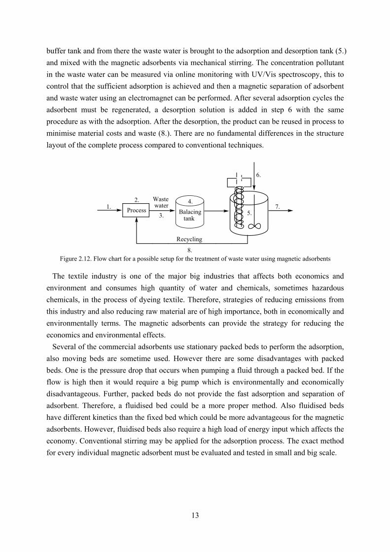
13
buffer tank and from there the waste water is brought to the adsorption and desorption tank (5.)
and mixed with the magnetic adsorbents via mechanical stirring. The concentration pollutant
in the waste water can be measured via online monitoring with UV/Vis spectroscopy, this to
control that the sufficient adsorption is achieved and then a magnetic separation of adsorbent
and waste water using an electromagnet can be performed. After several adsorption cycles the
adsorbent must be regenerated, a desorption solution is added in step 6 with the same
procedure as with the adsorption. After the desorption, the product can be reused in process to
minimise material costs and waste (8.). There are no fundamental differences in the structure
layout of the complete process compared to conventional techniques.
Balacingtank
Process
Wastewater
Recycling
1.2.
3.
4.
5.
6.
7.
8. Figure 2.12. Flow chart for a possible setup for the treatment of waste water using magnetic adsorbents
The textile industry is one of the major big industries that affects both economics and
environment and consumes high quantity of water and chemicals, sometimes hazardous
chemicals, in the process of dyeing textile. Therefore, strategies of reducing emissions from
this industry and also reducing raw material are of high importance, both in economically and
environmentally terms. The magnetic adsorbents can provide the strategy for reducing the
economics and environmental effects.
Several of the commercial adsorbents use stationary packed beds to perform the adsorption,
also moving beds are sometime used. However there are some disadvantages with packed
beds. One is the pressure drop that occurs when pumping a fluid through a packed bed. If the
flow is high then it would require a big pump which is environmentally and economically
disadvantageous. Further, packed beds do not provide the fast adsorption and separation of
adsorbent. Therefore, a fluidised bed could be a more proper method. Also fluidised beds
have different kinetics than the fixed bed which could be more advantageous for the magnetic
adsorbents. However, fluidised beds also require a high load of energy input which affects the
economy. Conventional stirring may be applied for the adsorption process. The exact method
for every individual magnetic adsorbent must be evaluated and tested in small and big scale.

14
2.2 UPDATED RESEARCH PROGRESS IN MAGNETIC PHOTOCATALYST Since the discovery of the photocatalytic properties of TiO2 in 1972 by Fujishima and
Honda [2.60], many new types of applications for TiO2 have been discovered, such as
photovoltaics [2.61]. Photocatalytic reactions have recently drawn much attention due
potential of producing hydrogen from water using solar light [2.62-2.64]. One of the main
applications for photocatalysts is the degradation of pollutants from waste water. This
technique is becoming increasingly important, especially for removing low trace contaminants
but also for bacteria and viruses [1.14, 1.15, 2.65-2.68]. The photocatalysts have the same
problems as many adsorbents, due to the size they are difficult to separate from aqueous
solutions. [1.17].
The size of the TiO2 particles have found to be important for the photocatalytic efficiency,
when decreasing TiO2 particle size the specific surface area increases, thus the contact area
increases which results in an increase of efficiency [2.69]. However, separation and recovery
of the TiO2 particles is difficult due to the small particle size. Therefore, incorporating TiO2 in
magnetic carriers is of great interest due to the effective separation and recovery that can be
achieved by magnetic separation. Further, the carriers can be fluidised by an external
magnetic field [2.70]. Several different types of magnetic carriers have recently been
investigated, such as barium ferrite [2.71], Fe2O3/SiO2 [2.72], magnetite [2.73], and magnetic
poly(methyl methacrylate) microspheres (mPMMA) [2.74].
In this chapter, several different types of magnetic carriers and the preparation of
photocatalysts using these carriers are discussed. Also, the degradation of different organic
contaminants using the magnetic photocatalysts is discussed. The sol-gel technique is the
most commonly used technique for the preparation of semi-conducting photocatalysts. There
are several articles and reviews addressing this technique. Therefore, in this chapter we focus
on alternative synthesis route for the preparation of photocatalysts.
2.2.1 DIFFERENT PREPARATION TECHNIQUES AND MAGNETIC CARRIERS Magnetic Fe2O3/SiO2/TiO2 nanoparticles were synthesised by Wang et al. [2.72]. The
synthesis route is presented in Figure 2.13. Fe3O4 particles were dispersed in water followed
by addition of tetraethyl orthosilicate (TEOS) at high pH. Subsequently, the Fe3O4/SiO2
particles were transferred to an autoclave and dispersed in hexane, water, and TBOT solution
and the autoclave was heated. The product was then calcinated under high temperature and
the Fe3O4 particles were transferred into Fe2O3.
Figure 2.13Scheme for the preparation of Fe2O3/SiO2/TiO2 magnetic photocatalyst catalyst [2.72]

15
Xuan et al. [2.70] successfully synthesised hollow Fe3O4/TiO2 spheres by using a
poly(styrene-acrylic acid) (PSA) core. First, PSA-spheres with a diameter of 270 nm were
dispersed in ethanol followed by addition of aqueous FeCl3. Second, the pH was adjusted to
9-10 using aqueous NH3 followed by addition of Na2SO3. Fe3O4 particles were formed on the
PSA surface. The PSA/Fe3O4 particles were dispersed in a mixture of butanol, ethanol, and
TBOT and stirred overnight to get a saturated adsorption of TBOT on the spheres.
Subsequently, water was added to hydrolyse the TBOT into amorphous titania. The
PSA/Fe3O4/TiO2 particles were dispersed in THF, this to dissolve the PSA and then
transferred to an autoclave. The amorphous titania was transformed to anastase form by
solvothermal heating with hexane in the autoclave. A scheme for this procedure is presented
in Figure 2.14.
Figure 2.14 Scheme for the preparation of hollow Fe3O4/SiO2 magnetic photocatalyst catalyst [2.70]
The preparation of black sand based magnetic photocatalyst was developed by Luo et al.
[2.74, 2.75]. Black sand (BS), a natural magnetic material, was used as magnetic core in the
preparation of magnetic photocatalyst. BS was dispersed in water with TEOS and was stirred
for a short while following by addition of ammonium hydroxide. The formed particles were
dried and then the BS/SiO2 was dispersed in a mixture of propanol and Titanium tetra-
isopropoxide (TPOT) followed by stirring and heating. The product was then calcinated in
order to transfer the TiO2 into crystalline phase. The synthesis process of BS/SiO2/TiO2 is
shown in Figure 2.15.
Figure 2.15. Scheme for the preparation of black sand/SiO2/TiO2 magnetic photocatalyst catalyst [2.74, 2.75]
TiO2 coated mPMMA microspheres were prepared by Chen et al. [2.76]. The magnetic
particles were oleic acid coated (OMP) and introduced with the PMMA microspheres via
modified suspension polymerisation. Described shortly, the OMP were mixed with
divinylbenzene, methyl methacrylate, benzoyl peroxide, and hexane and was let reacted under
heating. Then the formed mPMMA was magnetically separated from the solution.
Subsequently, the mPMMA was mixed with TiO2 powder and heated close to the glass
transition temperature of PMMA which resulted in a soft surface that the TiO2 could be
adhered to. The process is shown in Figure 2.16.

16
polymerisation TiO2
mPMMA/TiO2mPMMAOMP
Figure 2.16. Scheme for the preparation of mPMMA/TiO2 magnetic photocatalyst catalyst [2.76]
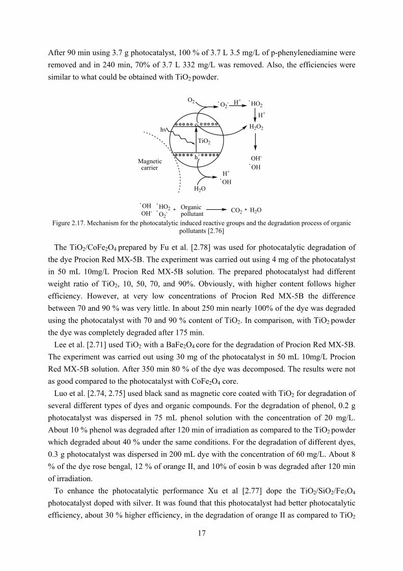
2.2.2 DEGRADATION OF ORGANIC POLLUTANTS BY MAGNETIC PHOTOCATALYSTS The general photocatalytic process is illustrated in Figure 2.17 and by equations 1-6. When
the TiO2 photocatalyst is irradiated with light, an electron in the valence band can be excited
to the conduction band and a hole in the valence band is generated (equation 1). The back
reaction (equation 1*) produces heat. Further, the free electron reacts with the oxygen and
forms the super oxide anion (equation 2). The hole in the valence band take one electron from
the water and H+ and ·OH is formed (equation 3). The super oxide anion combines with H+
and forms the ·HO2 (equation 4). The ·HO2 reacts with H+ and forms hydrogen peroxide which
then dissociates into hydroxyl radicals (equation 5, 6). [2.77]
Wang et al. [2.72] used the Fe2O3/SiO2/TiO2 for photocatalytic degradation of MB. After 80
min, 80 % of the 50 mL 25 mg/L MB was degraded using 0.1 g photocatalyst. The results
showed that the prepared photocatalyst had nearly the same photocatalytic ability compared
with TiO2 powder. This indicates that the SiO2 layer prevented the electrons from transferring
to the Fe2O3 that would reduce the photocatalytic ability.
Rhodamine B was photocatalytic degraded using hollow Fe3O4/TiO2 spheres prepared by
Xuan et al. [2.70]. In the experiment, 25 mg photocatalyst was used for the degradation of 60
mL 0.3 mg/L rhodamine B. After 80 min nearly 100 % of the dye was degradated. This
concludes that the hollow Fe3O4/TiO2 spheres are an excellent photocatalyst.
Photocatalytic degradation of p-phenylenediamine using TiO2-coated magnetic PMMA
microspheres was performed by Chen et al. [2.76]. Experiments using different concentrations
of p-phenylenediamine were performed. The experiments showed that the TiO2-coated
magnetic PMMA microspheres were excellent for removing low concentration contaminants.
17
After 90 min using 3.7 g photocatalyst, 100 % of 3.7 L 3.5 mg/L of p-phenylenediamine were
removed and in 240 min, 70% of 3.7 L 332 mg/L was removed. Also, the efficiencies were
similar to what could be obtained with TiO2 powder.
TiO2
h+
e-
hv
O2
H2O
Magneticcarrier
OH
O2-
H+
HO2
H2O2
OH-
OH
H+
H+
OrganicpollutantO2
-OHOH-
HO2 CO2 H2O
Figure 2.17. Mechanism for the photocatalytic induced reactive groups and the degradation process of organic pollutants [2.76]
The TiO2/CoFe2O4 prepared by Fu et al. [2.78] was used for photocatalytic degradation of
the dye Procion Red MX-5B. The experiment was carried out using 4 mg of the photocatalyst
in 50 mL 10mg/L Procion Red MX-5B solution. The prepared photocatalyst had different
weight ratio of TiO2, 10, 50, 70, and 90%. Obviously, with higher content follows higher
efficiency. However, at very low concentrations of Procion Red MX-5B the difference
between 70 and 90 % was very little. In about 250 min nearly 100% of the dye was degraded
using the photocatalyst with 70 and 90 % content of TiO2. In comparison, with TiO2 powder
the dye was completely degraded after 175 min.
Lee et al. [2.71] used TiO2 with a BaFe2O4 core for the degradation of Procion Red MX-5B.
The experiment was carried out using 30 mg of the photocatalyst in 50 mL 10mg/L Procion
Red MX-5B solution. After 350 min 80 % of the dye was decomposed. The results were not
as good compared to the photocatalyst with CoFe2O4 core.
Luo et al. [2.74, 2.75] used black sand as magnetic core coated with TiO2 for degradation of
several different types of dyes and organic compounds. For the degradation of phenol, 0.2 g
photocatalyst was dispersed in 75 mL phenol solution with the concentration of 20 mg/L.
About 10 % phenol was degraded after 120 min of irradiation as compared to the TiO2 powder
which degraded about 40 % under the same conditions. For the degradation of different dyes,
0.3 g photocatalyst was dispersed in 200 mL dye with the concentration of 60 mg/L. About 8
% of the dye rose bengal, 12 % of orange II, and 10% of eosin b was degraded after 120 min
of irradiation.
To enhance the photocatalytic performance Xu et al [2.77] dope the TiO2/SiO2/Fe3O4
photocatalyst doped with silver. It was found that this photocatalyst had better photocatalytic
efficiency, about 30 % higher efficiency, in the degradation of orange II as compared to TiO2
18
powder. After 20 min using 50 mg photocatalyst, 90 % of 0.1 L 17.5 mg/L orange II was
removed. In comparison with TiO2 powder were 70 % was removed after 20 min. The process
can be described with equation 1-8. Compared to the un-doped photocatalyst reaction 7 and 8
do not take place. The formation of extra super oxide anion boosts the photocatalytic
degradation.
2.2.3 CONCLUSIONS
In this chapter, several different magnetic carriers for the TiO2 photocatalyst have been
reviewed. The magnetic carriers provide an effective way of the separation of TiO2 which
otherwise is very difficult and expensive to separate. However, the photocatalyst need to be
more efficient and more research has to be put in the field before they will have industrial
applications. The photocatalyst have high potential for providing a cheap and efficient way of
removing pollutants from effluents.
19
2.3 UPDATED RESEARCH PROGRESS IN BIOSEPARATION USING MAGNETIC
TECHNOLOGY The magnetic technology has many applications in several areas. Biotechnology is perhaps
the most prominent field for the magnetic technology. The magnetic technology can be used
for drug delivery, enzyme immobilisation, protein separation and isolation [2.79, 2.80].
In this chapter, a brief review of the recent progress on the magnetic technology for
bioseparation is presented.
2.3.1 BIOSEPARATION
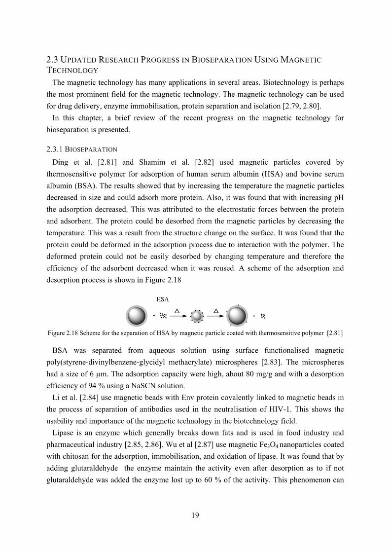
Ding et al. [2.81] and Shamim et al. [2.82] used magnetic particles covered by
thermosensitive polymer for adsorption of human serum albumin (HSA) and bovine serum
albumin (BSA). The results showed that by increasing the temperature the magnetic particles
decreased in size and could adsorb more protein. Also, it was found that with increasing pH
the adsorption decreased. This was attributed to the electrostatic forces between the protein
and adsorbent. The protein could be desorbed from the magnetic particles by decreasing the
temperature. This was a result from the structure change on the surface. It was found that the
protein could be deformed in the adsorption process due to interaction with the polymer. The
deformed protein could not be easily desorbed by changing temperature and therefore the
efficiency of the adsorbent decreased when it was reused. A scheme of the adsorption and
desorption process is shown in Figure 2.18
Figure 2.18 Scheme for the separation of HSA by magnetic particle coated with thermosensitive polymer [2.81] BSA was separated from aqueous solution using surface functionalised magnetic
poly(styrene-divinylbenzene-glycidyl methacrylate) microspheres [2.83]. The microspheres
had a size of 6 µm. The adsorption capacity were high, about 80 mg/g and with a desorption
efficiency of 94 % using a NaSCN solution.
Li et al. [2.84] use magnetic beads with Env protein covalently linked to magnetic beads in
the process of separation of antibodies used in the neutralisation of HIV-1. This shows the
usability and importance of the magnetic technology in the biotechnology field.
Lipase is an enzyme which generally breaks down fats and is used in food industry and
pharmaceutical industry [2.85, 2.86]. Wu et al [2.87] use magnetic Fe3O4 nanoparticles coated
with chitosan for the adsorption, immobilisation, and oxidation of lipase. It was found that by
adding glutaraldehyde the enzyme maintain the activity even after desorption as to if not
glutaraldehyde was added the enzyme lost up to 60 % of the activity. This phenomenon can

20
be used for controlling processes in the food industry and pharmaceutical industry more
accurate.
Recently, Luo et al [2.88] prepared cellulose microspheres with fabricated Fe3O4 particles in
the cellulose pores. The synthesised microspheres were used for adsorption with controlled
release of BSA. The release of BSA from the microspheres could be controlled by changing
pH. At pH 7.4, which is close to the pH in the human body, nearly 100 % of the BSA could be
released. However, the process was very slow, about 500 h. This could be exploited for slow
drug delivery which releases the active substance slowly.
Lin et al. [2.89] use amino-functionalised magnetic nanoparticles with trypsin immobilised
on the surface. These particles were used for digestion of BSA, myoglobin, and cytochrome c.
After the digestion the microspheres could easily be removed by magnetic separation and the
samples could be analysed. This is an interesting way of studying the digestion of proteins
and to be able to investigate the process without interference of the enzyme in the analysis.
Chiang [2.90] use Fe3O4 particles coated with glycidyl methacrylate-iminodiacetic acid for
extraction of protein from a specific fluorescent modified protein from E-coli bacteria. The
result shows that the magnetic particles were specific and were effective in adsorbing and
extracting this specific protein.
2.3.2 CONCLUSIONS
In this chapter, we have shown that the magnetic technology can be used not only for
environmental purpose but also in the field of biotechnology. By altering the coatings of the
magnetic particles different kinds of bio-molecules can be separated which can be exploited
when there is a need to separate in a moiety of bio-molecules. Without doubt, the magnetic
technology can provide the fast, specific, and efficient separation which is important in this
field.
21
CHAPTER 3: MAGNETIC MICROSPHERES FOR REMOVAL OF
METHYLENE BLUE FROM WASTEWATER Most dyes used in industries are considered hazardous, so, it is necessary to remove these
from effluents. Organic dyes show low biodegradability, hence, other methods than the
biological treatment is required for removing dyes from effluents [3.4]. Also, other
conventional techniques such as reverse osmosis and chemical coagulation have problems
with removing the colour completely [3.5].
Methylene blue (MB) is a cationic dye and is used in many areas, such as medicine and
biotechnology [3.6, 3.7]. Several materials have recently been studied to remove MB from
wastewater such as carbon nano-tubes, fly ash, red mud, and vermiculite [3.8-3.12]. However,
these methods do not provide an easy and quick separation process.
In this study, mesoporous magnetic microspheres have been synthesised and used for the
adsorption of MB. MB is a model dye and been used extensively in adsorption studies, hence
make a comparison between other methods convenient. The chemical structure of MB is
shown in Figure 2.6. Liu et al. [3.13] reported that the mesoporous magnetic microspheres
have good ability to adsorb the cationic dyes methyl violet and basic fuchsin. Further,
magnetic separation makes the reusage of the adsorbent convenient.
3.1 EXPERIMENTAL
3.1.1 MATERIALS
Porous polystyrene microspheres with a diameter of ~200 µm which were prepared in house
were used in this study. Chlorosulfonic acid and ferrous chloride were purchased from
Jingshanting Chemical Corporation of Shanghai, China. Dichloromethane, sodium hydroxide,
potassium chloride, anhydrous ethanol, and hydrogen peroxide were purchased from Lanling
Chemical Corporation of Linan in Zhejiang, China. Methylene blue was purchased from
Shanghai San’aisi Chemical Company, China. All the chemicals were used without further
treatment.
3.1.2 PREPARATION
3.1.2.1 PREPARATION OF SULFONATED MICROSPHERES
The porous magnetic microspheres were prepared according to the previous reported
method by Liu et al [3.13]. Into a 250 ml 3-neck round bottom flask 75 ml dichloromethane
and 5.78 g polystyrene microspheres were added. The solution was cooled using an ice bath,
subsequently, 0.8 ml chlorosulfonic acid in 75 ml dichloromethane was added dropwise into
the stirring solution. The solution was kept stirring for 24 h. The products were then filtrated
and washed with dichloromethane followed by drying in a vacuum oven for 12 h. Finally, the
microspheres were washed with deionised water and again dried in a vacuum oven.

22
3.1.2.2 PREPARATION OF MAGNETIC MICROSPHERES
Into a 250 ml 3-neck round bottom flask with a mechanical stirrer and argon inlet, 2.11 g of
the sulfonated microspheres as synthesised, were added with 80 ml 1 M FeCl2 solution. The
solution was kept stirring for 24 h. The products were then filtrated and washed three times
with deionised water and then transferred to another 3-neck round bottom flask with a
mechanical stirrer and a condenser, 20 ml deionised water and 80 ml 2 M NaOH was added.
The mixture was heated using an oil bath, when the temperature reached 75 °C, 20 ml 30 %
H2O2 was added dropwise into the mixture and the solution was kept stirring at 75 °C for 2 h.
The products were filtrated and washed three times with deionised water, one time with
anhydrous ethanol, and then dried in a vacuum oven.
3.1.3 CHARACTERISATION
Optical microscope photos were taken using a NOVEL XS-2100 microscope instrument
connected to a Canon powershot A630 with a Canon 640 52 mm objective. TGA were carried
out on a Perkin Elmer TGA-7 apparatus with a heating rate of 10 °C/min, starting from room
temperature to 700 °C. The surface areas were determined by nitrogen sorption porosimetry
on a micromeritics ASIC-2 apparatus. A 5.2 × 5.2 × 2.8 cm Fe-SrBr magnet was used for the
magnetic separation.
3.1.4 ADSORPTION AND DESORPTION OPTIMISATION
The adsorption optimisation was performed by varying pH and sonication time (contact
time adsorbent/MB) as shown in Table 3.1. In principle, into a small vial 0.2 g magnetic
microspheres were added with 4 mL of 1×10⁻⁶ M MB solution and then the solution was
sonicated for 2 min. The pH value of the system was regulated by using a specific pH buffer.
Subsequently, the magnetic microspheres were separated using a magnet and the solution was
collected and the adsorption efficiency were determined using an UNICO 3802 UV/Vis
spectrophotometer.
Table 3.1.4 Different parameters for the optimisation of the adsorption of MB onto magnetic microspheres
Run (pH, 0.2 g, 2min)
A01
pH 6
A02
pH 7
A03
pH 8
A04
pH 9
A05
pH 10
A06
pH 11
A07
pH 12
Run (Time, 0.2 g, pH 7)
A11
1 min
A12
2 min
A13
3 min
A14
4 min
A15
5 min
The desorption optimisation was performed by varying the parameters of EtOH/H2O
volume ratio, salt type, salt concentration, and sonication time, as shown in Table 3.2. In
principle, into a small vial 0.2 g magnetic microspheres were added with 4 mL of the

23
desorption solution and then the solution was sonicated for 2 min. Subsequently, the magnetic
microspheres were separated using a magnet and the solution was collected and the adsorption
optimisation efficiency were determined using a UV751GW UV/Vis spectrophotometer.
Table 3.2.5 Different parameters for the optimisation of the desorption of MB from microspheres
Run (E/W, 0.2 g, 2min) EW01 EW02 EW03 EW04 EW05
E/W(v/v) 2/8 4/6 6/4 8/2 10/0
Run (1 M salt, 0.2 g, 2min) EW11 EW12 EW13
E/W (0/10) NaCl KCl MnCl2
Run (1 M salt, 0.2 g, 2min) EW21 EW22 EW23
E/W (4/6) NaCl KCl MnCl2
Run (1 M salt, 0.2 g, 2min) EW31 EW32 EW33
E/W (6/4) NaCl KCla MnCl2
Run (KCl, 0.2 g, 2min) EW41 EW42 EW43 EW44
E/W (4/6) 0.1 M 0.5 M 1 M Saturated
Run (KCl, 0.2 g, 2min) EW51 EW52 EW53
E/W (6/4) 0.1 M 0.5 M Saturated
Run (Time, 0.2 g, Sat. KCl)
E/W (6/4)
EW61
1 min
EW62
2 min
EW763
3 min
EW64
4 min
EW65
5 min a Saturated KCl
3.2 RESULTS AND DISCUSSION
3.2.1 PREPARATION OF MAGNETIC MICROSPHERES
The porous polystyrene microspheres used in this study were prepared by free radical
suspension polymerisation with toluene as porogen according to the routes proposed by Liu et
al. [3.14]. The synthesised microspheres used for the preparation of the magnetic
microspheres had a diameter of ~200 µm.
First, the polystyrene microspheres were sulfonated in the presence of chlorosulfonic acid
and -SO3H were formed on the surface and on the pore walls. The sample was labelled as PS-
SO3H. Subsequently, the sulfonated microspheres were stirred in the presence of ferrous
chloride for 24 h to ensure full ion exchange. Finally, the ferrous ions were oxidised using
hydrogen peroxide and Fe3O4 particles were formed on the surface and in the pores of the
microspheres. A scheme of this process is shown in Figure 3.1. The weight fraction of Fe3O4
in the microspheres was very low, so the microspheres showed little magnetic response.
Therefore, the process described above was repeated. After each cycle, a sample was
collected and labelled as Mag 1 and Mag 2.
24
SO3-
SO3-
SO3-
SO3-
SO3-
+Na
+Na
+Na
+Na
+Na
SO3-
SO3-
SO3-
SO3-
SO3-
+H
+H
+H+H
+H
Fe2+CH2Cl2
SO3HCl FeCl2 NaOH
H2O2
SO3-
SO3-
SO3-
SO3-
SO3-
Fe2+
Fe2+
SO3-+H SO3
-SO3
- +Na
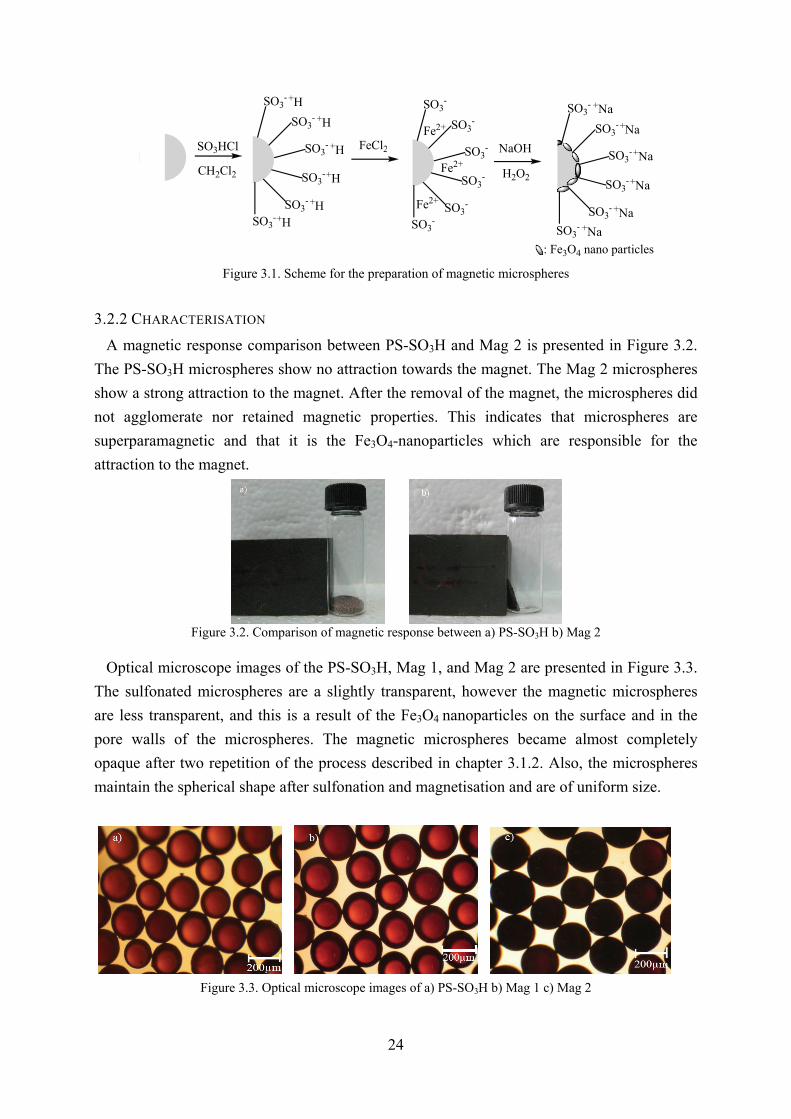
: Fe3O4 nano particles Figure 3.1. Scheme for the preparation of magnetic microspheres
3.2.2 CHARACTERISATION
A magnetic response comparison between PS-SO3H and Mag 2 is presented in Figure 3.2.
The PS-SO3H microspheres show no attraction towards the magnet. The Mag 2 microspheres
show a strong attraction to the magnet. After the removal of the magnet, the microspheres did
not agglomerate nor retained magnetic properties. This indicates that microspheres are
superparamagnetic and that it is the Fe3O4-nanoparticles which are responsible for the
attraction to the magnet.
Figure 3.2. Comparison of magnetic response between a) PS-SO3H b) Mag 2
Optical microscope images of the PS-SO3H, Mag 1, and Mag 2 are presented in Figure 3.3.
The sulfonated microspheres are a slightly transparent, however the magnetic microspheres
are less transparent, and this is a result of the Fe3O4 nanoparticles on the surface and in the
pore walls of the microspheres. The magnetic microspheres became almost completely
opaque after two repetition of the process described in chapter 3.1.2. Also, the microspheres
maintain the spherical shape after sulfonation and magnetisation and are of uniform size.
Figure 3.3. Optical microscope images of a) PS-SO3H b) Mag 1 c) Mag 2

25
The results from the TGA are presented in Figure 3.4, and shows that the major
decomposition region of the polystyrene microspheres is from 400 to 600 °C. The weigh-loss
between room temperature and 100 °C was attributed to the microspheres containing a small
amount of water. According to the results Mag 1 and Mag 2 had a weight fraction of Fe3O4-
nanoparticles of 23 % and 33 % respectively. However, Liu et al. [3.13] reported a weight
fraction Fe3O4-nanoparticles of Mag 1 to 9 % and Mag 4 to 25 %. The difference may be a
result from insufficient washing of the microspheres in the preparation of the microspheres.
0 100 200 300 400 500 600 7000
10
20
30
40
50
60
70
80
90
100
Wei
ght
(%)
Temperature (°C)
PS-SO3H
Mag 1 Mag 2
A
a) TGA
-
1 10 100 1000
0,0
0,2
0,4
0,6
0,8
1,0
1,2
b) Pore size distribution
macromesomicro
Log
diff
ere
ntal
vol
um
e (c
m3/g
)
Pore diameter (nm)
PS-SO3H
Mag 1 Mag 2
Figure 3.4. a) TGA and b) pore size distribution curves for PS-SO3H, Mag 1, and Mag 2
From the N2-sorption analysis the specific surface area of the PS-SO3H, Mag 1, and Mag 2
were calculated to 420 m2/g, 243 m2/g, and 175 m2/g respectively. Figure 3.4 b) shows the
pore size distribution for PS-SO3H, Mag 1, and Mag 2. The PS-SO3H had some micropores,
however, these pores were unavailable or closed in Mag 1 and 2, indicating that Fe3O4-
nanoparticles were formed in these pores. The average pore diameter of PS-SO3H was
calculated to 4.9 nm, for Mag 1: 5.9 nm, and for Mag 2: 6 nm. Further the average pore
volume decreases from 0.50 cm3/g to 0.36 cm3/g and 0.26 cm3/g for the magnetic
microspheres compared with the PS-SO3H microspheres. Indicating, Fe3O4-nanoparticles
were successfully formed in all of the pores of the microspheres.
3.2.3 ADSORPTION AND DESORPTION STUDY OF METHYLENE BLUE
An adsorption and desorption study using the porous magnetic microspheres for the removal
of MB were performed. This was to evaluate the performance of removing cationic dyes using
magnetic microspheres. In this experiment, the microspheres were adsorbing 4 mL 1×10⁻⁶ M
MB-solution under sonication at 80 % for 2 minutes at 22-25 °C. Liu et al. [3.13] reported
that a 4/6 MeOH/H2O with saturated KCl were effective as desorption solution for the
desorption of methyl violet and basic fuchsin. The same desorption solution was used in this
study. The adsorption and desorption efficiencies were evaluated by UV-spectrophotometer at
26
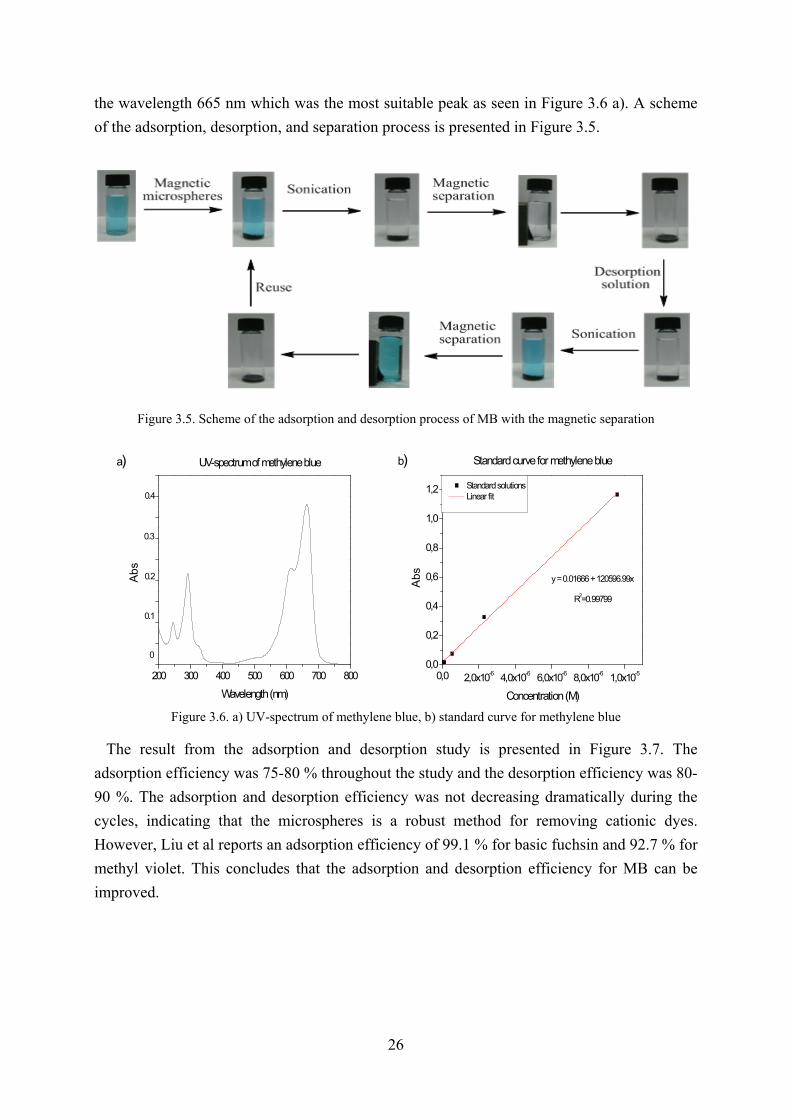
the wavelength 665 nm which was the most suitable peak as seen in Figure 3.6 a). A scheme
of the adsorption, desorption, and separation process is presented in Figure 3.5.
Figure 3.5. Scheme of the adsorption and desorption process of MB with the magnetic separation
200 300 400 500 600 700 800
a)
0.3
0.1
0
0.2
Abs
Wavelength (nm)
UV-spectrum of methylene blue
0.4
0,0 2,0x10⁻⁶ 4,0x10⁻⁶ 6,0x10⁻⁶ 8,0x10⁻⁶ 1,0x10-50,0
0,2
0,4
0,6
0,8
1,0
1,2
b)
Standard curve for methylene blue
Abs
Concentration (M)
Standard solutions Linear fit
R2=0.99799
y = 0.01666 + 120596.99x
Figure 3.6. a) UV-spectrum of methylene blue, b) standard curve for methylene blue
The result from the adsorption and desorption study is presented in Figure 3.7. The
adsorption efficiency was 75-80 % throughout the study and the desorption efficiency was 80-
90 %. The adsorption and desorption efficiency was not decreasing dramatically during the
cycles, indicating that the microspheres is a robust method for removing cationic dyes.
However, Liu et al reports an adsorption efficiency of 99.1 % for basic fuchsin and 92.7 % for
methyl violet. This concludes that the adsorption and desorption efficiency for MB can be
improved.

27
1 2 3 4 5 6 7 8 9 100
20
40
60
80
100
a)
Ad
sorp
tion
effi
cie
ncy
(%)
Adsorption cycle
Adsorption of methylene blue over several cycles
1 2 3 4 5 6 7 8 9 100
20
40
60
80
100
b) Desorption of methylene blue over several cycles
Des
orp
tion
eff
icie
ncy
(%)
Desorption cycle
Figure 3.7. Adsorption a) and desorption b) efficiencies of methylene blue [1×10⁻⁶ M] over several cycles. Sonication time 2 min at 80 %, 22-25 °C. MeOH/H2O 40/60 saturated KCl solution was used as desorbing solution. 3.2.4 ADSORPTION AND DESORPTION MECHANISM
The proposed adsorption and desorption mechanisms are presented in Figure 9 and 10. The
affinity of MB to –SO3- is higher than for Na+. The MB has a Cl- group which interacts with
the Na+ and forms Na+-Cl, i.e. an ion exchange is achieved. In the case of the desorption, an
ion exchange is also performed. However, using the co-solvent EtOH/H2O/KCl the process
can be speed up. The EtOH/H2O/KCl interfere the electrostatic interaction between MB and
-SO3-, thus the counterions are changed and the dye is desorbed from the microspheres.
Figure 3.8. Proposed mechanism for the adsorption of MB onto magnetic microspheres
Figure 3.9. Proposed mechanism for the desorption of MB from magnetic microspheres

28
3.2.5 ADSORPTION OPTIMISATION AND DESORPTION OPTIMISATION
The result from the adsorption optimisation is presented in Figure 3.10. The adsorption
efficiency is most likely pH-dependent. This is attributed to the change of charge and
ionisation that appear with pH change. The pH was changed in the range of pH 6-12. The
result from the study is shown in Figure 3.10 a). The result shows that there is no significant
change in adsorption efficiency between pH 6-12. However, with increasing pH a slight
faster adsorption rate was detected. The results show that the magnetic microspheres can be
used in a broad pH range which is important for industrial use where the pH might fluctuate
with time. Unfortunately, in acid environment, pH 1~3, the Fe3O4 particles start to dissolve.
Therefore should acid environment be avoided. However, after short exposure of strong acid
the microspheres retain the magnetic properties. It was found adsorption time is what affects
the adsorption efficiency the most. After 5 minutes of adsorption nearly 100 % of the MB was
adsorbed.
6 7 8 9 10 11 1290
92
94
96
98
100
A07
A06
A05A04
A03
A02
A01
a)
Ad
sorp
tion
effi
cie
ncy
(%)
pH
Adsorption at different pH
1 2 3 4 570
80
90
100b)
A15A14
A13
A12
A11
Adsorption time
Ad
sorp
tion
effi
cie
ncy
(%)
Adsorption time (min)
Figure 3.10. Result from the adsorption optimisation of pH a) and sonication time b)
The results from the desorption optimisation is presented in Figure 3.11 a)-g). In the
optimisation of the desorption, several types of salts, salt concentrations, and EtOH/H2O-
ratios were examined. As seen in Figure 3.11 a) the EtOH/H2O 10/0 had the highest
desorption efficiency. However, the efficiency was only about 40 % so further optimisation
was required. The dye is adsorbed ion exchange between the dye and the -SO3- and there are
two major ways of breaking this interaction. One is through polarity differences of the co-
solvent and the other one is ion exchange. Therefore was salts introduced to the desorption
solution. However, most salts are insoluble in EtOH so it was necessary to use an EtOH/H2O
ratio with H2O ≠ 0 to dissolve the salts.
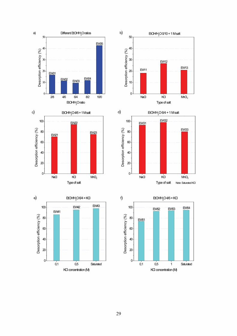
29
2/8 4/6 6/4 8/2 10/00
10
20
30
40
50
EW05
EW04EW03
EW01
Des
orp
tion
eff
icie
ncy
(%)
EtOH/H2O ratio
Different EtOH/H2O ratios
EW02
a)
NaCl KCl MnCl0
10
20
30
40
50
EW13
EW12
EW11
2
EtOH/H2O 0/10 + 1 M salt
Des
orp
tion
eff
icie
ncy
(%)
Type of salt
b)
NaCl KCl MnCl0
20
40
60
80
100
c)
EW23
EW22
EW21
2
EtOH/H2O 4/6 + 1 M salt
De
sorp
tion
eff
icie
ncy
(%
)
Type of salt
NaCl KCl MnCl0
20
40
60
80
100
d)
EW33
EW32EW31
De
sorp
tion
eff
icie
ncy
(%
)
Type of salt
EtOH/H2O 6/4 + 1 M salt
Note: Saturated KCl
2
0,1 0,5 Saturated0
20
40
60
80
100
e)
EW43EW42
EW41
EtOH/H2O 6/4 + KCl
Des
orpt
ion
effic
ienc
y (%
)
KCl concentration (M)
0,1 0,5 1 Saturated0
20
40
60
80
100
f)
EW54EW53EW52
EW51
EtOH/H2O 4/6 + KCl
Des
orpt
ion
effic
ienc
y (%
)
KCl concentration (M)
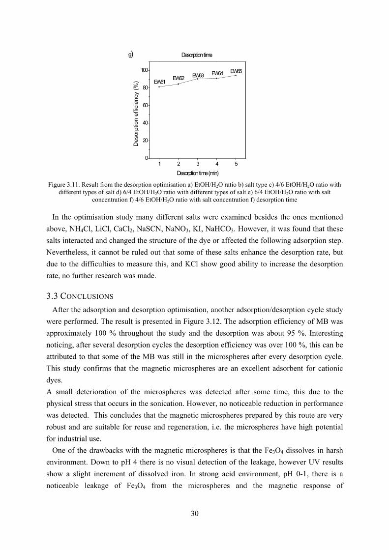
30
1 2 3 4 50
20
40
60
80
100
g)
EW65EW64EW63EW62EW61
Desorption time
De
sorp
tion
eff
icie
ncy
(%
)
Desorption time (min)
Figure 3.11. Result from the desorption optimisation a) EtOH/H2O ratio b) salt type c) 4/6 EtOH/H2O ratio with different types of salt d) 6/4 EtOH/H2O ratio with different types of salt e) 6/4 EtOH/H2O ratio with salt
concentration f) 4/6 EtOH/H2O ratio with salt concentration f) desorption time In the optimisation study many different salts were examined besides the ones mentioned
above, NH4Cl, LiCl, CaCl2, NaSCN, NaNO3, KI, NaHCO3. However, it was found that these
salts interacted and changed the structure of the dye or affected the following adsorption step.
Nevertheless, it cannot be ruled out that some of these salts enhance the desorption rate, but
due to the difficulties to measure this, and KCl show good ability to increase the desorption
rate, no further research was made.
3.3 CONCLUSIONS
After the adsorption and desorption optimisation, another adsorption/desorption cycle study
were performed. The result is presented in Figure 3.12. The adsorption efficiency of MB was
approximately 100 % throughout the study and the desorption was about 95 %. Interesting
noticing, after several desorption cycles the desorption efficiency was over 100 %, this can be
attributed to that some of the MB was still in the microspheres after every desorption cycle.
This study confirms that the magnetic microspheres are an excellent adsorbent for cationic
dyes.
A small deterioration of the microspheres was detected after some time, this due to the
physical stress that occurs in the sonication. However, no noticeable reduction in performance
was detected. This concludes that the magnetic microspheres prepared by this route are very
robust and are suitable for reuse and regeneration, i.e. the microspheres have high potential
for industrial use.
One of the drawbacks with the magnetic microspheres is that the Fe3O4 dissolves in harsh
environment. Down to pH 4 there is no visual detection of the leakage, however UV results
show a slight increment of dissolved iron. In strong acid environment, pH 0-1, there is a
noticeable leakage of Fe3O4 from the microspheres and the magnetic response of
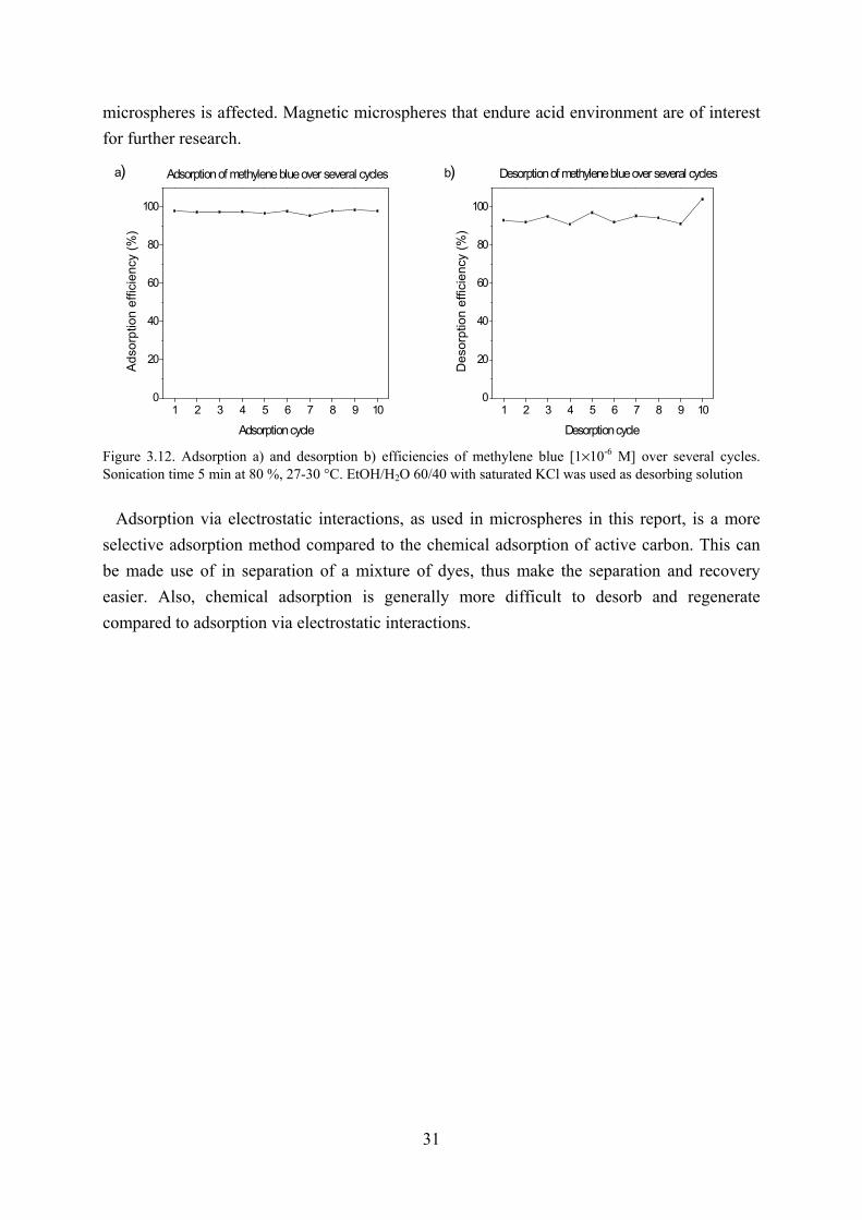
31
microspheres is affected. Magnetic microspheres that endure acid environment are of interest
for further research.
1 2 3 4 5 6 7 8 9 100
20
40
60
80
100
a) Adsorption of methylene blue over several cycles
Ad
sorp
tion
effi
cie
ncy
(%
)
Adsorption cycle
1 2 3 4 5 6 7 8 9 100
20
40
60
80
100
b) Desorption of methylene blue over several cycles
De
sorp
tion
effi
cie
ncy
(%
)
Desorption cycle
Figure 3.12. Adsorption a) and desorption b) efficiencies of methylene blue [1×10⁻⁶ M] over several cycles. Sonication time 5 min at 80 %, 27-30 °C. EtOH/H2O 60/40 with saturated KCl was used as desorbing solution
Adsorption via electrostatic interactions, as used in microspheres in this report, is a more
selective adsorption method compared to the chemical adsorption of active carbon. This can
be made use of in separation of a mixture of dyes, thus make the separation and recovery
easier. Also, chemical adsorption is generally more difficult to desorb and regenerate
compared to adsorption via electrostatic interactions.

32
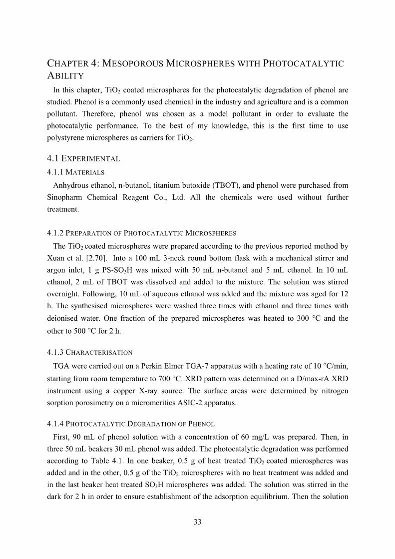
33
CHAPTER 4: MESOPOROUS MICROSPHERES WITH PHOTOCATALYTIC
ABILITY In this chapter, TiO2 coated microspheres for the photocatalytic degradation of phenol are
studied. Phenol is a commonly used chemical in the industry and agriculture and is a common
pollutant. Therefore, phenol was chosen as a model pollutant in order to evaluate the
photocatalytic performance. To the best of my knowledge, this is the first time to use
polystyrene microspheres as carriers for TiO2.
4.1 EXPERIMENTAL
4.1.1 MATERIALS
Anhydrous ethanol, n-butanol, titanium butoxide (TBOT), and phenol were purchased from
Sinopharm Chemical Reagent Co., Ltd. All the chemicals were used without further
treatment.
4.1.2 PREPARATION OF PHOTOCATALYTIC MICROSPHERES
The TiO2 coated microspheres were prepared according to the previous reported method by
Xuan et al. [2.70]. Into a 100 mL 3-neck round bottom flask with a mechanical stirrer and
argon inlet, 1 g PS-SO3H was mixed with 50 mL n-butanol and 5 mL ethanol. In 10 mL
ethanol, 2 mL of TBOT was dissolved and added to the mixture. The solution was stirred
overnight. Following, 10 mL of aqueous ethanol was added and the mixture was aged for 12
h. The synthesised microspheres were washed three times with ethanol and three times with
deionised water. One fraction of the prepared microspheres was heated to 300 °C and the
other to 500 °C for 2 h.
4.1.3 CHARACTERISATION
TGA were carried out on a Perkin Elmer TGA-7 apparatus with a heating rate of 10 °C/min,
starting from room temperature to 700 °C. XRD pattern was determined on a D/max-rA XRD
instrument using a copper X-ray source. The surface areas were determined by nitrogen
sorption porosimetry on a micromeritics ASIC-2 apparatus.
4.1.4 PHOTOCATALYTIC DEGRADATION OF PHENOL
First, 90 mL of phenol solution with a concentration of 60 mg/L was prepared. Then, in
three 50 mL beakers 30 mL phenol was added. The photocatalytic degradation was performed
according to Table 4.1. In one beaker, 0.5 g of heat treated TiO2 coated microspheres was
added and in the other, 0.5 g of the TiO2 microspheres with no heat treatment was added and
in the last beaker heat treated SO3H microspheres was added. The solution was stirred in the
dark for 2 h in order to ensure establishment of the adsorption equilibrium. Then the solution
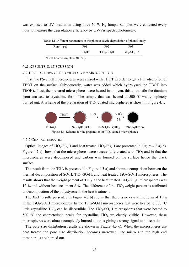
34
was exposed to UV irradiation using three 50 W Hg lamps. Samples were collected every
hour to measure the degradation efficiency by UV/Vis spectrophotometry.
Table 4.16 Different parameters in the photocatalytic degradation of phenol study
Run (type)
P01
SO3Ha
P02
TiO2-SO3H
P03
TiO2-SO3Ha
a Heat treated samples (300 °C)
4.2 RESULTS & DISCUSSION
4.2.1 PREPARATION OF PHOTOCATALYTIC MICROSPHERES
First, the PS-SO3H microspheres were stirred with TBOT in order to get a full adsorption of
TBOT on the surface. Subsequently, water was added which hydrolysed the TBOT into
Ti(OH)x. Last, the prepared microspheres were heated in an oven, this to transfer the titanium
from anastase to crystalline form. The sample that was heated to 500 °C was completely
burned out. A scheme of the preparation of TiO2-coated microspheres is shown in Figure 4.1.
500 C
2 h
TBOT H2O
PS-SO3H/TBOT PS-SO3H/Ti(OH)x PS-SO3H/TiO2PS-SO3H Figure 4.1. Scheme for the preparation of TiO2 coated microspheres
4.2.2 CHARACTERISATION
Optical images of TiO2-SO3H and heat treated TiO2-SO3H are presented in Figure 4.2 a)-b).
Figure 4.2 a) shows that the microspheres were successfully coated with TiO2 and b) that the
microspheres were decomposed and carbon was formed on the surface hence the black
surface.
The result from the TGA is presented in Figure 4.3 a) and shows a comparison between the
thermal decomposition of SO3H, TiO2-SO3H, and heat treated TiO2-SO3H microspheres. The
results shows that the weight percent of TiO2 in the heat treated TiO2-SO3H microspheres was
12 % and without heat treatment 8 %. The difference of the TiO2 weight percent is attributed
to decomposition of the polystyrene in the heat treatment.
The XRD results presented in Figure 4.3 b) shows that there is no crystalline form of TiO2
in the TiO2-SO3H microspheres. In the TiO2-SO3H microspheres that were heated to 300 °C
little crystalline TiO2 can be discernible. The TiO2-SO3H microspheres that were heated to
500 °C the characteristic peaks for crystalline TiO2 are clearly visible. However, these
microspheres were almost completely burned out thus giving a strong signal to noise ratio.
The pore size distribution results are shown in Figure 4.3 c). When the microspheres are
heat treated the pore size distribution becomes narrower. The micro and the high end
mesoporous are burned out.

35
Figure 4.2. Optical microscope images of a) TiO2-SO3H b) heat treated TiO2-SO3H
0 100 200 300 400 500 600 7000
10
20
30
40
50
60
70
80
90
100TGA
We
igh
t %
Temperature °C
PS-SO3H
TiO2-SO
3H
ΔTiO2-SO
3H
a)
10 20 30 40 50 60 70 80
TiO2-SO
3H
Δ300 °C TiO2-SO
3H
Δ500 °C TiO2-SO
3H
XRD
Re
lativ
e in
ten
sity
(a
.u.)
2θ degree
b)
1 10 100 1000
0,0
0,2
0,4
0,6
0,8
1,0
1,2
1,4
macromesomicro
c) Pore size distribution
PS-SO3H
ΔTiO2-SO
3H
TiO2-SO
3H
Lo
g d
iffe
ren
tal v
olu
me
(cm
3 /g)
Pore diameter (nm)
Figure 4.3 a) TGA curves for SO3H, TiO2-SO3H, and heat treated TiO2-SO3H b) XRD results for TiO2-SO3H, heat treated 300°C TiO2-SO3H, and heat treated 500°C TiO2-SO3H microspheres
4.2.3 PHOTOCATALYTIC DEGRADATION OF PHENOL The photocatalytic degradation of phenol processes is explained by Figure 4.4 and 4.5. In
Figure 4.4 a simplified scheme for the formation of the reactive species that reacts with the
phenol is presented. In Figure 4.5 a possible and simplified mechanism for the photocatalytic
degradation of phenol is presented. The complete and exact pathway for the degradation is

36
very complicated and has many steps and intermediates. However, the end products are water
and carbon dioxide.
TiO2
h+
e-
hv
O2
H2O
Mesoporousmicrosphere
OH
O2-
H+
O2- HO2
H2O2
OH-
OH
H+
H+
Figure 4.4. Simplified scheme for the photocatalytic induced formation of reactive species [2.76]
OH
OH
OH
OH
OH
OH
OH
OH
OH
OH
OHOpen ringfragments
CO2H2O
OHOH-
Figure 4.5. Simplified mechanism of the photocatalytic degradation of phenol [4.16]
A scheme for the setup used for the photocatalytic degradation of phenol is presented in
Figure 4.6. The UV-lamps produce heat so the beaker was cooled down in a water bath to
ensure constant temperature in the study.
hv
Figure 4.6. Scheme of the setup used for the degradation of phenol using TiO2 coated microspheres
The results from the study of the photocatalytic degradation of phenol are presented in
Figure 4.7 a)-c). As shown in Figure 4.7 a) it is obvious that the microspheres adsorb the
phenol. This phenomenon could be used for other purposes. Unfortunately, the photocatalytic
degradation of phenol using the TiO2 coated microspheres did not work. In Figure 4.7 b) there
is a noticeable increase of absorbance in time. This was attributed to that TiO2 was leaking

37
from the microspheres, which concludes that the interaction between the microspheres and
TiO2 were not strong. However, the heat treated microspheres showed no leakage of TiO2
from the microspheres.
200 250 3000,0
0,5
1,0
1,5
2,0
2,5
3,0
Ab
s
Wavelength (nm)
original after adsorption 1h 2h 3h
a) ΔSO3H
200 250 3000,0
0,5
1,0
1,5
2,0
2,5
3,0
Ab
s
Wavelength (nm)
original after adsorption 1h 2h 3h
b) SO3H-TiO
2
200 250 3000,0
0,5
1,0
1,5
2,0
2,5
3,0ΔTiO
2-SO
3H
Ab
s
Wavelength
original after adsorption 2h 4h 6h 8h
c)
Figure 4.7. Results from the photocatalytic degradation of phenol by a) heat treated sulfonated microspheres, b) TiO2 coated microspheres c) heat treated TiO2coated microspheres
4.3 CONCLUSIONS
Several different methods were examined for the preparation of photocatalytic magnetic
microspheres. The methods were the following: sol-gel technique, physical deposition of TiO2
by heat treatment, and the method described above. The methods were tried under different
conditions such as temperature and molar ratio. However, neither of them worked. The
magnetic microspheres could not be coated with TiO2, however the sulfonated microspheres
were successfully coated with TiO2. The Fe3O4-particles should not interfere with the TiO2-
coating. According to the literature, Fe3O4-particles can be coated with TiO2. The magnetic
microspheres have R-SO3Na groups on the surface and the sulfonated microspheres have R-
SO3H groups. This is probably the main reason why the magnetic microspheres could not be
38
coated with TiO2. Several attempts of replacing the Na with H were executed. However, only
strong acids at low pH (pH 0-2) were able to change the Na to H, at pH 3-4 there was no
change. Unfortunately, the Fe3O4 dissolves in strong acid environment and the magnetic
microspheres lose their magnetic ability. However, after treatment with acid the microspheres
could be coated with TiO2. This concludes that if the Fe3O4-particles can be protected from
the acid, TiO2 coated magnetic microspheres could be synthesised. There are several possible
ways of protecting the Fe3O4, for example to coat the particles with oleic acid or SiO2.
Another possibility is to synthesise magnetic microspheres using another method that the
Fe3O4-particles are on the inside of the microspheres and not on the surface as with the
method used in this thesis.
There are several possible reasons why the photocatalytic degradation of phenol did not
work. The microspheres were too large to disperse in the phenol solution and because of that
there was too little contact between the TiO2 and phenol. This could be solved by using nano-
sized microspheres which easily disperse in solution and have much greater surface area. The
setup used for the photocatalytic degradation was very simple. If a photo reactor would have
been used, the results would perhaps been successful. Also, using a glass beaker was not
optimal, a beaker made of quarts would have been better because then the solution could have
been exposed to more UV-light thus having higher efficiency.
The heat treated TiO2 microspheres were also used for photocatalytic degradation of the
anionic dye creosol red. The dye was pH and temperature sensitive and it was difficult to
stabilise these parameters so the dye was changes to phenol. However, the microspheres
showed quite good ability to adsorb this anionic dye and it could be desorbed using
EtOH/H2O 6/4 with saturated KCl. One interesting idea is to heat treat the magnetic
microspheres under N2 atmosphere thus making magnetic microspheres with active carbon
surface.

39
CHAPTER 5: MESOPOROUS MICROSPHERES FOR BIOSEPARATION Recently, all the genes in the human DNA was determined and identified in the HUGO-
project. The HUGO-project was completed ahead of the time schedule. This was a result of
the technological advances which took place during this time [5.1]. At present, the human
proteomes are being investigated and catalogued [5.2]. Again, the technological advances
probably make this work go faster and easier than planned. Quick, selective, and effective
separation techniques in the biotechnology field are of high importance in order to study and
fully understand the proteomes and the different processes in which the proteomes take part.
Mass spectrometry is the most used instrument for the characterisation of proteins.
However, many samples cannot be directly analysed and there is a need for purification and
preconcentration. For example in the enzymatic digestion of proteins were the digested
intermediates and product are in small quantities there is a need for selective adsorption in
order to investigate the process [5.3].
One promising separation technique is to use magnetic adsorbents in the separation of
proteomes [5.3-5.5].
Lysozyme (Figure 5.1) is used in this as a model protein for the separation of proteins using
magnetic microspheres.
Figure 5.1. Three dimensional structure of lysozyme [5.6]
5.1 EXPERIMENTAL
5.1.1 MATERIAL
Lysozyme was purchased from Sigma-Aldrich Co. H3BO3, NaOH, KCl, ethanol, methanol,
citric acid, were purchased from Sinopharm Chemical Reagent Co., Ltd. All chemicals were
used as received.
5.1.2 SEPARATION OF LYSOZYME
A 50 mL lysozyme solution (0.7 g/L) was prepared in a pH 9.6 buffer, which is near the pI
for lysozyme. Into a vial, 0.2 g microspheres and 4 mL of lysozyme solution were added. The
mixture was shaken for 30 min and then separated. The lysozyme was extracted from the
microspheres by adding a pH 13 buffer and again shaken for 30 min. The adsorption and
extraction efficiencies were determined using a UNICO 3802 UV/Vis spectrophotometer.
The adsorption and desorption process was optimised according to Table 5.1 and 5.2.
Parameters such as pH and amount of adsorbent were evaluated.
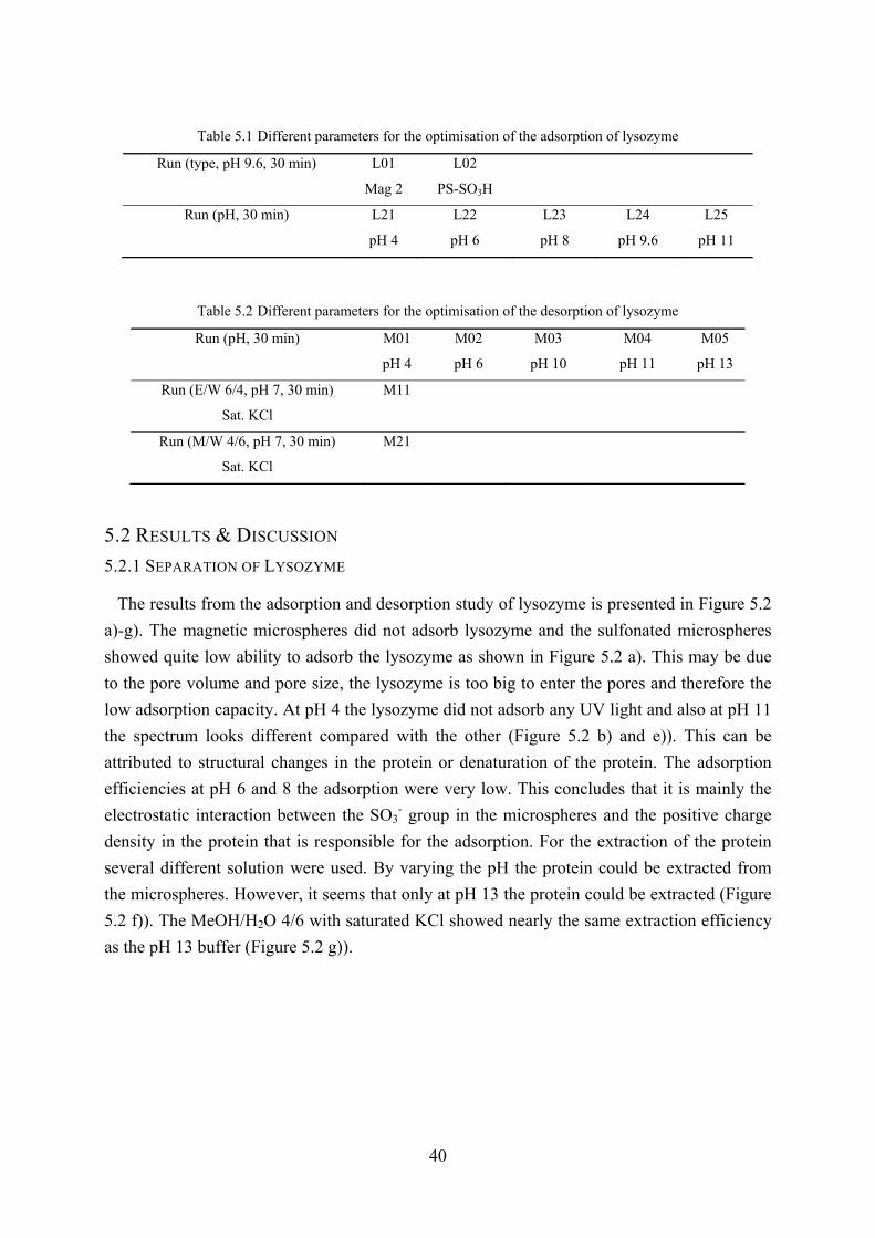
40
Table 5.17 Different parameters for the optimisation of the adsorption of lysozyme
Run (type, pH 9.6, 30 min)
L01
Mag 2
L02
PS-SO3H
Run (pH, 30 min)
L21
pH 4
L22
pH 6
L23
pH 8
L24
pH 9.6
L25
pH 11
Table 5.28 Different parameters for the optimisation of the desorption of lysozyme
Run (pH, 30 min)
M01
pH 4
M02
pH 6
M03
pH 10
M04
pH 11
M05
pH 13
Run (E/W 6/4, pH 7, 30 min)
Sat. KCl
M11
Run (M/W 4/6, pH 7, 30 min)
Sat. KCl
M21
5.2 RESULTS & DISCUSSION
5.2.1 SEPARATION OF LYSOZYME
The results from the adsorption and desorption study of lysozyme is presented in Figure 5.2
a)-g). The magnetic microspheres did not adsorb lysozyme and the sulfonated microspheres
showed quite low ability to adsorb the lysozyme as shown in Figure 5.2 a). This may be due
to the pore volume and pore size, the lysozyme is too big to enter the pores and therefore the
low adsorption capacity. At pH 4 the lysozyme did not adsorb any UV light and also at pH 11
the spectrum looks different compared with the other (Figure 5.2 b) and e)). This can be
attributed to structural changes in the protein or denaturation of the protein. The adsorption
efficiencies at pH 6 and 8 the adsorption were very low. This concludes that it is mainly the
electrostatic interaction between the SO3- group in the microspheres and the positive charge
density in the protein that is responsible for the adsorption. For the extraction of the protein
several different solution were used. By varying the pH the protein could be extracted from
the microspheres. However, it seems that only at pH 13 the protein could be extracted (Figure
5.2 f)). The MeOH/H2O 4/6 with saturated KCl showed nearly the same extraction efficiency
as the pH 13 buffer (Figure 5.2 g)).

41
200 250 3000,0
0,5
1,0
1,5
2,0
2,5
3,0
3,5
Ab
s
Wavelength (nm)
Original Magnetic microspheres Sulfonated microspheres
a) Adsorption of lysozyme
200 250 3000,0
0,5
1,0
1,5
2,0
2,5
3,0
3,5
Abs
Wavelength (nm)
original
pH 4b)
200 250 3000,0
0,5
1,0
1,5
2,0
2,5
Ab
s
Wavelength (nm)
original adsorption
Adsorption at pH 6c)
200 250 3000,0
0,5
1,0
1,5
2,0
2,5
3,0
3,5
Ab
s
Wavelength (nm)
original adsorption
Adsorption at pH 8d)
200 250 3000,0
0,5
1,0
1,5
2,0
2,5
3,0
3,5
Ab
s
Wavelength (nm)
original adsorption
Adsorption at pH 11e)
200 250 3000,0
0,5
1,0
1,5
2,0
2,5
3,0
3,5Lysozyme desorption at different pH
Abs
Wavelength (nm)
original adsorption ph4 ph6 ph10 ph11 ph13
f)
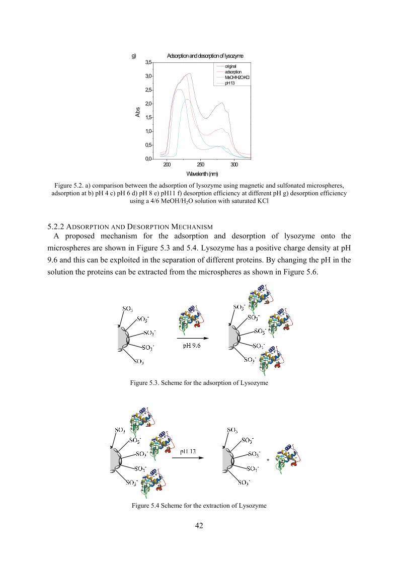
42
200 250 3000,0
0,5
1,0
1,5
2,0
2,5
3,0
3,5
Ab
s
Wavelenth (nm)
original adsorption MeOH/H2O KCl pH 13
Adsorption and desorption of lysozymeg)
Figure 5.2. a) comparison between the adsorption of lysozyme using magnetic and sulfonated microspheres,
adsorption at b) pH 4 c) pH 6 d) pH 8 e) pH11 f) desorption efficiency at different pH g) desorption efficiency using a 4/6 MeOH/H2O solution with saturated KCl
5.2.2 ADSORPTION AND DESORPTION MECHANISM A proposed mechanism for the adsorption and desorption of lysozyme onto the
microspheres are shown in Figure 5.3 and 5.4. Lysozyme has a positive charge density at pH
9.6 and this can be exploited in the separation of different proteins. By changing the pH in the
solution the proteins can be extracted from the microspheres as shown in Figure 5.6.
Figure 5.3. Scheme for the adsorption of Lysozyme
Figure 5.4 Scheme for the extraction of Lysozyme
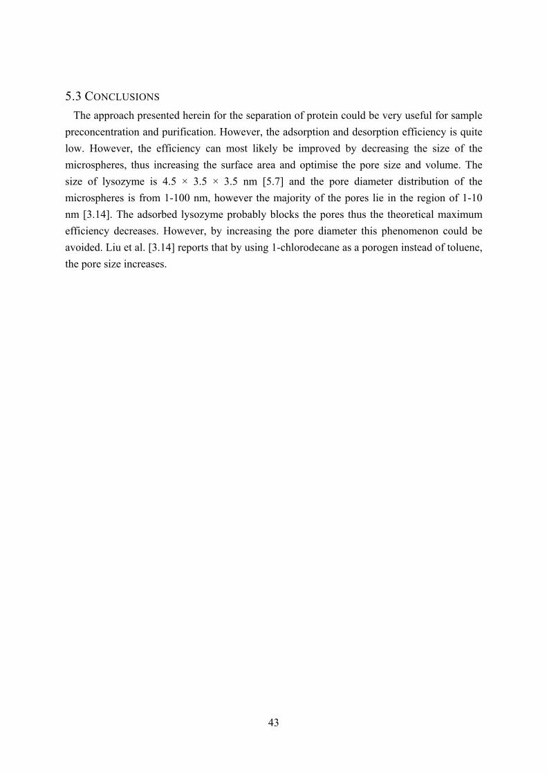
43
5.3 CONCLUSIONS
The approach presented herein for the separation of protein could be very useful for sample
preconcentration and purification. However, the adsorption and desorption efficiency is quite
low. However, the efficiency can most likely be improved by decreasing the size of the
microspheres, thus increasing the surface area and optimise the pore size and volume. The
size of lysozyme is 4.5 × 3.5 × 3.5 nm [5.7] and the pore diameter distribution of the
microspheres is from 1-100 nm, however the majority of the pores lie in the region of 1-10
nm [3.14]. The adsorbed lysozyme probably blocks the pores thus the theoretical maximum
efficiency decreases. However, by increasing the pore diameter this phenomenon could be
avoided. Liu et al. [3.14] reports that by using 1-chlorodecane as a porogen instead of toluene,
the pore size increases.

44
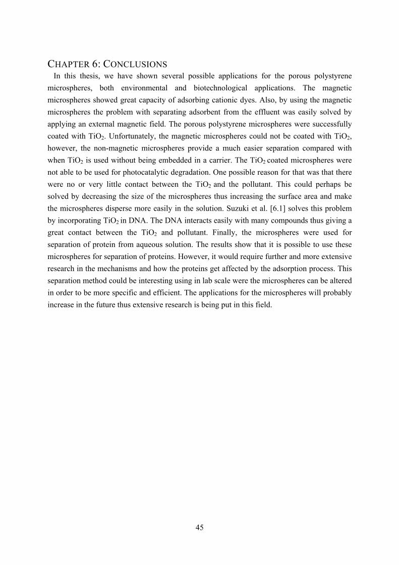
45
CHAPTER 6: CONCLUSIONS
In this thesis, we have shown several possible applications for the porous polystyrene
microspheres, both environmental and biotechnological applications. The magnetic
microspheres showed great capacity of adsorbing cationic dyes. Also, by using the magnetic
microspheres the problem with separating adsorbent from the effluent was easily solved by
applying an external magnetic field. The porous polystyrene microspheres were successfully
coated with TiO2. Unfortunately, the magnetic microspheres could not be coated with TiO2,
however, the non-magnetic microspheres provide a much easier separation compared with
when TiO2 is used without being embedded in a carrier. The TiO2 coated microspheres were
not able to be used for photocatalytic degradation. One possible reason for that was that there
were no or very little contact between the TiO2 and the pollutant. This could perhaps be
solved by decreasing the size of the microspheres thus increasing the surface area and make
the microspheres disperse more easily in the solution. Suzuki et al. [6.1] solves this problem
by incorporating TiO2 in DNA. The DNA interacts easily with many compounds thus giving a
great contact between the TiO2 and pollutant. Finally, the microspheres were used for
separation of protein from aqueous solution. The results show that it is possible to use these
microspheres for separation of proteins. However, it would require further and more extensive
research in the mechanisms and how the proteins get affected by the adsorption process. This
separation method could be interesting using in lab scale were the microspheres can be altered
in order to be more specific and efficient. The applications for the microspheres will probably
increase in the future thus extensive research is being put in this field.

46
47
REFERENCES
CHAPTER 1 [1.1] Lim B.R., Hu H.Y., Fujie K., Biological degradation and chemical oxidation characteristics of coke-oven
wastewater, Water Air Soil Pollut., 2003, 146, 23-33
[1.2] Arvin E., la Cour Jansen J., Henze M., Harremoes P., 2002, Third edition, Wastewater treatment biological
chemical process, ISBN: 3540422285, Berlin publishers New York
[1.3] Choi J.W., Yang K.S., Kim D.J., Lee C.E., Adsorption of zinc and toluene by alginate complex
impregnated with zeolite and activated carbon, Curr. Appl. Phys., 2009, 9, 694-697
[1.4] Wang K.Y., Yang Q., Chung T.S., Rajagopalan R., Enhanced forward osmosis from chemically modified
polybenzimidazole (PBI) nanofiltration hollow fiber membranes with a thin wall, Chem. Eng. Sci., 2009, 64,
1577-1584
[1.5] Jeona C.S., Baeka K., Parkb J.K., Ohc Y.K., Leec S.D., Adsorption characteristics of As(V) on iron-coated
zeolite, J. Hazard. Mater., 2009, 163, 804-808
[1.6] Solpan D., Kölge Z., Adsorption of methyl violet in aqueous solutions by poly(N-vinylpyrrolidone-co-
methacrylic acid) hydrogels, Radiat. Phys. Chem., 2006, 75, 120-128
[1.7] Mittal A., Gajbe V., Mittal J., Removal and recovery of hazardous triphenylmethane dye, methyl violet
through adsorption over granulated waste materials, J. Hazard. Mater., 2008, 150, 364-375
[1.8] Jeon D.J., Yeom S.H., Recycling wasted biomaterial, crab shells, as an adsorbent for the removal of high
concentration of phosphate, Bioresour. Technol., 2009, 100, 2646-2649
[1.9] Liua S.S., Leea C.K., Chena H.C., Wanga C.C., Juanga L.C., Application of titanate nanotubes for Cu(II)
ions adsorptive removal from aqueous solution, Chem. Eng. J., 2009, 147, 188-193
[1.10] Karaca S., Gurses A., Acıkyıldız M., Korucu M.E., Adsorption of cationic dye from aqueous solutions by
activated carbon, Microporous Mesoporous Mater., 2008, 115, 376-382
[1.11] Hamdaoui O., Batch study of liquid-phase adsorption of methylene blue using cedar sawdust and crushed
brick, J. Hazard. Mater., 2006, 135, 264-273
[1.12] Bansal R.C., Goyal M., 2005, First edition, Activated carbon adsorption, page 1, ISBN: 9780824753443,
CRC Press, U.K.
[1.13] Yang N., Zhu S.M., Zhang D., Xu S., Synthesis and properties of magnetic Fe3O4-activated carbon
nanocomposite particles for dye removal, Mater. Lett., 2008, 62, 645-647
[1.14] Bhatkhande D.S., Pangarkar V.G., Beenackers A.A.C.M., Photocatalytic degradation for environmental
applications - a review, J. Chem. Technol. Biotechnol., 2001, 77, 102-116
48
[1.15] Mills A., Punte L., Stephan M., An overview of semiconductor photocatalysis. J. Photochem. Photobiol.
A, 1997, 108, 1-35
[1.16] Mills A., Davies R. H., Worsley D., Water purification by semiconductor photocatalysis, Chem. Soc.
Revs., 1993, 22, 417-425
[1.17] Luo M., Bowden D., Brimblecombe P., Preparation of black sand-based magnetic photocatalysts for
photocatalytic oxidation of aqueous phenol, Appl. Catal., B, 2009, 87, 1-8
[1.18] Keller K., Friedmann T., Boxman A., The bioseparation needs for tomorrow, Trends Biotechnol., 2001,
19, 438-441
[1.19] Li Y., Xu X., Qi D., Deng C., Yang P., Zhang X., Novel Fe3O4@TiO2 core-shell microspheres for
selective enrichment of phosphopeptides in phosphoproteome analysis, J. Proteome Res., 2008, 7, 2526-2538
[1.20] Unsal E., Elmas B., Camili G.M., Tuncel M., Senel S., Tuncel A., Monodisperse-porous poly(styrene-co-
divinylbenzene) beads providing high column efficiency in reversed phase HPLC, J. Appl. Polym. Sci., 2005,
95, 1430-1438
[1.21] Coutinho F.M.B., Alves G.M., Neves A.F.S., Dias M.L., Chagas B.S., Modified suspension
polymerization of styrene-divinylbenzene for application in size exclusion chromatography, Macromol. Symp.,
2000, 151, 399-405
[1.22] Huang F.C., Ke C.H., Kao C.Y., Lee W.C., J. Appl. Polym. Sci., Preparation and application of partially
porous poly(styrene-divinylbenzene) particles for lipase immobilization, 2001, 80, 39-46
[1.23] Struck M., Widdecke H., Surface functionalization of polymer networks. Sulfonation of the internal
surface of macroporous styrene-divinylbenzene copolymers, Angew. Makromol. Chem., 1996, 235, 131-148
[1.24] Rhodes C.N., Brown D.R., Plant S., Dale J.A., Sulphonated polystyrene resins: acidities and catalytic
activities, React. Funct. Polym., 1999, 40, 187-193
CHAPTER 2 [2.1] Reiss G., Hütten A., Magnetic nanoparticles: applications beyond data storage, Nat. Mater., 2005, 4, 725-
726
[2.2] Rudge S.R., Kurtz T.L., Vessely C.R., Catterall L.G., Williamson D.L., Preparation, characterization, and
performance of magnetic iron-carbon composite microparticles for chemotherapy, Biomaterials, 2000, 21, 1411-
1420
[2.3] Giri S., Trewyn B.G., Stellmaker M.P., Lin V.S.Y., Stimuli-responsive controlled-release delivery system
based on mesoporous silica nanorods capped with magnetic nanoparticles, Angew. Chem. Int. Ed., 2005, 44,
5038-5044
49
[2.4] Nuñez L., Kaminski M.D., Transuranic separation using organophosphorus extractants adsorbed onto
superparamagnetic carriers, J. Magn. Magn. Mater., 1999, 194, 102-107
[2.5] Mornet S., Grasset F., Vasseur S., Veverka P., Goglio G., Demourgues A., Portier J., Pollert E., Duguet E.,
Magnetic nanoparticle design for medical applications, Prog. Solid State Chem., 2006, 34, 237-247
[2.6] Liu Q., Wang L., Xiao A., Yu H., Huo J., State key laboratory of chemical engineering, Collage of
materials science and chemical engineering, Zhejiang University, Hangzhou 310027, China, Templated
preparation of mesoporous magnetic microspheres for removal of cationic dyes from water, submitted to journal
[2.7] Yantasee W., Warner C.L., Sangvanicht T., Addleman R.S., Carter G., Wiacek R.J., Fryxell G.E.,
Timchalk C., Warner M.G., Removal of heavy metals from aqueous systems with thiol functionalized
superparamagnetic nanoparticles, Environ. Sci. Technol., 2007, 41, 5114-5119
[2.8] Mak S.Y., Chen D.H., Fast adsorption of methylene blue on polyacrylic acid-bound iron oxide magnetic
nanoparticles, Dyes Pigm., 2004, 61, 93-98
[2.9] Chang S.C., Anderson T.I., Bahrman S.E., Gruden C.L., Khijniak A.I., Adriaens P.,
Comparing recovering efficiency of immunomagnetic separation and centrifugation of mycobacteria in
metalworking fluids, J. Ind. Microbiol. Biotechnol., 2005, 32, 629-638
[2.10] Yavuz C.T., Mayo J.T., Yu W.W., Prakash A., Falkner J.C., Yean S., Cong L., Shipley H.J., Kan A.,
Tomson M., Natelson D., Colvin V.L., Low-field magnetic separation of monodisperse Fe3O4 nanocrystals,
Science, 2006, 314, 964-967
[2.11] Safarik I., Nymburska K., Safarikova M., Adsorption of water-soluble organic dyes on magnetic charcoal,
J. Chem. Tech. Biotechnol., 1997, 69, 1-4
[2.12] Liao M. H., Chen D. H., Preparation and characterization of a novel magnetic nano-adsorbent, J. Mater.
Chem., 2002, 12, 3654-3659
[2.13] Dyal A., Loos K., Noto M., Chang S. W., Spagnoli C., Shafi K.V. P. M., Ulman A., Cowman M., Gross
R. A., Activity of candida rugosa lipase immobilized on y-Fe2O3 magnetic nanoparticles, J. Am. Chem. Soc.,
2003, 125, 1684-1685
[2.14] Dresge C.T., Leonowicz M.E., Roth W.J., Vartuli J.C., Beck J.S., Ordered mesoporous molecular sieves
synthesized by a liquid-crystal template mechanism, Nature, 1992, 359, 710-712
[2.15] Lin Y.S., Wu S.H., Hung Y., Chou Y.H., Chang C., Lin M.L., Tsai C.P., Mou C.Y., Multifunctional
composite nanoparticles: magnetic, luminescent, and mesoporous, Chem. Mater., 2006, 18, 5170-5172
[2.16] Giri S., Trewyn B.G., Stellmaker M.P., Lin V.S.Y., Stimuli-responsive controlled-release delivery system
based on mesoporous silica nanorods capped with magnetic nanoparticles, Angew. Chem. Int. Ed., 2005, 44,
5038-5044
50
[2.17] Kim J., Lee J.E., Lee J., Yu J.H., Kim B.C., An K., Hwang Y., Shin C.H., Park J.G., Kim J., Hyeon T.,
Magnetic fluorescent delivery vehicle using uniform mesoporous silica spheres embedded with monodisperse
magnetic and semiconductor nanocrystals, J. Am. Chem. Soc., 2006, 128, 688-689
[2.18] Wu P.G., Zhu J.H., Xu Z.H., Template-assisted synthesis of mesoporous magnetic nanocomposite
particles, Adv. Funct. Mater., 2004, 14, 345-351
[2.19] Dong J., Xu Z.H., Wang F., Engineering and characterization of mesoporous silica-coated magnetic
particles for mercury removal from industrial effluents, Appl. Surf. Sci., 2008, 254, 3522-3530
[2.20] Zubieta C., Sierra M.B., Morini M.A., Schulz P.C., Albertengo L., Rodríguez M.S., The adsorption of
dyes used in the textile industry on mesoporous materials, Colloid Polym. Sci., 2008, 286, 377-384
[2.21] Deng Y.H., Qi D.W., Deng C.H, Zhang X.M., Zhao D.Y., Superparamagnetic high-magnetization
microspheres with an Fe3O4@SiO2 core and perpendicularly aligned mesoporous SiO2 shell for removal of
microcystins, J. Am. Chem. Soc., 2008, 130, 28-29
[2.22] Lawton L.A., Cornish B.J.P.A., MacDonald A.W.R., Removal of cyanobacterial toxins (microcystins) and
cyanobacterial cells from drinking water using domestic water filters, Water Res., 1998, 32, 633-638
[2.23] Liao M.H., Chen D.H., Preparation and characterization of a novel magnetic nano-adsorbent, J. Mater.
Chem., 2002, 12, 3654-3659
[2.24] Mak S.Y., Chen D.H., Binding and sulfonation of poly(acrylic acid) on iron oxide nanoparticles: a novel,
magnetic, strong acid cation nano-adsorbent, Macromol. Rapid Commun., 2005, 26, 1567-1571
[2.25] Huang S.H., Chen D.H., Rapid removal of heavy metal cations and anions from aqueous solutions by an
amino-functionalized magnetic nano-adsorbent, J. Hazard. Mater., 2009, 163, 174-179
[2.26] Chen D.H., Huang S.H., Fast separation of bromelain by polyacrylic acid-bound iron oxide magnetic
nanoparticles, Process Biochem., 2004, 39, 2207-2211
[2.27] Chang Y.C., Chen D.H., Adsorption kinetics and thermodynamics of acid dyes on a carboxymethylated
chitosan-conjugated magnetic nano-adsorbent, Macromol. Biosci., 2005, 5, 254-261
[2.28] Liu J.F., Zhao Z.S., Jiang G.B., Coating Fe3O4 Magnetic nanoparticles with humic acid for high efficient
removal of heavy metals in water, Environ. Sci. Technol., 2008, 42, 6949-6954
[2.29] Giménez J., Martínez M., Pablo J. d., Rovira M., Duro L., Arsenic sorption onto natural hematite,
magnetite, and goethite, J. Hazard. Mater., 2007, 141, 575-580
[2.30] Sarir M.S., Durrani M.I., Effect of the source and rate of humic acid on phosphorus transformations, J.
Agric. Biol. Sci., 2006, 1, 29-31
[2.31] Juang R.S., Shao H.J., A simplified equilibrium model for sorption of heavy metal ions from aqueous
solutions on chitosan, Water Res., 2002, 36, 2999-3008

51
[2.32] Ng J.C.Y., Cheung W.H., McKay G., Equilibrium studies of the sorption of Cu(II) ions onto chitosan, J.
Colloid Interf. Sci., 2002, 255, 64-74
[2.33] Karthikeyan G., Anbalagan K., Andal N.M., Adsorption dynamics and equilibrium studies of Zn(II) onto
chitosan, J. Chem. Sci., 2004, 116, 119-127
[2.34] Chang Y.C., Chang S.W., Chen D.H, Magnetic chitosan nanoparticles: studies on chitosan binding and
adsorption of Co(II) ions, React. Funct. Polym., 2006, 66, 335-341
[2.35] Chang Y.C., Chen D.H., Preparation and adsorption properties of monodisperse chitosan-bound Fe3O4
magnetic nanoparticles for removal of Cu(II) ions, J. Colloid Interf. Sci., 2005, 283, 446-451
[2.36] Ma W., Ya F.Q., Han M., Wang R., Characteristics of equilibrium, kinetics studies for adsorption of
fluoride on magnetic-chitosan particle, J. Hazard. Mater., 2007, 143, 296-302
[2.37] Wilson B., Samantaa M.K., Santhib K., Kumara K.P.S., Ramasamyc M., Suresh B., Significant delivery
of tacrine into the brain using magnetic chitosan microparticles for treating Alzheimer's disease, J. Neurosci.
Methods, 2009, 177, 427-433
[2.38] Bozkir A., Saka O.M., Chitosan nanoparticles for plasmid DNA delivery: effect of chitosan molecular
structure on formulation and release characteristics, Drug Deliv., 2004, 11, 107-112
[2.39] Majeti N.V., Kumar R., A review of chitin and chitosan applications, React. Funct. Polym., 2000, 46, 1-27
[2.40] Limeback H., A re-examination of the pre-eruptive and post-eruptive mechanism of the anti-caries effects
of fluoride: is there any caries benefit from swallowing fluoride?, Community Dent Oral Epidemiol., 1999,
27, 62-71
[2.41] Turner C. H., Owan I., Brizendine E. J., Zhang W., Wilson M. E., Dunipace A. J., High fluoride intakes
cause osteomalacia and diminished bone strength in rats with renal deficiency, Bone, 1996, 19, 595-601
[2.42] Ndiaye P.I., Moulin P., Domingwez L., Millet J.C., Charbit F., Removal of fluoride from electronic
industrial effluent by RO membrane separation, Desalination, 2005, 173, 25-32
[2.43] Hu K., James M.D., Nanofiltration membrane performance on fluoride removal from water, J. Membr.
Sci., 2006, 279, 529-538
[2.44] Singh B., Madhusudhanan S., Dubey V., Nath R., Rao N. B. S. N., Active carbon for removal of toxic
chemicals from contaminated water, Carbon, 1996, 34, 327-330
[2.45] Monser L., Adhoum N., Modified activated carbon for the removal of copper, zinc, chromium and
cyanide from wastewater, Sep. Purif. Technol., 2002, 26, 137-146
[2.46] Namasivayam C., Kadirvelu K., Uptake of mercury (II) from wastewater by activated carbon from an
unwanted agricultural solid by-product: coirpith, Carbon, 1999, 37, 79-84

52
[2.47] Malik P.K., Dye removal from wastewater using activated carbon developed from sawdust: adsorption
equilibrium and kinetics, J. Hazard. Mater., 2004, 113, 81-88
[2.48] Long R.Q., Yang R.T., Carbon nanotubes as superior sorbent for dioxin removal, J. Am. Chem. Soc.,
2001, 123. 2058-2059
[2.49] Peng X.J., Luan Z.K., Di Z.C., Zhang Z.G., Zhu C.L., Carbon nanotubes-iron oxides magnetic composites
as adsorbent for removal of Pb(II) and Cu(II) from water, Carbon, 2005, 43, 855-894
[2.50] Qu S., Huang F., Yu S., Chen G., Kong J., Magnetic removal of dyes from aqueous solution using multi-
walled carbon nanotubes filled with Fe2O3 particles, J. Hazard. Mater., 2008, 160, 643-647
[2.51] Gong J.L, Wang B., Zeng G.M., Yang C.P., Niu C.G., Niu Q.Y., Zhou W.J., Liang Y., Removal of
cationic dyes from aqueous solution using magnetic multi-wall carbon nanotube nanocomposite as adsorbent, J.
Hazard. Mater., 2009, 164, 1517-1522
[2.52] Gorria P., Sevilla M., Blanco J.A., Fuertes A.B., Synthesis of magnetically separable adsorbents through
the incorporation of protected nickel nanoparticles in an activated carbon, Carbon, 2006, 44, 1954-1957
[2.53] Wang X., Zhao C.M., Zhao P., Dou P.P., Ding Y., Xu P., Gellan gel beads containing magnetic
nanoparticles: an effective biosorbent for the removal of heavy metals from aqueous system, Bioresource
Technol., 2009, 100, 2301-2304
[2.54] Rocher V., Siaugue J.M., Cabuil V., Bee A., Removal of organic dyes by magnetic alginate beads, Water
Res., 2008, 42, 1290-1298
[2.55] Lim S.F., Chen J.P., Synthesis of an innovative calcium-alginate magnetic sorbent for removal of multiple
contaminants, Appl. Clay Sci., 2007, 253, 5772-5775
[2.56] Liu X.Q., Ma Z.Y., Xing J.M., Liu H.Z., Preparation and characterization of amino-silane modified
superparamagnetic silica nanospheres, J. Magn. Magn. Mater., 2004, 270, 1-6
[2.57] Analytical Chemistry Institute of Hangzhou University, The Handbook of Analytical Chemistry (I), 2nd
section, Chemical Industry Press, Hangzhou, Beijing, 1997, 308-314 (in Chinese)
[2.58] Suleiman J.S, Hu B., Peng H., Huang C., Separation/preconcentration of trace amounts of Cr, Cu and Pb
in environmental samples by magnetic solid-phase extraction with Bismuthiol-II-immobilized magnetic
nanoparticles and their determination by ICP-OES, Talanta, 2009, 77, 1579-1583
[2.59] Machado L.C.R., Lima F.W.J., Paniago R., Ardisson J.D., Sapag K., Lago R.M., Polymer coated
vermiculite-iron composites: novel floatable magnetic adsorbents for water spilled contaminants, Appl. Clay
Sci., 2006, 31, 207-215
[2.60] Fujishima A., Honda K., Electrochemical photolysis of water at a semiconductor electrode, Nature, 1972,
238, 37-38
53
[2.61] O'Regan B., Grätzel, M. A low-cost, high efficiency solar cell based on dye-sensitized colloidal TiO2
films, Nature, 1991, 353, 737-740
[2.62] Wang X.C., Maeda K., Thomas A., Takanabe K., Xin G., Carlsson J.M., Domen K., Antonietti M., A
metal-free polymeric photocatalyst for hydrogen production from water under visible light, Nat. Mater., 2009, 8,
76-80
[2.63] Maeda K., Teramura K., Lu D., Takata T., Saito N., Inoue Y., Domen K., Photocatalyst releasing
hydrogen from water, Nature, 2006, 440, 295
[2.64] Zou Z., Ye J., Sayama K., Arakawa H., Direct splitting of water under visible light irradiation with an
oxide semiconductor photocatalyst, Nature, 2001, 414, 625-627
[2.65] Tang J., Zou Z., Ye J., Efficient photocatalytic decomposition of organic contaminants over CaBi2O4
under visible-light irradiation, Angew. Chem. Int. Ed., 2004, 43, 4463-4466
[2.66] Fujihira M., Satoh Y., Osa T., Heterogeneous photocatalytic oxidation of aromatic compounds on TiO2,
Nature, 1981, 293, 206-208
[2.67] Sun S., Wang W., Zhang L., Zhou L., Yin W., Shang M., Visible light-induced efficient contaminant
removal by Bi5O7I, Environ. Sci. Technol., 2009, 43, 2005-2010
[2.68] Kim H.G., Hwang D.W., Lee J.S., An undoped, single-phase oxide photocatalyst working under visible
light, J. Am. Chem. Soc., 2004, 126, 8912-8913
[2.69] Chen X., Mao S.S., Titanium dioxide nanomaterials: synthesis, properties, modifications, and
applications, Chem. Rev., 2007, 107, 2891-2959
[2.70] Xuan S., Jiang W., Gong X., Hu Y., Chen Z., Magnetically separable Fe3O4/TiO2 hollow spheres:
fabrication and photocatalytic activity, J. Phys. Chem. C, 2009, 113, 553-558
[2.71] Lee S.W., Drwiega J., Wu C.Y., Mazyck D., Sigmund W.M., Anatase TiO2 nanoparticle coating on
barium ferrite using titanium bis-ammonium lactato dihydroxide and its use as a magnetic photocatalyst, Chem.
Mater., 2004, 16, 1160-1164
[2.72] Wang C., Yin L., Z L., Kang L., Wang X., Gao R., Magnetic (y-Fe2O3@SiO2)n@TiO2 functional hybrid
nanoparticles with activated photocatalytic ability, J. Phys. Chem. C, 2009, 113, 4008-4011
[2.73] Watson S., Beydoun D., Amal R., Synthesis of a novel magnetic photocatalyst by direct deposition of
nanosized TiO2 crystals onto a magnetic core, J. Photochem. Photobiol., A, 2002, 148, 303-313
[2.74] Luo M., Bowden D., Brimblecombe P., Removal of Dyes from Water Using a TiO2 Photocatalyst
Supported on Black Sand, Water Air Soil Pollut., 2009, 198, 233-241
[2.75] Luo M., Bowden D., Brimblecombe P., Preparation of black sand-based magnetic photocatalysts for
photocatalytic oxidation of aqueous phenol, Appl. Catal., B, 2009, 87, 1-8

54
[2.76] Chen Y.H., Liu Y.Y., Lin R.H., Yen F.S., Photocatalytic degradation of p-phenylenediamine with TiO2-
coated magnetic PMMA microspheres in an aqueous solution, J. Hazard. Mater., 2009, 163, 973-981
[2.77] Xu M.W., Bao S.J., Zhang X.G., Enhanced photocatalytic activity of magnetic TiO2 photocatalyst by
silver deposition, Mater. Lett., 2005, 59, 2194-2198
[2.78] Fu W., Yang H., Li M., Li M., Yang N., Zou G., Anatase TiO2 nanolayer coating on cobalt ferrite
nanoparticles for magnetic photocatalyst, Mater. Lett., 2009. 59, 3530-3534
[2.79] Pankhurst Q.A., Connolly J., Jones S.K., Applications of magnetic nanoparticles in biomedicine, J. Phys.
D. Appl. Phys., 2003, 36, 167-181
[2.80] Franzreb M., Siemann-Herzberg M., Hobley J.T., Thomas O.R.T., Protein purification using magnetic
adsorbent particles, Appl. Microbiol. Biotechnol., 2006, 70, 505-516
[2.81] Ding X.B., Sun Z.H., Zhang W.C., Peng Y.X., Wan G.X., Jiang Y.Y., Adsorption/desorption of protein on
magnetic particles covered by thermosensitive polymers, J. Appl. Polym. Sci., 2000, 77, 2915-2920
[2.82] Shamim N., Hong L., Hidajat K., Uddi M.S., Thermosensitive-polymer-coated magnetic nanoparticles:
Adsorption and desorption of Bovine Serum Albumin, J. Colloid Interface Sci., 2006, 304, 1-8
[2.83] Ma Z.Y., Guan Y.P., Liu H.Z., Affinity adsorption of albumin on Cibacron Blue F3GA coupled non-
porous micrometer-sized magnetic polymer microspheres, React. Funct. Polym., 2006, 66, 618-624
[2.84] Li Y., Migueles S.A., Welcher B., Svehla K., Phogat A., Louder M.K.,Wu X., Shaw G.M., Connors M..,
Wyatt R.T., Mascola J.R., Broad HIV-1 neutralization mediated by CD4-binding site antibodies, Nat. Med.,
2007, 13, 1032-1034
[2.85] Chang, S.W. Shaw, J.F., Yang K.H., Chang S.F., Shieh C.J., Studies of optimum conditions for covalent
immobilization of Candida rugosa lipase on poly(glutamic acid) by RSM, Bioresour. Technol., 2008, 99, 2800-
2805
[2.86] Kose O., Tuter M., Aksoy H.A., Immobilized Candida antarctica lipase catalyzed alcoholysis of cotton
seed oil in a solvent-free medium, Bioresour. Technol., 2002, 83, 125-129
[2.87] Wu Y., Wang Y., Luo G., Dai Y., In situ preparation of magnetic Fe3O4-chitosan nanoparticles for lipase
immobilization by cross-linking and oxidation in aqueous solution, Bioresour. Technol., 2009, 100, 3459-3464
[2.88] Luo X., Liu S., Zhou J., Zhang L., In situ synthesis of Fe3O4/cellulose microspheres with magnetic-
induced protein delivery, J. Mater. Chem., 2009, 19, 3538-3545
[2.89] Lin S., Yun D., Qi D., Deng C., Li Y., Zhang X., Novel microwave-assisted digestion by trypsin-
immobilized magnetic nanoparticles for proteomic analysis, J. Proteome Res., 2008, 7, 1297-1307
[2.90] Chiang C.L., Chen C.Y., Chang L.W., Purification of recombinant enhanced green fluorescent protein
expressed in Escherichia coli with new immobilized metal ion affinity magnetic absorbents, J. Chromatogr. B,
2008, 864, 116-122

55
CHAPTER 3 [3.1] Wang H., Gao Q., Hu J. High hydrogen storage capacity of porous carbons prepared by using activated
carbon, J. Am. Chem. Soc., 2009, 131, 7016-7022
[3.2] Kim S.N., Son W.J., Choi J.S., Ahn W.S., CO2 adsorption using amine-functionalized mesoporous silica
prepared via anionic surfactant-mediated synthesis, Microporous Mesoporous Mater., 2008, 115, 497-503
[3.3] Bibby A., Mercier L., Mercury(II) ion adsorption behavior in thiol-functionalized mesoporous silica
microspheres, Chem. Mater., 2002, 14, 1591-1597
[3.4] Karaca S., Gurses A., Acıkyıldız M., Korucu M.E., Adsorption of cationic dye from aqueous solutions by
activated carbon, Microporous Mesoporous Mater., 2008, 115, 376-382
[3.5] Rastogi K., Sahu J.N., Meikap B.C., Biswas M.N., Removal of methylene blue from wastewater using fly
ash as an adsorbent by hydrocyclone, J. Hazard. Mater., 2008, 158, 531-540
[3.6] Märkl B., Kerwel T.G., Wagner T., Anthuber M., Arnholdt H.M., Methylene blue injection into the rectal
artery as a simple method to improve lymph node harvest in rectal cancer, Mod. Pathol., 2007, 20, 797-801
[3.7] Kelley S.O., Barton J.K., Electrochemistry of methylene blue bound to a DNA-modified electrode,
Bioconjugate Chem., 1997, 8, 31-37,
[3.8] Gupta V.K., Suhas, Ali I., Sainit V. K., Removal of rhodamine B, fast green, and methylene blue from
wastewater using red mud, an aluminum industry waste, Ind. Eng. Chem. Res., 2004, 43, 1740-1747
[3.9] Zhao M.F., Tang Z.B., Liu P., Removal of methylene blue from aqueous solution with silica nano-sheets
derived from vermiculite, J. Hazard. Mater., 2008, 158, 43–51
[3.10] Yan Y.M., Zhang M.N., Gong K.P., Su L., Guo Z.X., Mao L.Q., Adsorption of methylene blue dye onto
carbon nanotubes: A route to an electrochemically functional nanostructure and its layer-by-layer assembled
nanocomposite, Chem. Mater., 2005, 17, 3457-3463
[3.11] Andini S., Cioffi R., Colangelo F., Montagnaro F., Santoro L., Adsorption of chlorophenol, chloroaniline
and methylene blue on fuel oil fly ash, J. Hazard. Mater., 2008, 157, 599-604
[3.12] Hong S., Wen C., He J., Gan F., Ho Y.S., Adsorption thermodynamics of methylene blue onto bentonite,
J. Hazard. Mater., Article in press, doi:10.1016/j.jhazmat.2009.01.014
[3.13] Liu Q., Wang L., Xiao A., Yu H., Huo J., State key laboratory of chemical engineering, Collage of
materials science and chemical engineering, Zhejiang University, Hangzhou 310027, China, Templated
preparation of mesoporous magnetic microspheres for removal of cationic dyes from water, submitted to journal
[3.14] Liu Q., Wang L., Xiao A., Yu H., Tan Q., Ding J., Ren G., Unexpected behavior of 1-chlorodecane as a
novel porogen in the preparation of high-porosity poly(divinylbenzene) microspheres, J. Phys. Chem. C, 2008,
112, 13171-13174

56
CHAPTER 4 [4.16] Nagaveni K., Sivalingam G., Hedge M.S., Madras G., Photocatalytic Degradation of Organic Compounds
over Combustion-Synthesized Nano-TiO2, Environ. Sci. Technol., 2004, 38, 1600-1604
CHAPTER 5 [5.1] Collins F.S., Green E.D., Guttmacher A.E., Guyer M.S., A vision for the future of genomics research,
Nature, 2003, 422, 835-847
[5.2] Pontén F., Jirström K., Uhlen M., The human protein atlas - a tool for pathology, J. Pathol., 2008, 216, 387-
393
[5.3] Li Y., Xu X., Qi D., Deng C., Yang P., Zhang X., Novel Fe3O4@TiO2 core-shell microspheres for selective
enrichment of phosphopeptides in phosphoproteome analysis, J. Proteome Res., 2008, 7, 2526-2538
[5.4] Tong X.D., Xue B., Sun Y., A novel magnetic affinity support for protein adsorption and purification,
Biotechnol. Progr., 2001, 17, 134-139
[5.5] Bucak S., Jones D.A.,, Laibinis P.E., Hatton T.A., Protein separations using colloidal magnetic
nanoparticles, Biotechnol. Progr., 2003, 19, 477-484
[5.6] Protein Data Bank, 2009, http://www.pdb.org, This work has been released into the public domain by the
copyright holder. This applies worldwide, Assessed: 2009-06-15
[5.7] Vertegel A.A., Siegel R.W., Dordick J.S., Silica nanoparticle size influences the structure and enzymatic
activity of adsorbed lysozyme, Langmuir, 2004, 20, 6800-6807
CHAPTER 6 [6.1] Suzuki H., Amano T., Toyooka T., Ibuki Y., Preparation of DNA-adsorbed TiO2 particles with high
performance for purification of chemical pollutans, Environ. Sci. Technol., 2008, 42, 8076-8082

57
PUBLICATIONS
Ericson M., Wang L., Recent research progress on application of magnetic adsorbents in
the treatment of wastewater, submitted to Adsorption
Ericson M., Wang L., Liu Q., Removal of methylene blue from wastewater by magnetic
microspheres, submitted to Journal of Magnetism and Magnetic Materials
Ericson M., Wang L., Liu Q., Porous polystyrene microspheres for the separation of
lysozyme from aqueous solution, submitted to Adsorption Science & Technology
Liu Q., Wang L., Xiao A., Yu H., Huo J., Ericson M., Templated preparation of
mesoporous magnetic microspheres for removal of cationic dyes from water, submitted to
Journal of Hazardous Materials
Liu Q., Wang L., Xiao A., Yu H., Huo J., Ericson M., Regeneration research of porous magnetic microspheres during treatment of wastewater containing cationic dyes, submitted to Separation Science and Technology
ACKNOWLEDGMENT I would like to thank all those people who helped me with this work, without them, I could
not have completed this project. In no specific order, Professor Wang Li, Liu Qingquan, Per-
Olof Persson, He Yingfang. Also, I would like to thank the group members at Zhejiang
University for the good working environment and helping me in the laboratory.
END NOTE All cited text in this thesis has been credited to the original authors. All data is my original
and have not been manipulated and can be repeated. All figures have been made by me.


TRITA-IM 2009:25 ISSN 1402-7615 Industrial Ecology, Royal Institute of Technology www.ima.kth.se
Recommended