648 | CANCER DISCOVERY�JUNE 2013 www.aacrjournals.org
RESEARCH BRIEF
Succinate Dehydrogenase Mutation Underlies Global Epigenomic Divergence in Gastrointestinal Stromal Tumor J. Keith Killian 1 , Su Young Kim 1 , Markku Miettinen 1 , Carly Smith 1 , Maria Merino 1 , Maria Tsokos 1 , Martha Quezado 1 , William I. Smith Jr 2 , Mona S. Jahromi 4 , Paraskevi Xekouki 3 , Eva Szarek 3 , Robert L. Walker 1 , Jerzy Lasota 1 , Mark Raffeld 1 , Brandy Klotzle 5 , Zengfeng Wang 1 , Laura Jones 1 , Yuelin Zhu 1 , Yonghong Wang 1 , Joshua J. Waterfall 1 , Maureen J. O’Sullivan 7 , Marina Bibikova 5 , Karel Pacak 3 , Constantine Stratakis 3 , Katherine A. Janeway 6 , Joshua D. Schiffman 4 , Jian-Bing Fan 5 , Lee Helman 1 , and Paul S. Meltzer 1
ABSTRACT Gastrointestinal stromal tumors (GIST) harbor driver mutations of signal transduc-tion kinases such as KIT, or, alternatively, manifest loss-of-function defects in the
mitochondrial succinate dehydrogenase (SDH) complex, a component of the Krebs cycle and electron transport chain. We have uncovered a striking divergence between the DNA methylation profi les of SDH-defi cient GIST ( n = 24) versus KIT tyrosine kinase pathway–mutated GIST ( n = 39). Infi nium 450K methylation array analysis of formalin-fi xed paraffi n-embedded tissues disclosed an order of mag-nitude greater genomic hypermethylation relative to SDH-defi cient GIST versus the KIT -mutant group (84.9 K vs. 8.4 K targets). Epigenomic divergence was further found among SDH -mutant paraganglioma/pheochromocytoma ( n = 29), a developmentally distinct SDH-defi cient tumor sys-tem. Comparison of SDH -mutant GIST with isocitrate dehydrogenase -mutant glioma, another Krebs cycle–defective tumor type, revealed comparable measures of global hypo- and hypermethylation. These data expose a vital connection between succinate metabolism and genomic DNA methylation during tumorigenesis, and generally implicate the mitochondrial Krebs cycle in nuclear epigenomic maintenance.
SIGNIFICANCE: This study shows that SDH defi ciency underlies pervasive DNA hypermethylation in multiple tumor lineages, generally defi ning the Krebs cycle as mitochondrial custodian of the methyl-ome. We propose that this phenomenon may result from a failure of maintenance CpG demethylation, secondary to inhibition of the TET 5-methylcytosine dioxgenase demethylation pathway, by inhibi-tory metabolites that accumulate in tumors with Krebs cycle dysfunction. Cancer Discov; 3(6); 648–57. ©2013 AACR.
Authors’ Affi liations: 1 National Cancer Institute-Center for Cancer Research; 2 Suburban Hospital; 3 Eunice Kennedy Shriver NICHD, Bethesda, Maryland; 4 University of Utah, Salt Lake City, Utah; 5 Illumina, Inc., San Diego, California; 6 Dana Farber Cancer Institute, Boston, Massachusetts; 7 Our Lady’s Children’s Hospital, Dublin, Ireland Note: Supplementary data for this article are available at Cancer Discovery Online (http://cancerdiscovery.aacrjournals.org/).
Corresponding Author: Paul S. Meltzer, Genetics Branch, National Cancer Institute, 37 Convent Drive MSC 4265, Bethesda, MD 20892-4265. Phone: 301-496-5266; Fax: 301-402-3241; E-mail: [email protected] doi: 10.1158/2159-8290.CD-13-0092 ©2013 American Association for Cancer Research.
on August 17, 2019. © 2013 American Association for Cancer Research.cancerdiscovery.aacrjournals.org Downloaded from
Published OnlineFirst April 2, 2013; DOI: 10.1158/2159-8290.CD-13-0092
JUNE 2013�CANCER DISCOVERY | 649
SDH Defi ciency Underlies Global Epigenome Divergence RESEARCH BRIEF
INTRODUCTION
DNA methylation profi les have been shown to carry clini-cal predictive and/or prognostic value for multiple tumor types, and thus epigenotype–phenotype correlation is a powerful approach in cancer discovery and translational research. Recently, a clinically relevant oncogenotype–epi-genotype correspondence has been established for some tumor mutation subtypes, and has provided novel insight into the mechanistic basis of cancer epigenomic reprogram-ming ( 1–3 ).
Gastrointestinal stromal tumor (GIST), the most com-mon mesenchymal tumor of the gastrointestinal tract, is alternatively driven by mutant cell surface KIT kinase path-way hyperactivation or mitochondrial metabolic derange-ment due to frequent mutation of succinate dehydrogenase complex (SDH) subunit genes SDHA , SDHB , SDHC , or SDHD ( 4–7 ). The distinction is important clinically because onco-genic KIT mutations are “actionable” and may be targeted by therapy directed at mutant cell surface tyrosine kinase receptors ( 8 ). In contrast, the tumorigenic biochemis try of SDH deficiency stems from within the mitochondria. Normally, SDH converts succinate to fumarate in the Krebs/tricarboxylic acid cycle while providing electrons for oxidative phosphorylation in the inner mitochondrial membrane ( 9 ).
Tumor suppression by the SDH complex is mediated by regulation of the level of succinate. Succinate accumulation within SDH-defi cient cells inhibits α-ketoglutarate (α-KG)-dependent dioxygenase-catalyzed reactions that generate succinate and CO 2 as byproducts. For instance, elevation of succinate levels unblocks the hypoxia-inducible fac-tor (HIF) angiogenic pathway by inhibiting HIF prolyl hydroxylation by prolyl hydroxylase (PHD; refs. 8–10 ). Other dioxygenases, including some required for chroma-tin maintenance and DNA methylome stability, have also been reported to be affected by such a succinate product inhibition mechanism ( 10 ). For example, succinate accu-mulation in SDH defi ciency was shown to be inhibitory for histone demethylation by JMJD3 ( 11 ). And more recently, SDH knockdown was found to elevate intracellular succi-nate levels and the succinate/α-KG ratio, which in turn was shown to antagonize TET2 dioxygenase-catalyzed oxida-tion of 5-methylcytosine (5-mC) to 5-hydroxymethylcyto-sine (5-hmC), i.e., the initial step in the DNA demethylation pathway ( 12 ). Currently, the effects of SDH defi ciency on tumor tissue DNA methylation programming are not known, but based on these prior studies, altered profi les may be hypothesized.
Thus, beyond an important clinical distinction, the oncogenotype duality of GIST tumor subtypes suggests an elegant natural model in which to evaluate for epigenotype correlation, and further explore the role of mitochondrial processes in epigenomic programming. In the current study, we analyze the DNA methylome profi les of GIST tumors as a function of SDH- versus kinase- driver-mutation subclass. We then compare the methylomes of multiple Krebs cycle–mutant tumors across disparate developmental lineages including GIST, paraganglioma, pheochromocytoma, and glioma.
RESULTS
GIST, Comparison Tumors, and Normal Reference Samples
Included in the GoldenGate methylation analysis were 186 samples: 63 GIST (24 SDH -mutant and 39 KIT kinase pathway-mutant); 21 glioma (7 IDH1 -mutant and 14 IDH1 R132 wild-type); 29 paraganglioma/pheochromocytoma (20 SDH -mutant and 9 wild-type); and 73 normal reference tis-sues including gastrointestinal muscularis mucosa, gastric mucosa, neuronal tissue, glial tissue, adrenal gland, and lym-phoid tissue ( Figs. 1–4 ; Supplementary Table S1).
Bimodal GIST Methylation Programming Target methylation profi les of GIST samples fi t a strongly
bimodal distribution ( Fig. 1B ). SDH mutation versus kinase pathway mutation status segregated perfectly with the 2 methylation subclasses ( Fig. 1B ). Also, SDH complex defi -ciency as determined by immunohistochemistry (IHC) seg-regated perfectly with both SDH mutation and methylation subclasses, as all SDH -mutant tumors, but no kinase pathway mutants, were SDH-defi cient by IHC.
Methyl Divergence of SDH -Mutant GIST To compare epigenomic divergence from baseline of the
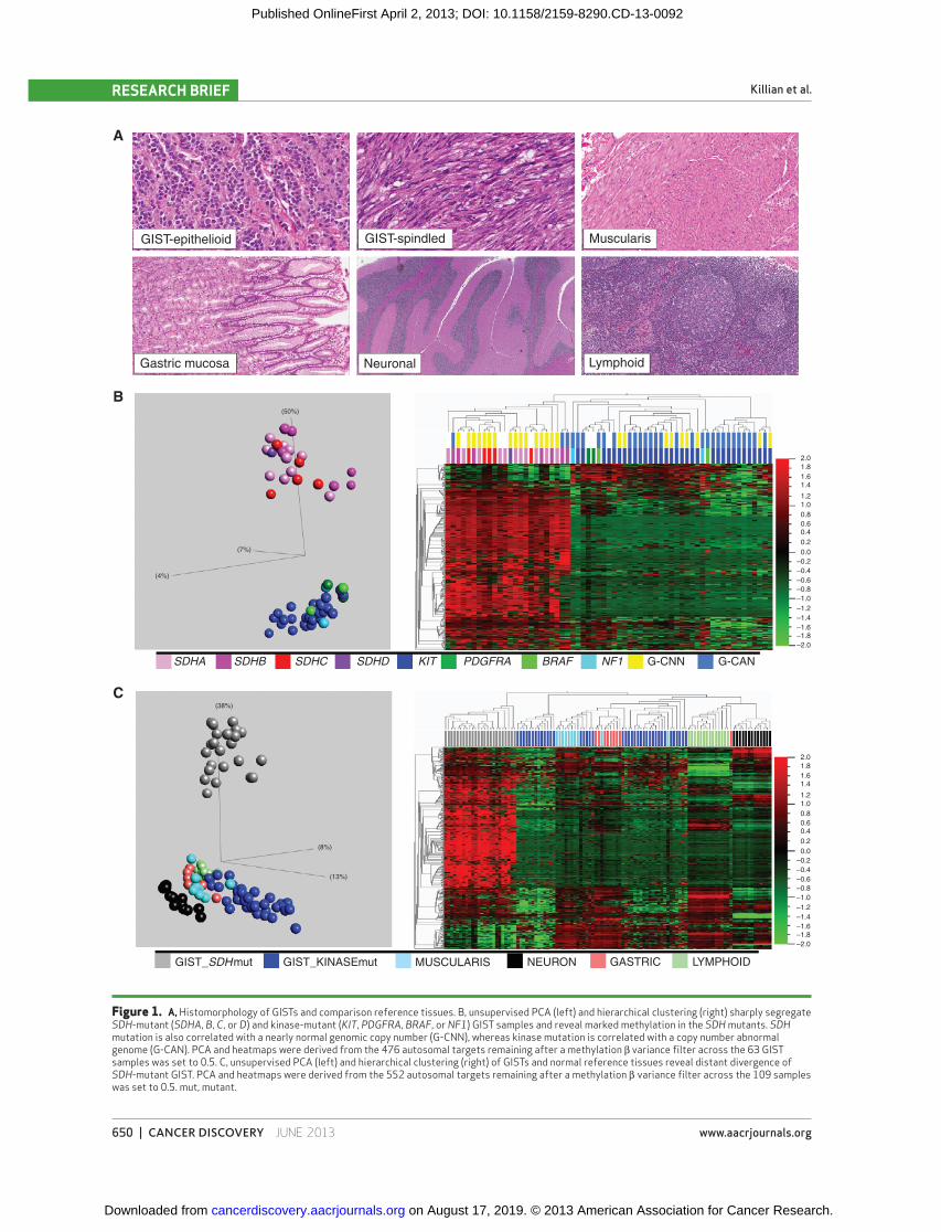
2 SDH subclasses, we analyzed the relatedness of subclass methylation profi le to those of multiple anatomically relevant normal tissues ( Fig. 1A and C ; Supplementary Table S2; refs. 13, 14 ). GIST is postulated to derive from the lineage of interstitial cell of Cajal (ICC), a mesodermal cell resident of the gastrointestinal tract involved in peristalsis and with both neuronal and mesenchymal characteristics ( 15, 16 ). Thus, neu-ronal cells and gut muscularis ( Fig. 1A ) were selected for the relatedness comparison. Also included were gut mucosa and lymphoid tissue, potential constituents of GIST specimens.
Unsupervised principal component analysis (PCA) and hierarchical clustering showed that the kinase-mutant GIST methylome signature most closely resembles that of all evalu-ated normal tissues, particularly muscularis tissue ( Fig. 1C ). In contrast, the SDH -mutant subclass comprised a distant out-group to all tissues ( Fig. 1C ). The marked divergence of the SDH-defi cient methylation profi le from multiple normal references argues against a purely clonal enrichment for the epigenome of a normal cell precursor population in normal gut, and instead supports a divergence during tumorigenesis. Thus, the kinase-mutant and SDH-defi cient GIST methyla-tion subclasses were respectively termed methyl-centrist and methyl-divergent.
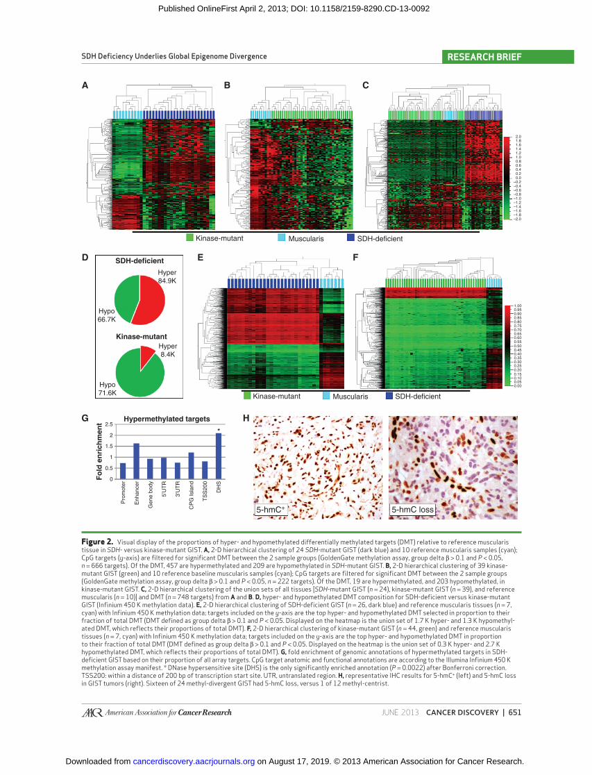
We subsequently quantifi ed the relative contributions of hypo- and hypermethylations to the aberrant methylation profi les of SDH - versus kinase-mutant GIST ( Fig. 2 ). Interest-ingly, we observed a comparable number of signifi cant CpG target hypomethylations in both subclasses: 209 and 203, respectively; in contrast, we observed 457 signifi cant hyper-methylations in the SDH mutants versus only 19 among KIT pathway mutants ( Fig. 2A–C ; Supplementary Table S2). Over-all, there is a comparable and substantial hypomethylation fraction in both oncogenotypes, but signifi cantly greater genomic hypermethylation in SDH mutants.
on August 17, 2019. © 2013 American Association for Cancer Research.cancerdiscovery.aacrjournals.org Downloaded from
Published OnlineFirst April 2, 2013; DOI: 10.1158/2159-8290.CD-13-0092
650 | CANCER DISCOVERY�JUNE 2013 www.aacrjournals.org
Killian et al.RESEARCH BRIEF
Figure 1. A, Histomorphology of GISTs and comparison reference tissues. B, unsupervised PCA (left) and hierarchical clustering (right) sharply segregate SDH -mutant ( SDHA , B , C , or D ) and kinase-mutant ( KIT , PDGFRA , BRAF , or NF1 ) GIST samples and reveal marked methylation in the SDH mutants. SDH mutation is also correlated with a nearly normal genomic copy number (G-CNN), whereas kinase mutation is correlated with a copy number abnormal genome (G-CAN). PCA and heatmaps were derived from the 476 autosomal targets remaining after a methylation β variance fi lter across the 63 GIST samples was set to 0.5. C, unsupervised PCA (left) and hierarchical clustering (right) of GISTs and normal reference tissues reveal distant divergence of SDH -mutant GIST. PCA and heatmaps were derived from the 552 autosomal targets remaining after a methylation β variance fi lter across the 109 samples was set to 0.5. mut, mutant.
GIST-epithelioid
A
B
C
GIST-spindled Muscularis
Lymphoid
2.0
(50%)
(7%)
(4%)
(38%)
(8%)
(13%)
1.81.6
1.01.2
1.4
0.80.60.40.20.0
–0.2–0.4–0.6–0.8–1.0–1.2–1.4–1.6–1.8–2.0
2.01.81.6
1.01.2
1.4
0.80.60.40.20.0
–0.2–0.4–0.6–0.8–1.0–1.2–1.4–1.6–1.8–2.0
Neuronal
SDHA
GIST_SDHmut GIST_KINASEmut MUSCULARIS NEURON GASTRIC LYMPHOID
SDHB SDHC SDHD KIT PDGFRA BRAF NF1 G-CNN G-CAN
Gastric mucosa
on August 17, 2019. © 2013 American Association for Cancer Research.cancerdiscovery.aacrjournals.org Downloaded from
Published OnlineFirst April 2, 2013; DOI: 10.1158/2159-8290.CD-13-0092
JUNE 2013�CANCER DISCOVERY | 651
SDH Defi ciency Underlies Global Epigenome Divergence RESEARCH BRIEF
Figure 2. Visual display of the proportions of hyper- and hypomethylated differentially methylated targets (DMT) relative to reference muscularis tissue in SDH - versus kinase-mutant GIST. A, 2-D hierarchical clustering of 24 SDH -mutant GIST (dark blue) and 10 reference muscularis samples (cyan); CpG targets ( y -axis) are fi ltered for signifi cant DMT between the 2 sample groups (GoldenGate methylation assay, group delta β > 0.1 and P < 0.05, n = 666 targets). Of the DMT, 457 are hypermethylated and 209 are hypomethylated in SDH -mutant GIST. B, 2-D hierarchical clustering of 39 kinase-mutant GIST (green) and 10 reference baseline muscularis samples (cyan); CpG targets are fi ltered for signifi cant DMT between the 2 sample groups (GoldenGate methylation assay, group delta β > 0.1 and P < 0.05, n = 222 targets). Of the DMT, 19 are hypermethylated, and 203 hypomethylated, in kinase-mutant GIST. C, 2-D hierarchical clustering of the union sets of all tissues [ SDH -mutant GIST ( n = 24), kinase-mutant GIST ( n = 39), and reference muscularis ( n = 10)] and DMT ( n = 748 targets) from A and B. D, hyper- and hypomethylated DMT composition for SDH-defi cient versus kinase-mutant GIST (Infi nium 450 K methylation data). E, 2-D hierarchical clustering of SDH-defi cient GIST ( n = 26, dark blue) and reference muscularis tissues ( n = 7, cyan) with Infi nium 450 K methylation data; targets included on the y -axis are the top hyper- and hypomethylated DMT selected in proportion to their fraction of total DMT (DMT defi ned as group delta β > 0.1 and P < 0.05. Displayed on the heatmap is the union set of 1.7 K hyper- and 1.3 K hypomethyl-ated DMT, which refl ects their proportions of total DMT). F, 2-D hierarchical clustering of kinase-mutant GIST ( n = 44, green) and reference muscularis tissues ( n = 7, cyan) with Infi nium 450 K methylation data; targets included on the y -axis are the top hyper- and hypomethylated DMT in proportion to their fraction of total DMT (DMT defi ned as group delta β > 0.1 and P < 0.05. Displayed on the heatmap is the union set of 0.3 K hyper- and 2.7 K hypomethylated DMT, which refl ects their proportions of total DMT). G, fold enrichment of genomic annotations of hypermethylated targets in SDH-defi cient GIST based on their proportion of all array targets. CpG target anatomic and functional annotations are according to the Illumina Infi nium 450 K methylation assay manifest. * DNase hypersensitive site (DHS) is the only signifi cantly enriched annotation ( P = 0.0022) after Bonferroni correction. TSS200: within a distance of 200 bp of transcription start site. UTR, untranslated region. H, representative IHC results for 5-hmC+ (left) and 5-hmC loss in GIST tumors (right). Sixteen of 24 methyl-divergent GIST had 5-hmC loss, versus 1 of 12 methyl-centrist.
Kinase-mutant
Hypermethylated targets
5-hmC+ 5-hmC loss
Muscularis SDH-deficient
Hyper84.9K
Hypo66.7K
Hypo71.6K
Hyper8.4K
SDH-deficient
A B C
D E
G H
F
Kinase-mutant
2.01.81.6
1.01.21.4
1.000.950.900.850.800.750.700.650.600.550.500.450.400.35
0.250.30
0.200.150.10
0.00
2.5
*2
1.5
1
0.5
Fo
ld e
nri
ch
me
nt
Pro
mot
er
Enh
ance
r
Gen
e bo
dy
5’U
TR
3’U
TR
CP
G Is
land
TS
S20
0
DH
S
0
0.05
0.80.60.40.20.0
–0.2–0.4–0.6–0.8–1.0–1.2–1.4–1.6–1.8–2.0
Kinase-mutant Muscularis SDH-deficient
on August 17, 2019. © 2013 American Association for Cancer Research.cancerdiscovery.aacrjournals.org Downloaded from
Published OnlineFirst April 2, 2013; DOI: 10.1158/2159-8290.CD-13-0092
652 | CANCER DISCOVERY�JUNE 2013 www.aacrjournals.org
Killian et al.RESEARCH BRIEF
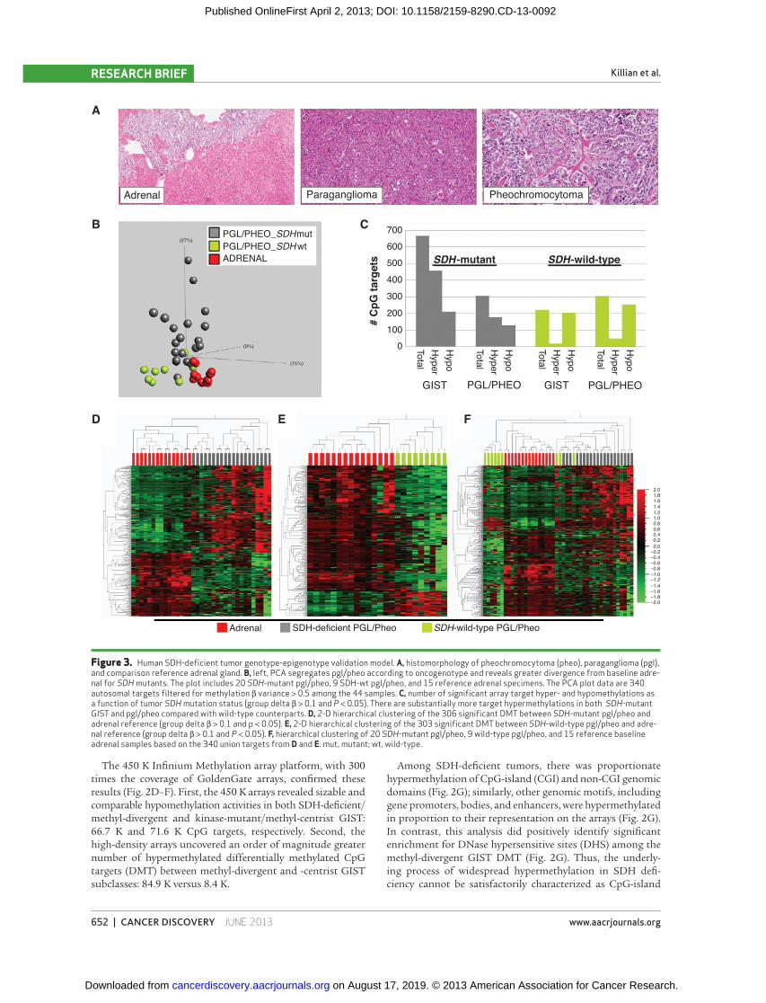
Figure 3. Human SDH-defi cient tumor genotype-epigenotype validation model. A, histomorphology of pheochromocytoma (pheo), paraganglioma (pgl), and comparison reference adrenal gland. B, left, PCA segregates pgl/pheo according to oncogenotype and reveals greater divergence from baseline adre-nal for SDH mutants. The plot includes 20 SDH -mutant pgl/pheo, 9 SDH-wt pgl/pheo, and 15 reference adrenal specimens. The PCA plot data are 340 autosomal targets fi ltered for methylation β variance > 0.5 among the 44 samples. C, number of signifi cant array target hyper- and hypomethylations as a function of tumor SDH mutation status (group delta β > 0.1 and P < 0.05). There are substantially more target hypermethylations in both SDH -mutant GIST and pgl/pheo compared with wild-type counterparts. D, 2-D hierarchical clustering of the 306 signifi cant DMT between SDH -mutant pgl/pheo and adrenal reference (group delta β > 0.1 and p < 0.05). E, 2-D hierarchical clustering of the 303 signifi cant DMT between SDH -wild-type pgl/pheo and adre-nal reference (group delta β > 0.1 and P < 0.05). F, hierarchical clustering of 20 SDH -mutant pgl/pheo, 9 wild-type pgl/pheo, and 15 reference baseline adrenal samples based on the 340 union targets from D and E. mut, mutant; wt, wild-type.
Adrenal SDH-deficient PGL/Pheo SDH-wild-type PGL/Pheo
Pheochromocytoma
SDH -mutant
GIST#
CpG
targ
ets
PGL/PHEO GIST PGL/PHEO
SDH-wild-type
700600500400300200100
0 TotalH
yperH
ypo
TotalH
yperH
ypo
TotalH
yperH
ypo
TotalH
yperH
ypo
PGL/PHEO_SDHmutPGL/PHEO_SDHwtADRENAL
Adrenal Paraganglioma
(27%)
(9%)
(15%)
2.01.81.6
1.01.21.4
0.80.60.40.20.0
–0.2–0.4–0.6–0.8–1.0–1.2–1.4–1.6–1.8–2.0
A
B
D E F
C
The 450 K Infi nium Methylation array platform, with 300 times the coverage of GoldenGate arrays, confi rmed these results ( Fig. 2D–F ). First, the 450 K arrays revealed sizable and comparable hypomethylation activities in both SDH-defi cient/methyl-divergent and kinase-mutant/methyl-centrist GIST: 66.7 K and 71.6 K CpG targets, respectively. Second, the high-density arrays uncovered an order of magnitude greater number of hypermethylated differentially methylated CpG targets (DMT) between methyl-divergent and -centrist GIST subclasses: 84.9 K versus 8.4 K.
Among SDH-defi cient tumors, there was proportionate hypermethylation of CpG-island (CGI) and non-CGI genomic domains ( Fig. 2G ); similarly, other genomic motifs, including gene promoters, bodies, and enhancers, were hypermethylated in proportion to their representation on the arrays ( Fig. 2G ). In contrast, this analysis did positively identify signifi cant enrichment for DNase hypersensitive sites (DHS) among the methyl-divergent GIST DMT ( Fig. 2G ). Thus, the underly-ing process of widespread hypermethylation in SDH defi -ciency cannot be satisfactorily characterized as CpG-island
on August 17, 2019. © 2013 American Association for Cancer Research.cancerdiscovery.aacrjournals.org Downloaded from
Published OnlineFirst April 2, 2013; DOI: 10.1158/2159-8290.CD-13-0092
JUNE 2013�CANCER DISCOVERY | 653
SDH Defi ciency Underlies Global Epigenome Divergence RESEARCH BRIEF
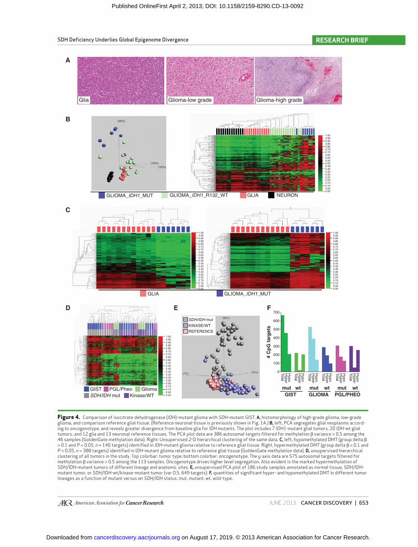
Figure 4. Comparison of isocitrate dehydrogenase ( IDH )-mutant glioma with SDH -mutant GIST. A, histomorphology of high-grade glioma, low-grade glioma, and comparison reference glial tissue. (Reference neuronal tissue is previously shown in Fig. 1A .) B, left, PCA segregates glial neoplasms accord-ing to oncogenotype, and reveals greater divergence from baseline glia for IDH mutants. The plot includes 7 IDH1 -mutant glial tumors, 20 IDH -wt glial tumors, and 12 glia and 13 neuronal reference tissues. The PCA plot data are 386 autosomal targets fi ltered for methylation β variance > 0.5 among the 46 samples (GoldenGate methylation data). Right: Unsupervised 2-D hierarchical clustering of the same data. C, left, hypomethylated DMT (group delta β > 0.1 and P < 0.05, n = 140 targets) identifi ed in IDH -mutant glioma relative to reference glial tissue. Right, hypermethylated DMT (group delta β > 0.1 and P < 0.05, n = 388 targets) identifi ed in IDH -mutant glioma relative to reference glial tissue (GoldenGate methylation data). D, unsupervised hierarchical clustering of all tumors in the study. Top colorbar: tumor type; bottom colorbar: oncogenotype. The y -axis data are 575 autosomal targets fi ltered for methylation β variance > 0.5 among the 113 samples. Oncogenotype drives higher level segregation. Also evident is the marked hypermethylation of SDH/IDH -mutant tumors of different lineage and anatomic sites. E, unsupervised PCA plot of 186 study samples annotated as normal tissue, SDH/IDH -mutant tumor, or SDH/IDH -wt/kinase-mutant tumor (var 0.5, 649 targets). F, quantities of signifi cant hyper- and hypomethylated DMT in different tumor lineages as a function of mutant versus wt SDH/IDH status. mut, mutant; wt, wild-type.
Glia
A
B
C
D E F
Glioma-low grade
GLIOMA_IDH1_MUT GLIOMA_IDH1_R132_WT GLIA NEURON
GLIA
GISTSDH/IDH mut
PGL/Pheo GliomaKinase/WT
SDH/IDH mut
KINASE/WTREFERENCE
700
600
500
400
300
200
100
0
GLIOMA_IDH1_MUT
Glioma-high grade
1.000.950.900.850.800.750.700.650.600.550.500.450.400.350.300.250.200.150.100.050.00
1.000.950.900.850.800.750.700.650.600.550.500.450.400.350.300.250.200.150.100.050.00
1.00
(7%) (10%)
(26%)
0.950.900.850.800.750.700.650.600.550.500.450.400.350.300.250.200.150.100.050.00
1.000.950.900.850.800.750.700.650.600.550.500.450.400.350.300.250.200.150.100.050.00
mut wt
# C
pG
targ
ets
GIST
TotalH
yperH
ypo
TotalH
yperH
ypo
TotalH
yperH
ypo
TotalH
yperH
ypo
TotalH
yperH
ypo
TotalH
yperH
ypo
mut wt
GLIOMA
mut wt
PGL/PHEO
(33%)
(10%)(12%)
on August 17, 2019. © 2013 American Association for Cancer Research.cancerdiscovery.aacrjournals.org Downloaded from
Published OnlineFirst April 2, 2013; DOI: 10.1158/2159-8290.CD-13-0092
654 | CANCER DISCOVERY�JUNE 2013 www.aacrjournals.org
Killian et al.RESEARCH BRIEF
methylator specifi c. In addition, differential methylation in these tumors is not randomly distributed, as evidenced by the large numbers of signifi cant, recurrent hypo- and hyper-methylated genomic targets (66.7 K and 84.9 K targets, respectively, Supplementary Table S3), principal component uniformity among samples, hierarchical clustering ( Fig. 2D and E ), and signifi cant enrichment for DHS ( Fig. 2G ).
Overall, the primary distinction observed by methylation microarray between SDH - and kinase pathway–mutant GIST was marked hypermethylation in the former. Neither subclass showed a signifi cant bias toward CGI methylation as a frac-tion of total hypermethylation ( Fig. 2G ).
Loss of 5-hmC in Methyl-Divergent GIST Mechanistically, accumulation of cytosine 5-methylation
in methyl-divergent tumors may arise from de novo meth-ylation and/or failed maintenance demethylation. It has been previously reported that elevated intracellular succinate levels result from SDH defi ciency and are toxic for the dioxygenase TET2, an enzyme required to catalyze DNA demethylation by conversion of 5-mC to 5-hmC. Thus, GIST tumors were scored for loss of 5-hmC by immunohistochemistry. Sixteen of 24 methyl-divergent versus 1 of 12 methyl-centrist tumors showed loss of 5-hmC ( P = 0.001). Thus, the fi nding of sig-nifi cant defi ciency of 5-hmC in SDH -null GIST ( Fig. 2H ) is consistent with a failure in TET2 maintenance demethylation in SDH-defi cient GIST. This potential connection in GIST between succinate accumulation, TET inhibition, and loss of 5-hmC is supported by the recent fi nding in melanoma that downregulation of TET family proteins leads to loss of 5-hmC ( 17 ).
Genomic Stability of Methyl-Divergent GIST We further evaluated the genomic copy number landscape
of GIST for ties to the identifi ed bimodal epigenomic diver-gence. The genomes of KIT -mutant/methyl-centrist GIST samples featured numerous and recurrent copy number aber-rations, including gains and losses of multiple chromosome arms similar to most types of cancer ( 18 ) and other published reports of GIST (refs. 19, 20 ; Fig. 1B and Supplementary Fig. S1). In contrast, methyl-divergent GIST samples had remarkably stable genomes, with either no copy number changes or ≤2 chromosome arm copy number changes ( Fig. 1B and Supplementary Fig. S1) often limited to a single somatic loss on 1p or 5p overlying germline SDHB or SDHA mutation. Thus, a highly altered epigenome is a unifying feature of SDH-defi cient GIST, and in some cases may be the only identifi able molecular aberration. No samples had both a normal genome and a normal methylome, and overall we observed an inverse correlation between karyotypic aberra-tion and epigenomic divergence in GIST samples ( Fig. 1B ).
Methyl Divergence in SDH-Defi cient Paraganglioma and Pheochromocytoma
We next sought to further validate and test the generality of the link between SDH mutation and methyl-divergence in tumorigenesis. As a model system, we analyzed other naturally occurring human SDH-defi cient tumor tissues, in particular SDH -mutant hereditary paraganglioma and pheo-chromocytoma (PGLs/Pheos). As is the case with GIST, a
subset of PGLs/Pheos occur in the setting of germline SDH mutation followed by a somatic second hit, thereby creating a genetic and functional SDH -null tumor. In contrast with the mesenchymal lineage of GIST, PGLs/Pheos are neuroen-docrine tumors of neural crest embryonic origin that arise in multiple anatomic sites outside the gut. We determined the methyl-divergence status for PGLs/Pheos based on com-parison with the adrenal reference tissue, including microdis-sected adrenal medulla, the postulated lineage of origin for Pheo ( Fig. 3A ). GoldenGate DNA methylation profi les from 29 PGLs/Pheos showed an elevated total number of target hypermethylations in the SDH -mutant versus -wild-type sub-group (177 targets versus 49 targets; Fig. 3B ; Supplementary Table S2). Moreover, as in GIST, there is an elevated ratio of hyper- to hypomethylated targets in the SDH -mutant group, whereas in the SDH -wild-type group, hypermethyla-tions are only a fraction of hypomethylations ( Fig. 3C–F ). Thus, human SDH-defi cient tumors from multiple ana-tomic origins, including stomach, adrenal, and carotid body and other paraganglia, manifest oncogenotype-dependent methyl-divergent profi les.
Comparison of SDH- and IDH-Defective Tumors Upstream of SDH in the Krebs cycle, isocitrate dehy-
drogenase (IDH) catalyzes the oxidative decarboxylation of isocitrate, producing α-KG and CO 2 . Mutation of IDH genes is coupled with heightened genomic methylation in several cancers. As postulated for elevated succinate levels stemming from SDH defi ciency, the result of IDH mutation is accumu-lation of a metabolite that is inhibitory for TET family dioxy-genase-mediated DNA demethylation. Using the GoldenGate methylation assay, we compared the epigenomic profi les of SDH -mutant GIST with those of IDH1 -mutant glial neoplasms (i.e., gliomas). Normal brain glial tissue served as the differ-ential methylation reference tissue for glioma ( Fig. 4A–C ). We identifi ed 388 and 140 hyper- and hypomethylations in IDH1 -mutant gliomas, compared with 457 and 209 in SDH -mutant GIST ( Fig. 4 ; Supplementary Table S2). Thus, IDH1 -mutant glioma and SDH -mutant GIST epigenomes have comparable proportions of hyper- and hypomethylation.
Subsequently, we conducted combined analysis on all the tumors in the study. Unsupervised hierarchical clustering assorted the gliomas, GIST, paragangliomas, and pheochro-mocytomas, not according to anatomic site of origin, but instead according to presence or absence of a Krebs cycle mutation ( Fig. 4D ); the heatmap further displays that hyper-methylated targets in SDH -mutant GIST are similarly hyper-methylated in other Krebs cycle–mutant tumors. Next, the PCA analysis showed the Krebs cycle–mutant tumors to lie along a shared principal axis orthogonal to the nonmutants, which more closely clustered with the normal tissues ( Fig. 4E ). Taken together, these data identify epigenomic homology of tumors from 4 divergent developmental lineages that have in common a mutation of a Krebs cycle enzyme.
DISCUSSION
In this study, we have found a striking correspondence between GIST oncogenotype and epigenotype, as evidenced by marked methyl-divergence of SDH mutants. Although
on August 17, 2019. © 2013 American Association for Cancer Research.cancerdiscovery.aacrjournals.org Downloaded from
Published OnlineFirst April 2, 2013; DOI: 10.1158/2159-8290.CD-13-0092
JUNE 2013�CANCER DISCOVERY | 655
SDH Defi ciency Underlies Global Epigenome Divergence RESEARCH BRIEF
impaired SDH function has previously been tied to tumori-genesis ( 21 ) and chromatin modifi cation ( 11 ), epigenomic profi ling of SDH-defi cient versus kinase pathway–mutant tumors has not been reported.
One of the fi rst described targets of pathologic succinate accumulation in cancer was the HIF-1 PHD, the inhibition of which leads to increased HIF-1α activity and result-ant tumorigenesis ( 22 ). Beyond unfettering HIF-1–medi-ated processes, succinate accumulation has been found to inhibit additional α-KG–dependent processes that generate succinate and CO2 as byproducts ( 10 ). For example, DNA demethylation through the oxidation of 5-mC to 5-hmC depends upon the dioxygenase TET2, and is inhibited by intracellular succinate accumulation; in the setting of SDH defi ciency, tumors may be expected to accumulate 5-mC or lose 5-hmC, analogous to epigenotype profi les in tumors harboring IDH mutations or TET protein downregulation, respectively.
Thus, our results suggest a mechanism for cancer-related DNA hypermethylation in SDH-deficient GIST that may be analogous to that proposed for IDH mutations in gliomas and leukemia. Rather than unscheduled de novo methyltransferase activity, we propose that the hypermeth-ylation phenomenon in this class of tumors may involve failure of maintenance DNA demethylation. Consistent with this idea, we found that SDH -mutant Pgl/Pheo and GIST have similar proportions of hyper- and hypomethyl-ated targets.
Importantly, we found that the methyl-divergence proc-ess is not random, as we uncovered tens of thousands of signifi cant, recurrent hypermethylations and hypomethyla-tions. Within the hypermethylated genomic compartment, we found DHS to be signifi cantly enriched. It is possi-ble that the identifi ed changes defi ne a succinate-sensitive hypermethyla tion genomic space in the GIST lineage. With this report, the methyl-divergence landscape of GIST tumors is now well characterized and clearly connected to the oncog-enotype. Although the consequences of this altered epige-netic state for tumor cell behavior remain uncertain and merit further exploration, the widespread perturbation that we have observed is unlikely to be phenotypically neutral. Our fi nding that younger patients with GIST with genome copy number-stable tumors have such dramatic epigenomic reprogramming seems to discount relative contributions from age-related epimutation and potential genomic dam-age from reactive oxygen species. The epigenomic similarities of diverse Krebs cycle–mutant tumors support the idea of a shared etiology stemming from their metabolite profi les. Thus, the current work defi nitively links SDH defi ciency to pervasive DNA hypermethylation and generally impli-cates the Krebs cycle as mitochondrial custodian of the methylome.
METHODS
Tissue Specimens Archival formalin-fi xed paraffi n-embedded (FFPE) tumor and
reference tissues were provided by the Pediatric and Wildtype GIST Clinic ( http://www.pediatricgist.cancer.gov/ ) at the NIH ( 6 ). Sam-ples were reviewed by a pathologist, and regions of characteristic
histomorphology were needle microdissected. Tissue cores were lysed and processed to yield genomic DNA (gDNA) as previously described ( 23, 24 ).
Immunophenotyping SDHB and 5-hmC IHC staining and analysis were conducted in a
central laboratory for uniformity of results. For SDHB, clone 21A11 (Abcam) was used at 1:1,000 dilution. For 5-hmC, polyclonal rabbit anti 5-hmC (Active Motif) was used at 1:2,000 dilution. IHC was conducted on a Bond-Max autostainer [Leica Microsystems with high pH antigen retrieval (AR2, pH 8.0)]. Loss of 5-hmC staining was defi ned as a ≥50% reduction in staining of tumor cell versus peritu-moral normal fi brovascular stroma.
Genomic Copy Number Analysis Tumor gDNAs were analyzed for chromosomal copy number aber-
rations using a commercial 180 K-feature array comparative genomic hybridization (aCGH) assay (Agilent Inc.). Array fl uorescence intensi-ties were imported to Nexus 6.0 (Biodiscovery) and analyzed using standard segment gain/loss settings. Genomic copy number near normal is defi ned as ≤2 gross chromosome arm gains or losses; genomic copy number aberration is defi ned as ≥3 gross chromosome arm gains or losses. Representative aCGH results are displayed in Supplementary Fig. S1.
DNA Methylation Arrays
GoldenGate. gDNA was bisulfi te converted and assayed using the GoldenGate Cancer Panel I methylation assay (Illumina, Inc.) as described previously ( 23 , 25 ). Briefl y, this assay measures DNA methy lation at 1536 distinct CpG targets distributed among 818 genes. Methylation β values were extracted from Cy3 and Cy5 signal intensities using BeadStudio software (Illumina, Inc.), and samples were excluded that did not pass array signal intensity controls. Gold-enGate Methylation β data are provided in Supplementary Table S2. These methylation β data may also be retrieved from Gene Expression Omnibus (GEO), accession number GSE34387.
Infi nium 450 K Methylation Assay for FFPE Samples. We used the EZ DNA Methylation kit (Cat# 5004, Zymo Research) for bisulfi te conversion of gDNA extracted from FFPE samples. For optimized results, we used 250 ng of gDNA and followed the manu-facturer’s recommendations. Namely, gDNA was denatured by addition of NaOH-containing M-Dilution buffer and incubated for 15 minutes at 37°C. Freshly prepared CT -conversion reagent (Zymo Research) containing sodium bisulfi te was added to the denatured DNA and samples were incubated for 16 hours at 50°C in a ther-mocycler and denatured every 60 minutes by heating to 95°C for 30 seconds. After bisulfi te conversion, the DNA was bound to a Zymo-Spin I-96 Binding Plate, washed with M-Wash Buffer, and desulphonated on the binding plate using M-desulphonation rea-gent. The bisulfi te-converted DNA was eluted from the plate wells in 10 μL elution buffer.
Bisulfi te-converted DNA was restored using Infi nium HD FFPE DNA Restore Kit (Cat#WG-321-1002, Illumina) following the manu-facturer’s recommendations. The process restores degraded FFPE DNA to a state that is amplifi able by the Infi nium whole-genome amplifi cation protocol. After DNA restoration, the Infi nium Methy-lation assay was carried out as described previously ( 26 ). In brief, bisulfi te converted and restored DNA (∼8 μL) was used in the whole-genome amplifi cation reaction. After amplifi cation, the DNA was fragmented enzymatically, precipitated, and resuspended in hybridization buffer. All subsequent steps were conducted follow-ing the standard Infi nium protocol (User Guide part #15019519 A). Fragmented DNA was dispensed onto the HumanMethylation450
on August 17, 2019. © 2013 American Association for Cancer Research.cancerdiscovery.aacrjournals.org Downloaded from
Published OnlineFirst April 2, 2013; DOI: 10.1158/2159-8290.CD-13-0092
656 | CANCER DISCOVERY�JUNE 2013 www.aacrjournals.org
Killian et al.RESEARCH BRIEF
BeadChips, and hybridization conducted in a hybridization oven for 20 hours. After hybridization, these arrays were processed through a primer extension and an IHC staining protocol to allow detection of a single-base extension reaction. Finally, BeadChips were coated and then imaged on an Illumina iScan. 450 K methylation array data are provided via GEO accession number GSE34387.
Mutation Identifi cation For GIST tumors, genomic DNA libraries were constructed and
SDHA , SDHB , SDHC , SDHD , KIT , BRAF , NF1 , and PDGFRA genes were sequenced using a custom capture assay as previously described ( 7 ). IDH1 status of gliomas was determined by Sanger sequencing of the R132 codon. SDH status of Pgls and Pheos was annotated as previously determined clinically, and results validated as part of this study.
Methylation Data Analysis Beta values were imported to Qlucore Omics Explorer (QOE v2.2).
For unsupervised cluster analyses, autosomal targets were selected, and then variance and normalization settings were dynamically tuned to produce the representative PCA plots and heatmaps shown in fi g-ures. For PCA plots, data normalization in QOE was set to mean = 0, var = 1; for 2-dimensional (2D) heatmaps, data normalization method in QOE is indicated by the green–red color scale in the fi gure. Statistical signifi cance of target differential methylation between var-ious comparison groups is P < 0.05 and group delta β > 0.1. In 450 K methylation GIST group comparisons, annotation as SDH-defi cient/mutant versus KIT kinase pathway–mutant was based fi rstly upon sequencing and secondly upon SDHB IHC for SDH/KIT wild-type samples.
Disclosure of Potential Confl icts of Interest B. Klotzle and M. Bibikova are employees of Illumina, Inc.
J.D. Schiffman is a consultant/advisory board member of Affymetrix, Inc. J.-B. Fan is an employee of Illumina, Inc. No potential confl icts of interest were disclosed by the other authors .
Authors’ Contributions Conception and design: J.K. Killian, M. Miettinen, C.A. Stratakis, L.J. Helman, P.S. Meltzer Development of methodology: J.K. Killian, M. Miettinen, R. Walker, Z. Wang, J.-B. Fan Acquisition of data (provided animals, acquired and managed patients, provided facilities, etc.): S.Y. Kim, M. Miettinen, C. Smith, M. Merino, M. Tsokos, M. Quezado, W.I. Smith Jr, M.S. Jahromi, E. Szarek, J. Lasota, B. Klotzle, Z. Wang, L. Jones, M. Bibikova, K. Pacak, K.A. Janeway, J.D. Schiffman, J.-B. Fan, L.J. Helman, P.S. Meltzer Analysis and interpretation of data (e.g., statistical analysis, biostatistics, computational analysis): J.K. Killian, M. Miettinen, M. Quezado, M.S. Jahromi, J. Lasota, Y.J. Zhu, Y. Wang, J. Waterfall, C.A. Stratakis, K.A. Janeway, J.D. Schiffman, P.S. Meltzer Writing, review, and/or revision of the manuscript: J.K. Killian, S.Y. Kim, M. Miettinen, M. Tsokos, M. Quezado, M.S. Jahromi, E. Szarek, J. Lasota, M. Raffeld, J. Waterfall, M.J. O’Sullivan, K. Pacak, K.A. Janeway, J.D. Schiffman, L.J. Helman, P.S. Meltzer Administrative, technical, or material support (i.e., reporting or organizing data, constructing databases): C. Smith, M.S. Jahromi, P. Xekouki, J. Lasota, M. Bibikova Study supervision: C.A. Stratakis, P.S. Meltzer
Grant Support This work was supported by grants from the Intramural Research
Program of NIH, the National Cancer Institute, and the Center for Cancer Research.
Received December 12, 2012; revised April 1, 2013; accepted April 23, 2013; published OnlineFirst April 2, 2013.
REFERENCES 1. Noushmehr H , Weisenberger DJ , Diefes K , Phillips HS , Pujara K ,
Berman BP , et al. Identifi cation of a CpG island methylator phenotype that defi nes a distinct subgroup of glioma . Cancer Cell 2010 ; 17 : 510 – 22 .
2. Figueroa ME , Abdel-Wahab O , Lu C , Ward PS , Patel J , Shih A , et al. Leukemic IDH1 and IDH2 mutations result in a hypermethylation phenotype, disrupt TET2 function, and impair hematopoietic dif-ferentiation . Cancer Cell 2010 ; 18 : 553 – 67 .
3. Pansuriya TC , van Eijk R , d’Adamo P , van Ruler MA , Kuijjer ML , Oosting J , et al. Somatic mosaic IDH1 and IDH2 mutations are associated with enchondroma and spindle cell hemangioma in Ollier disease and Maffucci syndrome . Nat Genet 2011 ; 43 : 1256 – 61 .
4. Hirota S , Isozaki K , Moriyama Y , Hashimoto K , Nishida T , Ishiguro S , et al. Gain-of-function mutations of c-kit in human gastrointestinal stromal tumors . Science 1998 ; 279 : 577 – 80 .
5. Pantaleo MA , Astolfi A , Indio V , Moore R , Thiessen N , Heinrich MC , et al. SDHA loss-of-function mutations in KIT-PDGFRA wild-type gastrointestinal stromal tumors identifi ed by massively parallel sequencing . J Natl Cancer Inst 2011 ; 103 : 983 – 7 .
6. Janeway KA , Kim SY , Lodish M , Nose V , Rustin P , Gaal J , et al. Defects in succinate dehydrogenase in gastrointestinal stromal tumors lacking KIT and PDGFRA mutations . Proc Natl Acad Sci U S A 2011 ; 108 : 314 – 8 .
7. Miettinen M , Killian JK , Wang ZF , Lasota J , Lau C , Jones L , et al. Immunohistochemical loss of succinate dehydrogenase subunit A (SDHA) in gastrointestinal stromal tumors (GISTs) signals SDHA germline mutation . Am J Surg Pathol 2013 ; 37 : 234 – 40 .
8. Demetri GD , Benjamin RS , Blanke CD , Blay JY , Casali P , Choi H , et al. NCCN Task Force report: management of patients with gastrointes-tinal stromal tumor (GIST)–update of the NCCN clinical practice guidelines . J Natl Compr Canc Netw 2007 ; 5 : S1 – 29 ; quiz S30 .
9. Gottlieb E , Tomlinson IP . Mitochondrial tumour suppressors: a genetic and biochemical update . Nat Rev Cancer 2005 ; 5 : 857 – 66 .
10. Frezza C , Pollard PJ , Gottlieb E . Inborn and acquired metabolic defects in cancer . J Mol Med 2011 ; 89 : 213 – 20 .
11. Cervera AM , Bayley JP , Devilee P , McCreath KJ . Inhibition of suc-cinate dehydrogenase dysregulates histone modifi cation in mamma-lian cells . Mol Cancer 2009 ; 8 : 89 .
12. Xiao M , Yang H , Xu W , Ma S , Lin H , Zhu H , et al. Inhibition of alpha-KG-dependent histone and DNA demethylases by fumarate and succinate that are accumulated in mutations of FH and SDH tumor suppressors . Genes Dev 2012 ; 26 : 1326 – 38 .
13. Miettinen M , Lasota J . Gastrointestinal stromal tumors: review on morphology, molecular pathology, prognosis, and differential diag-nosis . Arch Pathol Lab Med 2006 ; 130 : 1466 – 78 .
14. Rosai J . Gastrointestinal stromal tumor and its mimics . Int J Surg Pathol 2010 ; 18 : 79S – 87S .
15. Rosai J . GIST: an update . Int J Surg Pathol 2003 ; 11 : 177 – 86 . 16. Min KW . Gastrointestinal stromal tumor: an ultrastructural inves-
tigation on regional differences with considerations on their histo-genesis . Ultrastruct Pathol 2010 ; 34 : 174 – 88 .
17. Lian CG , Xu Y , Ceol C , Wu F , Larson A , Dresser K , et al. Loss of 5-hydroxymethylcytosine is an epigenetic hallmark of melanoma . Cell 2012 ; 150 : 1135 – 46 .
18. Beroukhim R , Mermel CH , Porter D , Wei G , Raychaudhuri S , Donovan J , et al. The landscape of somatic copy-number alteration across human cancers . Nature 2010 ; 463 : 899 – 905 .
19. Belinsky MG , Skorobogatko YV , Rink L , Pei J , Cai KQ , Vanderveer LA , et al. High density DNA array analysis reveals distinct genomic profi les in a subset of gastrointestinal stromal tumors . Genes Chro-mosomes Cancer 2009 ; 48 : 886 – 96 .
20. Astolfi A , Nannini M , Pantaleo MA , Di Battista M , Heinrich MC , Santini D , et al. A molecular portrait of gastrointestinal stromal
on August 17, 2019. © 2013 American Association for Cancer Research.cancerdiscovery.aacrjournals.org Downloaded from
Published OnlineFirst April 2, 2013; DOI: 10.1158/2159-8290.CD-13-0092
JUNE 2013�CANCER DISCOVERY | 657
SDH Defi ciency Underlies Global Epigenome Divergence RESEARCH BRIEF
tumors: an integrative analysis of gene expression profi ling and high-resolution genomic copy number . Lab Invest 2010 ; 90 : 1285 – 94 .
21. Selak MA , Armour SM , MacKenzie ED , Boulahbel H , Watson DG , Mansfi eld KD , et al. Succinate links TCA cycle dysfunction to onco-genesis by inhibiting HIF-alpha prolyl hydroxylase . Cancer Cell 2005 ; 7 : 77 – 85 .
22. Denko NC . Hypoxia, HIF1 and glucose metabolism in the solid tumour . Nat Rev Cancer 2008 ; 8 : 705 – 13 .
23. Killian JK , Bilke S , Davis S , Walker RL , Jaeger E , Killian MS , et al. A methyl-deviator epigenotype of estrogen receptor-positive breast
carcinoma is associated with malignant biology . Am J Pathol 2011 ; 179 : 55 – 65 .
24. Killian JK , Bilke S , Davis S , Walker RL , Killian MS , Jaeger EB , et al. Large-scale profi ling of archival lymph nodes reveals pervasive remodeling of the follicular lymphoma methylome . Cancer Res 2009 ; 69 : 758 – 64 .
25. Bibikova M , Fan JB . GoldenGate assay for DNA methylation profi l-ing . Methods Mol Biol 2009 ; 507 : 149 – 63 .
26. Bibikova M , Le J , Barnes B , Saedinia-Melnyk S , Zhou L , Shen R , et al. Genome-wide DNA methylation profi ling using Infi nium(R) assay . Epigenomics 2009 ; 1 : 177 – 200 .
on August 17, 2019. © 2013 American Association for Cancer Research.cancerdiscovery.aacrjournals.org Downloaded from
Published OnlineFirst April 2, 2013; DOI: 10.1158/2159-8290.CD-13-0092
2013;3:648-657. Published OnlineFirst April 2, 2013.Cancer Discovery J. Keith Killian, Su Young Kim, Markku Miettinen, et al. Divergence in Gastrointestinal Stromal TumorSuccinate Dehydrogenase Mutation Underlies Global Epigenomic
Updated version
10.1158/2159-8290.CD-13-0092doi:
Access the most recent version of this article at:
Material
Supplementary
http://cancerdiscovery.aacrjournals.org/content/suppl/2013/04/02/2159-8290.CD-13-0092.DC1
Access the most recent supplemental material at:
Cited articles
http://cancerdiscovery.aacrjournals.org/content/3/6/648.full#ref-list-1
This article cites 26 articles, 4 of which you can access for free at:
Citing articles
http://cancerdiscovery.aacrjournals.org/content/3/6/648.full#related-urls
This article has been cited by 19 HighWire-hosted articles. Access the articles at:
E-mail alerts related to this article or journal.Sign up to receive free email-alerts
Subscriptions
Reprints and
To order reprints of this article or to subscribe to the journal, contact the AACR Publications Department at
Permissions
Rightslink site. Click on "Request Permissions" which will take you to the Copyright Clearance Center's (CCC)
.http://cancerdiscovery.aacrjournals.org/content/3/6/648To request permission to re-use all or part of this article, use this link
on August 17, 2019. © 2013 American Association for Cancer Research.cancerdiscovery.aacrjournals.org Downloaded from
Published OnlineFirst April 2, 2013; DOI: 10.1158/2159-8290.CD-13-0092
Recommended