ISSN: 2319-8753
International Journal of Innovative Research in Science,
Engineering and Technology
(An ISO 3297: 2007 Certified Organization)
Vol. 2, Issue 9, September 2013
Copyright to IJIRSET www.ijirset.com 4852
Synthesis, Characterization of novel Cu(II)
complexes of isatin derivatives as potential
cytotoxicity, DNA binding, cleavage and
antibacterial agents
Ramadoss Gomathi 1, Andy Ramu *
Research Scholar, Dept. of Inorg. Chem., School of Chemistry, Madurai Kamaraj University, Madurai-21, India1
Professor, Dept. of Inorg. Chem., School of Chemistry, Madurai Kamaraj University, Madurai-21, India*
Abstract: Schiff-base ligands are potential anti-cancer, anti-bacterial and anti-viral agents and this activity tends to
increase in metal(II) Schiff-base complexes. Some oxindole-Schiff base copper(II) complexes have shown potential
antitumor activity towards deferent cells, inducing apoptosis in a process modulated by the ligand. Here, six novel
copper(II) complexes with schiff base ligands were isolated and characterized by IR, 1H-NMR, UV-visible , CV, EPR
and ESI-Mass. All the complexes are soluble in DMF and DMSO. Elemental analysis and molar conductance values
indicate that the complexes are non-electrolytes. All the complexes adopt octahedral geometry around the metal ions.
DNA binding activities of the complexes L1-Cu to L6-Cu a spectroscopy are studied by UV-vis. and also cleavage
studies of complexes have been done by agarose gel electrophoresis method. In-vitro biological activities of the free
ligands and its Cu(II) complexes are screened against few Gram +ve and Gram –ve bacteria by disc diffusion technique.
Cytotoxicity experiments carried out toward human Liver HepG2 cells confirmed its pro apoptosis property.
Interestingly, compounds, L4-Cu and L6-Cu were found to be excellent anticancer activity against HepG2: liver cells
and L4-Cu was most significant cytotoxicity activity against MAIT cells .
Keywords: isatin, 2, 2-diphenylethanamine, Cu(II) complexes, antibacterial activity, DNA binding, cleavage and
anticancer studies.
I. INTRODUCTION
The synthesis and study of inorganic compounds containing biologically relevant ligands are encouraged by the
importance of metal ions in a variety of biochemical processes [l-3]. The therapeutic application of metal complexes in
modern medicine was arguably initiated by the discovery of the anticancer properties of cisplatin [4, 5]. In fact,
cisplatin and the later compounds carboplatin, and oxaliplatin enjoy the status of the world's best-selling anticancer
drugs. Copper is the greatest importance for life and essential for photosynthesis and mitochondrial respiration, for
carbon and nitrogen metabolism, for oxidative stress protection, and is required for cell wall synthesis, to name only a
many of its cellular tasks. Copper has long been used to control the growth of organisms in wood products as well as in
aquaculture, agriculture and medicine. The multifaceted role of copper in biological systems is established by several
studies. In particular the involvement of copper in human diseases has been described from a medicinal-chemical and a
biochemical view focusing on the molecular physiology of Cu transport [6-8]. A number of Cu(II) chelate complexes
that exhibit cytotoxic activity through cell apoptosis or enzyme inhibition have been reviewed. Such complexes
containing bi-Schiff bases as ligands are effective in reducing tumor size, delaying of metastasis, and significantly
increasing the survival of the hosts. Current interest in Cu complexes is stemming from their potential use as
antimicrobial, antiviral, anti-inflammatory, antitumor agents, enzyme inhibitors, or chemical nucleases has been studied
[9]. Moreover, several authors have brought to attention the antiviral and antibacterial activity of Cu(II) complexes. For
instance, it was shown that the infectivity of influenza A virus is reduced after exposure on copper surfaces [10].
Copper(II) complexes are regarded as the most promising alternatives to cisplatin as anticancer drugs; an idea

ISSN: 2319-8753
International Journal of Innovative Research in Science,
Engineering and Technology
(An ISO 3297: 2007 Certified Organization)
Vol. 2, Issue 9, September 2013
Copyright to IJIRSET www.ijirset.com 4853
supported by a considerable number of research articles describing the synthesis, DNA binding and cytotoxic activities
of numerous copper(II) complexes[11, 12]. In addition, the fundamental role of copper(II) complexes as important
bioactive compounds in vitro and in vivo aroused an ever-increasing interest in these agents as potential drugs for
therapeutic intervention in various diseases [13-17].
Isatin and its derivatives are unique members in the Schiff base family. The simple isatin based Schiff base compounds
having, acyl, aroyl and heteroacroyl Schiff bases have additional donor sites >C=O, >C=N-, etc. These donor sites
make them more flexible and versatile. This versatility has made them excellent chelating agents that can form a variety
of complexes with various transition and inner transition metals and has attracted the attention of many researchers [18,
19]. Isatin, an endogenous indole and its derivatives exhibit a wide range of biological activities [20]. Isatin-based
Schiff base copper(II) complex is related to the antiviral drug, methisazone. Significant interest in the design of metal
compounds such as drugs and diagnostic agents is termed medicinal inorganic chemistry [21, 22]. Application of
electroanalytical techniques includes determination of reaction mechanisms. Metal–isatin binary complexes were
advantageous over simple isatin in chemotherapy and found to act as anticancer agents, especially Schiff-base
transition metal complexes derived from isatin [23].
This created a great interest in researchers to synthesize variety of isatin derivatives and screened them for their diverse
biological activities such as anticancer, anti-HIV, anthelmintic, antimycobacterial, anti-inflammatory, antidiabetic,
antimicrobial, trypanocidal as well antimalarial activities [24]. Information obtained from this study will be helpful to
understand the mechanism of isatin derivatives interaction with DNA, and should be useful to develop excellent
anticeancer activity and new therapeutic reagents for some diseases. The free ligand and its complexes have been tested
for in vitro antimicrobial activity against seven different bacteria and four different fungi by minimum inhibitory
concentration (MIC).
II. EXPERIMENTAL A Materials and Methods
All chemicals were purchased from Sigma-Aldrich, E-Merk and used as received without purification. isatin, 2,2-
diphenylethanamine, DMSO, Calf Thymus (CT) DNA and pUC-19 plasmid DNA purchased from Sigma-Aldrich G.R
grade, Bangalore. Metal chloride [CuCl2.2H2O] and solvents were purchased from E-Merk,A.R grade, Mumbai.
C, H and N analyses of the free Schiff base ligands and their complexes were performed in CHN analyzer Elementar
Vario EL III. Metal contents were analyzed by the standard procedures. Hand-Held Meter LF330 was used to measure
the molar conductance of the free Schiff base ligands and metal complexes in DMSO (1x10⁻³
M). The electronic spectra
were recorded in DMSO solutions using Shimatzu Model 160 UV-visible spectrophotometer. The IR spectra of the
complexes were recorded on a JASCO V-550 UV-Vis spectrophotometer in KBr pellets. 1H NMR spectra were
recorded on BRUKER DPX-300 High performance Digital FT-NMR spectrometer in DMSO-d6 using TMS as internal
standard. Electrospray ionisation mass spectrometry (ESI-MS) analysis was performed in the positive ion mode on a
liquid chromatography-ion trap mass spectrometer (LCQ Fleet, Thermo Fisher Instruments Limited, US. Magnetic
susceptibility measurement of the powdered samples was carried out by the Gouy balance. EPR measurements were
carried out by using a Varian E4 X-band spectrometer equipped with 100Hz modulation. Cyclic Voltammetric
measurements were carried out in a Bio-Analytical System (BAS) model CV-50W electrochemical analyzer.
B Synthesis of Schiff base ligands and its Cu(II) complexes
Synthesis of Schiff base ligands: (L1-L6)
L1: (E)-3-(2,2-diphenylethylimino)indolin-2-one
L2: (E)-3-(2,2-diphenylethylimino)-5-fluoroindolin-2-one
L3: (E)-5-chloro-3-(2,2-diphenylethylimino)indolin-2-one
L4: (E)-5-bromo-3-(2,2-diphenylethylimino)indolin-2-one
L5: (E)-3-(2,2-diphenylethylimino)-5-methylindolin-2-one
L6: (E)-3-(2,2-diphenylethylimino)-5-nitroindolin-2-one
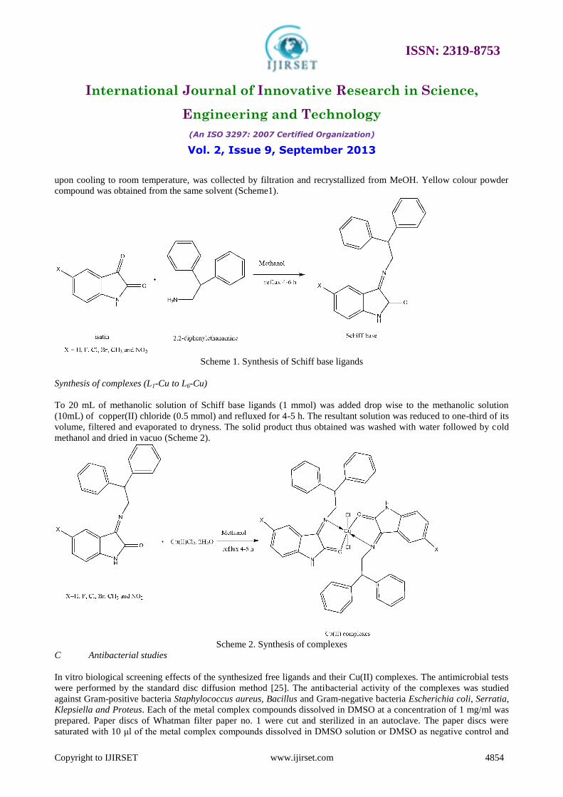
5-substituted isatin (1mmol) with 2, 2-diphenylethanamine (1mmol) were dissolved in 50mL of absolute MeOH, three
drops of glacial acetic acid was added and the resulting solution was refluxed for 4-6 hr. The compound precipitated
ISSN: 2319-8753
International Journal of Innovative Research in Science,
Engineering and Technology
(An ISO 3297: 2007 Certified Organization)
Vol. 2, Issue 9, September 2013
Copyright to IJIRSET www.ijirset.com 4854
upon cooling to room temperature, was collected by filtration and recrystallized from MeOH. Yellow colour powder
compound was obtained from the same solvent (Scheme1).
Scheme 1. Synthesis of Schiff base ligands
Synthesis of complexes (L1-Cu to L6-Cu)
To 20 mL of methanolic solution of Schiff base ligands (1 mmol) was added drop wise to the methanolic solution
(10mL) of copper(II) chloride (0.5 mmol) and refluxed for 4-5 h. The resultant solution was reduced to one-third of its
volume, filtered and evaporated to dryness. The solid product thus obtained was washed with water followed by cold
methanol and dried in vacuo (Scheme 2).
Scheme 2. Synthesis of complexes
C Antibacterial studies
In vitro biological screening effects of the synthesized free ligands and their Cu(II) complexes. The antimicrobial tests
were performed by the standard disc diffusion method [25]. The antibacterial activity of the complexes was studied
against Gram-positive bacteria Staphylococcus aureus, Bacillus and Gram-negative bacteria Escherichia coli, Serratia,
Klepsiella and Proteus. Each of the metal complex compounds dissolved in DMSO at a concentration of 1 mg/ml was
prepared. Paper discs of Whatman filter paper no. 1 were cut and sterilized in an autoclave. The paper discs were
saturated with 10 μl of the metal complex compounds dissolved in DMSO solution or DMSO as negative control and

ISSN: 2319-8753
International Journal of Innovative Research in Science,
Engineering and Technology
(An ISO 3297: 2007 Certified Organization)
Vol. 2, Issue 9, September 2013
Copyright to IJIRSET www.ijirset.com 4855
were placed aseptically in the Petri dishes containing Nutrient agar media inoculated with the above mentioned six
bacteria separately. The petridishes were incubated at 37 0C and the inhibition zones were recorded after 24 h of
incubation. The inhibition zone formed by these compounds against the particular test bacterial strain determined the
antibacterial activities of the synthetic compounds.The mean value obtained for three individual replicates was used to
calculate the zone of growth inhibition of each sample.
D Nuclease studies
The concentration of CT-DNA was determined by UV absorbance at 260 nm (ε = 6600 M-1
cm-1
). CT DNA free from
protein contamination was confired from its absorbance values at 260 nm, 280 nm and ratio A260/A280 was found to be
1.87 [26].
E Absorption studies
The UV–Vis absorption spectroscopy studies and the DNA binding experiments were performed at room temperature.
The purity of the CT-DNA was verified by taking the ratio of the obsorbance values at 260 and 280 nm in the
respective buffer, which was found to be 1.8:1, indicating that the DNA was sufficiently free of protein. The DNA
concentration per nucleotide was determined by absorption spectroscopy using the molar extinction coefficient value of
6600 dm3 mol
-1 cm
-1 at 260 nm. The complexes were dissolved in a mixed solvent of 5% DMSO and 95% phosphate
buffered saline for all the experiments. Absorption titration experiments were performed with a fixed concentration of
the compounds (30 µM) while gradually increasing the concentration of DNA (5–50 µM). While measuring the
absorption spectra, an equal amount of DNA was added to both the test solution and the reference solution to eliminate
the absorbance of DNA itself. For metal complexes, the intrinsic binding constant (Kb) was determined from the
spectral titration data using the following equation [27].
[DNA] / (Ɛa-Ɛf) = [DNA]/ (Ɛb-Ɛf) + 1/Kb (Ɛb-Ɛf)
Where, Ɛa, Ɛb and Ɛf are the molar extinction coefficients of the free complexes in solution, complex in the fully bound
from with CT-DNA and complex bound to DNA at a definite concentration respectively. In the plot of [DNA] / (Ɛa-Ɛf)
versus [DNA], Kb was calculated.
F Cleavage studies
pUC19 DNA at pH 7.2 in Tris-HCL buffered solution was used to perform agarose gel electrophoresis techniques.
Oxidative cleavage of DNA was examined by keeping the concentration of the 30µM of complexes and 2µL of pUC19
DNA and this was made up the volume to 16µL with 5mM Tris-HCl/5mM NaCl buffer solution. The resulting mixtures
were incubated at 37 C̊ for 2 h and followed by electrophoresed for 2 h at 50 V in Tris-aetate-EDTA (TAE) buffer
using 1% agarose gel containing 1.0 µg/ml ethidium bromide (EB) and photographed under UV light [28].
G Cytotoxic activity evaluation
3-(4, 5-dimethylthiazol-2- yl)- 2,5- diphenyltetrazolium bromide (MTT) assay
Cytotoxic effect of the four new complexes on human liver cancer cells (HepG2) were assayed by the 3-(4, 5-
dimethylthiazol-2- yl)- 2,5- diphenyltetrazolium bromide (MTT) assay[29]. The assay was carried out according to the
instruction provided by the vendor. Briefly, cells were harvested from the logarithmic phase of cultures and re-
suspended in Dulbecco’s Modified Eagles Medium (DMEM) supplemented with 10% fetal bovine serum (FBS). The
cell counts were adjusted and equal number of cells were plated into each well of 96-well cell culture plates and
allowed to grow overnight at 37 oC, in presence of 5% CO2. The cells were treated with test substances at various
concentrations ranging between 0.7 µM to 2.5 µM for 72h. In vehicle control culture wells, a maximum of 0.5%
DMSO was added. Culture medium was renewed at every 24h with fresh culture medium supplemented with the test
substances. Thereafter, 0.5 µM of MTT reagent was added to each well and the microplate was incubated further for 4h
at 37 oC in presence of 5% CO2. Finally, the cells were solubilized by adding solubilizing solution and allowed to
incubate at 37 oC overnight. After complete solubilization of the formazan crystals the absorbance was read at 540 nm

ISSN: 2319-8753
International Journal of Innovative Research in Science,
Engineering and Technology
(An ISO 3297: 2007 Certified Organization)
Vol. 2, Issue 9, September 2013
Copyright to IJIRSET www.ijirset.com 4856
in a microplate reader (BioRad, USA). The results (mean OD ± SD) obtained from quadruplicate wells were used in
calculation to determine the cytotoxicity (50% of inhibitory concentration, IC50) of the test compounds.
Tryphan blue exclusion assay
The trypan blue dye exclusion assay is the most commonly utilized test for cell viability [30]. The value of this
procedure is limited since the number of blue-staining cells increases following addition of the dye, requiring that cells
be counted within 3-5 min [30]. For cell growth cycle viability studies, a uniform suspension of cells was inoculated
into triplicate 75 cm2 tissue culture flasks and maintained in darkness in a standard CO2 incubation chamber. Replicate
samples from all flasks were counted each day for 7days using dye exclusion assays of cell viability. Comparative cell
viability stain experiments were conducted three times. Following analysis of variance, data from all experiments were
pooled for further statistical analysis For trypan blue staining, 200 µL of cells was aseptically transferred to a 1.5-mL
clear Eppendorf tube and incubated for 3 min at room temperature with an equal volume of 0.4% (w/v) trypan blue
solution prepared in 0.81 % NaCl and 0.06 % (w/v) dibasic potassium phosphate. Cells were counted using a dual-
chamber hemocytometer and a light microscope. Viable and nonviable cells were recorded separately, and the means of
three independent cell counts were pooled for analysis.
III. RESULTS AND DISCUSSION
The bidentate NO type of Schiff base ligands (L1-L6) and its Cu(II) complexes with 5-substituted isatin and 2,2-
diphenylethanamine were synthesized and characterized by various spectral techniques. The synthesized Cu(II)
complexes were found to be air stable, amorphous nature, moisture free and soluble only in DMF and DMSO.
A Elemental analysis and conductivity measurements
The synthesized schiff base ligands (L1 to L6) and their Cu(II) complexes were analyzed for their physico-chemical
properties like melting point (m.p.), color, yield, elemental analysis and conductivity which are given in table.1. The
elemental analytical data of ligands and their complexes are well agreed with their calculated values, showing that 2:1
(ligand : metal) stoichiometry ratio. The observed low conductivity values (15.81-36.55Ω -1
cm2
mol-1
) were accounted
for the dissociation and hence the complexes are found as non-electrolytes [31].
B Vibrational spectral studies
Vibrational spectra of free Schiff base ligands (L1-L6) were compared to investigate the mode of binding present in the
synthesized Cu(II) complexes. The FT-IR spectral data are summarized in Table 2. The IR spectrum of the free ligand
(L1 – L6) showed broad band’s 3151 – 3195 cm
-1, which can be attributed to υ(NH) stretching vibration of the isatin
moiety The ligands showed strong bands around at 1614-1621 cm-1
which assigned to azomethine moiety. In the
spectra of the complexes, this peak is slightly shifted to lower frequency around 1600-1584 cm-1
. This suggested that
coordination of the metal is through the azomethine nitrogen atom [32]. The strong intensity bands of ligands were
observed at the region 1714-1735 cm-1
of the spectra indicating carbonyl group. The positions of these bands were
shifted to lower region 1658-1683 cm-1
the spectra indicating the involvement of υ(C=O) with metal centre during
complexation. The ligands bind with the Cu(II) ions in a bidentate manner through azomethine –N and carbonyl –O
atoms respectively. Further, the two new bands appeared in the far infrared region at 455-462 cm-1
and 536-570 cm-1
were assigned to υ(M-N) and υ(M-O) respectively[33]. Thus, the IR spectral results provide evidence for bidentate
complexation of Schiff bases with metals.

ISSN: 2319-8753
International Journal of Innovative Research in Science,
Engineering and Technology
(An ISO 3297: 2007 Certified Organization)
Vol. 2, Issue 9, September 2013
Copyright to IJIRSET www.ijirset.com 4857
Table1. Composition and physical characteristics of L1 –L6 and their Cu(II) complexes
Ligands /
complexes
Molecular
Formula
Color Found (Calculated) % M.P
( 0C)
Yield
(%)
Ω
(Ohm-1
cm2
M-1
) M C H N
L1
C22H18N2O
Yellow - 81.62
(80.96)
4.90
(5.56)
8.01
(8.58)
130 95 -
L2 C22H17FN2O yellow - 75.82
(76.73)
5.41
(4.98)
7.45
(8.13)
124 90 -
L3 C22H17ClN2O
yellow - 72.75
(73.23)
4.20
(4.75)
7.03
(7.76)
120 90 -
L4
C22H17BrN2O
yellow - 64.23
(65.20)
4.70
(4.23)
7.62
( 6.91)
118
85 -
L5
C23H20N2O
Yellow - 80.30
(81.15)
5.31
(5.92)
8.01
(8.23)
132 80 -
L6 C22H17N3O3 Yellow - 70.68
(71.15)
4.21
(4.61)
11.81
(11.31)
148 80 -
L1-Cu C44H36Cl2CuN4O2
Light
brown
8.51
(8.07)
66.09
(67.13)
4.21
(4.61)
6.34
(7.12)
258 85 26.43
L2-Cu C44H34Cl2CuF2N4O2
Dark
green
7.02
(7.72)
63.04
(64.20)
4.71
(4.16)
7.05
(6.81)
280 85 24.11
L3-Cu C44H34Cl4CuN4O2 Dark
green
7.31
(7.42)
60.74
(61.73)
3.42
(4.00)
5.90
(6.54)
245 80 36.55
L4-Cu C44H34Br2Cl2CuN4O2
Dark
brown
5.36
(6.72)
56.08
(55.92)
3.07
(3.63)
5.71
(5.93)
260 85 15.81
L5-Cu C46H40Cl2CuN4O2 brown
7.81
(7.79)
66.83
(67.77)
4.31
(4.95)
6.44
(6.87)
250 75 18.03
L6-Cu C44H34Cl2CuN6O6
Dark
geen
7.22
(7.24)
60.31
(60.24)
3.58
(3.91)
8.92
(9.58)
256 70 22.54
Table 2. Vibration spectral data for the Cu(II) complexes and in KBr disc (cm-1
)
Compounds υ (NH) of
indole ring
Lactonyl, υ(C=O)
of indole ring
υ(C=N) υ (M-N) υ(M-O)
L1-Cu 3195 1686 1592 455 543
L2-Cu 3192 1678 1600 458 536
L3-Cu 3211 1691 1554 462 539
L4-Cu 3180 1683 1598 460 570
L5-Cu 3182 1697 1570 457 563
L6-Cu 3179 1689 1583 458 543
ISSN: 2319-8753
International Journal of Innovative Research in Science,
Engineering and Technology
(An ISO 3297: 2007 Certified Organization)
Vol. 2, Issue 9, September 2013
Copyright to IJIRSET www.ijirset.com 4858
C NMR Spectra
The 1H-NMR (300 MHz, CDCl3, δ/ppm) spectrum of the Schiff base exhibited the following signals around at 9.13-
9.45 NH protons, 6.7-8.2 aromatic protons. In addition to these two singlet peaks observed at 4.03-4.8 (-CH) and 5.0-
5.21 (-CH2) for methine and methene protons of ligands L1-L6.
The 13
C NMR (300 MHz, CDCl3, δ/ppm) spectra provide further support for the structural characterization of the Schiff
bases. The signals around at 164.03-164-91 (C=O, isatin), 158-95-159.01 (C=N, isoniazid), 110.36-144.82 (aromatic
carbons), 59.32-59.57 (methine carbon) and 52.41-52.88 (methane carbon) for L1-L6.
D Mass spectra
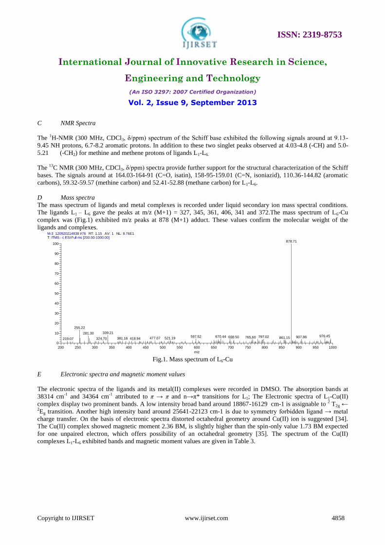
The mass spectrum of ligands and metal complexes is recorded under liquid secondary ion mass spectral conditions.
The ligands L1 – L6 gave the peaks at m/z (M+1) = 327, 345, 361, 406, 341 and 372.The mass spectrum of L6-Cu
complex was (Fig.1) exhibited m/z peaks at 878 (M+1) adduct. These values confirm the molecular weight of the
ligands and complexes. M-3_120920214838 #76 RT: 1.15 AV: 1 NL: 8.76E1T: ITMS - c ESI Full ms [200.00-1000.00]
200 250 300 350 400 450 500 550 600 650 700 750 800 850 900 950 1000
m/z
0
10
20
30
40
50
60
70
80
90
100
Re
lative
Ab
un
da
nce
878.71
255.22
339.21281.30976.45597.52 670.44 797.02698.50 907.86765.60 861.15477.07 521.19381.16324.70 418.94219.07
Fig.1. Mass spectrum of L6-Cu
E Electronic spectra and magnetic moment values
The electronic spectra of the ligands and its metal(II) complexes were recorded in DMSO. The absorption bands at
38314 cm-1
and 34364 cm-1
attributed to π → π and n→π* transitions for L1; The Electronic spectra of L1-Cu(II)
complex display two prominent bands. A low intensity broad band around 18867-16129 cm-1 is assignable to 2 T2g ←
2Eg transition. Another high intensity band around 25641-22123 cm-1 is due to symmetry forbidden ligand → metal
charge transfer. On the basis of electronic spectra distorted octahedral geometry around Cu(II) ion is suggested [34].
The Cu(II) complex showed magnetic moment 2.36 BM, is slightly higher than the spin-only value 1.73 BM expected
for one unpaired electron, which offers possibility of an octahedral geometry [35]. The spectrum of the Cu(II)
complexes L1-L6 exhibited bands and magnetic moment values are given in Table 3.
ISSN: 2319-8753
International Journal of Innovative Research in Science,
Engineering and Technology
(An ISO 3297: 2007 Certified Organization)
Vol. 2, Issue 9, September 2013
Copyright to IJIRSET www.ijirset.com 4859
Table 3. Electronic spectra, spectral parameters and magnetic moment with suggested structures
of ligands and their complexes
Compound π→ π* n→π* LMCT d-d Assignment Suggested µeff
(cm-1
) (cm-1
) Structure (B.M)
L1-Cu 38610 34013 23752 17301 2B1g →
2B2g ,
2Eg Octahedral 1.95
L2-Cu 37593 34013 23474 16808 2B1g →
2B2g ,
2Eg Octahedral 1.76
L3-Cu 37174 34129 25380 18621 2 T2g ←
2Eg Octahedral 2.34
L4-Cu 37037 31250 22883 16129 2B1g →
2B2g ,
2Eg Octahedral 1.87
L5-Cu 37313 31545 22883 16806 2B1g →
2B2g ,
2Eg Octahedral 2.10
L6-Cu 37174 31250 22123 17482 2 T2g ←
2Eg Octahedral 2.13
F ESR spectra
The X-band EPR spectrum of the copper(II) complexes were recorded in the solid state at room temperature. The
complex has a well resolved g׀׀ and broadened g region and various Hamiltonian parameters have been calculated (g׀׀
=1.93; g⊥=1.819; A102= ׀׀x104 for L1-Cu, g2.392= ׀׀; g⊥=2.031; A120= ׀׀x10
4 for L2-Cu, g2.011= ׀׀; g⊥=1.982; A׀׀
=108x104 for L3-Cu, g2.221= ׀׀; g⊥=2.103; A112= ׀׀x10
4 for L4-Cu, g1.902= ׀׀; g⊥=2.001; A107= ׀׀x10
4 for L5-Cu
and g2.131= ׀׀; g⊥=1.944; A116= ׀׀x104) the trend g׀׀ > g⊥ observed in this complex indicate that the unpaired electron
is most likely to be in the dx2-y
2 orbital [36].
G Cyclic voltammetry
A cyclic voltammogram of Cu(II) complex is value presented in Table 4. Voltammogram displays a reduction peak at
Epc= -2.8V with an associated oxidation peak at Epa= -0.4V at a scan rate of 50mV/s. The peak separation of this
couple (∆Ep) is 0.7V and increases with scan rate. The ∆Ep value increases at different scan scan rates respectively.
Thus, the analyses of cyclic voltametric responses at different scan rate give the evidence for quasi-reversible one
electron reduction. The most significant feature of the Cu(II) complex is the Cu(II)/Cu(I) couple. The ratio of cathodic
to anodic peak height was less than one. However, the peak current increases with the increase of the square root of the
scan rates. This establishes the electrode process as diffusion controlled [37].
ISSN: 2319-8753
International Journal of Innovative Research in Science,
Engineering and Technology
(An ISO 3297: 2007 Certified Organization)
Vol. 2, Issue 9, September 2013
Copyright to IJIRSET www.ijirset.com 4860
Table. 4. Electrochemical parameters for Cu(II), Co(II) and Ni(II) complexes
Compound Redox couple Epa (V) Epc(V) ΔEp(V) Ipa/Ipc
L1-Cu Cu(II)/Cu(I) 0.8 1.4 0.6 1.02
L2-Cu Cu(II)/Cu(I) 0.7 1.6 0.9 0.85
L3-Cu Cu(II)/Cu(I) 0.7 1.5 0.8 0.81
L4-Cu Cu(II)/Cu(I) 0.9 1.9 1.0 0.97
L5-Cu Cu(II)/Cu(I) 0.8 1.7 0.9 1.03
L6-Cu Cu(II)/Cu(I) 0.6 1.4 0.8 0.73
H DNA binding studies
Of all the techniques used, electronic absorption spectroscopy is one of the most common techniques for the
investigation of the mode of interaction of metal complexes with CT-DNA [38]. Hence, a complete electronic spectral
study was conducted with the new complexes and CT-DNA. The absorption spectra of L1-Cu to L6-Cu complexes in
the absence and presence of CT-DNA are given in Fig. 2. With increasing CT-DNA concentration for the L1-Cu
complexe, the hypochromism in the band at the found 435 and 445 nm reaches as high as 54.05% and 65.42%
respectively. Other Cu(II) complexes also exhibit the similar results during the addition of increasing concentration of
DNA, complexes showed hypochromicity and a red-shifted charge transfer peak maxima in the absorption spectra. The
intrinsic binding constant Kb is obtained from the ratio of slope to the intercept from the plots of [DNA]/(εa–εf) versus
[DNA]. The Kb values are shown in table 5. Hence the above phenomenon is indicative of most probable binding mode
of Cu(II) complexes for L1 to L6 with calf thymus DNA. It should be noted that significant effect on the absorption
bands of the molecule in the presence of double helical DNA, is characteristic of groove binder [39].
Fig.2. Absorption spectra of Cu(II) complex for L1, in the absence and in the presence of the CT-DNA. [DNA]=30 μM,
[complex] = 0 to 30 μM. The arrow indicates Absorption intensity decrease with increasing addition of the CT-DNA.

ISSN: 2319-8753
International Journal of Innovative Research in Science,
Engineering and Technology
(An ISO 3297: 2007 Certified Organization)
Vol. 2, Issue 9, September 2013
Copyright to IJIRSET www.ijirset.com 4861
Table 5. Absorption properties ofCu (II) complexes with CT-DNA.
Complex λ max (nm) Δλ (nm) Hypochromicity (%) Kbx104 ((mol L
-1)
-1)
L1-Cu 265, 298 4 56.25, 62.41 5.31
L2-Cu 260, 293 3 43.13, 48.16 5.80
L3-Cu 285, 297 2 47.53, 63.18 5.31
L4-Cu 266, 298 5 62.15, 71.36 8.25
L5-Cu 267,296 3 50.19, 55.16 5.04
L6-Cu 268,287 4 41.71, 55.13 5.78
I DNA cleavage studies
The DNA cleavage activities of Cu(II) complexes have been studied by gel electrophoresis and a representative
pictograph is shown in Fig. 3. The results showed that the supercoiled pUC19 DNA in buffer medium (pH=7.2; Tris-
HCl/NaCl) was converted into open circular form due to the formation of metal chelation. During the cleavage process,
the smallest fragments moved quickly towards anode than the larger fragments. Bromophenol blue was used as a
photosesitizer that can be activated on irradiation by UV. The completion of gel electrophoresis experiment clearly
indicated that the intensity of the treated DNA samples has diminished due to the cleavage of DNA. These results
indicated that the metal ions played an important role in the cleavage of DNA [40].
Fig.3. Cleavage of supercoiled pUC19 (10µM) by the Cu(II) complexes in the presence of Tri Acetate EDTA (TEA)
buffer at 37 C̊. Lane 1; DNA+H2O2, Lane 2; L2-Cu, Lane 3; L3-Cu, Lane 4; L5-Cu, Lane 5, L6-Cu, Lane 7; DNA-control.
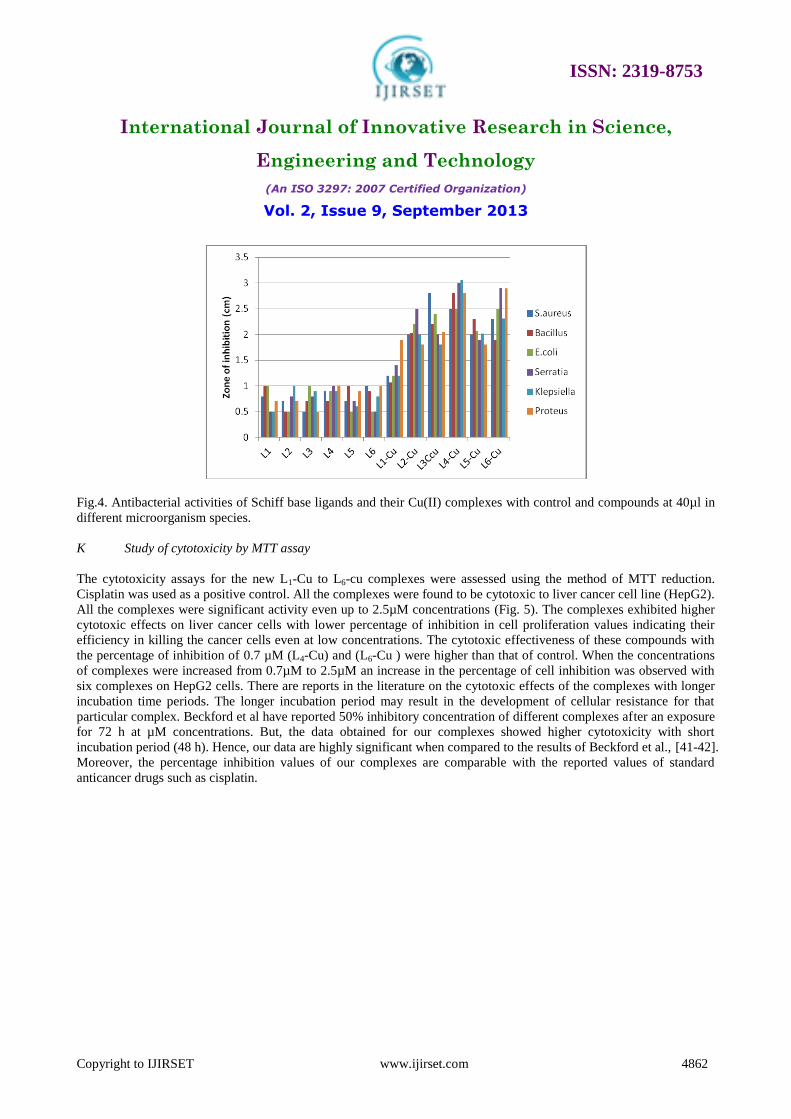
J In-vitro antimicrobial assay
The antimicrobial results are shown in Figure 4. From the antibacterial studies it is inferred that, the Schiff base was
found to be potentially active against Gram-positive bacteria Staphylococcus aureus, Bacillus and Gram-negative
bacteria Escherichia coli, Serratia, Klepsiella and Proteus. Some of the complexes were shown high antibacterial
activity against Escherichia coli and B. subtilis. L4-Cu complex was excellent antibacterial acvivity against all the
Gram +ve and Gram –ve bacteria.
ISSN: 2319-8753
International Journal of Innovative Research in Science,
Engineering and Technology
(An ISO 3297: 2007 Certified Organization)
Vol. 2, Issue 9, September 2013
Copyright to IJIRSET www.ijirset.com 4862
Fig.4. Antibacterial activities of Schiff base ligands and their Cu(II) complexes with control and compounds at 40µl in
different microorganism species.
K Study of cytotoxicity by MTT assay
The cytotoxicity assays for the new L1-Cu to L6-cu complexes were assessed using the method of MTT reduction.
Cisplatin was used as a positive control. All the complexes were found to be cytotoxic to liver cancer cell line (HepG2).
All the complexes were significant activity even up to 2.5µM concentrations (Fig. 5). The complexes exhibited higher
cytotoxic effects on liver cancer cells with lower percentage of inhibition in cell proliferation values indicating their
efficiency in killing the cancer cells even at low concentrations. The cytotoxic effectiveness of these compounds with
the percentage of inhibition of 0.7 µM (L4-Cu) and (L6-Cu ) were higher than that of control. When the concentrations
of complexes were increased from 0.7µM to 2.5µM an increase in the percentage of cell inhibition was observed with
six complexes on HepG2 cells. There are reports in the literature on the cytotoxic effects of the complexes with longer
incubation time periods. The longer incubation period may result in the development of cellular resistance for that
particular complex. Beckford et al have reported 50% inhibitory concentration of different complexes after an exposure
for 72 h at µM concentrations. But, the data obtained for our complexes showed higher cytotoxicity with short
incubation period (48 h). Hence, our data are highly significant when compared to the results of Beckford et al., [41-42].
Moreover, the percentage inhibition values of our complexes are comparable with the reported values of standard
anticancer drugs such as cisplatin.

ISSN: 2319-8753
International Journal of Innovative Research in Science,
Engineering and Technology
(An ISO 3297: 2007 Certified Organization)
Vol. 2, Issue 9, September 2013
Copyright to IJIRSET www.ijirset.com 4863
Fig. 5. Plots of Percentage inhibition in cell proliferation against various percentage of complexes
L In vitro culture of MAIT cells and proliferation assay for L4-Cu complex
Mucosal-associated invariant T (MAIT) cells are very abundant in humans and have antimicrobial specificity, but their
functions remain unclear. Cytotoxic activity detected for L4-Cu complex by MTT assay (Fig. 6). The cells were
exposed to various concentrations of MAIT for 24 h. (B) Antiproliferative effects detected by Tryphan blue exclusion
assay. Cells were treated with MAIT ranging in doses from 15 to 50'g/ml for 7 days. Control cells were treated with
0.1% DMSO. 0 'g/ml ( ), 15 'g/ml (*), 25 'g/ml (X) , 50 'g/ml (O). All the experiments were repeated three times, and
the values and bars represent mean and S.D., respectively. Hence, our data, L4-Cu complex was excellent cytotoxic
activity comparing other complexes.
Fig. 6. Cytotoxic activity activity detected for L4-Cu by MTT assay. The cells were exposed to various concentrations
of MAIT for 24 h. (B) Antiproliferative effects detected by Tryphan blue exclusion assay. Cells were treated with
MEIT ranging in doses from 15 to 50'g/ml for 7 days. Control cells were treated with 0.1% DMSO . 0 'µl ( ), 15 'µl (*),
25 'µl (X), 50 'µl (O). All the experiments were repeated three times, and the values and bars represent mean and S.D.,
respectively.

ISSN: 2319-8753
International Journal of Innovative Research in Science,
Engineering and Technology
(An ISO 3297: 2007 Certified Organization)
Vol. 2, Issue 9, September 2013
Copyright to IJIRSET www.ijirset.com 4864
IV. CONCLUSION
A series of new Cu(II) complexes of 5-substituted isatin with 2,2-diphenylethanamine have been prepared and
characterized by various spectral techniques. The UV-vis, IR, NMR ESI-Mass and EPR data showed that Cu(II)
complexes adopt octahedral geometry. From DNA binding results showed that all the complexes are moderate
intercalators. DNA cleavage indicated that the L4-Cu(II) complex has better cleaver other than complexes. The
antimicrobial actions of Cu(II) complexes, the zone of inhibition for L4-Cu and L6-Cu were excellent activity against
Staphylococcus aureus and Serratia. This study can be extended to investigate the toxicity and pharmacokinetic aspects
to get clear insight into the therapeutic utility of these compounds. Moreover, all the new complexes were screened for
antitumor activity against Hep G2 cancer cell lines, and they were found to exhibit excellent cytotoxicity to cancer cell
without affecting the normal cells. In all the above experimental results, we observed that complex L4-Cu and L6-Cu
have the excellent activity, which may be due to the 5th
position attached to bromine and nitro compound of isatin.
ACKNOWLEDGEMENTS
We sincere acknowledge the financial support received from JRF meritorious fellowship Ref. No. 17-03-2010; UGC
No.F4-1/2006(BSR)/7-119/2007(BSR) UGC, New Delhi for carrying out this research work. The authors express their
sincere thanks to Head and Country Manager, Department of Drug discovery Network partner, CRC group, USA for
providing antitumor studies.
REFERENCES
[1] R. H. Holm and J. A. Ibers, Modeling coordination sites in metallobiomolecules, Science, vol. 209, 1980, pp.223-235.
[2] E. I. Solomon, in Metal Ions in Biology, ed. T. G. Spiro, Wiley Interscience, New York, 1981, vol. 3, p. 41
[3] O.Farvcr, I. PcchI, (1984) in Copm Prorcms aed Copper Enqmes (Lonric. R.cd.) Vol. I. p.183. CRC Prm. Boca Raron [4] G. J. Brewer, “The risks of copper toxicity contributing to cognitive decline in the aging population and to Alzheimer's disease,” Journal of the
American College of Nutrition, vol. 28, no. 3, 2009, pp. 238–242.
[5] Daniel, K.G., P. Gupta, R.H. Harbach, W.C. Guida, and Q.P. Dou, Organic copper complexes as a new class of proteasome inhibitors and apoptosis inducers in human cancer cells. Biochem Pharmacol.6: 1139-1151 (2004)
[6] S. Puig and D. J. Thiele, “Molecular mechanisms of copper uptake and distribution,” Current Opinion in Chemical Biology, vol. 6, no. 2, pp.
171–180, 2002. [7] L. Tripathi, P. Kumar, and A. K. Singhai, Role of chelates in treatment of cancer. Indian Journal of Cancer, Vol. 44, 2007, pp. 62–71
[8] Jane E. Weder, Carolyn T. Dillona, Trevor W. Hambleya, Brendan J. Kennedya, Peter A. Laya, J. Ray Biffinb, Hubert L. Regtopb and Neal M. Davies, Copper complexes of non-steroidal anti-inflammatory drugs: an opportunity yet to be realized, Coordination Chemistry Reviews Vol. 232,
2002, pp. 95-126.
[9] J. O. Noyce, H. Michels, and C. W. Keevil, Inactivation of Influenza A Virus on Copper versus Stainless Steel Surfaces, Applied and Environmental Microbiology, Apr. 2007, p. 2748–2750
[10] Tamura, H.; Imai, H.; Kuwahara, J.; Sugiura, Y. (1987). A new antitumor complex: bis(acetato)bis(imidazole)
copper(II). J. Am. Chem. Soc. 109 (22): 6870–687 [11] S.S. Bhat, A.A. Kumbhar, H. Heptullah, A.A. Khan, V.V. Gobre, S.P. Gejji and V. Puranik, Synthesis, Electronic Structure, DNA and Protein
Binding, DN, A Cleavage, and Anticancer Activity of Fluorophore-Labeled Copper(II) Complexes, Inorganic Chemistry, Vol. 50, 2011, pp 545–558
[12] V. Rajendiran, R. Karthk, M. Palaniandavar, H. Stoeckli-Evans, VS. Periasamy, M.A. Akbarsha , B.S. Srinag , H. Krishnamurthy . Mixed-Ligand Copper(II)-phenolate Complexes: Effect of Coligand on Enhanced DNA and Protein Binding, DNA Cleavage, and Anticancer Activity.
Inorganic Chemistry, Vol. 46, 2007, pp.8208-8221.
[13] M.Gerhard, M.A. Jakupec, O. Zava, P.J. Dyson, V.B. Arion, B.K. Keppler, Highly Cytotoxic Copper(II) Complexes with Modified Paullone Ligands, Inorganic Chemistry, Vol. 49, 2010, pp. 302-311
[14] Hegg, E.L.; Burstyn, J.N. "Toward the Development of Metal-Based Synthetic Nucleases and Peptidases: A Rationale and Progress Report in
Applying the Principles of Coordination Chemistry." Coord. Chem. Rev. 1998, 173, 133-165. [15] F. Mancin, P. Scrimin, P. Tecilla and U. Tonellato, Artificial Metallonucleases, Chemistry Communications, 2005, pp.2540-2548.
[16]Yan An, Si-Dong Liu, Shu-Yi Deng, Liang-Nian Ji, and Zong-Wan Mao, Cleavage of double-strand DNA by linear and triangular trinuclear
copper complexes, Journal of Inorganic Biochemistry Vol.100, 2006, 1586–1593 [17] P.U. Maheswari, S. Roy, H.D. Dulk, S. Barends, G.V. Wezel, B. Kozlevcar, P. Gamez, J. Reedijk, The Square-Planar Cytotoxic
[CuII(pyrimol)Cl] Complex Acts as an Efficient DNA Cleaver without Reductant, Journal of American Chemical Society, Vol.128, 2006, 710-711.
[18] J. E. Weder, C. T. Dillon, T. W. Hambley, B. J. Kennedy, P. A. Lay, J. R. Biffin, H. L. Regtop, N. M. Davvies, Copper complexes of non-steroidal antiinflammatory drugs: an opportunity yet to be realized. Coord. Chem. Rev. 232, 2002, pp. 95-126.
[19] F. Tisato, C. Marzano, M. Porchia, M. Pellei and C. Santini , Copper in diseases and treatments, and copper-based anticancer strategies,
Medicinal Research Reviews. Vol. 30, 2010, pp.708- 749 [20] F. Lebon, N. Boggetto, M. Ledecq, F. Durant, Z. Benatallah, S. Sicsic, R. Lapouyade, O. Kahn, A. Mouithys-Mickalad, G. Deby-Dupont, M.
Reboud-Ravaux, Metal-organic compounds: a new approach for drug discovery:N1-(4-methyl-2-pyridyl)-2,3,6-trimethoxybenzamide copper(II)
complex as an inhibitor of human immunodeficiency virus 1 protease, Biochemical Pharmacology, Vol. 63, 2002, 1863-1873. [21] S.G Kucukguzel, I. Kucukgusel, E. Tater, S. Rollas and F.Sahin, Synthesis of some novel heterocyclic compounds derived from diflunisal
hydrazide as potential anti-infective and anti-inflammatory agents, European Journal Medicinal Chemistry, Vol. 42, 2007, 893.

ISSN: 2319-8753
International Journal of Innovative Research in Science,
Engineering and Technology
(An ISO 3297: 2007 Certified Organization)
Vol. 2, Issue 9, September 2013
Copyright to IJIRSET www.ijirset.com 4865
[22] W.Liua, , K. Bensdorf , , A. Hagenbachb, , U. Abramb, B. Niuc, A. Mariappanc, European Journal Medicinal Chemistry, Synthesis and biological studies of silver N-heterocyclic carbene complexes derived from 4,5-diarylimidazole, Vol.46, 2011, pp.5927
[23] S.N. Pandeya, D. Siram, G. Nath, E. Declercq, Synthesis, antibacterial, antifungal and anti-HIV activities of Schiff and Mannich bases derived
from isatin derivatives and N-[4-(4′-chlorophenyl)thiazol-2-yl] thiosemicarbazide, European Journal of Pharmaceutical Science, Vol.9, 1999, pp.25-31
[24] W. L. Drew, A. L. Barry, R. O’Toole, J. C. Sherris, Reliability of the Kirby-Bauer Disc Diffusion Method for Detecting Methicillin-Resistant Strains of Staphylococcus aureus, Applied and Environmental Microbiology, Vol. 24, 1972, 240. [25] C.Hee, K. C. Kong, S. T. Von, P. B.Paul, E.Thirthagiri, H. Hamadae and M. Chikira, Synthesis, characterization, DNA-binding study and anticancer properties of ternary metal(II) complexes of edda and an intercalating ligand, Dalton Transactions, Vol. 447, 2008, 447–454 . [26] V. Alverdi , L. Giovagnini, C. Marzano, R. Seraglia, F. Bettio, S. Sitran, R. Graziani, “Characterization studies and cytotoxicity assays of Pt(II) and Pd(II) dithiocarbamate complexes by means of FT-IR, NMR spectroscopy and mass spectrometry,” Journal of Inorganic Biochemistry, vol. 98, no. 6, pp. 1117–1128, 2004. [27] F. Arjmand, S. Parveena, M. Afzal, L. Toupet and T.B. Hadda, Molecular drug design, synthesis and crystal structure determination of Cu
II–Sn
IV heterobimetallic core: DNA binding and cleavage studies, European Journal of Medicinal Chemistry, Vol.49, 2012, pp.141-
150 [28] X. Qiao, Z.Ying Ma, C.Z. Xie, F. Xue and Y.W. Zhang, Study on potential antitumor mechanism of a novel Schiff Base copper(II) complex:Synthesis, crystal structure, DNA binding, cytotoxicity and apoptosis induction activity, Journal of Inorganic Biochemistry, Vol. 105, 2011, pp. 728–737 [29] T. Mossman, Rapid colorimetric assay for cellular growth and survival: application to proliferation and cytotoxicity assays. Journal oflmmunologicalMethods, Vol.65, 1983, 55-63 [30] B.B. Mishell,S.M. Shiigi, In Selected Methods in Cellular Immunology; Freeman and Co.: San Francisco, 1980, pp 16-19 [31] M. Tofazzal, H. Tarafder, Manaf A. Ali1, D. Juan Wee, Kasbollah Azahari, Sidik Silong and Karen A. Complexes of a tridentate ONS Schiff
base. Synthesis and biological properties, Transition Metal Chemistry, Vol. 25, 2000, 456-460. [32] K. Nakamoto, Infrared and Raman Spectra of Inorganic and Coordination Compounds, Wiley, New York, 1986.
[33] L.J. Bellamy, Infrared Spectra of Complex Molecules, second ed., Mathuen, London, 1958
[34] D. A. Kulkarni, S. A. Patil and P. S. Badami, Electrochemical Properties of some Transition Metal Complexes: Synthesis, Characterization and In-vitro antimicrobial studies of Co(II), Ni(II), Cu(II), Mn(II) and Fe(III) Complexes, International Journal of Electrochemical Science, Vol. 4, 2009,
pp.717 – 729
[35] C.L. Klein, R.J. Majeste, L. M. Trefonas,lc and C. J. O’Connor, Magnetic Properties and Molecular Structure of Copper( 11) Complexes of Pyrazinecarboxylic Acid, Inorganic Chemistry, Vol. 21, I982, pp.1891-1897
[36] R.L. Dutta, A. Syamal, Electron spin resonance responses, In Elements of Magnetochemistry, 2nd Ed.; East-West Press: New Delhi, (1999).
206–250. [37] A.J. Bard, L.R. Izatt (Eds), Electrochemical Methods: Fundamentals and Applications, 2nd ed., (Wiley, New York, 2001)
[38] C. V. Kumar, J. K. Barton, N. J. Turro, Photophysics of Ruthenium Complexes Bound to Double Helical DNA, Journal of American Chemical
Society, Vol. 107, 1985, pp.5518-5523 [39] E.Ramachandran, P. Kalaivani, R. Prabhakaran, M. Zeller, J.H. Bartlett, P. O. Adero, T. R.Wagner, K. Natarajan, Synthesis, characterization,
crystal structure and DNA binding studies of Pd(II) complexes containing thiosemicarbazone and triphenylphosphine/triphenylarsine Inorganica
Chimica Acta, Vol. 385, 2012, pp. 94–99
[40] N. Raman, K. Pothiraj, T. Baskaran, DNA-binding, oxidative DNA cleavage, and coordination mode of later 3d transition metal complexes of a
Schiff base derived from isatin as antimicrobial agents, J. Coor. Chem., Vol.64 2011, pp. 3900–3917
[41] F.A. Beckford, M. Shaloski, G. Leblanc, J. Thessing, L.C.L. Alleyne, A.A. Holder, L. Li, N.P. Seeram, Microwave synthesis of mixed ligand diimine–thiosemicarbazone complexes ofruthenium(II): biophysical reactivity and cytotoxicity, Dalton
Transactions, 2009, pp. 10757-10764.
[42] P. Mura, M. Camalli, L. Messori, F. Piccioli, P. Zanello, M. Corsini, Synthesis, Structural Characterization, Solution Chemistry, and Preliminary Biological Studies of the Ruthenium(III) Complexes [TzH][trans-RuCl4(Tz)2] and [TzH][trans-RuCl4(DMSO)(Tz)]â(DMSO), theThiazole
Analogues of Antitumor ICR and NAMI-A, Inorganic Chemistry, Vol.43, 2004, 3863-3870.
Recommended