The 3’UTR of the West Nile Virus genomic RNA is a potential
antiviral target site
Sara Ramos-Lorente, Isabel Pérez-Jover, Cristina Romero-López* and Alfredo Berzal-
Herranz*
1 Instituto de Parasitología y Biomedicina “López-Neyra”, IPBLN-CSIC
PTS Granada. Av del Conocimiento 17, 18016 Granada, Spain
* Corresponding author: [email protected]; [email protected]
1

The 3’UTR of the West Nile Virus genomic RNA is a potential antiviral
target site
2
Translation
cyclization complex
Site 2 Site 12
Site 2 Site 13Site 2
Site 1
Site 14
Site 2 Site 11
40S
40S
3’UTR 5’UTR
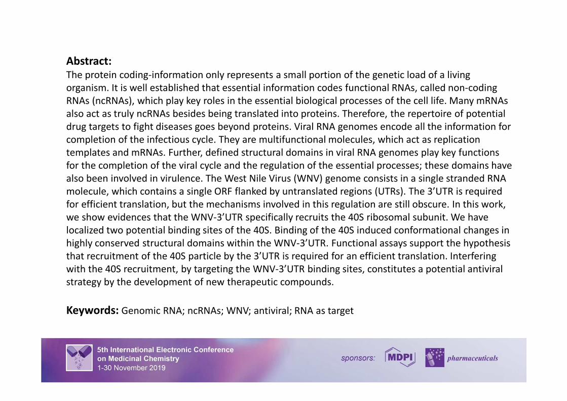
Abstract:
The protein coding-information only represents a small portion of the genetic load of a living
organism. It is well established that essential information codes functional RNAs, called non-coding
RNAs (ncRNAs), which play key roles in the essential biological processes of the cell life. Many mRNAs
also act as truly ncRNAs besides being translated into proteins. Therefore, the repertoire of potential
drug targets to fight diseases goes beyond proteins. Viral RNA genomes encode all the information for
completion of the infectious cycle. They are multifunctional molecules, which act as replication
templates and mRNAs. Further, defined structural domains in viral RNA genomes play key functions
for the completion of the viral cycle and the regulation of the essential processes; these domains have
also been involved in virulence. The West Nile Virus (WNV) genome consists in a single stranded RNA
molecule, which contains a single ORF flanked by untranslated regions (UTRs). The 3’UTR is required
for efficient translation, but the mechanisms involved in this regulation are still obscure. In this work,
we show evidences that the WNV-3’UTR specifically recruits the 40S ribosomal subunit. We have
localized two potential binding sites of the 40S. Binding of the 40S induced conformational changes in
highly conserved structural domains within the WNV-3’UTR. Functional assays support the hypothesis
that recruitment of the 40S particle by the 3’UTR is required for an efficient translation. Interfering
with the 40S recruitment, by targeting the WNV-3’UTR binding sites, constitutes a potential antiviral
strategy by the development of new therapeutic compounds.
Keywords: Genomic RNA; ncRNAs; WNV; antiviral; RNA as target
3
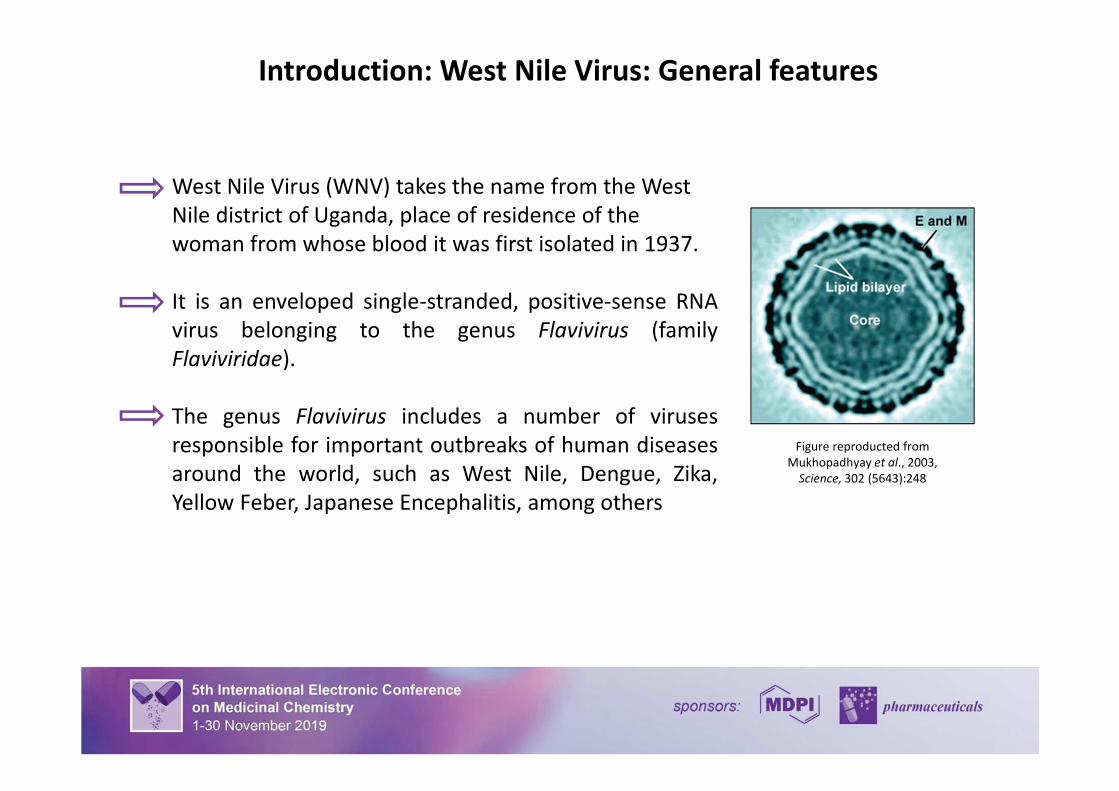
Introduction: West Nile Virus: General features
Figure reproducted from
Mukhopadhyay et al., 2003,
Science, 302 (5643):248
West Nile Virus (WNV) takes the name from the West
Nile district of Uganda, place of residence of the
woman from whose blood it was first isolated in 1937.
It is an enveloped single-stranded, positive-sense RNA
virus belonging to the genus Flavivirus (family
Flaviviridae).
The genus Flavivirus includes a number of viruses
responsible for important outbreaks of human diseases
around the world, such as West Nile, Dengue, Zika,
Yellow Feber, Japanese Encephalitis, among others
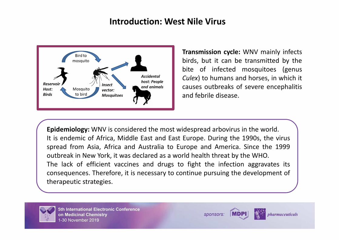
Introduction: West Nile Virus
Transmission cycle: WNV mainly infects
birds, but it can be transmitted by the
bite of infected mosquitoes (genus
Culex) to humans and horses, in which it
causes outbreaks of severe encephalitis
and febrile disease.
Epidemiology: WNV is considered the most widespread arbovirus in the world.
It is endemic of Africa, Middle East and East Europe. During the 1990s, the virus
spread from Asia, Africa and Australia to Europe and America. Since the 1999
outbreak in New York, it was declared as a world health threat by the WHO.
The lack of efficient vaccines and drugs to fight the infection aggravates its
consequences. Therefore, it is necessary to continue pursuing the development of
therapeutic strategies.
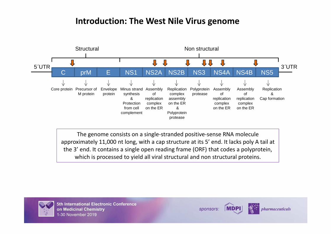
Introduction: The West Nile Virus genome
The genome consists on a single-stranded positive-sense RNA molecule
approximately 11,000 nt long, with a cap structure at its 5’ end. It lacks poly A tail at
the 3’ end. It contains a single open reading frame (ORF) that codes a polyprotein,
which is processed to yield all viral structural and non structural proteins.
C prM E NS1 NS2A NS2B NS3 NS4A NS4B NS55´UTR 3´UTR
Structural Non structural
Core protein Replication&
Cap formation
Precursor ofM protein
Envelopeprotein
Minus strandsynthesis
&Protectionfrom cell
complement
Assemblyof
replicationcomplex
on the ER
Replicationcomplexassemblyon the ER
& Polyproteinprotease
Assemblyof
replicationcomplex
on the ER
Assemblyof
replicationcomplex
on the ER
Polyproteinprotease
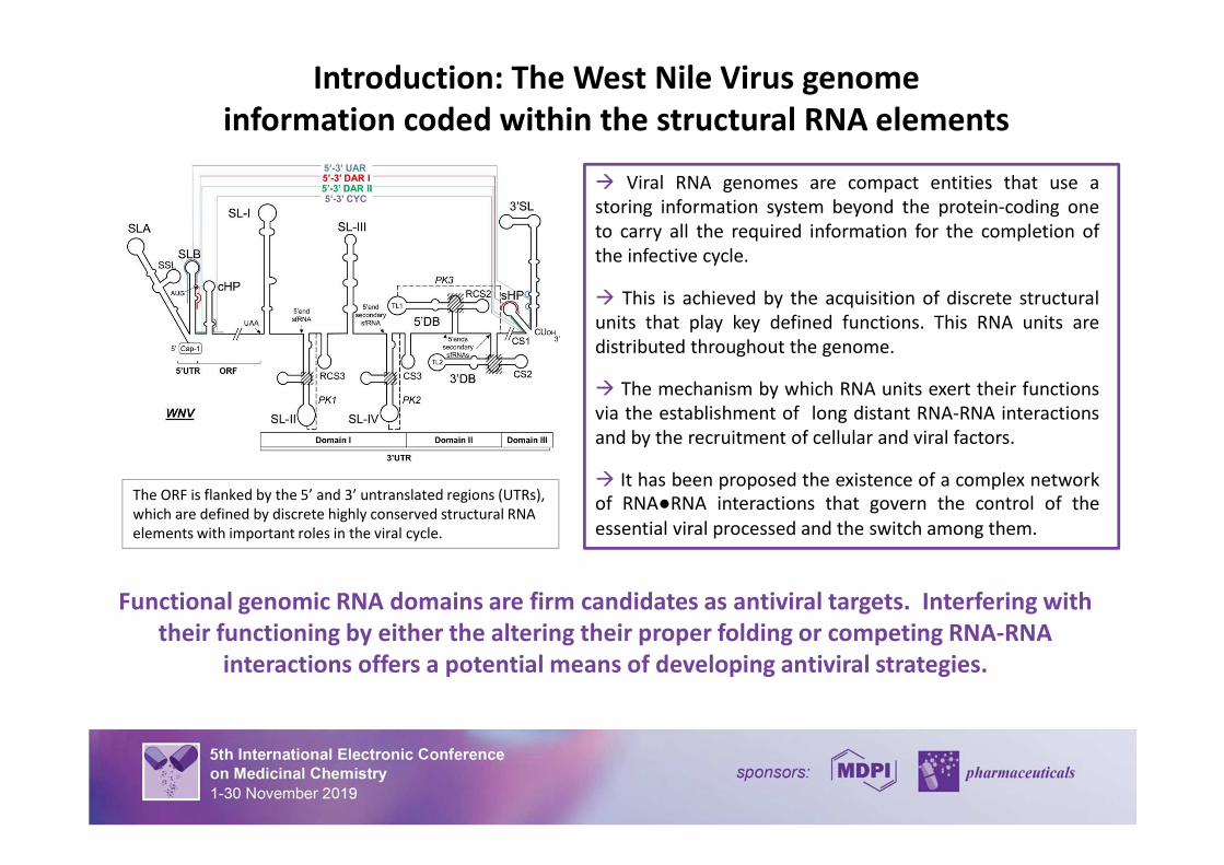
Introduction: The West Nile Virus genome
information coded within the structural RNA elements
The ORF is flanked by the 5’ and 3’ untranslated regions (UTRs),
which are defined by discrete highly conserved structural RNA
elements with important roles in the viral cycle.
� It has been proposed the existence of a complex network
of RNA●RNA interactions that govern the control of the
essential viral processed and the switch among them.
� Viral RNA genomes are compact entities that use a
storing information system beyond the protein-coding one
to carry all the required information for the completion of
the infective cycle.
� The mechanism by which RNA units exert their functions
via the establishment of long distant RNA-RNA interactions
and by the recruitment of cellular and viral factors.
� This is achieved by the acquisition of discrete structural
units that play key defined functions. This RNA units are
distributed throughout the genome.
Functional genomic RNA domains are firm candidates as antiviral targets. Interfering with
their functioning by either the altering their proper folding or competing RNA-RNA
interactions offers a potential means of developing antiviral strategies.
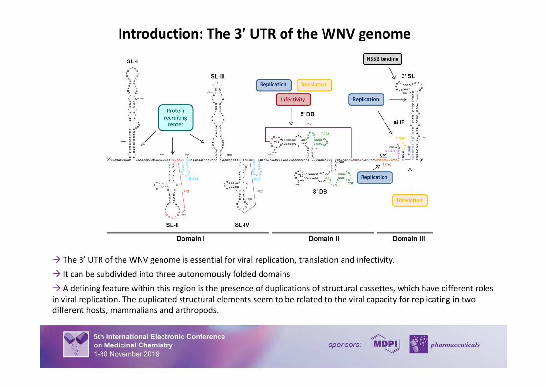
Introduction: The 3’ UTR of the WNV genome
� The 3’ UTR of the WNV genome is essential for viral replication, translation and infectivity.
� It can be subdivided into three autonomously folded domains
� A defining feature within this region is the presence of duplications of structural cassettes, which have different roles
in viral replication. The duplicated structural elements seem to be related to the viral capacity for replicating in two
different hosts, mammalians and arthropods.
Introduction: Role of the 3’ UTR in the WNV translation
� We have recently shown that hepatitis C virus (HCV), a closely related virus
belonging to the family Flaviviridae, recruits the ribosomal subunit 40S by a highly
conserved structural RNA element placed at the 3’ end of the viral genome.
� In this work, we have studied whether the 3’ UTR of the WNV genome recruits
the ribosomes as a means to regulate the viral translation.
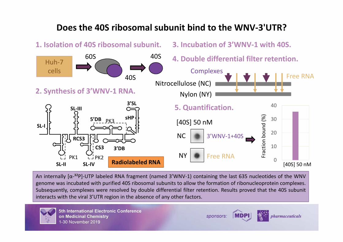
Does the 40S ribosomal subunit bind to the WNV-3'UTR?
1. Isolation of 40S ribosomal subunit.
2. Synthesis of 3’WNV-1 RNA.
4. Double differential filter retention.
Nitrocellulose (NC)
Nylon (NY)
ComplexesFree RNA
5. Quantification.
An internally [α-³²P]-UTP labeled RNA fragment (named 3’WNV-1) containing the last 635 nucleotides of the WNV
genome was incubated with purified 40S ribosomal subunits to allow the formation of ribonucleoprotein complexes.
Subsequently, complexes were resolved by double differential filter retention. Results proved that the 40S subunit
interacts with the viral 3’UTR region in the absence of any other factors.
Huh-7
cells
60S
40S
40S
0
10
20
30
40
Fra
ctio
nb
ou
nd
(%)
[40S] 50 nM
NC
NY
[40S] 50 nM
3’WNV-1+40S
Free RNA
PK3
SL-IV
CS3 3’DB
RCS3
PK1
SL-II
SL-III3’SL
PK2
sHP5’DB
SL-I
Radiolabeled RNA
3. Incubation of 3’WNV-1 with 40S.
11
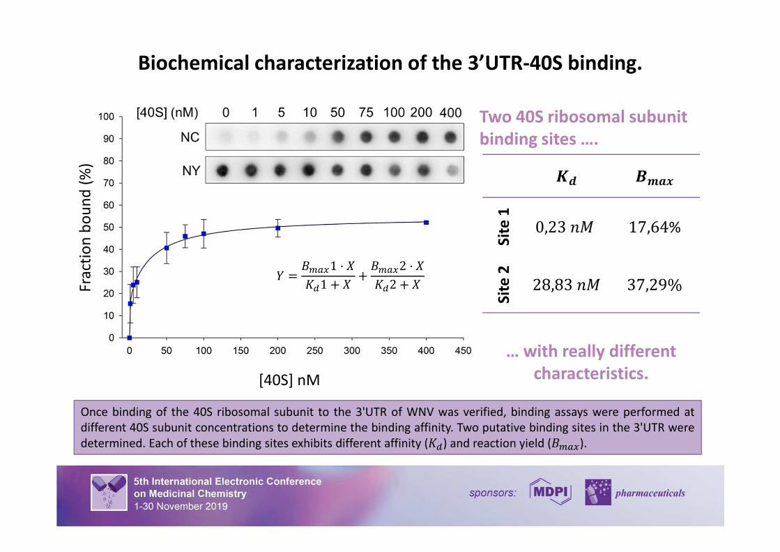
Biochemical characterization of the 3’UTR-40S binding.
Once binding of the 40S ribosomal subunit to the 3'UTR of WNV was verified, binding assays were performed at
different 40S subunit concentrations to determine the binding affinity. Two putative binding sites in the 3'UTR were
determined. Each of these binding sites exhibits different affinity (��) and reaction yield (����).
Two 40S ribosomal subunit
binding sites ….
… with really different
characteristics.
Fra
ctio
n b
ou
nd
(%
)
[40S] nM
�� ��
Sit
e1
0,23�� 17,64%
Sit
e2
28,83�� 37,29%� �
����1 � �
��1 � ������2 � �
��2 � �
12
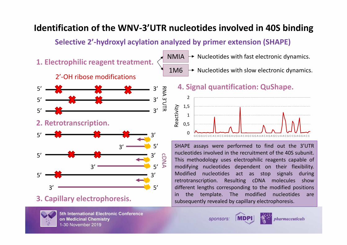
SHAPE assays were performed to find out the 3’UTR
nucleotides involved in the recruitment of the 40S subunit.
This methodology uses electrophilic reagents capable of
modifying nucleotides dependent on their flexibility.
Modified nucleotides act as stop signals during
retrotranscription. Resulting cDNA molecules show
different lengths corresponding to the modified positions
in the template. The modified nucleotides are
subsequently revealed by capillary electrophoresis.
2. Retrotranscription.
cDN
A
5’ 3’
3’ 5’
3’5’
3’ 5’
3. Capillary electrophoresis.
3’5’
3’ 5’
0
0,5
1
1,5
2
G C G G U C U C A C A C C A G G A U G U A C A G C G G A U A C A G A G C U A A C G C C G A G G A C C
Re
act
ivit
y
4. Signal quantification: QuShape.
Identification of the WNV-3’UTR nucleotides involved in 40S binding
1. Electrophilic reagent treatment.NMIA
1M6
Nucleotides with fast electronic dynamics.
Nucleotides with slow electronic dynamics.
Selective 2’-hydroxyl acylation analyzed by primer extension (SHAPE)
2’-OH ribose modifications
RN
A 3
’UT
R
3’5’
5’ 3’
5’ 3’
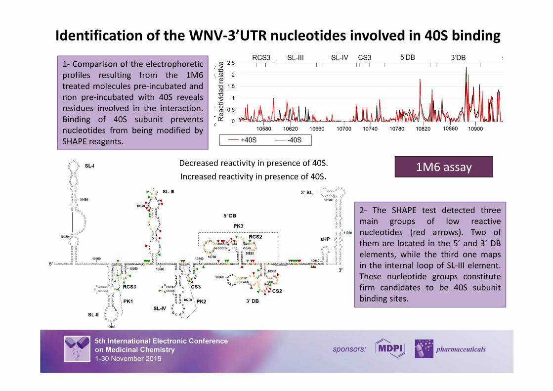
2- The SHAPE test detected three
main groups of low reactive
nucleotides (red arrows). Two of
them are located in the 5’ and 3’ DB
elements, while the third one maps
in the internal loop of SL-III element.
These nucleotide groups constitute
firm candidates to be 40S subunit
binding sites.
1313
Re
lati
ve r
ea
ctiv
ity
131313131313
Decreased reactivity in presence of 40S.
Increased reactivity in presence of 40S.
Identification of the WNV-3’UTR nucleotides involved in 40S binding
1- Comparison of the electrophoretic
profiles resulting from the 1M6
treated molecules pre-incubated and
non pre-incubated with 40S reveals
residues involved in the interaction.
Binding of 40S subunit prevents
nucleotides from being modified by
SHAPE reagents.
1M6 assay
2- The SHAPE test detected three
main groups of low reactive
nucleotides (red arrows). Two of
them are located in the 5’ and 3’ DB
elements, while the third one maps
in the internal loop of SL-III element.
These nucleotide groups constitute
firm candidates to be 40S subunit
binding sites.
Decreased reactivity in presence of 40S.
Increased reactivity in presence of 40S.
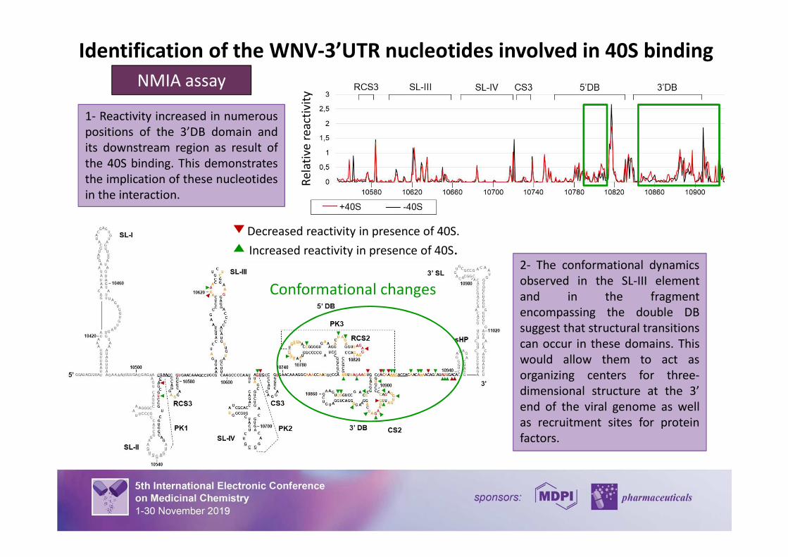
1- Reactivity increased in numerous
positions of the 3’DB domain and
its downstream region as result of
the 40S binding. This demonstrates
the implication of these nucleotides
in the interaction.
Re
lati
ve r
ea
ctiv
ity
NMIA assay
Identification of the WNV-3’UTR nucleotides involved in 40S binding
Conformational changes
2- The conformational dynamics
observed in the SL-III element
and in the fragment
encompassing the double DB
suggest that structural transitions
can occur in these domains. This
would allow them to act as
organizing centers for three-
dimensional structure at the 3’
end of the viral genome as well
as recruitment sites for protein
factors.
Decreased reactivity in presence of 40S.
Increased reactivity in presence of 40S.
Kd
(nM
)
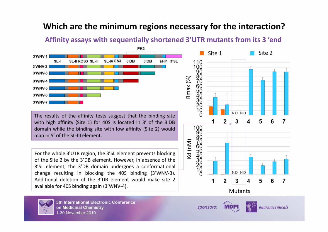
Which are the minimum regions necessary for the interaction?
The results of the affinity tests suggest that the binding site
with high affinity (Site 1) for 40S is located in 3’ of the 3'DB
domain while the binding site with low affinity (Site 2) would
map in 5’ of the SL-III element.
Affinity assays with sequentially shortened 3’UTR mutants from its 3 ’end
For the whole 3'UTR region, the 3’SL element prevents blocking
of the Site 2 by the 3’DB element. However, in absence of the
3’SL element, the 3’DB domain undergoes a conformational
change resulting in blocking the 40S binding (3’WNV-3).
Additional deletion of the 3’DB element would make site 2
available for 40S binding again (3’WNV-4).Mutants
Site 1 Site 2
Bm
ax
(%)
16
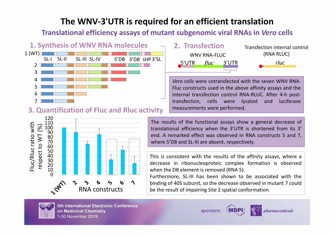
The WNV-3'UTR is required for an efficient translation
Translational efficiency assays of mutant subgenomic viral RNAs in Vero cells
1. Synthesis of WNV RNA molecules
0102030405060708090
100110120
Flu
c/R
luc
rati
o w
ith
resp
ect
toW
T (
%)
2. Transfection
3. Quantification of Fluc and Rluc activity
fluc5’UTR 3’UTR
Vero cells were cotransfected with the seven WNV RNA-
Fluc constructs used in the above affinity assays and the
internal transfection control RNA-RLUC. After 4-h post-
transfection, cells were lysated and luciferase
measurements were performed.
RNA constructs
The results of the functional assays show a general decrease of
translational efficiency when the 3’UTR is shortened from its 3’
end. A remarked effect was observed in RNA constructs 5 and 7,
where 5’DB and SL-III are absent, respectively.
WNV RNA-FLUC
Transfection internal control
(RNA RLUC)1 (WT)
2
3
4
5
6
7
SL-III SL-IV 5’DB 3’DB 3’SLsHPSL-IISL-Irluc
This is consistent with the results of the affinity assays, where a
decrease in ribonucleoproteic complex formation is observed
when the DB element is removed (RNA 5).
Furthermore, SL-III has been shown to be associated with the
binding of 40S subunit, so the decrease observed in mutant 7 could
be the result of impairing Site 2 spatial conformation.
0102030405060708090
100110
Flu
c/R
luc
rati
o w
ith
resp
ect
to
WT
(%
)
17
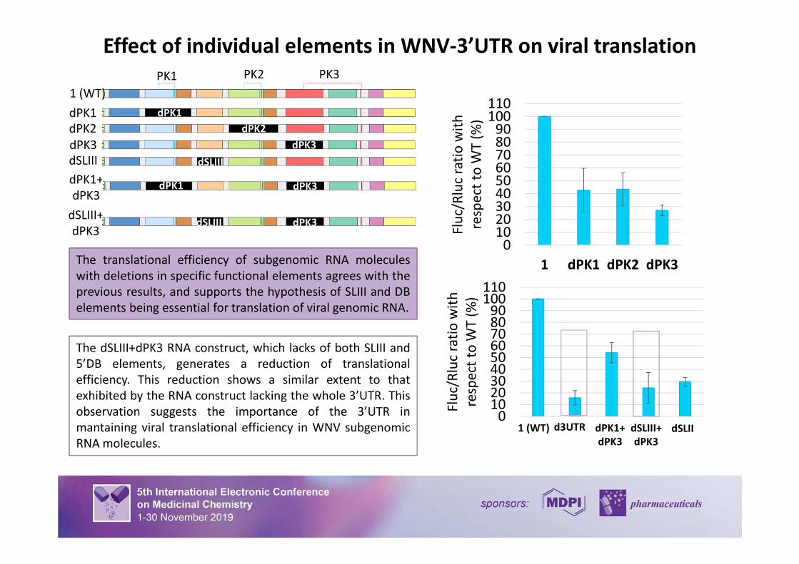
Effect of individual elements in WNV-3’UTR on viral translation
The translational efficiency of subgenomic RNA molecules
with deletions in specific functional elements agrees with the
previous results, and supports the hypothesis of SLIII and DB
elements being essential for translation of viral genomic RNA.
1 (WT)
dPK1
dPK2
dPK3
dSLIII
dPK1+
dPK3
dSLIII+
dPK3
PK1 PK2 PK3
dPK1
dPK2
dPK3
dPK3dPK1
dSLIII
dSLIII dPK3
dPK1 dPK3
d3UTR dSLIII+
dPK3
dSLIIdPK1+
dPK3
1 (WT)
The dSLIII+dPK3 RNA construct, which lacks of both SLIII and
5’DB elements, generates a reduction of translational
efficiency. This reduction shows a similar extent to that
exhibited by the RNA construct lacking the whole 3’UTR. This
observation suggests the importance of the 3’UTR in
mantaining viral translational efficiency in WNV subgenomic
RNA molecules.
0102030405060708090
100110
1 dPK1 dPK2 dPK3
Flu
c/R
luc
rati
o w
ith
resp
ect
toW
T (
%)
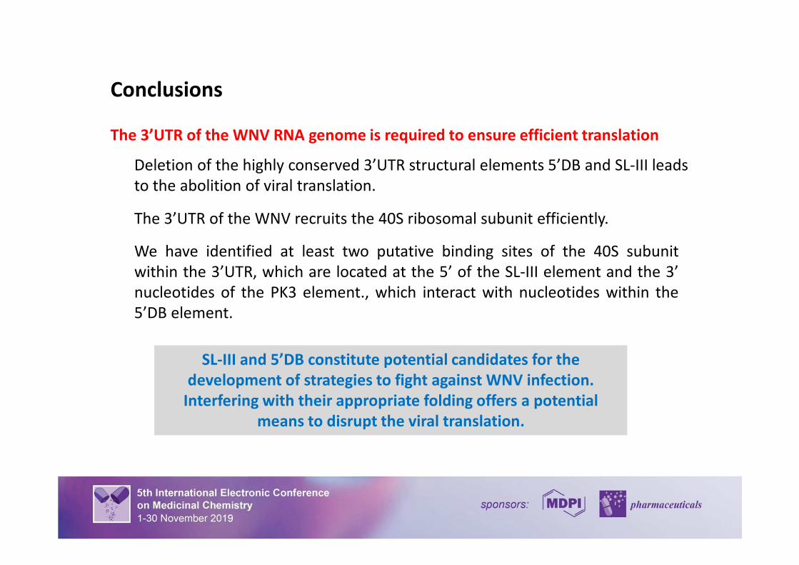
Conclusions
18
The 3’UTR of the WNV RNA genome is required to ensure efficient translation
Deletion of the highly conserved 3’UTR structural elements 5’DB and SL-III leads
to the abolition of viral translation.
The 3’UTR of the WNV recruits the 40S ribosomal subunit efficiently.
We have identified at least two putative binding sites of the 40S subunit
within the 3’UTR, which are located at the 5’ of the SL-III element and the 3’
nucleotides of the PK3 element., which interact with nucleotides within the
5’DB element.
SL-III and 5’DB constitute potential candidates for the
development of strategies to fight against WNV infection.
Interfering with their appropriate folding offers a potential
means to disrupt the viral translation.
Acknowledgments
19
Recommended