Teresa Camp-Rogers, MDDepartment of Emergency Medicine
Virginia Commonwealth University Medical CenterRichmond, Virginia
The Basic 12 LeadElectrocardiogram

Objectives
• How do they work?• Common mistakes• How to read an EKG• Artifacts• Cases
What is an EKG?
• Tracing of electrical activity of the heart
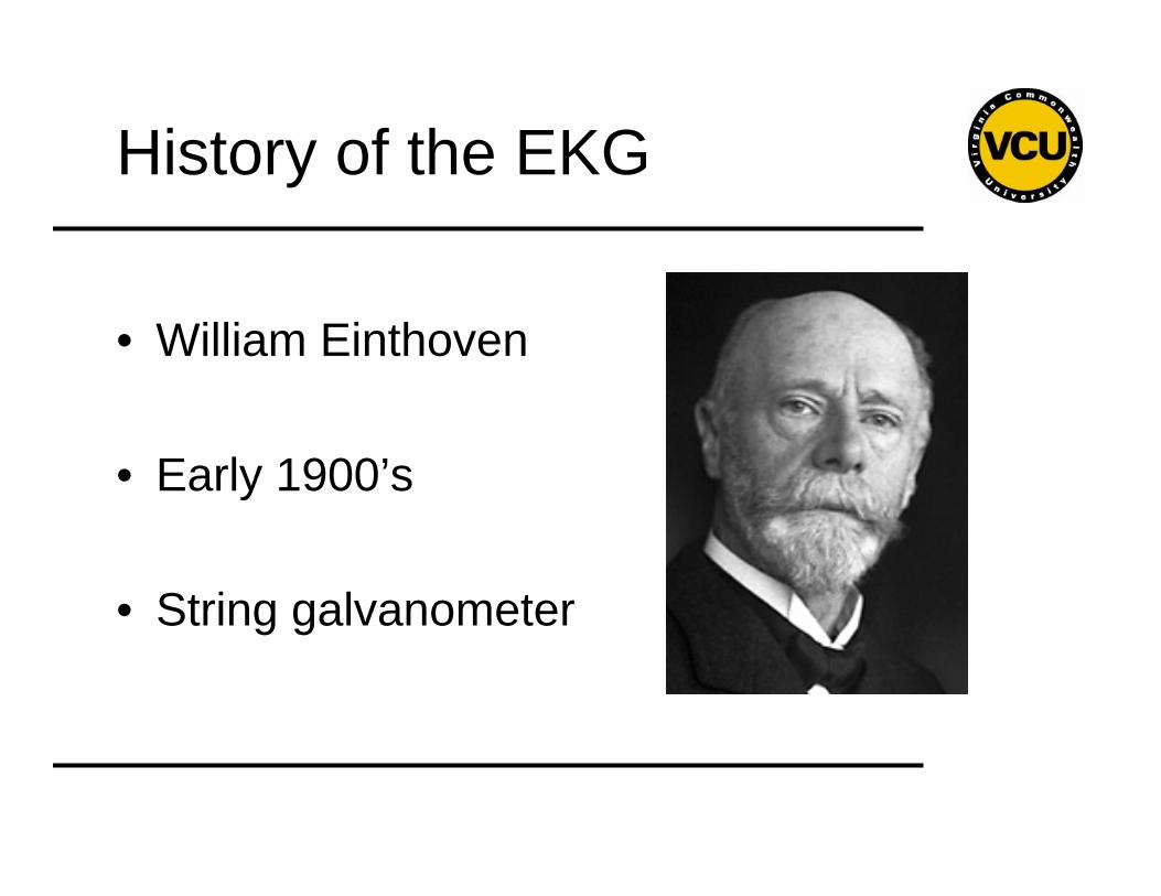
History of the EKG
• William Einthoven
• Early 1900’s
• String galvanometer
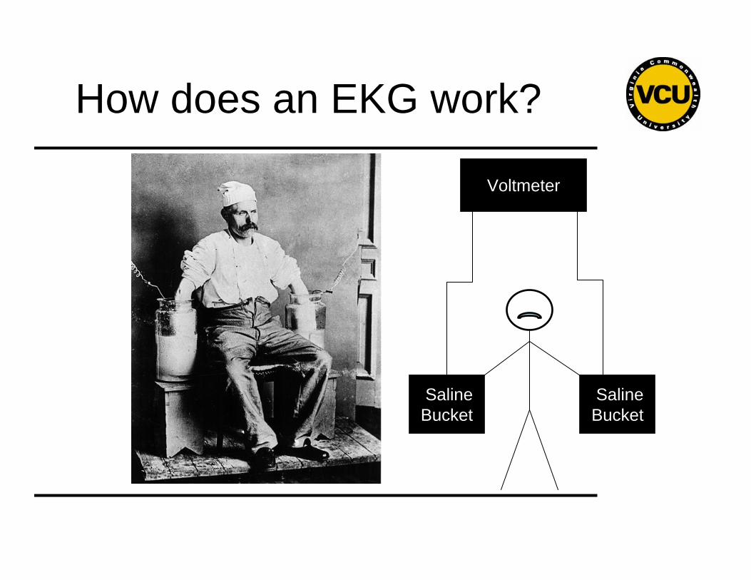
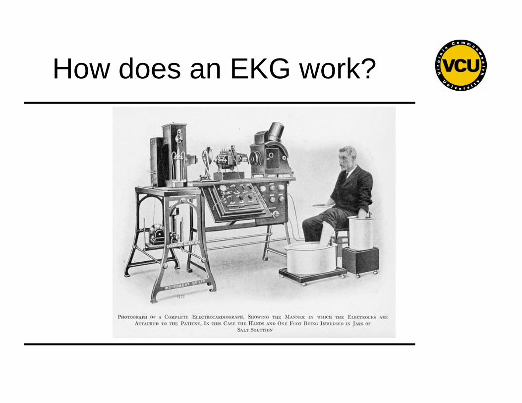
How does an EKG work?
Voltmeter
Saline Bucket
Saline Bucket
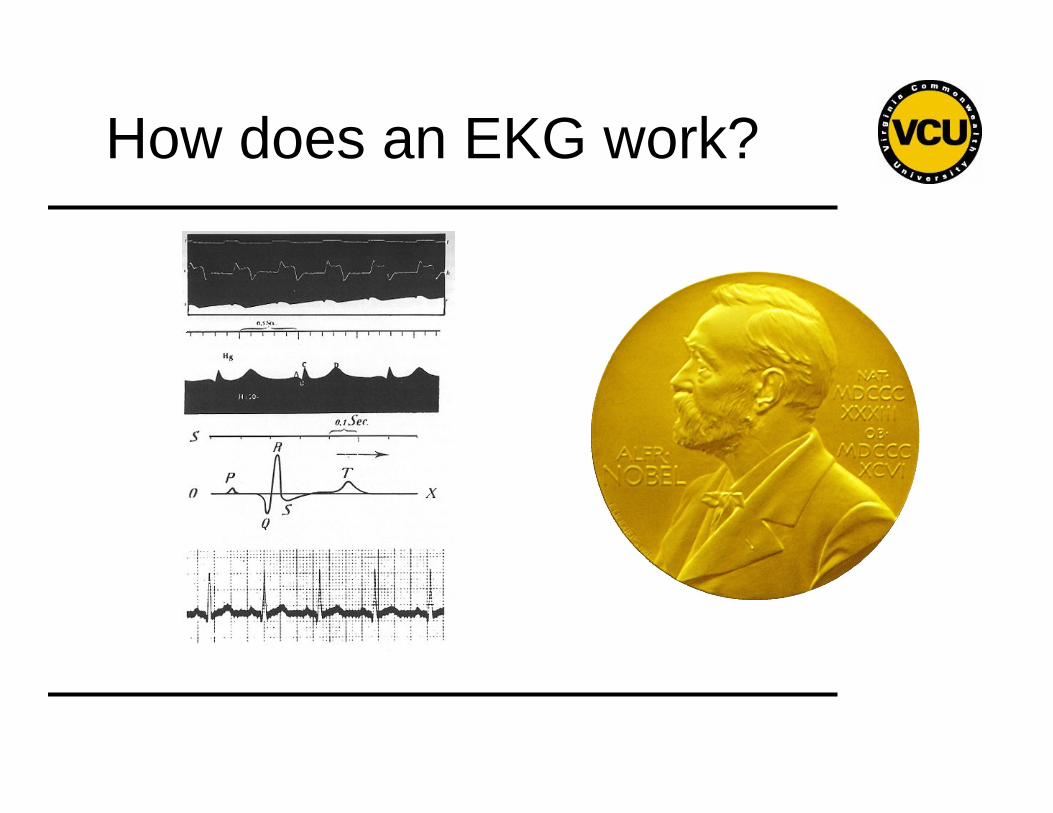
How does an EKG work?
How does an EKG work?
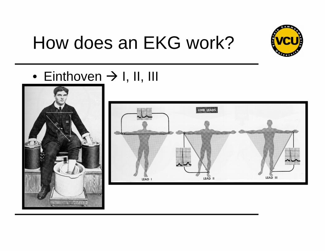
How does an EKG work?
• Einthoven I, II, III
How does an EKG work?
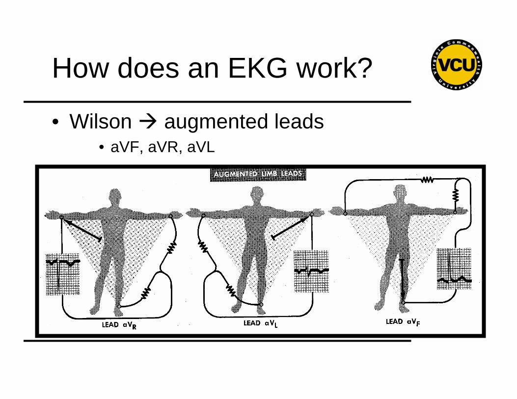
• Wilson augmented leads• aVF, aVR, aVL
How does an EKG work?
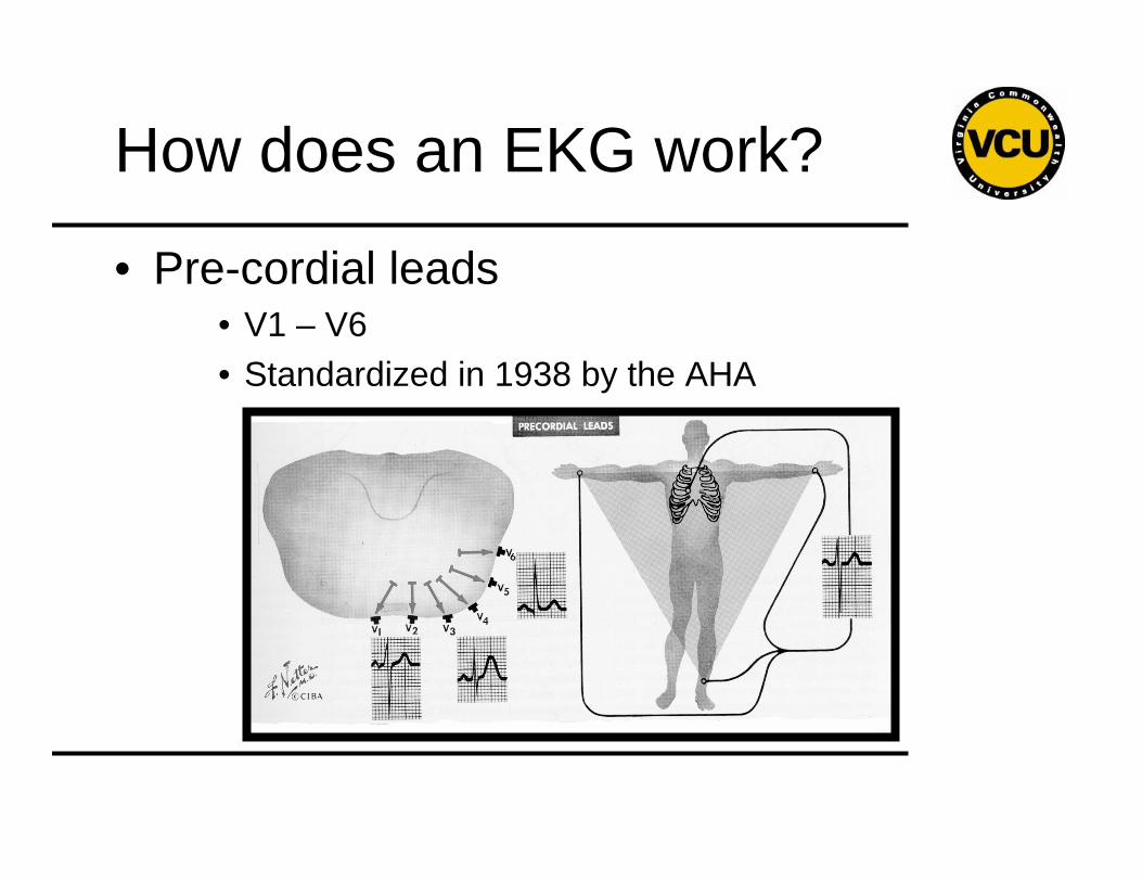
• Pre-cordial leads• V1 – V6• Standardized in 1938 by the AHA
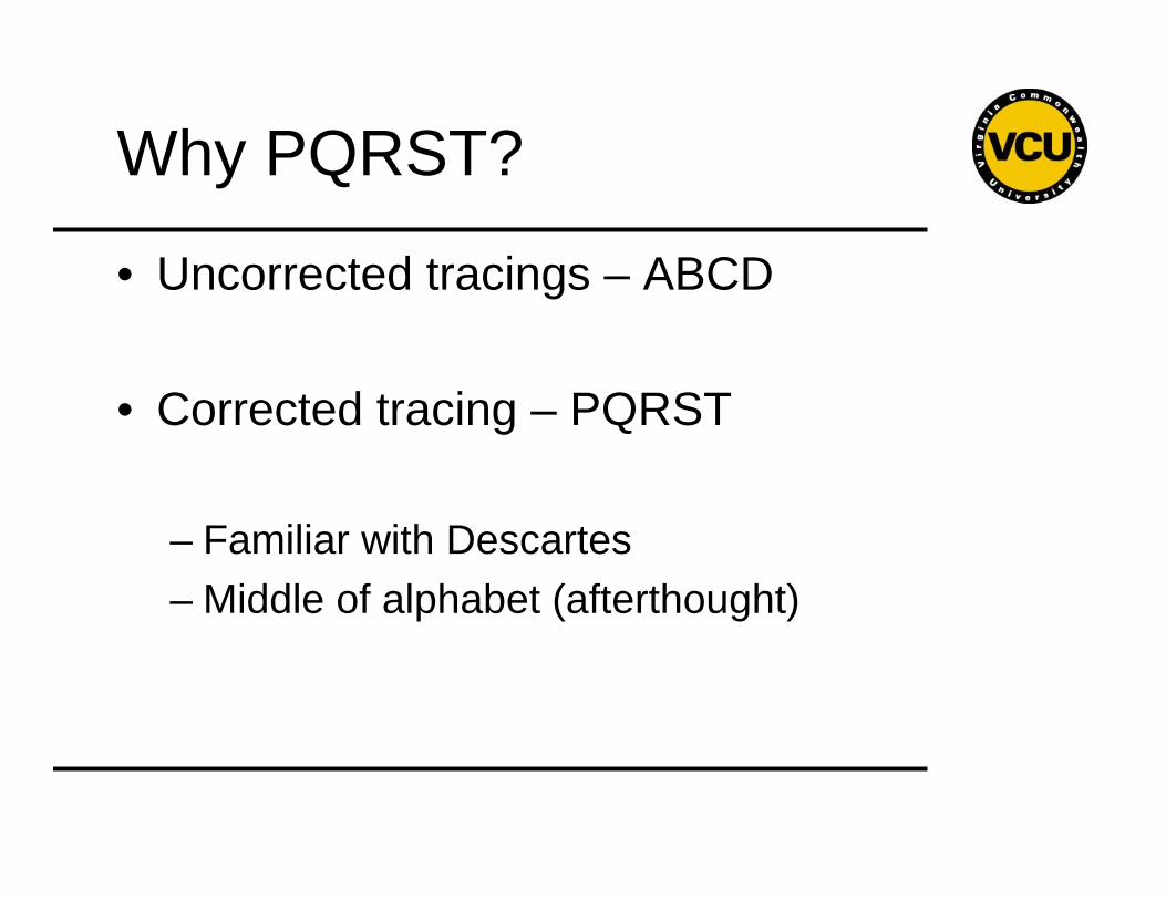
Why PQRST?
• Uncorrected tracings – ABCD
• Corrected tracing – PQRST
– Familiar with Descartes– Middle of alphabet (afterthought)
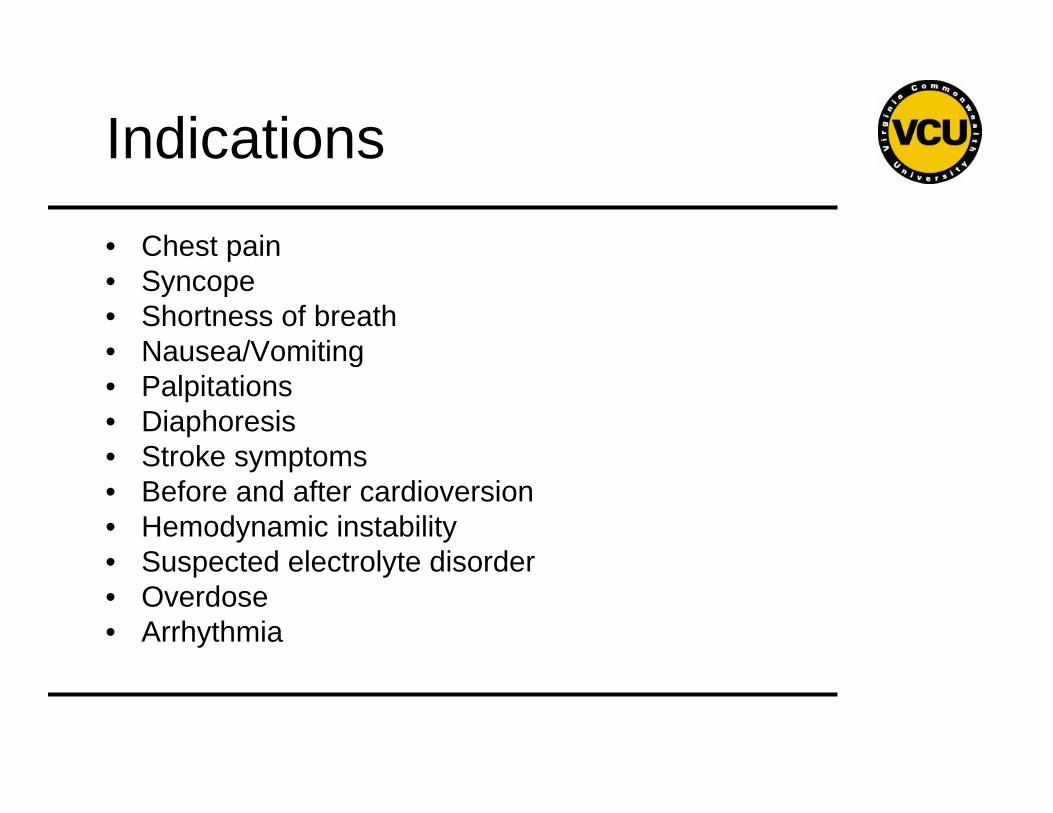
Indications
• Chest pain• Syncope• Shortness of breath• Nausea/Vomiting• Palpitations• Diaphoresis• Stroke symptoms• Before and after cardioversion• Hemodynamic instability• Suspected electrolyte disorder• Overdose• Arrhythmia
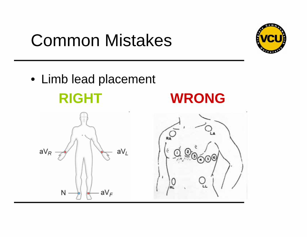
Common Mistakes
• Limb lead placementRIGHT WRONG
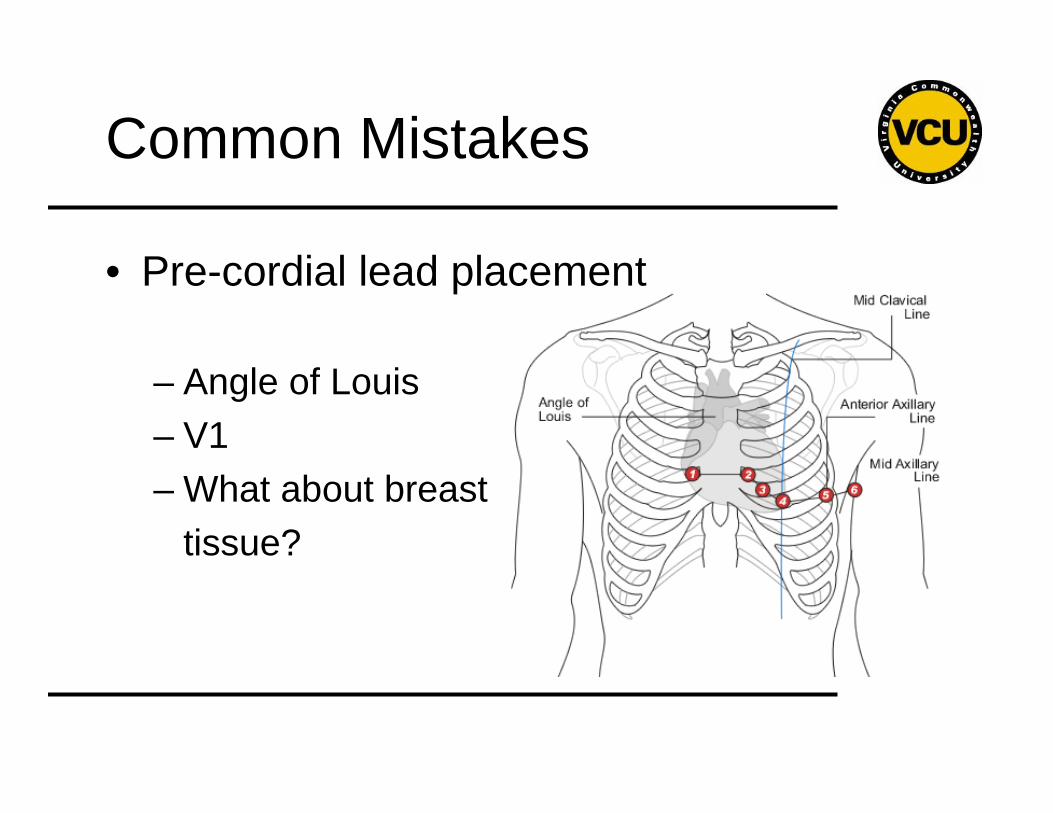
Common Mistakes
• Pre-cordial lead placement
– Angle of Louis– V1– What about breast
tissue?
How to read an EKG
• The Paper
• The Waveform
• The Plan
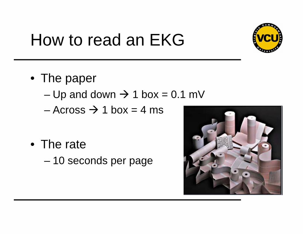
How to read an EKG
• The paper– Up and down 1 box = 0.1 mV– Across 1 box = 4 ms
• The rate – 10 seconds per page
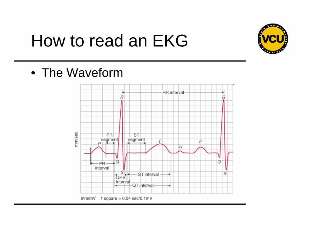
How to read an EKG
• The Waveform
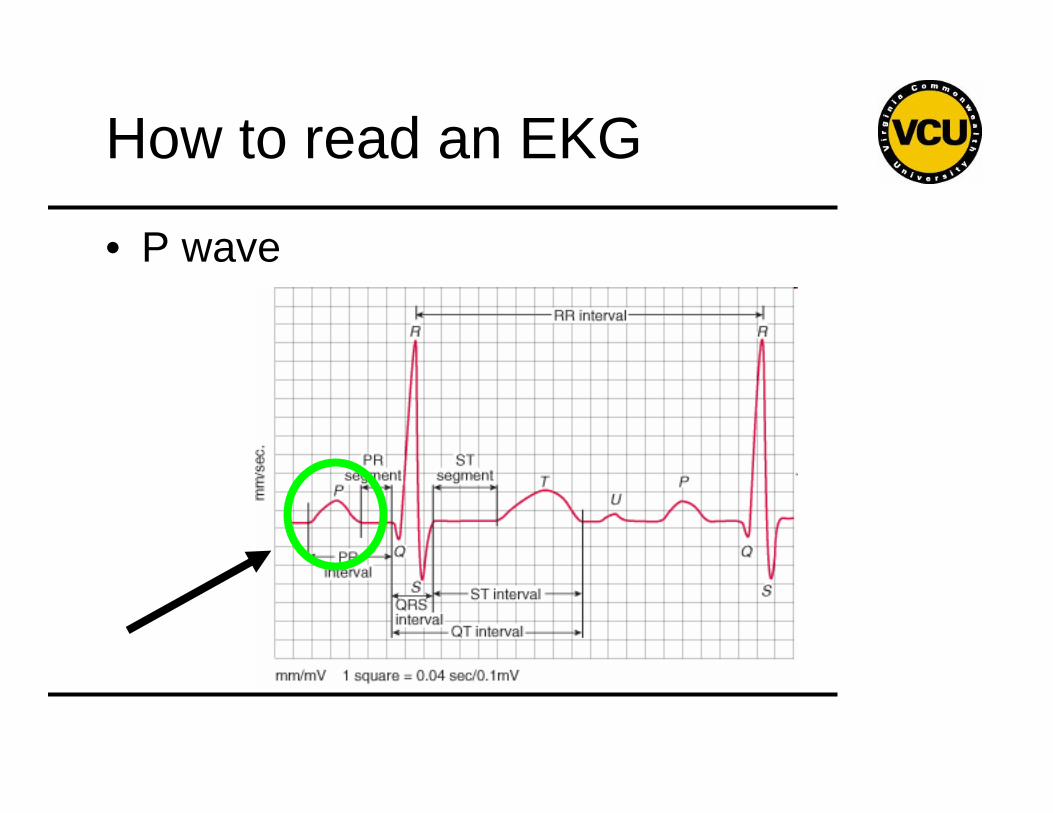
How to read an EKG
• P wave
How to read an EKG
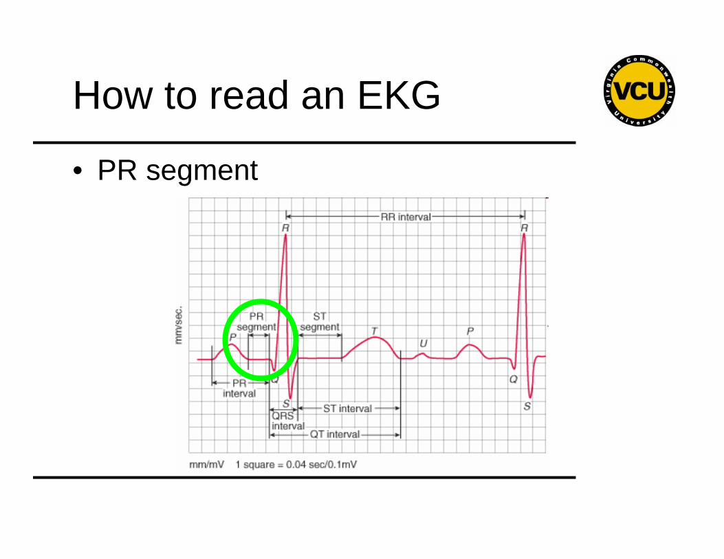
• PR segment
How to read an EKG
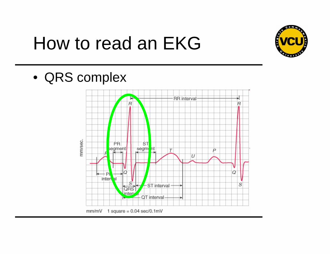
• QRS complex
How to read an EKG
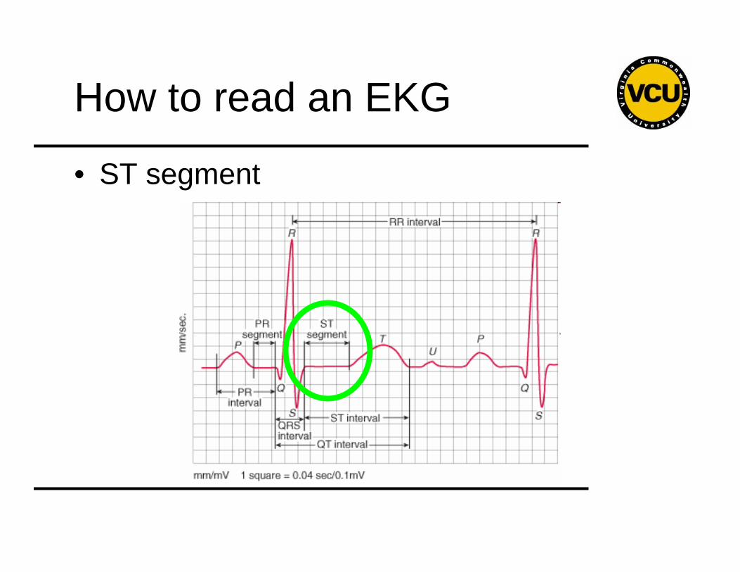
• ST segment
How to read an EKG
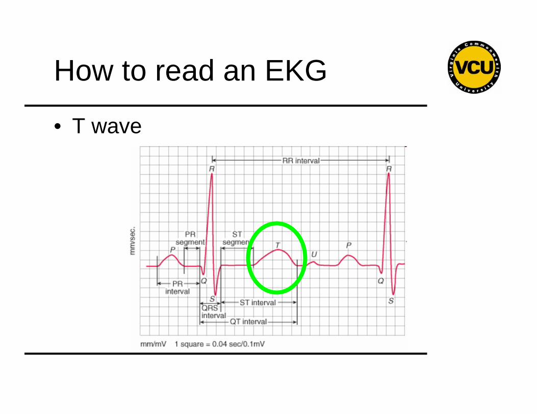
• T wave
How to read an EKG
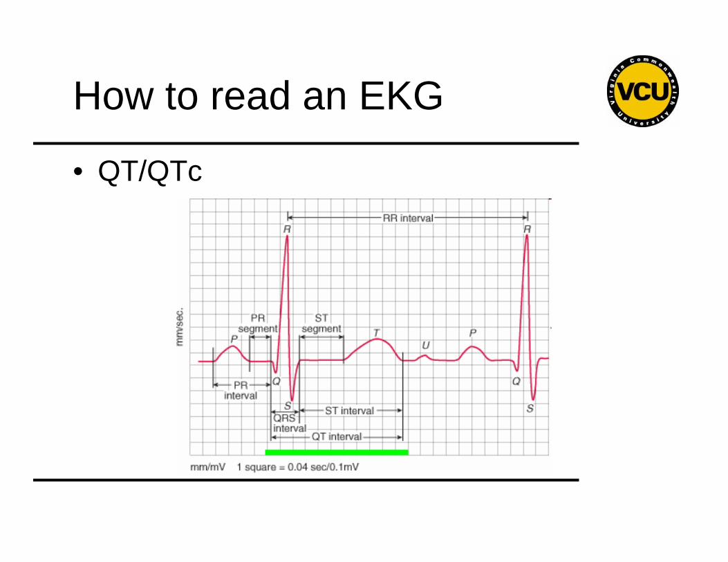
• QT/QTc
How to read an EKG
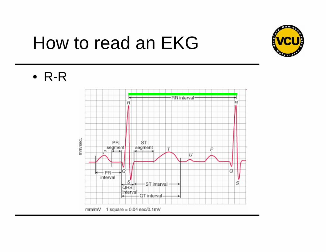
• R-R
The Plan
• Rate
• Rhythm
• Axis
• Interval
• Disease
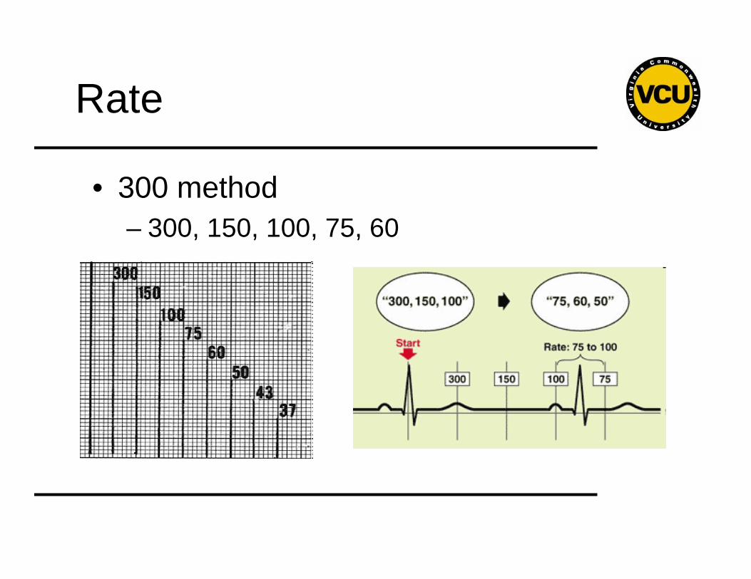
Rate
• 300 method – 300, 150, 100, 75, 60
Rate
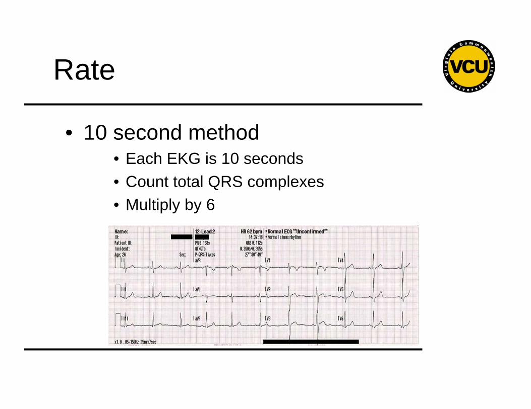
• 10 second method• Each EKG is 10 seconds• Count total QRS complexes • Multiply by 6
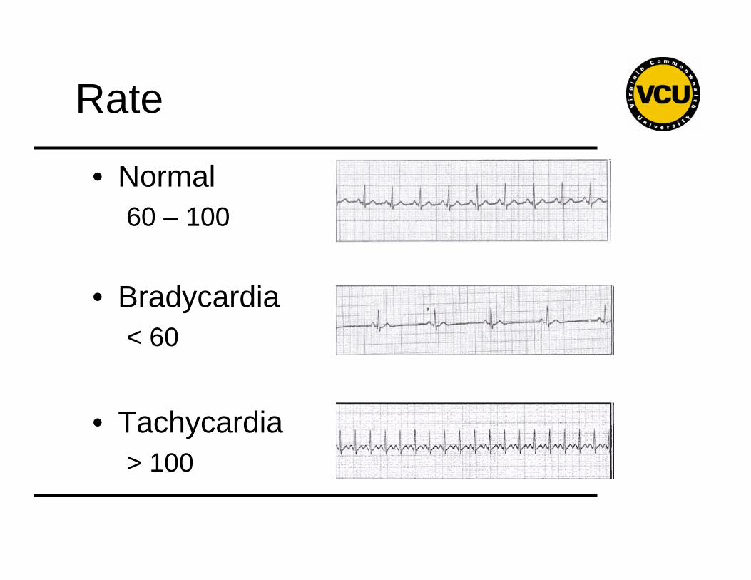
Rate
• Normal 60 – 100
• Bradycardia< 60
• Tachycardia> 100
Rhythm
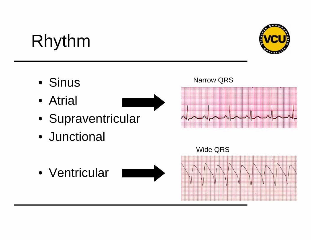
• Sinus• Atrial• Supraventricular• Junctional
• Ventricular
Narrow QRS
Wide QRS
Rhythm
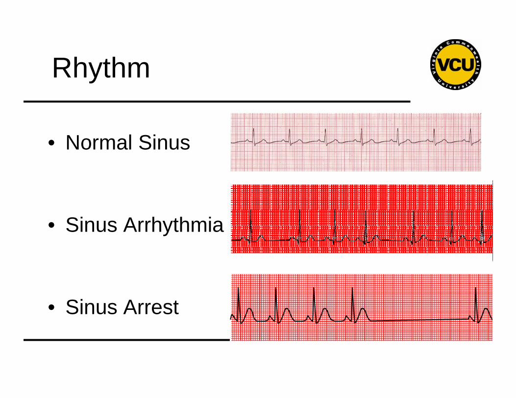
• Normal Sinus
• Sinus Arrhythmia
• Sinus Arrest
Rhythm
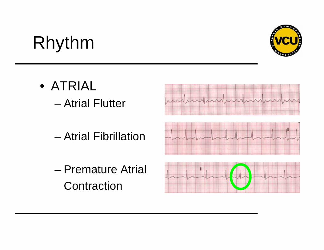
• ATRIAL– Atrial Flutter
– Atrial Fibrillation
– Premature AtrialContraction
Rhythm
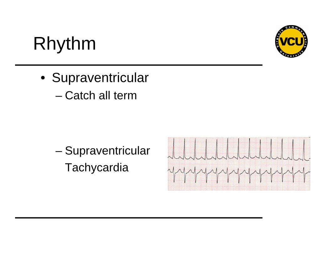
• Supraventricular– Catch all term
– Supraventricular Tachycardia
Rhythm
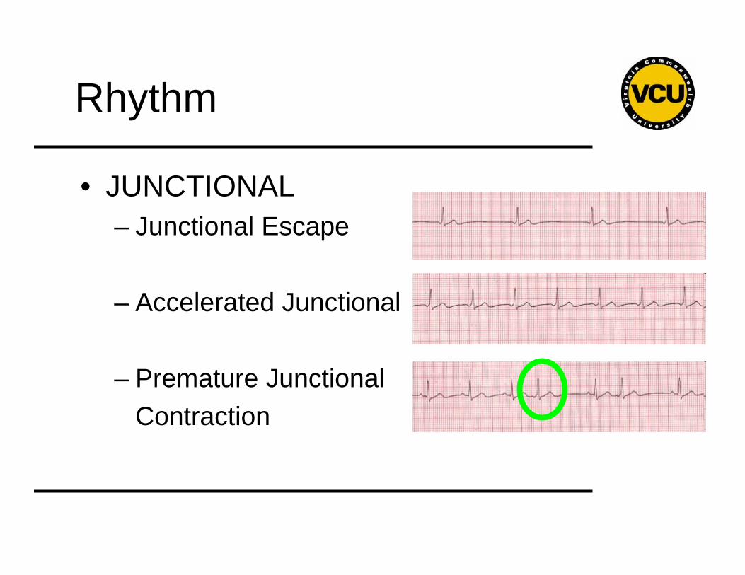
• JUNCTIONAL– Junctional Escape
– Accelerated Junctional
– Premature Junctional Contraction
Rhythm
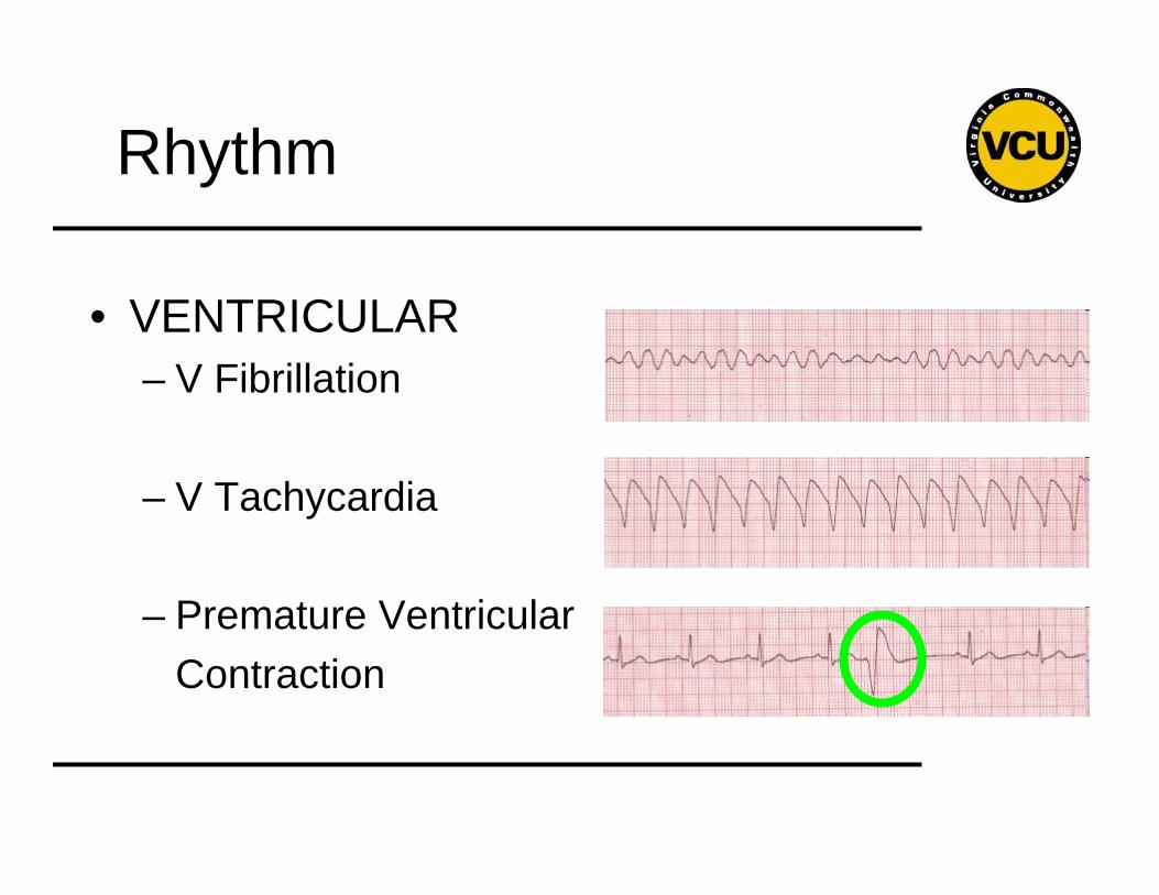
• VENTRICULAR– V Fibrillation
– V Tachycardia
– Premature Ventricular Contraction
Axis
• General direction of electrical activity
• Will not change your management
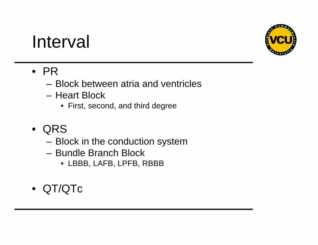
Interval• PR
– Block between atria and ventricles– Heart Block
• First, second, and third degree
• QRS– Block in the conduction system– Bundle Branch Block
• LBBB, LAFB, LPFB, RBBB
• QT/QTc
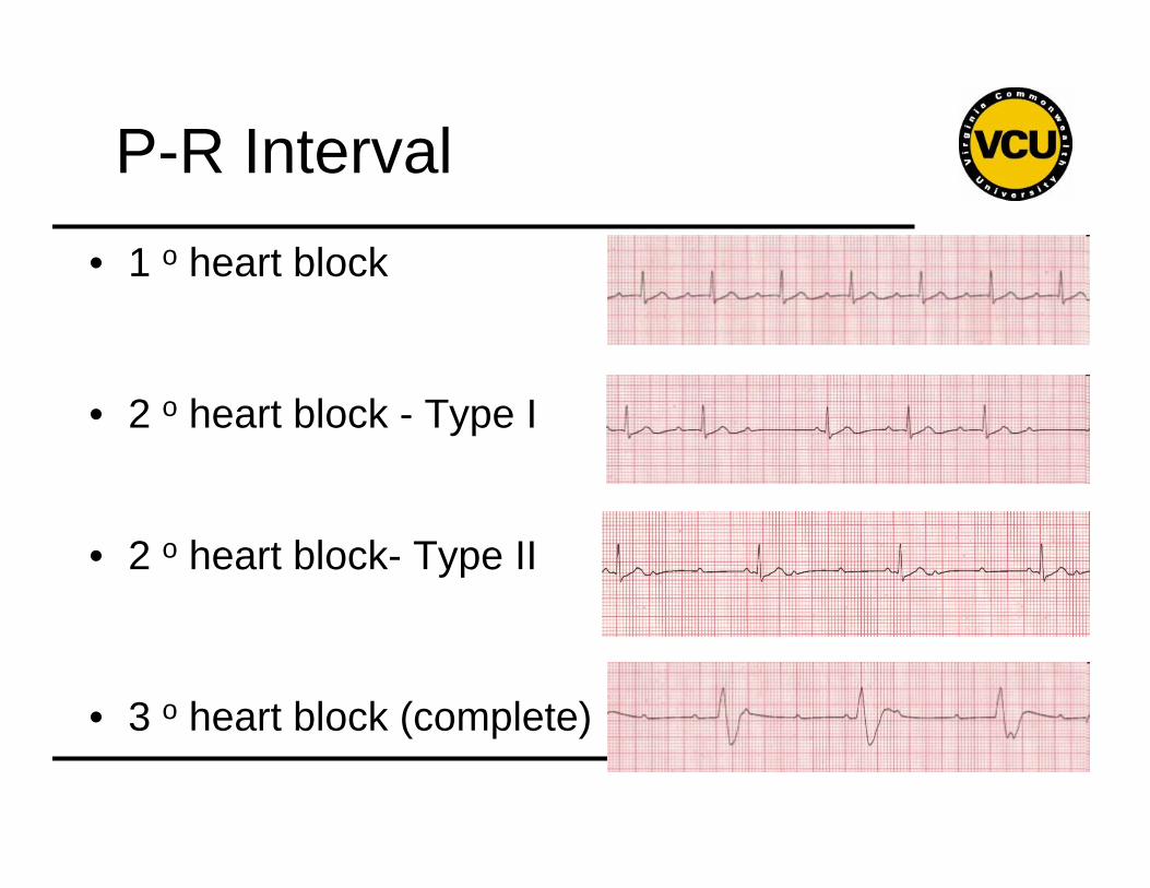
P-R Interval• 1 o heart block
• 2 o heart block - Type I
• 2 o heart block- Type II
• 3 o heart block (complete)
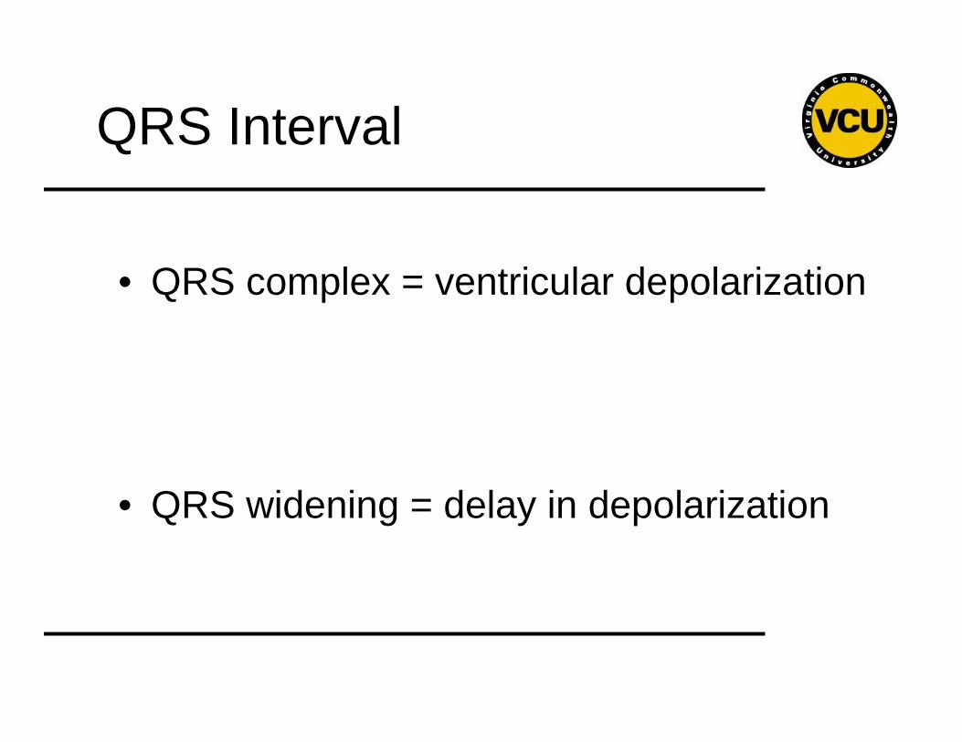
QRS Interval
• QRS complex = ventricular depolarization
• QRS widening = delay in depolarization
QRS Interval
• Causes of QRS widening
– Ventricular rhythm
– Damage to the conduction system• BBB• MI
– Metabolic/Drugs
QRS Interval
QT/QTc Interval
• QT
– Normal
– Prolonged
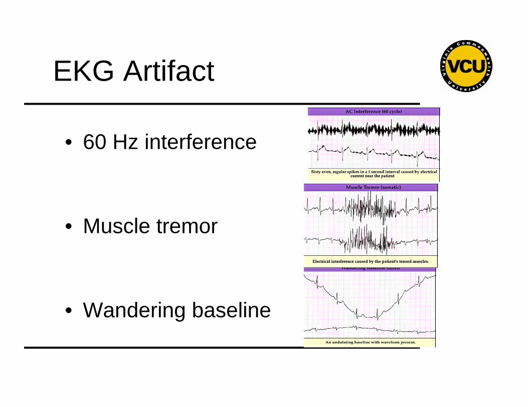
EKG Artifact
• 60 Hz interference
• Muscle tremor
• Wandering baseline
12 leads
• So far we’ve just done basic rhythm recognition with a single lead.
• What about the other 11 leads?
12 leads
• Each lead represents a different view of the heart
• More = better.
12 leads
• II p waves
• Axis
• Diseases– Myocardial infarction– PE– Hyperkalemia– Pericarditis
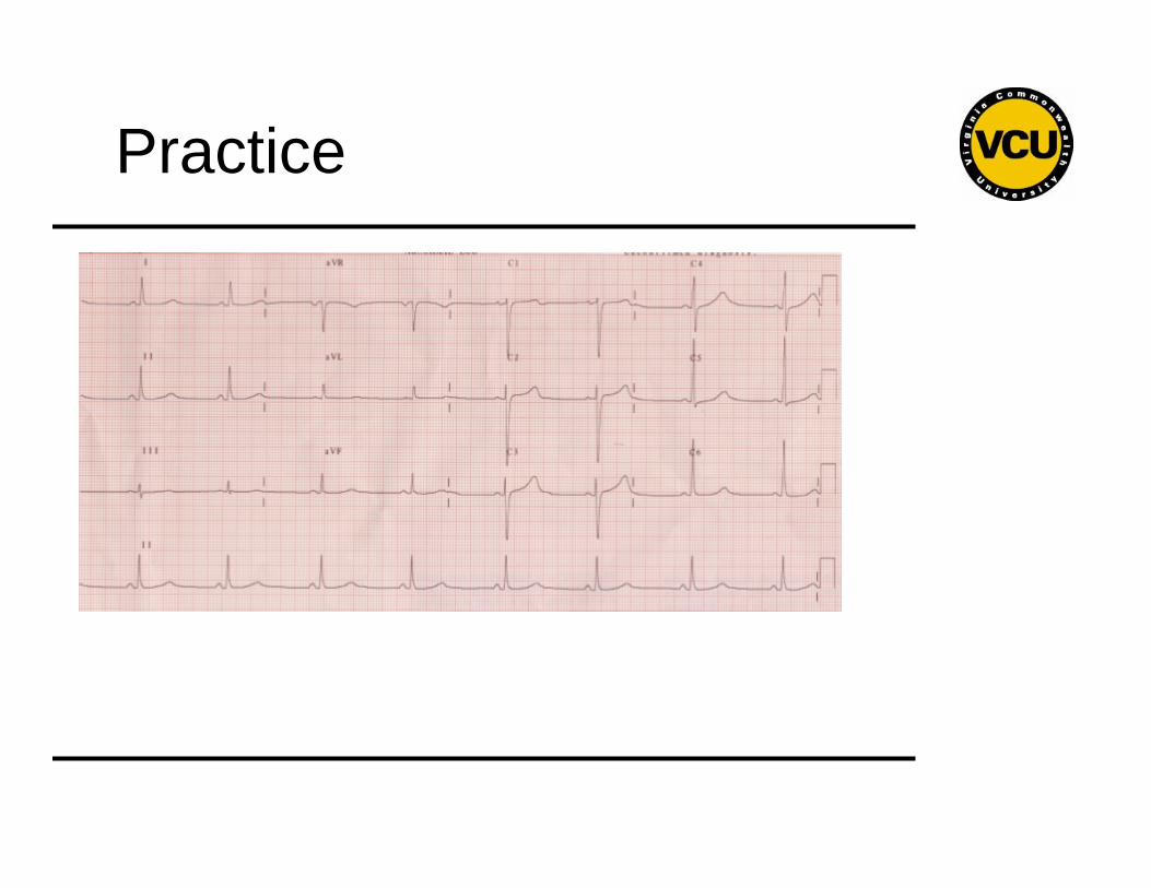
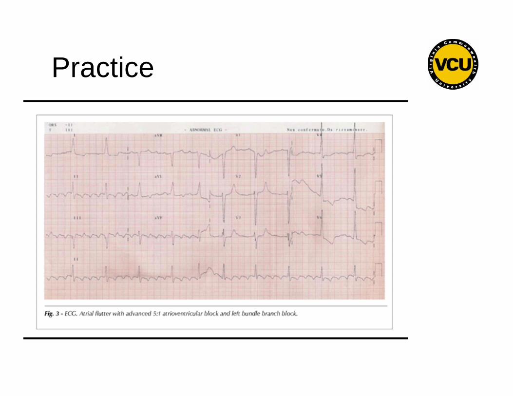
Practice
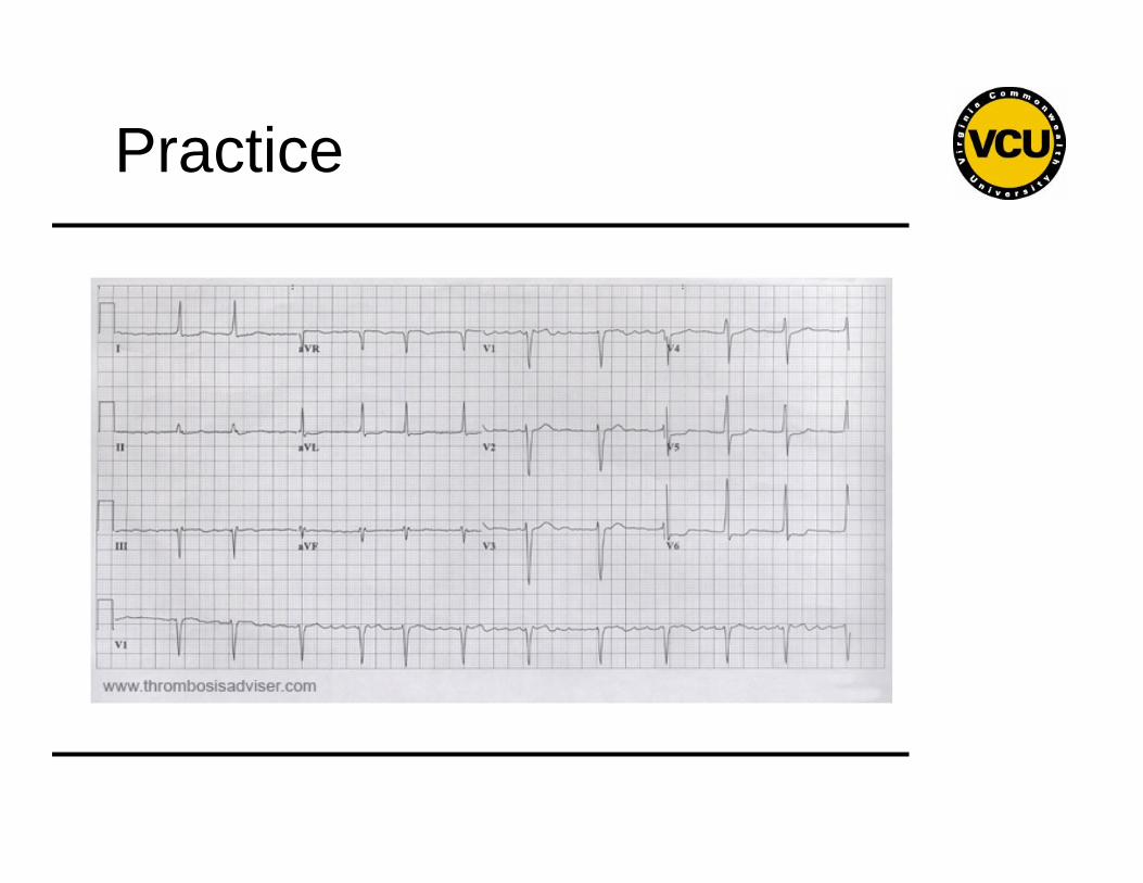
Practice
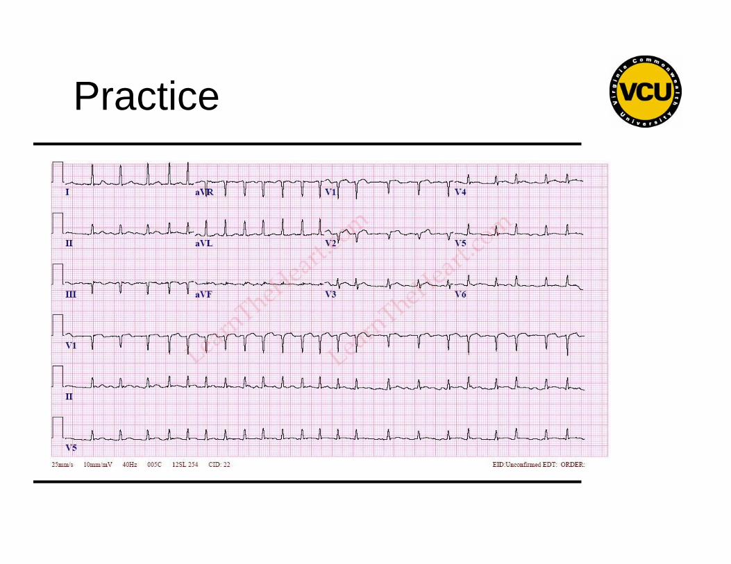
Practice
Practice
Recommended