The Cardiovascular System
Chapter 15

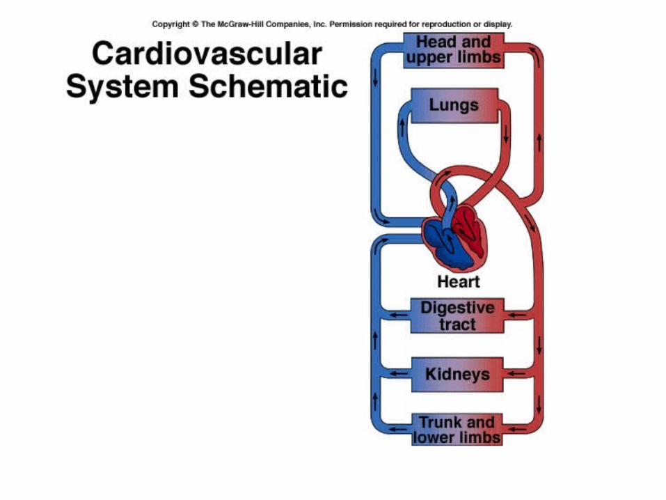
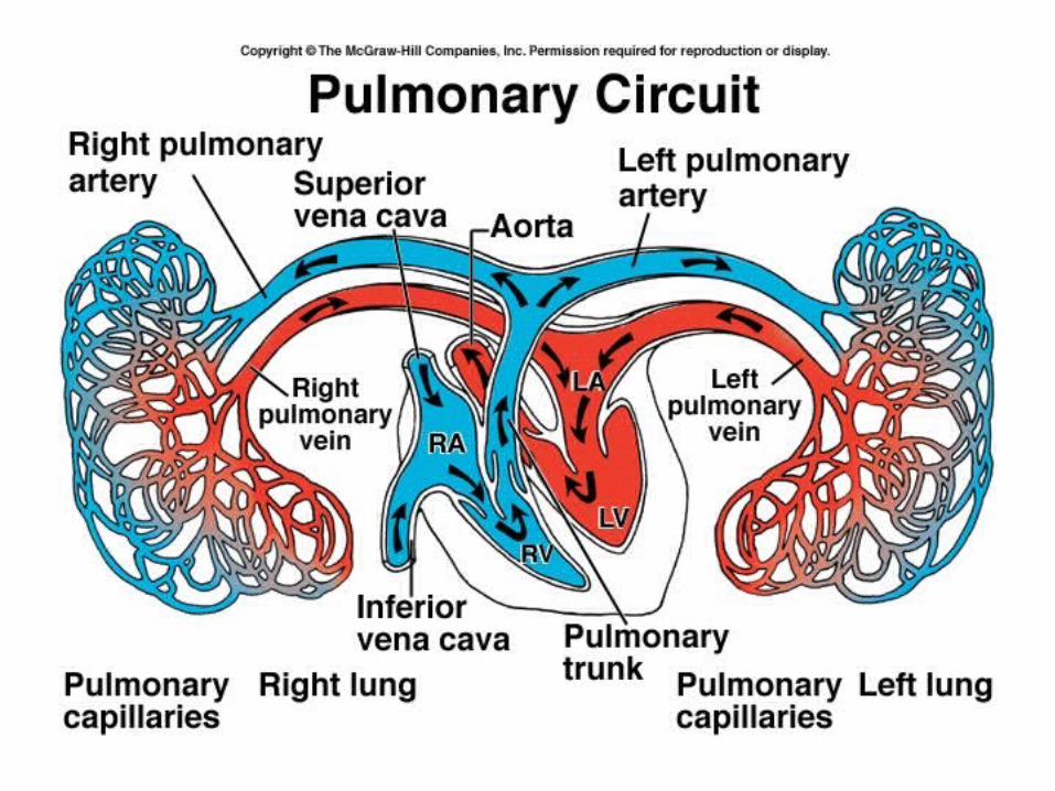
Heart is two pumps in one:
Right side – pulmonary circulation
Left side – systemic circulation
Heart→ Arteries → Arterioles → Capillaries → Venules→ Veins → Heart
Artery – any vessels that carries blood away from the heart.
Vein – any vessels that carries blood toward the heart

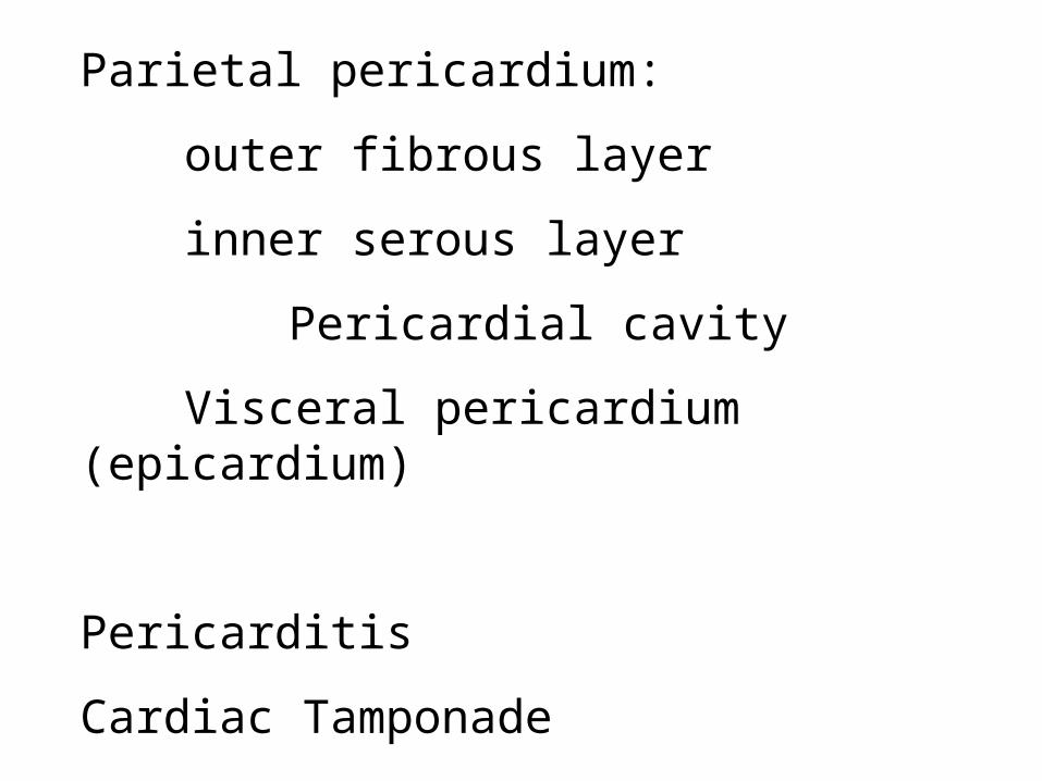
Parietal pericardium:
outer fibrous layer
inner serous layer
Pericardial cavity
Visceral pericardium (epicardium)
Pericarditis
Cardiac Tamponade
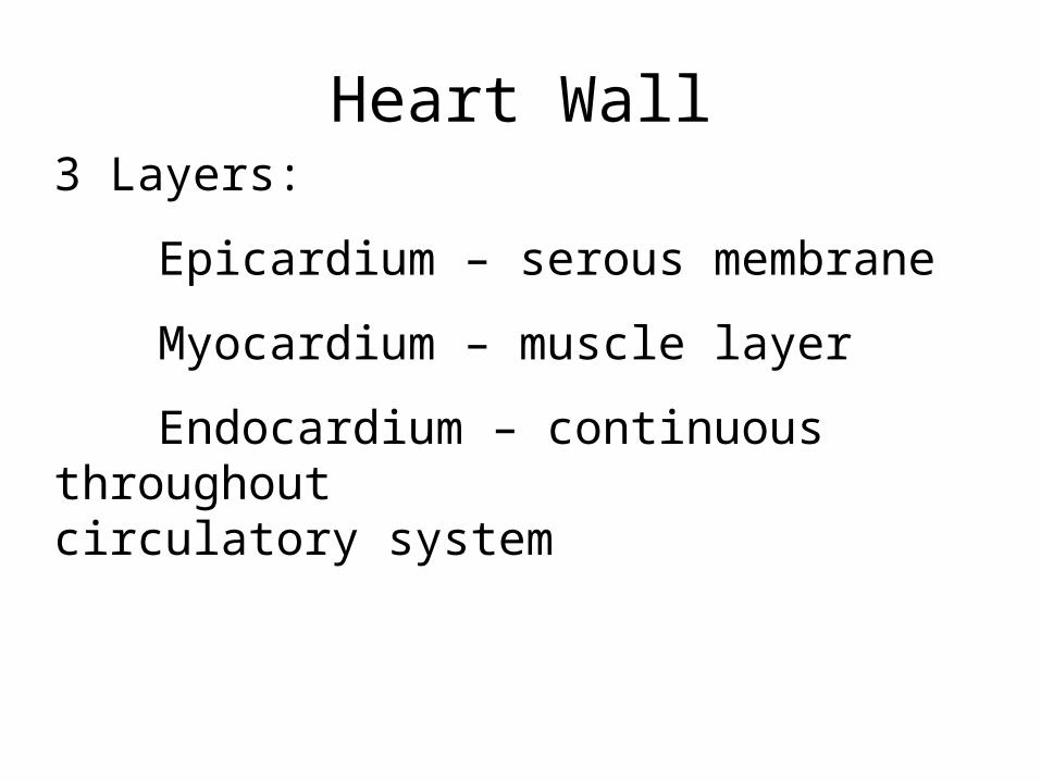
Heart Wall3 Layers:
Epicardium – serous membrane
Myocardium – muscle layer
Endocardium – continuous throughout circulatory system


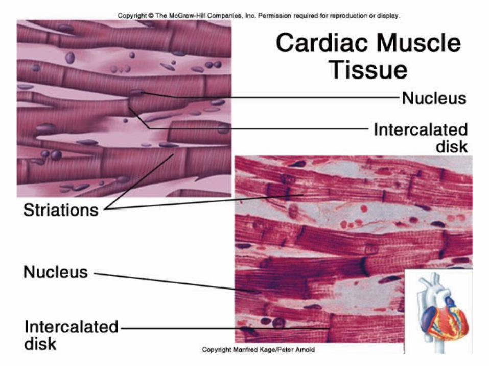
Cardiac Muscle :
involuntary, striated
Intercalated discs:
gap junctions
functional syncytium
desmosomes – “spot welds”


Anastomoses – collateral circulation
Ischemia – reduced blood flow
Hypoxia – reduced oxygen supply
Angina pectoris – “strangled chest”
Myocardial infarction – death of an area of tissue due to interrupted blood flow
Cardiac cycle
One complete heart beat:
systole (contraction) and
diastole (relaxation) of both atria and systole and diastole of both ventricles


Heart Murmurs – abnormal sounds caused by the flow of blood.Mitral stenosisMitral valve prolapse
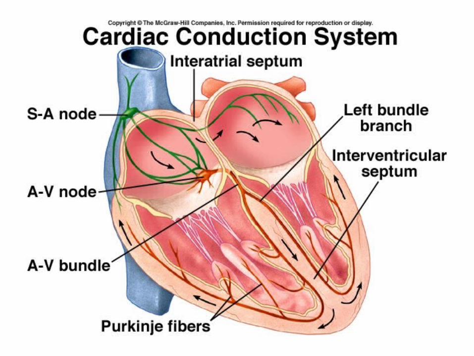

Conduction system of the heart
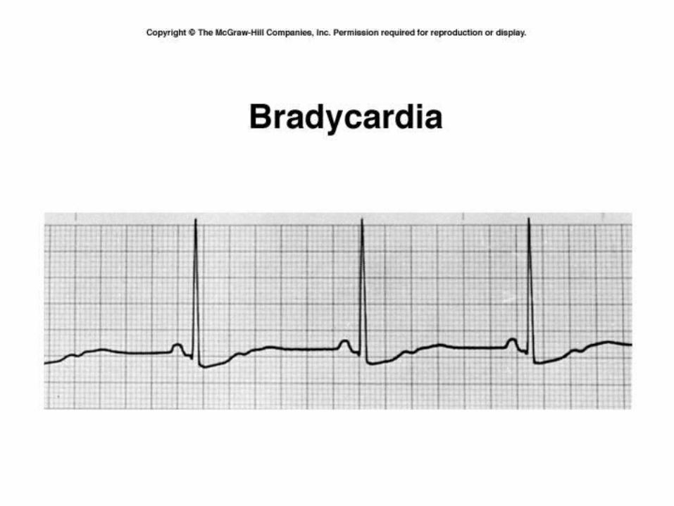
Sinoatrial (SA) node – “pacemaker’ →
Atrioventricular (AV) node →
Atrioventricular (AV) Bundle - Bundle of His→
Purkinje fibers – conduction myofibers
Ectopic pacemaker

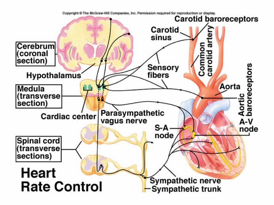
Regulation of Heart Rate
• Sympathetic N.S. increases heart rate and force of contraction – secrete epinephrine –accelerator nerves
• Parasympathetic N.S. decrease heart rate and force of contraction through the vagus nerve. Sends continuous impulses. Secretes acetylcholine
Other factors that influence heart rate
• Temperature
• Ions – K+ and Ca++
• Hormones
• Hypoxia, acidosis and alkalosis slow heart
• Age
• gender
• Physical fitness
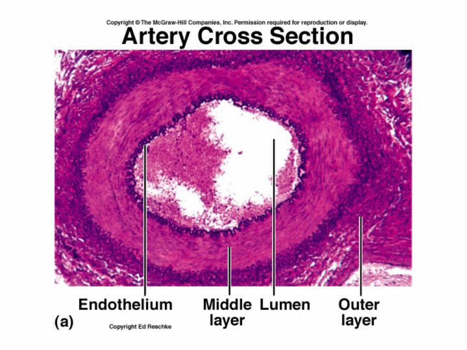
Anatomy of blood vessels
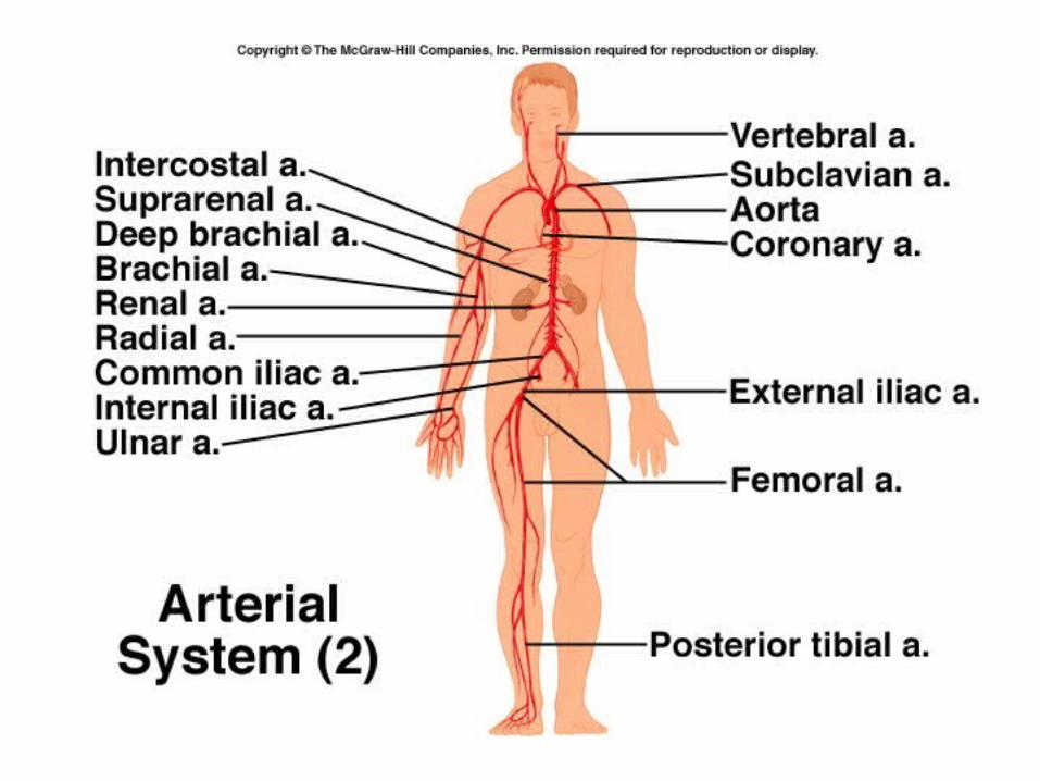
• Arteries carry blood away from the heart
• Hole is called the lumen
• Three layers or tunics:– Tunica interna (intima)– Tunica media– Tunica externa (adventitia)

Tunica interna
• Simple squamous epithelium called endothelium
• Secretes biochemicals that inhibit platelet aggregation
• Also substances that dilate or constrict vessels

Tunica media
• Bulk of vessel wall
• Smooth muscle fibers
• Innervated by the sympathetic N.S. – vasoconstriction; decreased impulses = vasodilation
• Thick layer of elastic connective tissue
Tunica externa
• Thin layer
• Connective tissue
• Attaches artery to surrounding tissue
• Contains tiny vessels – vasa vasorum that form capillaries and provide blood to external cells of the vessel

Arteries
• Large arteries are elastic (conducting) arteries – pressure reservoirs
• Medium arteries are muscular (distributing) arteries – more smooth muscle

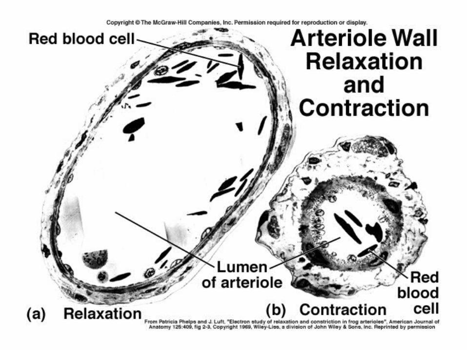
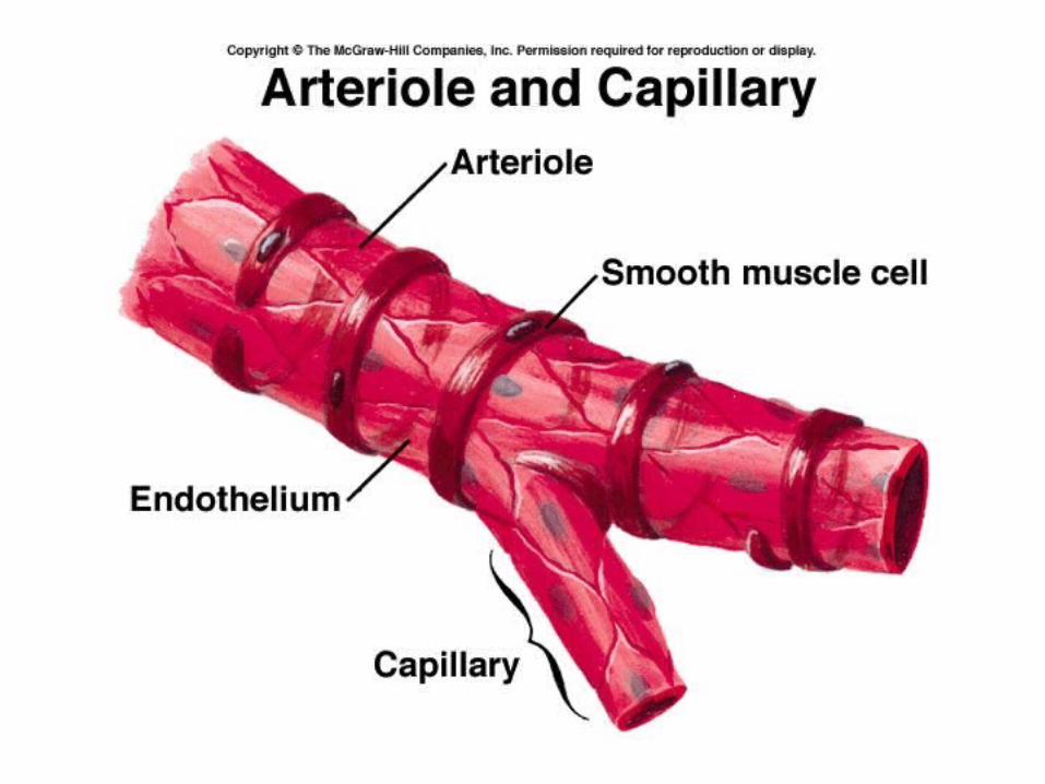
Arterioles
• Have all three layers, but thin as they divide
• Branches called metarterioles join capillaries
• Arteriovenous shunts
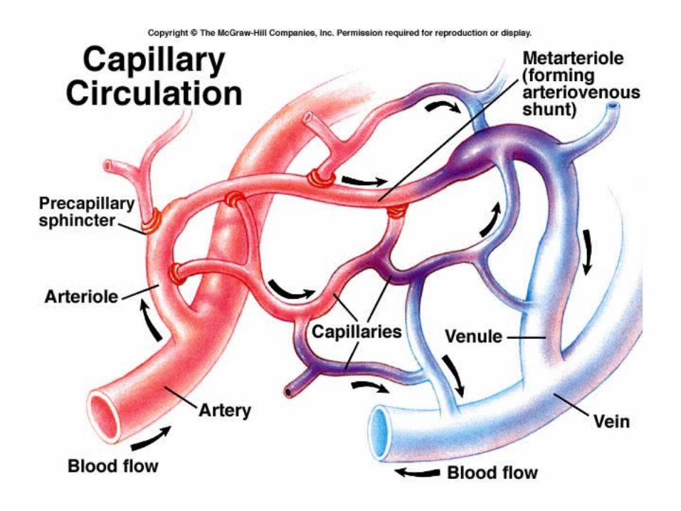
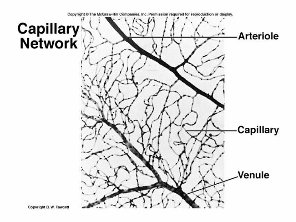
Capillaries
• Only a single layer of endothelium and a basement membrane
• Connect arterioles and venules
• Microcirculation
• Functional part of system for exchange of gases, wastes and nutrients
• True capillaries begin at a precapillary sphincter

Types of capillaries
• Continuous - intercellular clefts, but otherwise uninterrupted

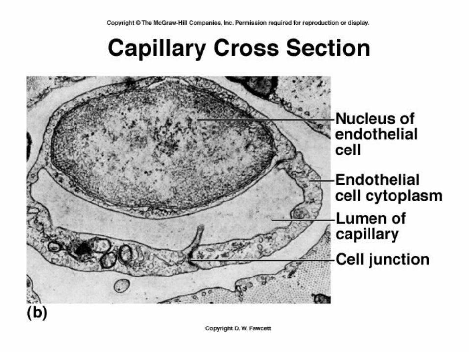

Types of capillaries
• Continuous - intercellular clefts, but otherwise uninterrupted
• Fenestrated capillaries – have “windows” or pores – act in filtration
• Sinusoids or discontinuous capillaries have spaces between cells, and basement membrane is incomplete or absent
• Tight junctions – form a barrier

Venules
• Small vessels that join capillaries and veins
• Add layers
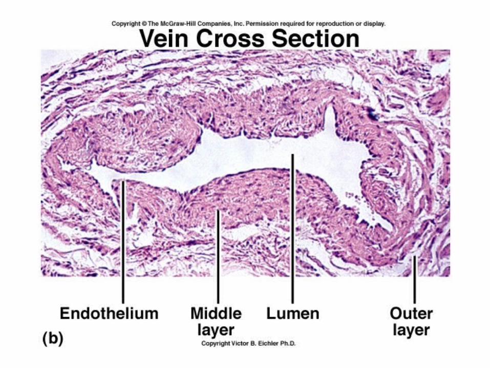
Veins
• Have same three tunics as arteries, but have a thinner tunica media
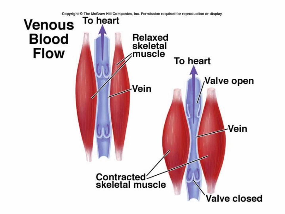
• Contain valves
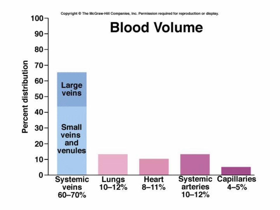
• Act as blood reservoirs

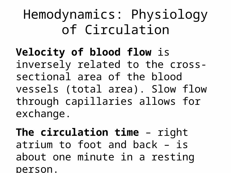
Hemodynamics: Physiology of Circulation
Velocity of blood flow is inversely related to the cross-sectional area of the blood vessels (total area). Slow flow through capillaries allows for exchange.
The circulation time – right atrium to foot and back – is about one minute in a resting person.

Blood pressure
• Pressure on walls of a vessel
• Arterial pressure rises and falls as the left ventricle contracts
• Highest during systole – Systolic pressure
• Lowest during diastole – Diastolic pressure
• Mean arterial pressure is about 93 mm Hg
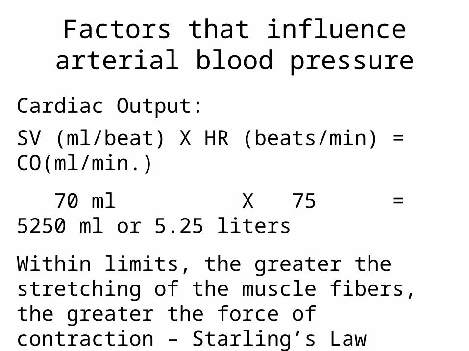
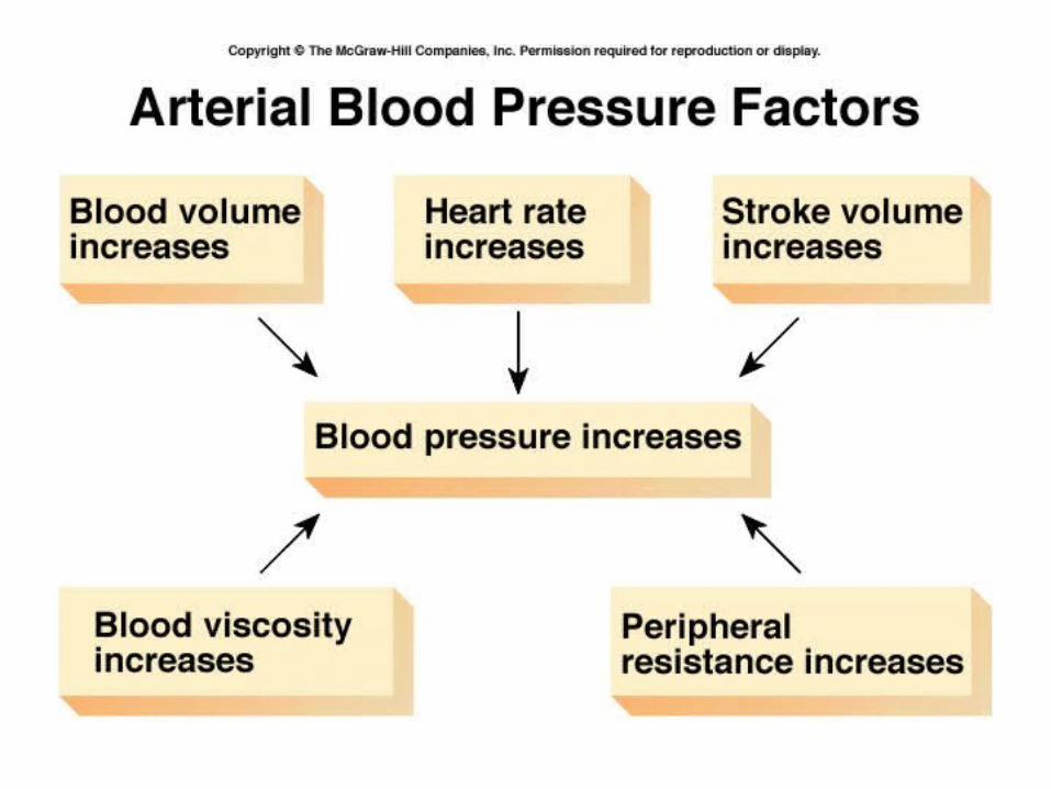
Factors that influence arterial blood pressure
Cardiac Output:
SV (ml/beat) X HR (beats/min) = CO(ml/min.)
70 ml X 75 = 5250 ml or 5.25 liters
Within limits, the greater the stretching of the muscle fibers, the greater the force of contraction – Starling’s Law
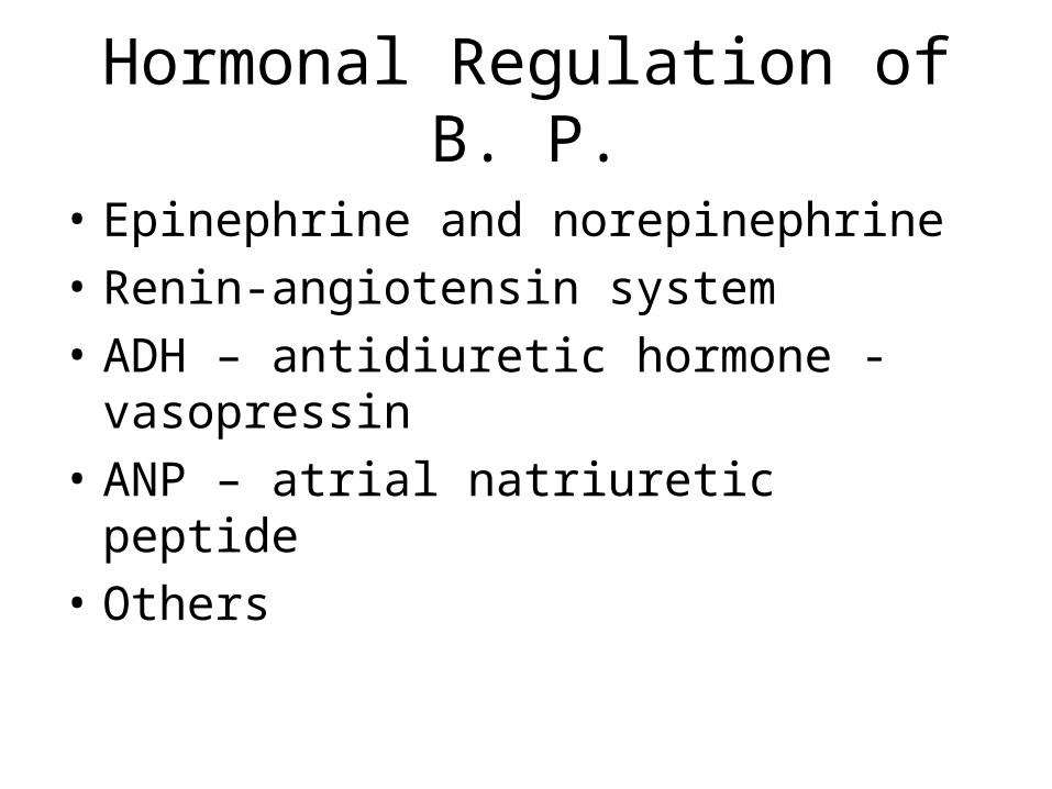
Hormonal Regulation of B. P.
• Epinephrine and norepinephrine
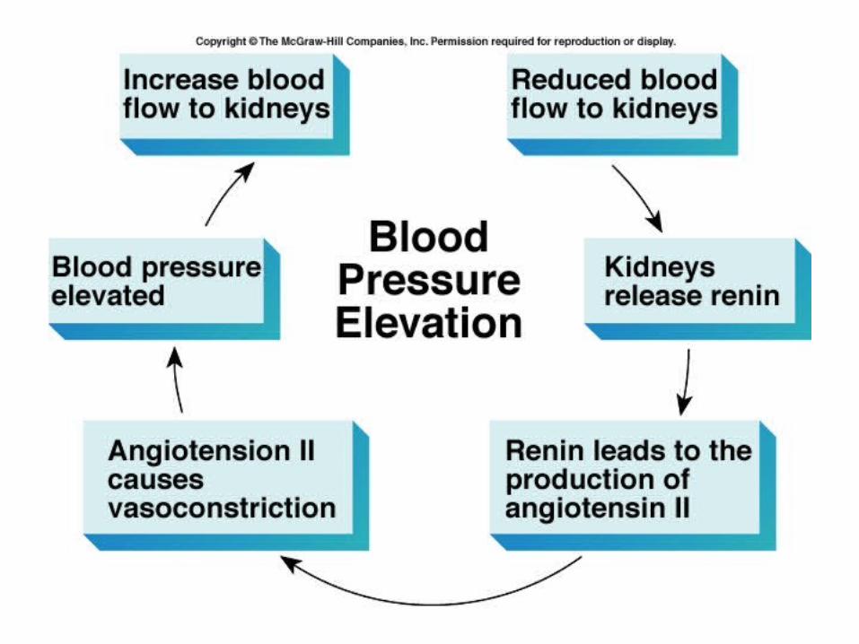
• Renin-angiotensin system
• ADH – antidiuretic hormone - vasopressin
• ANP – atrial natriuretic peptide
• Others
Hepatic Portal Circulation
• Drains spleen, stomach, pancreas, gallbladder and small and large intestines
• 2nd capillary bed in liver
• Glucose is removed, and stored as glycogen
• Blood is detoxified
• Leaves through hepatic vein → inferior vena cava


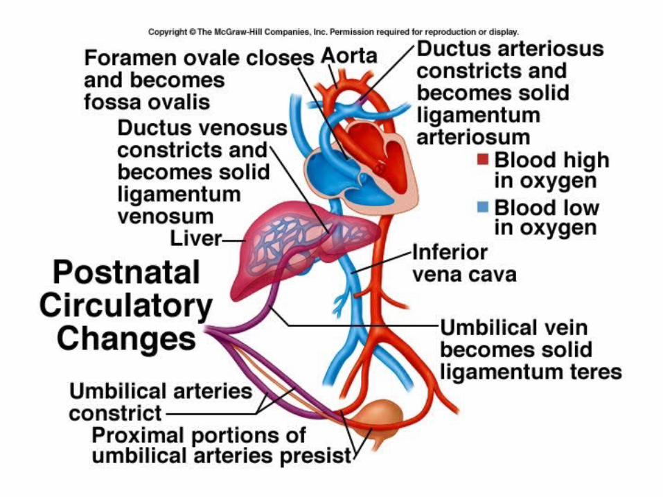
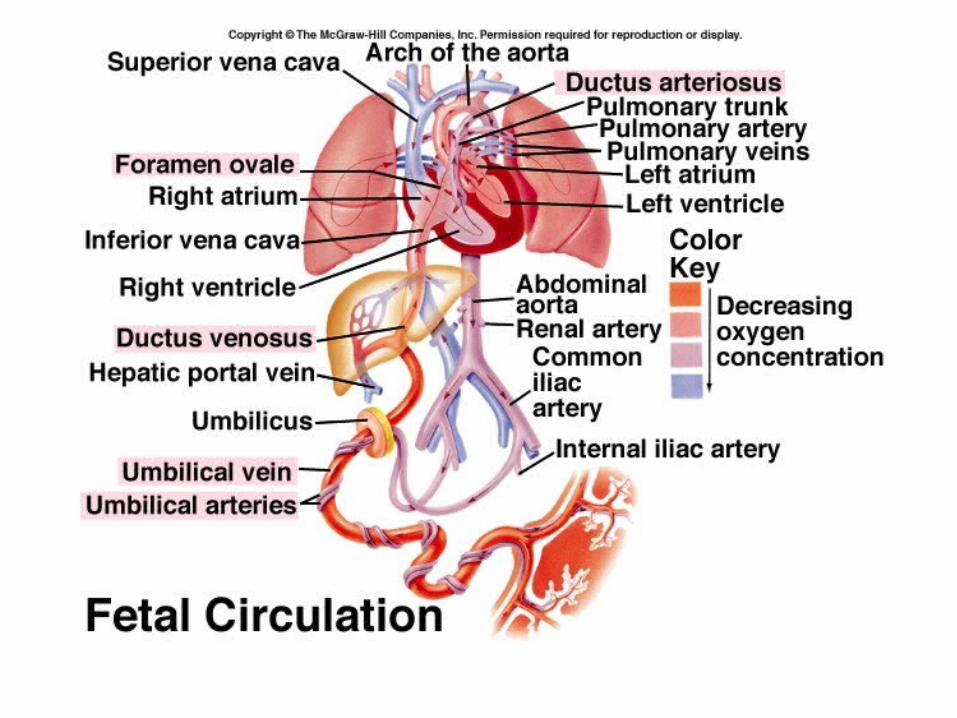
Fetal Circulation
• Obtains oxygen and nutrients from maternal circulation
• Two arteries off internal iliac arteries run through umbilical cord
• Umbilical vein returns oxygenated blood
• Several shunts in fetal circulation:

•Ductus venosus – bypasses fetal liver and dumps blood from umbilical vein into inferior vena cava.
•Foramen ovale – hole in atrial septum, blood passes from right atrium to left atrium, bypassing the developing lungs
•Ductus arteriosus – connects pulmonary artery with aorta
•If does not close – patent ductus arteriosus – get mixing of venous and arterial blood.
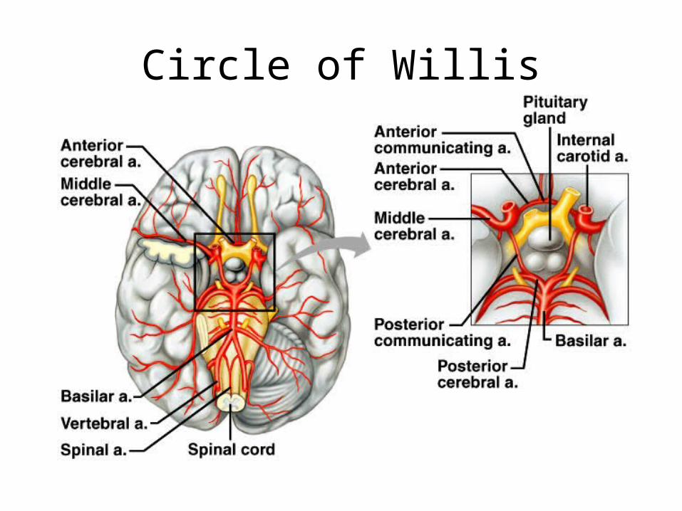
Circle of Willis
Recommended