Brigham Young University Brigham Young University
BYU ScholarsArchive BYU ScholarsArchive
Theses and Dissertations
2015-11-01
The Detection and Molecular Evolution of Francisella tularensis The Detection and Molecular Evolution of Francisella tularensis
Subspecies Subspecies
Mark K. Gunnell Brigham Young University - Provo
Follow this and additional works at: https://scholarsarchive.byu.edu/etd
Part of the Microbiology Commons
BYU ScholarsArchive Citation BYU ScholarsArchive Citation Gunnell, Mark K., "The Detection and Molecular Evolution of Francisella tularensis Subspecies" (2015). Theses and Dissertations. 5696. https://scholarsarchive.byu.edu/etd/5696
This Dissertation is brought to you for free and open access by BYU ScholarsArchive. It has been accepted for inclusion in Theses and Dissertations by an authorized administrator of BYU ScholarsArchive. For more information, please contact [email protected], [email protected].

The Detection and Molecular Evolution of
Francisella tularensis Subspecies
Mark K. Gunnell
A dissertation submitted to the faculty of Brigham Young University
in partial fulfillment of the requirements for the degree of
Doctor of Philosophy
Richard A. Robison, Chair Byron J. Adams Joel S. Griffitts
William R. McCleary Brent L. Nielsen
Department of Microbiology and Molecular Biology
Brigham Young University
November 2015
Copyright © 2015 Mark K. Gunnell
All Rights Reserved

ABSTRACT
The Detection and Molecular Evolution of
Francisella tularensis Subspecies
Mark K. Gunnell Department of Microbiology and Molecular Biology, BYU
Doctor of Philosophy
Francisella tularensis is the etiological agent of tularemia, a zoonotic disease with worldwide prevalence. F. tularensis is a highly pathogenic organism and has been designated as a potential biothreat agent. Currently there are four recognized subspecies of F. tularensis: tularensis (type A), holarctica (type B), mediasiatica, and novicida. In addition, genomic studies have further subdivided type A tularensis into two subclassifications, type A.I and type A.II. These two subclassifications differ in geographic distribution with type A.I appearing mainly in the Eastern United States and type A.II appearing mainly in the Western United States. Because of differences of virulence among the subspecies, it is important to be able to quickly identify each of the subspecies rapidly and accurately. This work describes the development of a multiplex real-time polymerase chain reaction (PCR) assay which was shown to be ~98% successful at identifying the known subspecies of F. tularensis. Furthermore, F. tularensis is thought be a genome in decay (losing genes) because of the relatively large number of pseudogenes present in its genome. We hypothesized that the observed frequency of gene loss/pseudogenes may be an artifact of evolution in response to a changing environment, and that genes involved in virulence should be under strong positive selection. Eleven arbitrarily chosen virulence genes were screened for positive selection along with 10 arbitrarily chosen housekeeping genes. Analyses of selection yielded one housekeeping gene and 7 virulence genes which showed significant evidence of positive selection. Our results suggest that while the loss of functional genes through disuse could be accelerated by negative selection, the genome decay in Francisella could also be the byproduct of adaptive evolution, as evidenced by several of its virulence genes which are undergoing strong, positive selection. Keywords: Francisella tularensis, real-time PCR, detection, genome decay, genome sequencing natural selection, virulence, TreeSAAP

ACKNOWLEDGEMENTS
I would like to thank Dr. Richard A. Robison for his guidance, encouragement and
mentoring throughout my academic career. I would also especially like to thank Dr. Byron J.
Adams for serving on my advisory committee and for his guidance and thoughtful review of my
manuscripts. Thanks also to the rest of my advisory committee: Drs. Joel Griffitts, William R.
McCleary, and Brent Nielsen.
I would like to thank Dr. Bruce Harper for inspiring me and providing the avenue to
pursue this degree as well as Dr. Angelo Madonna for his guidance and encouragement. Thanks
also to Shawn Slater for helping to talk though concepts and helping to refine the ideas presented
in this work. Most importantly, I would like to thank my family: my parents for instilling in me
from a very young age, a desire for knowledge and the importance of education, my children,
Spencer and Emily, for understanding when I had to be away working and at school, and my
wife, Jennifer, for her encouragement, support, understanding and love.
For the literature review presented in Chapter 1, “The Genetic Diversity and Evolution of
Francisella tularensis with Comments on Detection by PCR,” (1) I thank my co-authors: Byron
J. Adams and Richard A. Robison. Additionally, I thank Dr. Angelo Madonna of Dugway
Proving Ground for his guidance and leadership in the production of this work.
For the research in Chapter 2, “A multiplex real-time PCR assay for the detection and
differentiation of Francisella tularensis,” (2) I thank my co-authors: Charity D. Lovelace,
Benjamin A. Satterfield, Emily A. Moore, Kim L. O’Neill, and Richard A. Robison. Also, I
thank the Utah Department of Health, the New Mexico Department of Health, Dr. Fran Nano at
the University of Victoria, Rocky Mountain Laboratories, and Dugway Proving Ground for help
in obtaining the Francisella isolates, without which this work would have been impossible. I

thank Dr. Paul Keim of Northern Arizona University for his MLVA database. Finally, I thank
Scott Jonas of Dugway Proving Ground for expert guidance in performing the MLVA assays, as
well as Dr. Angelo Madonna also of Dugway Proving Ground for guidance and technical help.
For the research in Chapter 3 “Natural selection in virulence genes of Francisella
tularensis,” I thank my coauthors: Richard A. Robison and Byron J. Adams. Also, I thank the
Utah Department of Health and the New Mexico Department of Health for help in obtaining the
isolates used in this study, and for the thoughtful and constructive criticisms of the anonymous
reviewers, as well as Dr. Angelo Madonna of Dugway Proving Ground who provided expert
guidance and assistance in completing this work.

v
Table of Contents
TITLE PAGE ................................................................................................................................... i
ABSTRACT .................................................................................................................................... ii
ACKNOWLEDGEMENTS ........................................................................................................... iii
Table of Contents ............................................................................................................................ v
List of Tables ............................................................................................................................... viii
List of Figures ................................................................................................................................ ix
Chapter 1 The Genetic Diversity and Evolution of Francisella tularensis with Comments on
Detection by PCR ........................................................................................................................... 1
1.1 Abstract ................................................................................................................................. 1
1.2 Introduction ........................................................................................................................... 1
1.3 Genetic Diversity .................................................................................................................. 6
1.3.1 Detection ...................................................................................................................... 11
1.4 Evolution ............................................................................................................................. 18
1.5 Concluding Remarks ........................................................................................................... 20
Chapter 2 A multiplex real-time PCR assay for the detection and differentiation of Francisella
tularensis subspecies ..................................................................................................................... 23
2.1 Summary ............................................................................................................................. 23
2.2 Introduction ......................................................................................................................... 24
2.3 Methods............................................................................................................................... 26

vi
2.3.1 Bacterial strains and culture conditions ....................................................................... 26
2.3.2 Preparation of DNA ..................................................................................................... 26
2.3.3 Primer and probe design .............................................................................................. 27
2.3.4 PCR cycling conditions................................................................................................ 28
2.3.5 MLVA analysis ............................................................................................................ 29
2.4 Results ................................................................................................................................. 30
2.4.1 Assay validation ........................................................................................................... 30
2.4.2 Assay sensitivity .......................................................................................................... 32
2.4.3 Characterization of isolates .......................................................................................... 32
2.5 Discussion ........................................................................................................................... 34
Chapter 3 Natural selection in virulence genes of Francisella tularensis .................................... 38
3.1 Abstract ............................................................................................................................... 38
3.2 Introduction ......................................................................................................................... 39
3.3 Materials and Methods ........................................................................................................ 43
3.3.1 Bacterial strains and culture conditions ....................................................................... 43
3.3.2 Genome sequencing and annotation ............................................................................ 43
3.3.3 Analysis of selection .................................................................................................... 45
3.4 Results and Discussion ....................................................................................................... 46
3.4.1 Genome sequencing and annotation ............................................................................ 46
3.4.2 Analysis of selection .................................................................................................... 49

vii
3.5 Conclusions ......................................................................................................................... 63
Chapter 4 Conclusions and Future Work ...................................................................................... 64
Supplementary Tables ................................................................................................................... 67
Supplementary Figures ................................................................................................................. 70

viii
List of Tables
Table 1 F. tularensis genome sequences analyzed ....................................................................... 27
Table 2 Primer and probe sequences ............................................................................................ 28
Table 3 Scoring matrix for triplex assay ....................................................................................... 29
Table 4 Other isolates tested ......................................................................................................... 31
Table 5 Subspecies of F. tularesnsis and their worldwide distribution ........................................ 40
Table 6 Genomes used for analysis of selection ........................................................................... 44
Table 7 Summary of sequenced and aligned F. tularensis genomes ............................................ 47
Table 8 Summary of annotation results for F. tularensis genomes .............................................. 48
Table 9 Virulence genes randomly chosen for TreeSAAP analysis ............................................. 51
Table S 1 Assay results ................................................................................................................. 67

ix
List of Figures
Figure 1 Maximum likelihood tree inferring the phylogenetic relationships of the F. tularensis
subspecies. ...................................................................................................................................... 4
Figure 2 Whole genome alignment of F. tularensis subspecies ................................................... 10
Figure 3 Sensitivities of multiplex assays ..................................................................................... 34
Figure 4 Maximum likelihood tree inferring the phylogenetic relationships of the F. tularensis
subspecies ..................................................................................................................................... 41
Figure 5 Selection on FTL_1134. ................................................................................................. 52
Figure 6 Selection on FTT_0683 (pilD). ...................................................................................... 54
Figure 7 Selection on FTT_0881c (rocE). .................................................................................... 56
Figure 8 Selection on FTT_0504 (sucC). ..................................................................................... 57
Figure 9 Selection on FTT_0936c (bioF). .................................................................................... 59
Figure 10 Selection on FTT_0766 (deoD). ................................................................................... 60
Figure 11 Selection on FTT_1125 (metQ). ................................................................................... 61
Figure S 1 Mauve alignment of Francisella genomes .................................................................. 70
Figure S 2 Representative MLVA analysis .................................................................................. 71
Figure S 3 Sensitivities of singleplex assays ................................................................................ 73
Figure S 4 Predicted secondary structure of FTL_1134. .............................................................. 75
Figure S 5 Predicted secondary structure of FTT_0683 (pilD). ................................................... 77
Figure S 6 Predicted secondary structure of FTT_0881 (rocE). ................................................... 79
Figure S 7 Predicted secondary structure of FTT_0504 (sucC). .................................................. 81

x
Figure S 8 Predicted secondary structure of FTT_0936c (bioF). ................................................. 83
Figure S 9 Predicted secondary strucutre of FTT_0766 (deoD). .................................................. 85
Figure S 10 Predicted secondary structure of FTT_1125 (metQ). ................................................ 88

1
Chapter 1 The Genetic Diversity and Evolution of Francisella tularensis with Comments on
Detection by PCR
1.1 Abstract
Francisella tularensis has been the focus of much research over the last two decades
mainly because of its potential use as an agent of bioterrorism. F. tularensis is the causative
agent of zoonotic tularemia and has a worldwide distribution. The different subspecies of F.
tularensis vary in their biogeography and virulence, making early detection and diagnosis
important in both the biodefense and public health sectors. Recent genome sequencing efforts
reveal aspects of genetic diversity, evolution and phylogeography previously unknown for this
relatively small organism, and highlight a role for detection by various PCR assays. This review
explores the advances made in understanding the evolution and genetic diversity of F. tularensis
and how these advances have led to better PCR assays for detection and identification of the
subspecies.
1.2 Introduction
Francisella tularensis is a small, non-motile, Gram-negative coccobacillus and is the
causative agent of the zoonotic disease tularemia. This facultative intracellular pathogen was
first discovered in Tulare County California in 1911 where it caused a plague-like illness in local
rodents (3). F. tularensis is able to cause disease in rabbits, squirrels, and other mammals,
including humans (4). The transmission of F. tularensis to humans is mediated through
arthropod vectors such as ticks and deer flies, by the ingestion of contaminated food or water, or
by inhalation of aerosolized bacteria (5). F. tularensis subsp. tularensis is highly infectious. It is
2
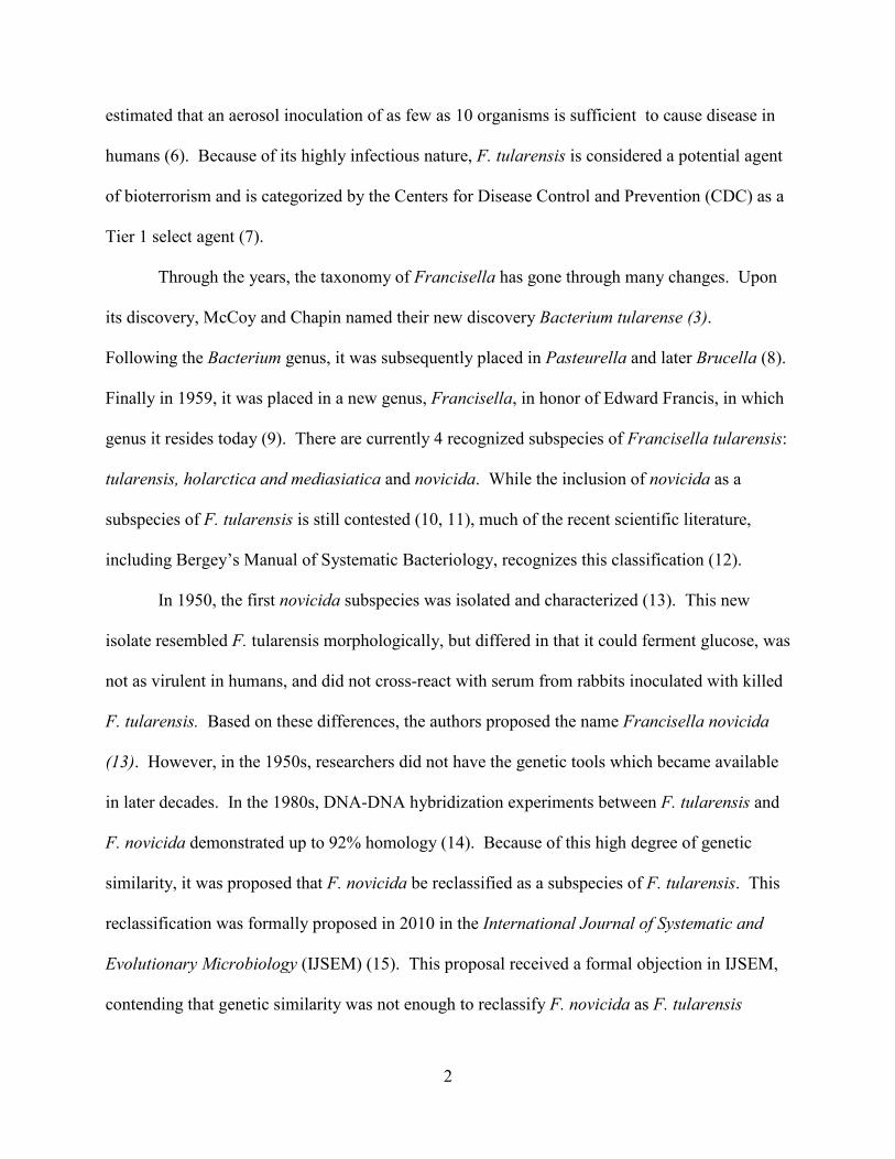
estimated that an aerosol inoculation of as few as 10 organisms is sufficient to cause disease in
humans (6). Because of its highly infectious nature, F. tularensis is considered a potential agent
of bioterrorism and is categorized by the Centers for Disease Control and Prevention (CDC) as a
Tier 1 select agent (7).
Through the years, the taxonomy of Francisella has gone through many changes. Upon
its discovery, McCoy and Chapin named their new discovery Bacterium tularense (3).
Following the Bacterium genus, it was subsequently placed in Pasteurella and later Brucella (8).
Finally in 1959, it was placed in a new genus, Francisella, in honor of Edward Francis, in which
genus it resides today (9). There are currently 4 recognized subspecies of Francisella tularensis:
tularensis, holarctica and mediasiatica and novicida. While the inclusion of novicida as a
subspecies of F. tularensis is still contested (10, 11), much of the recent scientific literature,
including Bergey’s Manual of Systematic Bacteriology, recognizes this classification (12).
In 1950, the first novicida subspecies was isolated and characterized (13). This new
isolate resembled F. tularensis morphologically, but differed in that it could ferment glucose, was
not as virulent in humans, and did not cross-react with serum from rabbits inoculated with killed
F. tularensis. Based on these differences, the authors proposed the name Francisella novicida
(13). However, in the 1950s, researchers did not have the genetic tools which became available
in later decades. In the 1980s, DNA-DNA hybridization experiments between F. tularensis and
F. novicida demonstrated up to 92% homology (14). Because of this high degree of genetic
similarity, it was proposed that F. novicida be reclassified as a subspecies of F. tularensis. This
reclassification was formally proposed in 2010 in the International Journal of Systematic and
Evolutionary Microbiology (IJSEM) (15). This proposal received a formal objection in IJSEM,
contending that genetic similarity was not enough to reclassify F. novicida as F. tularensis

3
subsp. novicida, but that the phenotypic differences were sufficient enough to justify separate
species designation (16).
Finally, in a rebuttal to the objection of Johansson et al., Busse et al. (17), stood by their
initial recommendation for reclassification, asserting that the genetic similarity meets the
definition of a subspecies (18). Furthermore, Busse et al. acknowledge the phenotypic
differences between F. tularensis and F. novicida, but contend that the 11 phenotypic differences
noted are not sufficient enough for a new species (17). There are many other examples of
bacteria with a greater percentage of phenotypic differences which are classified as the same
species (e.g. the various biovars of Pseudomonas fluorescens) (17). Despite this evidence, a
formal reclassification has yet to occur. Based on the high genetic similarity, and taking into
account the relatively few phenotypic differences, we also propose the reclassification of F.
novicida as a subspecies of F. tularensis, and will refer to it as such throughout this work.
Each subspecies is predominantly associated with a specific geographic distribution and
severity of disease. The subspecies tularensis is typically found in North America (19) while the
subspecies holarctica is found across much of the Northern Hemisphere (20). The subspecies
mediasiatica has only been isolated from the central Asian republics of the former Soviet Union
(21) and the subspecies novicida has been isolated from North America and Australia (14, 22).
Phylogenetic relationships among these subspecies are inferred in Figure 1.

4
Figure 1 Maximum likelihood tree inferring the phylogenetic relationships of the F. tularensis subspecies. Tree was constructed by concatenating 10 housekeeping genes (recA, gyrB, groEL, dnaK, rpoA1, rpoB, rpoD, rpoH, fopA, and sdhA) followed by alignment with Clustal W and generation of the tree with MEGA 5.2. Bootstrap values are indicated at the nodes except where support was less than 0.65.
The two subspecies most associated with human disease are tularensis and holarctica.
These are often abbreviated simply as Type A and Type B tularensis, respectively. Type A
tularensis causes a more severe form of tularemia while the presentation of type B tularemia is
somewhat milder (23, 24). The subspecies mediasiatica is fully virulent in mice, yet is believed
to be of relatively mild virulence in humans (21, 25). Similar to the subspecies mediasiatica, the
subspecies novicida is fully virulent in mice, yet rarely causes disease in humans (14).

5
Genetic analyses by multiple-locus variable-number tandem repeat analysis (MLVA) has
identified further sub classifications and geographic structure of Type A and Type B tularensis.
The major subdivisions of Type A tularensis include Type A.I and Type A.II, with the former
generally isolated from the eastern United States and the latter generally isolated from the
western United States (20). This biogeographic separation is correlated with the geographic
distribution of specific vectors, hosts, and other abiotic factors such as elevation and rainfall (26,
27). The major divisions of Type B tularensis also display geographic structure, with Type B.I
isolated from Eurasia, Type B.II isolated from North America and Scandinavia, Type B.III
isolated from Eurasia and North America, Type B.IV isolated from North America and Sweden,
and Type B.V isolated from Japan (20). Unlike type A tularensis, the distribution of Type B
tularensis has not been shown to correlate with the distribution of any specific vectors (26).
The F. tularensis subsp. holarctica isolated from Japan was first differentiated from other
F. tularensis subspecies based on its ability to ferment glucose (28). These isolates were further
differentiated by demonstrating a reduced virulence from the subspecies tularensis, displaying a
virulence similar to that of the subspecies holarctica (29). As genomic tools became more
widely available, this division was confirmed by microarray analysis (21), restriction fragment
length polymorphism (RFLP) analysis (30), and multiple-locus variable number tandem repeat
analysis (MLVA) (20, 31). Genetic analyses have hinted that these isolates from Japan
underwent a unique evolutionary process in a restricted area, separate from other F. tularensis
subspecies (31). Because of the phenotypic differences, the genetic differences, and the apparent
isolated evolution, it has been proposed that these strains from Japan be classified as another
subspecies of F. tularensis called F. tularensis subsp. japonica (32). However, since relatively

6
few isolates from Japan have been analyzed, we recommend that this designation not be adopted
at this time.
1.3 Genetic Diversity
The first complete genome of Francisella tularensis was sequenced in 2005 (33). This
first sequence was the classical type strain of Francisella tularensis subsp. tularensis
representing the Type A.I sub classification. Since then, numerous other whole and partial
genomes of F. tularensis have been sequenced: F. tularensis subsp. holarctica strain OSU18
(Type B) (34), a European isolate of Type A tularensis (35), F. tularensis subsp. novicida strain
U112 (36), a Type A.II tularensis (WY96-3418) (37), F. tularensis subsp. mediasiatica (10) and
at least 10 more comprising the 4 subspecies of F. tularensis (38-41). With the advent of
improved massively parallel sequencing technologies, more genomes continue to be sequenced
at an ever-increasing rate (42). In all, there are currently 16 complete genomes of Francisella
tularensis deposited in GenBank and even more partial genomes. This collection of genomic
information allows for the comparative analysis of these genomes and provides insight into the
evolution of F. tularensis genome architecture.
Even before the first Francisella genome was completed in 2005, studies analyzing the
genomic diversity of F. tularensis were plentiful. Because of its potential use as a bioweapon
and for public health reasons, rapid identification of F. tularensis became paramount (7). Early
DNA based techniques focused on 16S rDNA typing. This proved difficult since among the 4
subspecies, the 16S rDNA genes exhibit between 98.5 – 99.9% similarity, the result of only 6
nucleotide differences among the most divergent strains (43). Other DNA based techniques for
identification such as PCR, which is both rapid and accurate, helped spur further interest in the

7
genetic diversity of the F. tularensis subspecies (21, 44). A genome wide microarray that
analyzed 27 strains of all four subspecies confirmed the limited genetic variation within the
subspecies, but identified 8 variable regions that were used to develop a subspecies-specific PCR
assay (21). Another microarray study analyzing the genetic diversity of 11 Type A isolates and 6
Type B isolates from various localities around the United States identified 13 regions of
difference, including segments of several genes with implications for virulence (45). While
microarray and other studies revealed valuable information about the regional distribution and
differences in virulence, complete genome sequences reveal a more complete picture (20, 21,
45).
The first completed genome sequence of F. tularensis yielded insights to previously
undiscovered features of its genetic makeup. Some of the genetic features discovered included
previously uncharacterized virulence genes encoding type IV pili and iron acquisition systems
(33). The complete sequence also revealed a duplication of an approximately 30 kb region
previously identified as a pathogenicity island containing 17 open reading frames (ORFs),
perhaps shedding light on the enhanced virulence of Type A tularensis (33, 46, 47). Finally,
analysis of this genome indicated the loss of several biosynthetic pathways, which helps explain
the fastidious nutritional requirements of F. tularensis and suggests the need to infect a host
during its life cycle (33).
The first comparative genomic study of F. tularensis was of the Type A (Schu S4) and
Type B (OSU18) strains. This study revealed an extensive genomic similarity of 97.63%,
indicating that the differences in virulence between the two strains are likely not due to large
differences in gene content (34). This degree of sequence identity was confirmed among the
remaining subspecies as well (10, 25, 36). Perhaps the most striking difference between these

8
two strains is the vast amount of genomic rearrangement. These rearrangements can mostly be
attributed to homologous recombination using insertion (IS) elements (34).
After the genome sequence of F. tularensis subsp. novicida was complete, a 3-way
comparison between three of the subspecies (tularensis, holarctica, and novicida) was possible.
Again, a high degree of sequence identity among the subspecies was confirmed, as was the large
amount of genomic rearrangement (36). Even though the length and the gene content of the
novicida subspecies (1.91 Mb and 1,731 protein coding genes) are both greater than that of the
tularensis subspecies (1.89 Mb and 1,445 protein coding genes) and the holarctica subspecies
(1.89 Mb and 1,380 protein coding genes), these human pathogenic strains contain 41 genes
which the non-human pathogenic strains (novicida) do not (36). Initial comparisons of these
genomes revealed that the human pathogenic strains carry 2 copies of the Francisella
Pathogenicity Island (FPI) while the non human pathogenic strains carry only 1 copy, shedding
further light on the differences in virulence among the subspecies (47).
Many studies have been completed comparing the various subsets of available F.
tularensis genomes. A comparison of the genomes of two holarcitca subspecies, the live vaccine
strain (LVS) and strain FSC200, sought to uncover the mode of attenuation for LVS (48), which
was attenuated through the repeated passage of a holarctica strain between the 1930s and 1950s
in the former Soviet Union (34, 49). The genomes of the LVS and FSC200 strains differ by only
0.08% but the LVS strain was able to confer immunity to infection with F. tularensis subsp.
tularensis in BALB/c mice (48, 49). While the exact nature of genomic modifications leading to
LVS attenuation were not found, comparison with other more virulent Type A strains revealed
some candidate genes which could be targeted in the development of a future vaccine (48).
When the sequence of F. tularensis subsp. holarctica FTNF002-00 was completed and compared
9
to both LVS and the OSU18 strains, it was found to have greater than 99.9% sequence similarity
(38). Other studies have shown a stable genome architecture among Type B strains, but
FTNF002-00 carries a 3.9 kb inversion compared to other Type B strains (34, 38, 50).
Other whole genome comparisons focused on comparing different strains of Type A
tularensis. A comparison between F. tularensis subsp. tularensis Schu S4 (Type A.I) and
WY96-3481 (Type A.II) revealed only one whole gene difference, a hypothetical protein with an
unknown function (37). Despite the fact that these two strains are very closely related, there
were still many other differences, including numerous single nucleotide polymorphisms (SNPs),
small indels, differences in IS elements, and even 31 large chromosomal rearrangements (37).
Many of the chromosomal rearrangements are frequently bordered by IS elements, providing a
mechanism for the translocations (10, 37, 39). Another genome comparison of a Type A.I
clinical isolate to the Schu S4 genome showed that except for some minor changes, the genomes
were virtually identical, suggesting a high degree of sequence conservation within the Type A.I
subgroup (39). The genome of another Type A.I strain (TI0902) isolated from a cat in Virginia,
United States, is also highly similar to Schu S4 as it only differs by 103 SNPs (40). Other
researchers compared a European isolate of Type A.I tularensis (which is typically restricted to
North America) to Schu S4 and found that the two were virtually identical, with only 8 SNP and
3 variable number tandem repeat (VNTR) differences (35). The fact that these two strains are so
alike suggests that the European isolates are descended from the Schu S4 strain and did not
evolve independently in Europe (35).
The completion of a fourth subspecies genome of F. tularensis, the mediasiatica
subspecies, enabled full genome comparisons of the four subspecies of F. tularensis. It was
demonstrated that the subspecies mediasiatica and tularensis are highly similar, which raises

10
more questions about their differences in virulence (10, 21, 51). Phylogenetic analysis of the
complete genomes of the subspecies mediasiatica also demonstrated that it is a monophyletic
taxon of F. tularensis, contradicting previous evidence suggesting that the subspecies
mediasiatica was not a member of the F. tularensis clade (52). However, since isolates of the
mediasiatica subspecies are rare, it is difficult to know the true genetic diversity within the
subspecies. Figure 2 shows the overall genome architecture of representative strains of F.
tularensis, highlighting the large-scale genomic rearrangements between the subspecies.
Figure 2 Whole genome alignment of F. tularensis subspecies Whole genome alignment of representative strains from each of the four subspecies of Francisella tularensis using Mauve (53) highlighting differences in the macro genome architecture relative to the reference strain (A). Colored blocks represent homologous sections of each genome. A) F. tularensis subsp. tularensis Schu S4. B) F. tularensis subsp. holarctica LVS. C) F. tularensis subsp. mediasiatica FSC147. D) F. tularensis subsp. novicida U112.
The evolution of the Francisellacaea is complicated by the discovery of Francisella-like
endosymbionts (FLEs) of ticks, which have an unknown pathogenicity in humans (54-57).
While these endosymbionts lack sufficient evidence to be classified as F. tularensis, they are
similar enough to cross react with many molecular-based methods of detection (58). Because of
the potential to misidentify FLEs as F. tularensis, which could impact the diagnosis of tularemia
A
B
C
D

11
in public heath settings, many have cautioned about the use of PCR assays for the detection of F.
tularensis (59, 60). Despite this caution, PCR remains the standard of practice for the detection
and identification of F. tularensis subspecies (44).
1.3.1 Detection
The ability to accurately detect and diagnose F. tularensis infection carries significant
implications in public health and bioterror (2, 7). Because of the different pathogenic profiles
and biogeography of the various subspecies of F. tularensis, it is important to be able to
accurately discriminate among them (2). Polymerase chain reaction (PCR) has become the
method of choice for the identification of various pathogens because it is rapid, sensitive and
highly specific (61-63). Detection and differentiation of the subspecies of F. tularensis by PCR
is complicated by the lack of significant variability in their genomes (34, 43). Various methods
for the detection of F. tularensis have been reviewed in the last decade, however much more
work has since been completed on the detection of F. tularensis using PCR (44, 64).
1.3.1.1 Conventional PCR
Since 2008, research on the use of conventional PCR for the detection of F. tularensis
has dropped off considerably, with only a handful of publications on the subject. In alignment
with an earlier review (64), the gene tul4 was a popular choice to detect all subspecies of F.
tularensis (65, 66). Since F. tularensis is a potential agent of bioterrorism, some assays included
the multiplex detection of other biothreat agents. One such study developed two multiplex
assays to detect “Tier 1” select agents; one assay for DNA based organisms (Variola Major,
Bacillus anthracis, Yersinia pestis, Francisella tularensis, and Varicella zoster virus) and another

12
assay with a reverse transcriptase for RNA based viruses (Ebola virus, Lassa fever virus, Rift
Valley fever, Hantavirus Sin Nombre and the four serotypes of Dengue virus) (65). A major
drawback to these multiplex assays however, is the use of a reporter dye and a colormetric
detection system, because a positive result is unable to distinguish between the agents. The
assay is intended only as a broad screening tool and further testing is required to differentiate
between the organisms comprising the assay. Furthermore, since the genome of Variola Major
(the causative agent of Smallpox) is so highly regulated, testing was completed with a plasmid
control containing a small segment of the Variola Major genome (65).
Real-time PCR is known for being efficient and sensitive, but is not ideal for
multiplexing beyond a 4- or 6-plex reaction because of the limited number of fluorescent
channels available on most instrument platforms (67, 68). Researchers have overcome this
limitation by using modified primers to bind the PCR products of a 15-plex reaction to
fluorescent beads that can then be analyzed by a flow cytometer for the simultaneous detection
of 11 pathogens with similar sensitivities to real-time reactions (69). While effective, flow
cytometers can be large, difficult to use, and costly. The Luminex Corporation (Austin, TX) has
developed a similar, yet easier to use technology in their MAGPIX® system. Rather than a flow
cell, the MAGPIX® uses a magnet to capture fluorescently labeled magnetic beads and a CCD
camera to capture images of up to 50 different analytes (70, 71). Because of its relatively low
cost and ease of use, the MAGPIX® may be more ideally suited for integration in clinical labs
for the simultaneous detection of multiple pathogens (70).
While it may be useful to detect broad categories of pathogens, because of the virulence
status of various subspecies of F. tularensis, it is also important to be able to differentiate among
them as well. Using the tul4 gene and variations in the pilA gene, researchers were able to

13
differentiate the four subspecies of F. tularensis (66). Another study used suppression
subtractive hybridization (SSH) to identify regions of difference between the genomes of Type
A.I and Type A.II tularensis. This information was used to create a conventional PCR assay to
differentiate between Type A.I, Type A.II, Type B, and F. tularensis subsp. novicida isolates
(72). Later, this same assay was adapted to a real-time PCR platform (73).
1.3.1.2 Real-time PCR
Real-time PCR is a popular choice for the detection of F. tularensis because it is
sensitive, reliable, cost-effective, and eliminates the need for time consuming gels, though this
time commitment has been significantly reduced with the introduction of rapid dry gels (74). A
popular method of real-time PCR incorporates the use of SYBR Green which will fluoresce upon
binding double stranded DNA. Thus, the fluorescent signal will increase as PCR progresses and
more amplicons are synthesized. SYBR green is a popular alternative to other real-time
technologies because of its relatively low cost (75). However, it is not ideal for multiplex
reactions since the dye will bind to all double stranded DNA in the reaction and produce a
fluorescent signal. Sellek et al. (75) developed an assay to detect F. tularensis from soil using
the tul4 gene, previously used in conventional PCR assays (65, 66). However, the assay was
only validated with F. tularensis subsp. holarctica and subsp. novicida. Lacking were
representatives from the subsp. tularensis and mediasiatica. Furthermore, positive fluorescent
signals were obtained from other non-related bacteria. These were later ruled out as true
positives after analyzing the PCR products on a gel and finding only primer dimers (75).
Genome comparisons aided the development of SYBR green assays (76-78). Woubit et
al. (78) compared several genomes from the Escherichia, Francisella, Salmonella, Shigella,

14
Vibrio, and Yersinia genera to develop a series of 27 assays to detect and differentiate these
common food and biothreat pathogens. With respect to Francisella, the assays were so specific
that assays intended to detect all subspecies of Francisella were only able to detect the tularensis
and novicida subspecies (78).
The propensity of PCR assays to cross-react with environmental, non-pathogenic
Francisella or other closely related organisms (59) requires the development of more specific
assays to avoid false positives or incorrect diagnoses. To solve this problem, results from
resequencing microarrays were compared to identify SNPs along the phylogeny of F. tularensis
and build real-time PCR assays capable of differentiating Type A.I, A.II, A.Ia, A.Ib, Type B.I,
and B.II tularensis (76). Similarly, another group analyzed publically available whole genome
sequences to identify defining SNPs and small insertion/deletion elements (INDELs) to design a
series of 35 assays capable of distinguishing the four subspecies of F. tularensis and the major
subtypes of Type A and Type B tularensis, including Type A.I, A.II, and B.I, B.II, B.III, B.IV,
and B.V (77). Both assays were able to accurately assign isolates to the correct subspecies and
clade while avoiding any cross-reactivity to near neighbors (although the former includes only
one novicida strain in the analysis).
Another method for the real-time detection of F. tularensis is the 5’ nuclease or
TaqMan® assay. These assays incorporate fluorescently labeled DNA probes specific to the
template DNA resulting in even more specific identification than the SYBR Green assays,
eliminating the need to perform a melt curve analysis. Strategies for single-plex real-time assays
for the detection of F. tularensis with TaqMan® assays are varied. Gene targets include a gene
for an outer membrane protein, FopA, a single-copy gene for detection and quantification of all
subspecies of F. tularensis (79), the 16S rRNA gene to detect all subspecies of F. tularensis (80,
15
81), the insertion element ISFtu2, which is unique to Francisella species (82), intergenic regions
of differentiation to distinguish Type A.I from Type A.II tularensis (73), and SNP-based assays
to differentiate the species and subspecies of Francisella isolates (83). Some assays can be used
in concert with others to detect a wide variety of agents. These include biothreat agents (80) or
other organisms with similar disease presentations (81), while others were used solely for the
differentiation of subspecies and subpopulations of F. tularensis (73, 83). The advantage of
using a single-copy gene for detection is the ability to quantify the amount of the agent, which
can be useful in clinical and diagnostic settings (79). Conversely, multicopy-genes such as the
16S rRNA gene and the ISFtu2 gene should achieve lower detection limits, which is ideal given
the low infectious dose of F. tularensis (6, 80, 82). A significant drawback of using the 16S
rRNA gene for detection is that since it is so conserved, there is some cross reactivity with near
neighbors and other Francisella-like species, requiring further confirmatory analyses (43, 80).
Multiplex real-time TaqMan® assays incorporate the added convenience of running
multiple reactions in a single tube using probes labeled with various fluorophores. However, as
mentioned previously, multiplexing with TaqMan® assays is generally limited to a 4- or 6 plex
reaction because of the limited number of fluorescent channels on the instruments (67, 68). One
multiplex assay is a 2-plex assay designed from genome comparisons to detect the four
subspecies of F. tularensis but does not differentiate among them. Another multiplex assay is
capable of differentiating the four F. tularensis subspecies with only a 3-plex assay. This assay
was developed using both unique and shared genome regions among the subspecies with the
addition of a scoring matrix (2).
Since F. tularensis has the potential to be used as a bioweapon, a commercial market has
arisen for field-ready detection of biothreat agents, including Bacillus anthracis, Francisella

16
tularensis, Yersinia pestsis, Brucella species, and others. A comparison of one such commercial
instrument, the RAZOR®, (BioFire Defense; previously Idaho Technologies, Salt Lake City,
UT) and another instrument designed for laboratory use, the Applied Biosystems 7300/7500
system (Thermo Fisher Scientific, Grand Island, NY) used assays developed for B. anthracis,
Brucella species, F. tularensis, and Y. pestis, comparing sensitivities and specificities of the two
platforms. Results showed that for all agents, the sensitivities were between 10-100 fg of target
DNA per reaction, and no cross reactivity was observed with other closely related bacteria (84).
Run time on the RAZOR® was notably shorter than that of the 7300/7500 instrument.
Another diagnostic tool, the FilmArray® system (BioFire Defense, Salt Lake City, UT),
uses a lab-in-a-pouch approach to process raw samples and detect 17 biothreat pathogens with an
array of single-plex real-time PCR assays in about an hour (85). An evaluation of the Biothreat
Panel using DNA samples from B. anthracis, F. tularensis, and Y. pestis indicated sensitivities of
250 genome equivalents or lower and the authors conclude that the system is both sensitive and
selective (85). However, since the FilmArray® system is designed to be a complete sample to
answer system, sensitivities may vary when tested with whole organisms in different matrices
like blood or serum rather than purified DNA.
Another evaluation compared the FilmArray® system with TaqMan® Array Cards
developed for the detection of biothreat agents (86, 87). Here, researchers tested for B.
anthracis, F. tularensis, and Y. pestis in the blood of murine infection models. Results showed
that blood culture was the most sensitive means of detection followed by the FilmArray and
Array Cards for B. anthracis, and F. tularensis. All three methods demonstrated similar
detection levels for Y. pestis (87). While blood culture was the most sensitive means of detection
for two of the three agents tested, it requires much more time for detection compared to the PCR

17
assays. Each of these methods for detection carries drawbacks and benefits and must be weighed
appropriately to ensure the best possible outcome.
1.3.1.3 Other PCR assays
Recently, other PCR-based assays have been developed for the detection of F. tularensis
and other bacteria. One such assay involves analyzing PCR products with electrospray
ionization-mass spectrometry (ESI-MS). In this technique, the actual base composition of the
PCR products are identified and compared to a library of sequences for identification rather than
relying on the fluorescent signal obtained from real-time PCR (88). This PCR/ESI-MS
technique has been applied to the wide-spread identification of biothreat agents, respiratory
pathogens, and other pathogenic bacteria and viruses (88, 89). Others have used this technology
specifically for identifying F. tularensis from natural sources (90) and even for typing the
subspecies of F. tularensis (91).
Recombinase Polymerase Amplification (RPA) is a PCR-like assay in which
amplification is carried out at one temperature (isothermal) instead of cycling temperatures as in
PCR. Recently, RPA assays have been applied to the detection of F. tularensis and other
biothreat agents (92-94). Two of these assays showed comparable sensitivities to real-time PCR
assays with an instrument run time of about 10 minutes (92, 93). A third assay using
electrochemical detection rather than fluorescent probes seemed less sensitive than other assays,
with detection levels on the order of 104 copies/µL (94).
Finally, as the cost of sequencing continues to fall, more sequencing-based detection
assays are being used to detect biological agents such as F. tularensis. One such assay used a
pyrosequencing method to sequence the variable region of 16S rDNA to identify and group F.

18
tularensis isolates by subspecies (95). The results from analyzing the SNPs in 16S rDNA are
more distinctive than SNP analysis from real-time PCR. Another sequencing assay was
multiplexed for the detection and strain typing of B. anthracis, F. tularensis, and Y. pestis by
interrogating 10 loci per pathogen (96). While sequencing assays provide some promise for the
rapid detection and classification of F. tularensis, there is a noticeable lack of information on the
sensitivity or detection limits of these assays. In the world of clinical diagnostics and
biodefense, the ability to detect low quantities of F. tularensis and other agents is paramount.
1.4 Evolution
Numerous studies have been conducted on the evolution of the subspecies of F.
tularensis to define specific clades and to reveal their evolutionary history. Before next
generation whole genome sequencing was widely available, various techniques were used to
recover the phylogenetic relationships among strains of F. tularensis, such as microarrays (21,
45), MLVA (20), and sequencing specific genes or other genetic loci (52, 97). One of the
earliest of these studies produced a phylogenetic tree in which the subspecies tularensis and
mediasiatica shared a major clade along with the Japanese isolates of the holarctica subspecies
(21). A later analysis provided better resolution, differentiating the tularensis and mediasiatica
subspecies, and grouping the Japanese isolates of the holarctica subspecies with the other
holarctia subspecies (20). These authors also determined that F. tularensis subsp. holarctia
appears to have recently spread globally from a single geographic origin, while F. tularensis
subsp tularensis appears to have experienced most of its evolutionary history in North America,
and may even have originated in the central United States (98). However, F. tularensis subsp.
tularensis is now clearly distributed beyond North America into parts of Europe (35).

19
The finding that the subspecies holarctica recently spread from a single origin seems
likely because of the small amount of genetic diversity within the subspecies, that has been
identified by a variety of molecular methods (26, 48, 99-101). However, the precise area of
origin of the subspecies holarctica is unknown. Based on phylogenetic analyses, there are two
competing hypothesis as to its origin: 1) the subspecies holarctica originated in Asia or 2) the
subspecies holarctica originated in North America before spreading around the Northern
Hemisphere (102). There appears to be more evidence for the origination of the subspecies
holarctica in North America, though this may be due to the lack of Asian isolates for analysis.
Regardless, it appears that the holarctica subspecies is a highly fit clone that originated from a
single source and spread throughout the Northern Hemisphere (100, 102). However, if F.
tularensis subsp. tularensis originated in North America (20, 98) and the subspecies holarctica is
descended from the tularensis subspecies (97), then it seems likely that the subspecies holarctica
may have originated in North America as well. This hypothesis is supported by the fact that
sequences of various housekeeping genes and some outer membrane proteins from the
subspecies tularensis and holarctica align well, while those from the subspecies novicida and
mediasiatica do not (52).
It is generally accepted that F. tularensis subsp. novicida is the oldest of the F. tularensis
subspecies and evidence suggests that F. tularensis subsp. novicida and Francisella philomiragia
share a common, aquatic ancestor (97, 103, 104). These two species are generally considered
non-pathogenic to humans. However, their association with aquatic sources is further
substantiated in that documented human infections by these two species have occurred in near-
drowning victims (14, 105). Furthermore, F. philomiragia contains one copy of the FPI, similar

20
to F. tularensis subsp. novicida while the remaining subspecies of F. tularensis contain 2 copies
(47, 104).
Molecular evidence suggests that the four subspecies of F. tularensis have evolved by
vertical descent (97). A common method of acquiring genetic variation in bacteria is through
horizontal gene transfer. This is well documented in many species of bacteria, and especially in
the conference of antibiotic resistance (106-109). However, in the subspecies of F. tularensis,
genetic variation, including antibiotic resistance seems to have arisen by mutation rather than the
acquisition of new genes through horizontal gene transfer (110-112).
An in silico analysis has recently shown that the non human-pathogenic F. tularensis
subsp. novicida possesses a CRISPER/Cas system to defend against invading genetic elements.
This finding further supports the hypothesis that mutation is responsible for much of the
evolution of F. tularensis (113, 114). Analyses of the other three virulent subspecies of F.
tularensis (tularensis, holarctica, and mediasiatica), reveal that the genes responsible for the
CRISPER/Cas system are non-functional (114). This is somewhat puzzling since deletion of the
CRISPER/Cas system in other pathogens such as Neisseria meningitidis, Camphylobacter jejuni,
Legionella pneumophila, and Pseudomonas aeruginosa result in decreased virulence. It is
hypothesized that in the case of F. tularensis, other mutations in the genome have compensated
for the degeneration of the CRISPER/Cas system in the virulent subspecies of F. tularensis
(115).
1.5 Concluding Remarks
The genetic diversity of the subspecies of F. tularensis appears to be quite limited.
Genome comparisons among the subspecies reveal similarities greater than 95% (10, 25). Many

21
of the differences in the genomes of F. tularensis are large-scale genomic rearrangements and a
duplication of the pathogenicity island in the tularensis, holarctica, and mediasiatica subspecies
(34, 47). However, because the mediasiatica subspecies is so rare, assessments of its true
genetic diversity must be considered preliminary.
There are many pros and cons to the various PCR detection methods and the individual
user’s needs should dictate which method to use. Conventional PCR is easy and inexpensive but
is known for being time consuming because of the need to run gels. However, since the
introduction of rapid dry gels, the time commitment usually associated with gels has been
shortened considerably. Utilizing fast PCR technology in combination with rapid dry gels, it is
possible to get a result in approximately 50 minutes (74). In general, conventional PCR has
fallen out of favor with many researchers. However, this approach allows for large multiplex
reactions for the detection of many organisms at once, especially when coupled with another
detection system such as the MAGPIX® (70, 71).
Real-time PCR is one of the most popular methods for detection because it is simple, cost
effective, and sensitive. SYBR Green assays are inexpensive and accurate and can even be
multiplexed with the incorporation of a melting curve analysis. TaqMan® assays are more
expensive than SYBR Green assays, but carry an additional layer of specificity with the
sequence of the probe. Multiplexing with TaqMan® assays is possible, but usually only up to a
4- or 6-plex because of the limited number of available fluorescent channels on most instruments
(67, 68). The limited amount of multiplexing with TaqMan® assays can be overcome by setting
up an array of single-plex reactions similar to the FilmArray® system (85).
Many current PCR assays lack the specificity to differentiate between environmental,
non-pathogenic Francisella and other closely related organisms such as FLEs (58, 59). Perhaps
22
in these situations, it would be wise to use whole genome sequencing assays for the detection of
Francisella subspecies (95, 96)
As whole genome sequencing has become more widely available, genome comparisons
between the subspecies of F. tularensis are possible and shed further light on the genetic
diversity and evolution of this pathogen. It is apparent that the more virulent subspecies of F.
tularensis have evolved from F. tularensis subsp. novicida primarily by genomic decay, genomic
rearrangements, and the duplication of the FPI (36). Many of the interrupted genes
(pseudogenes) in the virulent subspecies of F. tularensis are metabolic genes, further supporting
an intracellular life cycle, while other interrupted genes include secreted effector proteins that
may have led to excessive virulence, furthering the patho-adaption of F. tularensis as an
intracellular pathogen (10, 106, 116, 117).
23
Chapter 2 A multiplex real-time PCR assay for the detection and differentiation of Francisella
tularensis subspecies
2.1 Summary
Francisella tularensis is the etiological agent of tularaemia, a zoonotic disease with
world-wide prevalence. F. tularensis is a highly pathogenic organism and has been designated a
category A biothreat agent by the Centers for Disease Control and Prevention (CDC).
Tularaemia is endemic in much of the United States, Europe, and parts of Asia. It is transmitted
by numerous vectors and vehicles such as deer flies, ticks, and rabbits. Currently, there are four
recognized subspecies of F. tularensis: tularensis (Type A), holarctica (Type B), mediasiatica,
and novicida. Within the Type A classification there are two subclassifications, Type A.I and
A.II, each with a specific geographic distribution across the United States. Type B tularensis is
found in both the United States and Europe. Because of virulence differences among subtypes,
it is important that health departments, hospitals, and other government agencies be able to
quickly identify each subtype. The purpose of this study was to develop a multiplex real-time
PCR assay for the identification and discrimination of Type A.I, Type A.II, Type B, and novicida
subspecies of F. tularensis. The assay was validated using 119 isolates of F. tularensis, 3 of its
nearest neighbours, and 14 other bacterial pathogens. This assay proved to be ~98 % successful
at identifying the known subspecies of F. tularensis, and could prove to be a useful tool in the
characterization of this important pathogen.
24
2.2 Introduction
Francisella tularensis is a Gram-negative facultative intracellular bacterial pathogen and
is the causative agent of the zoonotic disease, tularaemia. As few as 10 organisms can cause
disease via the aerosol route (6). Due to its high infectivity, ease of dissemination and ability to
cause illness and death, F. tularensis has long been considered a potential bioweapon by Japan,
the former Soviet Union, and the United States (118). With the advent of the CDC Select Agent
Program (a series of rules and regulations governing the possession and transfer of organisms
that could be used as bioweapons), F. tularensis has been classified as a category A potential
agent of bioterrorism. It was estimated by the World Health Organization (WHO) that 50 kg of
F. tularensis dispersed as an aerosol over a highly populated area of 5 million people would
result in 250 000 cases of tularaemia with 19 000 deaths (118).
Francisella tularensis is a member of the γ-subclass of the proteobacteria currently
consisting of three accepted subspecies: tularensis (Type A), holarctica (Type B), and
mediasiatica. The subspecies differ in their geographic distribution as well as virulence (20,
100). Much of the scientific literature, including this work, refers to Francisella novicida as a
fourth subspecies of F. tularensis (97, 100, 102). Types A and B are most associated with
human disease with Type A being the more virulent. The mediasiatica subspecies is more
commonly found in the Central Asian republics of the former Soviet Union and little is known
about its ability to cause disease in humans (21). The novicida subspecies is more associated
with water and rarely causes human disease (119). The natural reservoir of F. tularensis remains
largely unknown; though there is growing evidence that amoeba may play an important role in
harboring the bacterium (120-122).

25
The genome of F. tularensis is highly conserved among the four subspecies. The 16S
rRNA genes exhibit 98.5 % to 99.9 % similarity (43). Even with this high degree of sequence
similarity, each of the subspecies demonstrates notable differences in virulence. Within the Type
A tularensis, multi-locus variable-number tandem repeat analysis (MLVA) revealed a
subdivision: Type A.I and Type A.II (20). An apparent geographical separation exists between
these two subtypes. Type A.I isolates are primarily found in the Central and Eastern portions of
the United States, while Type A.II isolates are generally found in the Western portion (19).
Molins-Schneekloth et al.(72), using suppression subtractive hybridization (SSH), have
successfully identified genetic markers used for the differentiation of Type A.I and A.II
tularensis isolates.
Many molecular methods have been used for the identification of F. tularensis such as
Pulsed Field Gel Electrophoresis (PFGE), amplified fragment length polymorphism
fingerprinting (AFLP), 16S rRNA gene sequencing (123), RFLP (30), MLVA (20, 102), and
PCR (59, 124-126). Many of these techniques can be labour intensive and cumbersome to
perform, especially on a large number of samples. The previous PCR assays developed lack the
convenience of real-time detection and are not performed in multiplex. Since tularaemia is
endemic in many areas of the United States, and the potential exists for F. tularensis to be used
as a bioweapon, rapid techniques are necessary to aid in the accurate identification and
differentiation of F. tularensis subtypes. The goal of this study was to develop a multiplex real-
time PCR assay for the rapid identification of F. tularensis isolates relevant to the subspecies
commonly found in the United States and Europe.

26
2.3 Methods
2.3.1 Bacterial strains and culture conditions
The isolates used in this study are a part of a select agent archive housed at Brigham
Young University and maintained by Dr. Richard Robison. The collection largely consists of
isolates obtained from the State Health Departments of Utah and New Mexico over the past two
decades. All F. tularensis isolates were grown on modified Mueller Hinton agar
(MMHA)(Becton Dickinson and Company) for 3-4 days with 5 % CO2 at 35 ºC. MMHA was
prepared by autoclaving the Mueller Hinton base, which was chocolatized by adding 5 % sheep
blood while the medium was approximately 80 °C. After the medium cooled to 50 °C, 10 mL of
10 % glucose and 20 mL of IsoVitaleX were added to 1 L. For near neighbours, genomic DNA
was obtained from the Critical Reagents Program (CRP)
(www.jpeocbd.osd.mil/packs/Default.aspx?pg=1205).
2.3.2 Preparation of DNA
Total genomic DNA was extracted from each isolate using the MagNA Pure System
(Roche) and the MagNA Pure LC DNA Isolation Kit III (Roche) according to the manufactures
directions. Briefly, cells grown on MMHA agar were suspended in 250 μL of Tris/EDTA buffer
[10 mM Tris/HCl (pH 8.0), 1 mM EDTA] (TE buffer) containing 1.8 μg lysozyme μL-1 and
incubated for 1 h at 37 °C. To this tube, 270 μL of bacterial lysis buffer and 100 μL of
proteinase K were added and the tube was incubated for 10 min at 65 °C. Samples were then
incubated in boiling water for 10 min to inactivate pathogens. DNA was eluted in a total volume
of 100 µL. DNA concentration was measured using a PicoGreen assay (Invitrogen) and TBS-

27
380 fluorometer (Turner Biosystems ). For optimization purposes, DNA stock solutions were
diluted to a concentration of approximately 50 ng µL-1.
2.3.3 Primer and probe design
Whole genome sequences of F. tularensis subspecies holarctica strains OSU18
(accession number CP000437), LVS (AM233362), and FTNF002-00 (CP000803), subspecies
novicida strain U112 (CP000439), subspecies tularensis strains WY96-3418 (CP000608),
FSC198 (AM286280), and Schu S4 (AJ749949), and subspecies mediasiatica strain FSC147
(CP000915) were obtained from GenBank (www.ncbi.nlm.nih.gov/genbank/) (Table 1). These
genomes were aligned to each other using the genome alignment tool Mauve (53). With the
holarctica genomes set as the reference sequences, the genomes were analyzed for regions of
non-homology (Figure. S1). The process was repeated with each of the other genomes set as the
reference and analyzed. Only in the holarctia (nucleotides 800268-800721 of the FTNF002-00
strain) and novicida (nucleotides 1579889-1580210 of the U112 strain) genomes were unique
regions identified using this method.
Table 1 F. tularensis genome sequences analyzed Subspecies Other designation Type Accession # Reference holarctica FTNF002-00 B CP000803 Barabote et al., 2009 tularensis WY96-3418 A.II CP000608 Beckstrom-Sternberg et al., 2007 tularensis FSC198 A AM286280 Chaudhuri et al., 2007 novicida U112 CP000439 Rohmer et al., 2007 holarctica OSU18 B CP000437 Petrosino et al., 2006 holarctica LVS B AM233362 Unpublished tularensis Schu S4 A.I AF749949 Larsson et al., 2005 mediasiatica FSC147 CP000915 Larsson et al., 2009
For the A.I and A.II subtypes, the RD8 (A.I) and RD5 (A.II) regions described in Molins-
Schneekloth et al. (72) were selected for Taqman probe design. Putative sequences were then
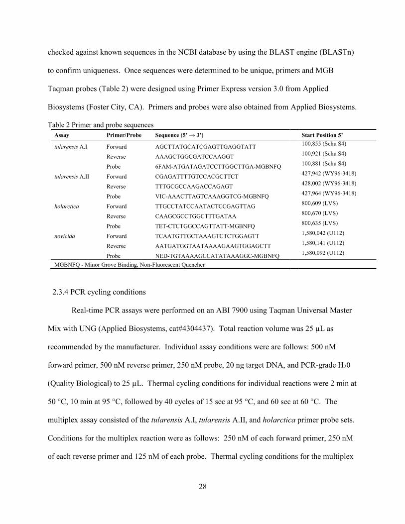
28
checked against known sequences in the NCBI database by using the BLAST engine (BLASTn)
to confirm uniqueness. Once sequences were determined to be unique, primers and MGB
Taqman probes (Table 2) were designed using Primer Express version 3.0 from Applied
Biosystems (Foster City, CA). Primers and probes were also obtained from Applied Biosystems.
Table 2 Primer and probe sequences Assay Primer/Probe Sequence (5’ → 3’) Start Position 5’
tularensis A.I Forward AGCTTATGCATCGAGTTGAGGTATT 100,855 (Schu S4)
Reverse AAAGCTGGCGATCCAAGGT 100,921 (Schu S4)
Probe 6FAM-ATGATAGATCCTTGGCTTGA-MGBNFQ 100,881 (Schu S4)
tularensis A.II Forward CGAGATTTTGTCCACGCTTCT 427,942 (WY96-3418)
Reverse TTTGCGCCAAGACCAGAGT 428,002 (WY96-3418)
Probe VIC-AAACTTAGTCAAAGGTCG-MGBNFQ 427,964 (WY96-3418)
holarctica Forward TTGCCTATCCAATACTCCGAGTTAG 800,609 (LVS)
Reverse CAAGCGCCTGGCTTTGATAA 800,670 (LVS)
Probe TET-CTCTGGCCAGTTATT-MGBNFQ 800,635 (LVS)
novicida Forward TCAATGTTGCTAAAGTCTCTGGAGTT 1,580,042 (U112)
Reverse AATGATGGTAATAAAAGAAGTGGAGCTT 1,580,141 (U112)
Probe NED-TGTAAAAGCCATATAAAGGC-MGBNFQ 1,580,092 (U112)
MGBNFQ - Minor Grove Binding, Non-Fluorescent Quencher
2.3.4 PCR cycling conditions
Real-time PCR assays were performed on an ABI 7900 using Taqman Universal Master
Mix with UNG (Applied Biosystems, cat#4304437). Total reaction volume was 25 µL as
recommended by the manufacturer. Individual assay conditions were are follows: 500 nM
forward primer, 500 nM reverse primer, 250 nM probe, 20 ng target DNA, and PCR-grade H20
(Quality Biological) to 25 µL. Thermal cycling conditions for individual reactions were 2 min at
50 °C, 10 min at 95 °C, followed by 40 cycles of 15 sec at 95 °C, and 60 sec at 60 °C. The
multiplex assay consisted of the tularensis A.I, tularensis A.II, and holarctica primer probe sets.
Conditions for the multiplex reaction were as follows: 250 nM of each forward primer, 250 nM
of each reverse primer and 125 nM of each probe. Thermal cycling conditions for the multiplex

29
assay were 2 min at 50 °C, 10 min at 95 °C, followed by 40 cycles of 15 sec at 95 °C and 60 sec
at 58 °C. A positive signal was determined by the crossing of a fluorescence threshold of 0.2
before cycle 40. Data analysis was performed using SDS v2.3 (Applied Biosystems). All tests
were performed in at least triplicate to ensure reproducibility.
While a novicida specific primer probe set was initially designed and tested singularly, it
was later determined that it could be omitted from the multiplex reaction. Rather than use all
four assays in the multiplex reaction, shared genomic markers among the subspecies allowed a
scoring matrix (Table 3) to be used to differentiate the subspecies using only three of the assays.
Table 3 Scoring matrix for triplex assay Ft Type A.I assay A.II assay holarctica assay Ft A.I Positive Negative Negative Ft A.II Negative Positive Negative
Ft holarctica Positive Positive Positive Ft novicida Positive Positive Negative
Ft - Francisella tularensis
2.3.5 MLVA analysis
Multi-locus variable-number tandem repeat analysis was performed as described
previously (20, 102). Briefly, the 11 marker MLVA was set up in five PCR mixes. Mix 1A
contained three labeled primers, mix 1B contained one labeled primer, mix 2 contained 3 labeled
primers, mix 3A contained three labeled primers, and mix 3B contained one labeled primer.
After PCR amplification of the targets, mixes 1A and 1B were combined with water in a final
ratio of 2:1:97 respectively, to produce mix 1. PCR products of mix 2 were diluted with water in
a final ratio of 1:49. The PCR products of mix 3A and 3B were diluted with water in a final ratio
of 2:1:97 to produce mix 3. The resulting three mixes were subjected to capillary electrophoresis
on a 16 capillary 3130xl (Applied Biosystems). Each sample was run with 2.25 µL per well
MapMarker 1000 (Bioventures) as a nucleic acid size standard. Resulting peaks (Figure. S2)

30
were compared to a MLVA database kindly provided by Paul Keim for subspecies identification.
Positive identifications were called if a minimum of 8 of 11 peaks were present with at least one
uniquely identifying peak to distinguish the isolate from others.
2.4 Results
The goal of this work was to address some of the shortfalls of previous PCR assays for F.
tularensis and develop a multiplex real-time PCR assay useful for the rapid identification and
characterization of F. tularensis isolates commonly found in the United States and Europe.
Markers unique to each of the F. tularensis subspecies could not be identified by us or others
(72). Molins-Schneekloth et al. (72) identified a genetic marker that was unique to type A.I
tularensis, subspecies holarctica and subspecies novicida, and another marker unique to type
A.II tularensis, subspecies holarctica and subspecies novicida. Based on these differences we
found it more reasonable and economical to use a multiplex assay with a scoring matrix to type
the four different types of F. tularensis (Table 3).
The development of this real-time PCR assay has broad application across the fields of
medical surveillance and CDC select agent detection. The different types and subspecies of F.
tularensis differ not only in their capacity to cause disease but in their geographic distribution as
well (100).
2.4.1 Assay validation
For the validation of the multiplex PCR assay, results from 119 Francisella isolates were
compared to other PCR assays (126) and the MLVA data from this study (Table S1). On 32
(~27 %) of the isolates, insufficient MLVA data was obtained for an identification. The

31
experiment was repeated 3 times with the same result. Even the U112 strain, which is widely
known to be a novicida isolate, was inconclusive using the MLVA technique. This illustrates the
need for additional typing methodologies.
Of the 87 isolates for which the MLVA was successful, 85 (~98 %) isolates had the same
subspecies identification as the multiplexed PCR assay. The assay was also tested against 3 near
neighbours, Wolbachia persica, Francisella philomiragia 25015, and F. philomiragia 25016;
each with a negative result. The assay also showed no cross-reactivity with other laboratory
species (Table 4).
Table 4 Other isolates tested Brucella abortus Burkholderia pseudomallei Clostridium botulinum Escherichia coli Francisella philomiragia Mycobacterium avium Mycobacterium tuberculosis Mycobacterium ulcerans Pasteurella multocida Salmonella choleraesuis Shigella dysenteriae Staphlococcus aureus Streptococcus pneumoniae Streptococcus pyogenes Wolbachia persica Yersinia pestis
Multiplex PCR results for isolates 70000907 and 6168 did not match up with the MLVA
data. While the multiplexed PCR assay categorized isolate 70000907 as novicida, the MLVA
analysis grouped it as type A.I. This is consistent with another PCR assay (126), which
classified this isolate simply as Type A. The multiplex PCR data classified isolate 6168 as Type
B while both the MLVA and the Kugeler et al., (126) PCR assay classified it as novicida. The

32
reasons for these discrepancies are not immediately clear but may be due to genomic
rearrangement. These discrepancies are a focus of ongoing research.
2.4.2 Assay sensitivity
Serial tenfold dilutions of genomic F. tularensis DNA were assayed to estimate the
detection limits when used in singleplex and multiplex reactions. In singleplex reactions, the
Type A.II tularensis was the most sensitive at 25 fg. Type A.I, Type B and novicida subspecies
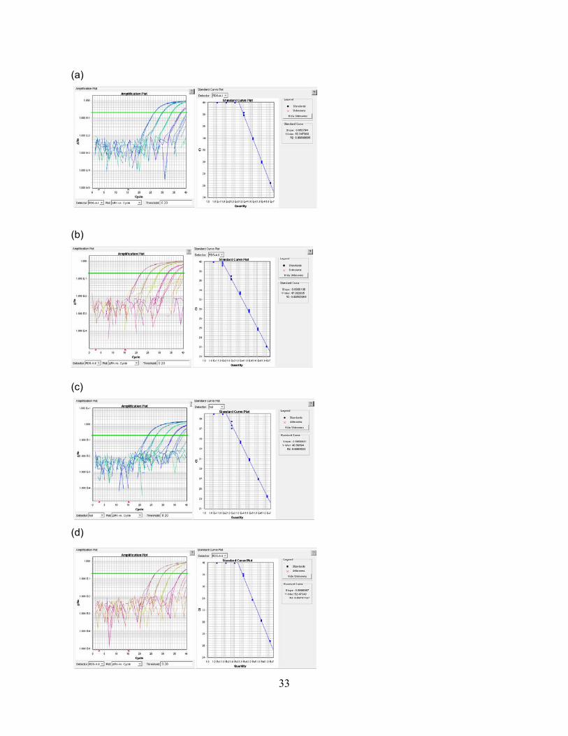
all had the same detection limit at 250 fg (Figure. S3). Assuming a genome weight of 2.2 fg,
limits of detection were ~11 organisms for the Type A.II, and ~114 organisms for the Type A.I,
Type B and novicida assays. In the multiplex reactions, limits of detection were reduced by one
log10 step; the Types A.I, A.II and novicida all had limits of detection at 250 fg while Type B
was at 2.5 pg (Figure. 3). Again, with a genome weight of 2.2 fg, this translates to ~114
organisms for the Type A.I, Type A.II and novicida assays, and ~1 136 organisms for the Type B
assay. Each of the assays were run in triplicate with identical results.
2.4.3 Characterization of isolates
We used this multiplex assay to characterize F. tularensis isolates, the majority of which
were natural isolates from Utah and New Mexico. Of all the isolates tested (Table S1) 5.0 %
were F. tularensis Type A.I, 76.5 % were F. tularensis Type A.II, 6.7 % were identified as F.
tularensis Type B, and 11.8 % were identified as F. tularensis subspecies novicida.
33
(a)
(b)
(c)
(d)

34
Figure 3 Sensitivities of multiplex assays Tenfold serial dilutions of F. tularensis chromosomal DNA were tested to determine individual assay sensitivities. (a) Type A.I, (b) Type A.II, (c) Type B, (d) novicida (analysed using the A.II detector). No Template Control (NTC) samples were negative for each sample processed. Calculated PCR efficiencies (a) 81.6%, (b) 92.0%, (c) 89.7% (d) 86.7%.
2.5 Discussion
Many PCR assays for F. tularensis and its subspecies have been developed. An assay developed
by Broekhuijsen et al., (21) is capable of discriminating the four subspecies of F. tularensis.
However, it does not have the convenience of real-time detection and necessitates running the
products on a gel to visualize amplicons up to 3 kilobases in length. Furthermore, the assay is
not multiplexed nor can it discriminate between Type A.I and Type A.II tularensis. Kugeler et
al. (126) developed a real-time PCR assay for F. tularensis that was able to distinguish Type A
from Type B tularensis, but it could not differentiate Type A.I from Type A.II, nor could it
identify the novicida subspecies. Finally, Molins-Schneekloth et al. (72) were able to develop a
multiplex PCR assay that could differentiate Type A.I tularensis from Type A.II, but the assay
was not real-time, nor could it differentiate among Type B tularensis or the novicida subspecies.
This work describes the first multiplexed, real-time PCR assay for the characterization of the
major types of F. tularensis found in the United States and around the world: Type A.I, Type
A.II, Type B, and subspecies novicida. Since type A.I tularensis is usually found on the East
Coast of the United States, an outbreak of type A.I on the West Coast could be an indication of
an intentional release. This multiplex assay could help law enforcement agencies to identify
possible bioterrorism events as well as guide the administration of therapeutics by health
officials.
A major hurdle for creating Francisella tularensis PCR assays specific for subspecies is that
their genomes are highly similar. The 16S rRNA genes which are typically used for

35
identification of microorganisms exhibit 98.5% to 99.9% similarity across all subspecies which
equates to 2 to 23 nucleotides difference (43). A recent study further highlighted the similarities
of F. tularensis subspecies. Their results show that a pairwise alignment of sequenced draft
genomes of low and high virulence subspecies exhibited over 95 % similarity (25). Using
various regions of difference identified by SSH (72) and comparative genomic data using Mauve
software (53), we were able to identify genomic regions conducive to the creation of real-time
PCR assays.
To validate the assays, we tested them against other laboratory strains (Table 4) in addition to
two near-neighbours from the same genus: F. philomiragia as well as the nearest neighbour of F.
tularensis, Wolbachia persica (43). The results showed no cross-reactivity among these other
species, demonstrating that the identified regions were unique to the subspecies of F. tularensis.
Furthermore, the subspecies identification based on the scoring matrix (Table 3) show a
successful identification of ~98 % of the isolates, compared to the MLVA data. This
discrepancy was not an artifact of the multiplex assay, as it was also seen in the singleplex
reaction. Despite these slight differences, we believe that this multiplex assay remains a
powerful, rapid, presumptive screening test for the subspecies of F. tularensis. In addition, this
work further underscores the need for multiple identification assays to definitively identify these
closely related subspecies.
In singleplex reactions, we demonstrated a sensitivity based on serial 10-fold dilutions of stock
DNA of 11 organisms for the Type A.II, and 114 organisms for the Type A.I, Type B, and
novicida assays. These sensitivities are within an order of magnitude of other published, real-
time F. tularensis PCR assays (125, 126). In the triplex reactions, the Type A.I, Type A.II, and
novicida assays yielded a sensitivity of 114 organisms and 1 136 organisms for the Type B

36
reaction. The sensitivities remained the same for the Type A.I and novicida assays with an order
of magnitude decrease in sensitivity for the Type A.II and Type B assays. Another multiplex
real-time PCR assay for F. tularensis demonstrated sensitivities as low as 0.5 genome
equivalents (127). This increased sensitivity may be explained by two factors: 1) the assay
targeted the 16S rDNA, of which multiple copies are present in the genome and 2) the multiplex
assays only included two assays thus reducing the chance of competitive PCR problems. When
Tomaso et al. (127) included three assays in their multiplex assay, no amplification curves were
observed.
In larger multiplex reactions, it is not uncommon to see a decrease in sensitivity. In a quadruplex
real-time PCR assay for Yesinia pestis, Stewart et al. (128) saw a 10 fold decrease in the
sensitivity when their assay was moved from singlplex to quadruplex: from 150 pg to 1.5 ng.
The results for our triplex assay are similar to these results showing a ten-fold decrease in
sensitivity for two of the assays when used in a multiplex format: from 114 organisms to 1 136
organisms.
This multiplex assay was used to characterize a large collection of F. tularensis isolates. Many
of the natural isolates were obtained from the Utah and New Mexico Departments of Health.
Some standard laboratory strains of F. tularensis, such as Schu S4, LVS, U112 and other
novicida variants were included for reference. The characterization including all isolates except
the standard strains and novicida variants returned 104 isolates characterized. Of those, the vast
majority (91 isolates or ~88 %) were identified as Type A.II. Four isolates (~3.8 %) were
identified as Type A.I, seven isolates (~6.7 %) were identified as Type B, and one isolate (~0.9
%) was identified as novicida. This one isolate identified as novicida however is likely a

37
misidentification because another PCR assay (Kugler et al., 2006) as well as the MLVA data
identified it as Type A.
It is not surprising that the vast majority of the tested isolates were Type A.II since most of the
isolates used in this study were obtained from the Utah and New Mexico Departments of Health.
This distribution of isolates is consistent with previous data (19, 100) suggesting that Type A.II
tularensis is predominant in the western United Sates and that Type A.I tularensis is
predominantly found in the eastern United States.
F. tularensis is classified as a Category A select agent by the CDC. Due to its potential use as a
bioterrorism weapon, its endemic status in various locations around the world, and differences in
virulence among the various types of tularensis, it is important that government agencies, health
departments, and hospitals be able to rapidly identify each subtype. The assay presented in this
work is a rapid, single-tube, multiplex, real-time PCR assay that can be used to quickly screen
individual samples or adapted for high-throughput applications in either 96- or 384-well formats.
We believe that this assay will be an invaluable tool in the presumptive identification and
characterization of F. tularensis isolates during outbreaks of disease or possible bioterrorism
events.

38
Chapter 3 Natural selection in virulence genes of Francisella tularensis
3.1 Abstract
A fundamental tenet of evolution is that alleles that are under negative selection are often
deleterious and confer no evolutionary advantage. Negatively selected alleles are removed from
the gene pool and are eventually extinguished from the population. Conversely, alleles under
positive selection do confer an evolutionary advantage and lead to an increase in the overall
fitness of the organism. These alleles increase in frequency until they eventually become fixed
in the population. Francisella tularensis is a zoonotic pathogen and a potential biothreat agent.
The most virulent type of F. tularensis, Type A, is distributed across North America with Type
A.I occurring mainly in the east and Type A.II appearing mainly in the west. F. tularensis is
thought to contain a genome in decay (losing genes) because of the relatively large number of
pseudogenes present in its genome. We hypothesized that the observed frequency of gene
loss/pseudogenes may be an artifact of evolution in response to a changing environment, and that
genes involved in virulence should be under strong positive selection. To test this hypothesis we
sequenced and compared whole genomes of Type A.I and A.II isolates. We analyzed a subset of
virulence and housekeeping genes from several F. tularensis subspecies genomes to ascertain the
presence and extent of positive selection. Eleven previously identified virulence genes were
randomly chosen and screened for positive selection along with 10 randomly chosen
housekeeping genes. Analyses of selection yielded one housekeeping gene and 7 virulence
genes which showed significant evidence of positive selection at loci implicated in cell surface
structures and membrane proteins, metabolism and biosynthesis, transcription, translation and
cell separation, and substrate binding and transport. Our results suggest that while the loss of

39
functional genes through disuse could be accelerated by negative selection, the genome decay in
Francisella could also be the byproduct of adaptive evolution, as evidenced by several of its
virulence genes which are undergoing strong, positive selection.
3.2 Introduction
Francisella tularensis is a Gram-negative facultative intracellular pathogen and is the
causative agent of the zoonotic disease, tularemia (rabbit fever). While many mammals are
susceptible to the disease, they are not known to act as environmental reservoirs (129).
Arthropods such as ticks and deer flies are likely the primary vectors in the spread of tularemia,
with human infection usually resulting from the bite of an insect or inhalation of aerosolized
bacteria (27, 130). While there are no confirmed reservoirs for F. tularensis, it has been
demonstrated that some strains cause the rapid encystment of Acanthamoeba castellanii and are
able to survive within these amoeba cysts for up to three weeks, thus increasing the
environmental persistence of the bacteria (122, 129).
F. tularensis is highly virulent with an infectious dose of around 10 organisms (6).
Because of its highly infectious nature, F. tularensis has, in the past, been characterized as a
bioweapon by many nations (118). The Centers for Disease Control and Prevention (CDC) has
classified F. tularensis as a Tier 1 potential agent of bioterrorism. While there is a vaccine for
tularemia, it is still classified as an investigational new drug (IND) and is only available for high
risk personnel such as the military and certain laboratory workers (131). In order to develop a
licensed vaccine, it is imperative that the virulence mechanisms of F. tularensis be investigated
and understood.

40
There are currently three recognized species within the Francisella genus: F. tularensis,
F. philomiragia, and F. novicida. It has been suggested that F. novicida be reclassified as a
subspecies of F. tularensis, and much of the scientific literature, including Bergey’s manual of
Systemic Bacteriology, already reflects this change (2, 97, 100, 130, 132). Excluding F.
novicida, there are three recognized subspecies of F. tularensis: tularensis, holarctica, and
mediasiatica. Two of the subspecies of Francisella tularensis are often abbreviated simply as
Type A (tularensis) and Type B (holarctica). Furthermore, Type A tularensis has been further
divided into A.I and A.II types based on geographic distribution and genome architecture (20).
Type A tularensis is generally found in North America with Type A.I dominating the central and
eastern portions of the continent while Type A.II is typically found in the western United States
(19). Type B tularensis (or holarctica) exhibits a worldwide prevalence, while the mediasiatica
subspecies appears to be confined to the central Asian republics of the former Soviet Union (20,
133) (Table 5). The phylogenetic relationships of these subspecies are shown in Figure 4. Type
A tularensis is the most virulent subspecies of F. tularensis (23, 24).
Table 5 Subspecies of F. tularesnsis and their worldwide distribution Subspecies Type Distribution tularensis A.I Eastern United States tularensis A.II Western United States holarctica B North America, Eruope, Japan mediasiatica N/A Central Asain Republics novicida N/A Worldwide

41
Figure 4 Maximum likelihood tree inferring the phylogenetic relationships of the F. tularensis subspecies Tree was constructed by concatenating 10 housekeeping genes (recA, gyrB, groEL, dnaK, rpoA1, rpoB, rpoD, rpoH, fopA, and sdhA) followed by alignment with Clustal W and generation of the tree with MEGA 5.2. Taxa names use the shorthand described in Table 2.
Virulence genes are those whose products contribute to an organism’s ability to infect
and colonize a host. Virulence genes are often located within mobile genetic elements such as
plasmids, transposons, bacteriophages and pathogenicity islands, which enhance genetic
diversity through the exchange with other bacteria (134). F. tularensis possesses a pathogenicity
island of about 30 kb in length consisting of 17 open reading frames flanked by inverted repeat
sequences (47). The Francisella Pathogenicity Island (FPI) has been shown to be required for
intra-macrophage growth (46), phagosome escape and general virulence of F. tularensis (135).
The subspecies novicida is most similar to the ancestral lineage of the F. tularensis subspecies

42
and contains only one copy of the FPI while the other more pathogenic subspecies contain two
copies of the FPI (47). It should be noted that the inverted repeats flanking the FPI provide a
direct means for this duplication to take place.
Despite the varied mechanisms for sharing genes among bacteria, molecular evidence
suggests that the four subspecies of F. tularensis have evolved and acquired virulence genes by
vertical descent rather than by horizontal gene transfer (97). A whole genome analysis of
horizontal gene transfer (HGT) in F. tularensis identified 30 candidate genes as having been
acquired through HGT (136), but none of them were putative virulence genes. Genetic variation
in the subspecies of F. tularensis seems to have arisen by mutation rather than HGT (110-112).
Furthermore, the subspecies novicida has been shown to possess a CRISPER/Cas system to
defend against invading genetic elements, further supporting the notion that mutation and
selection are the driving factors of evolution in F. tularensis virulence rather than the acquisition
of mobile genetic elements by HGT (113, 114).
Whole genome comparisons between pathogenic and non-pathogenic subspecies of F.
tularensis revealed that the pathogenic subspecies evolved from the non-pathogenic subspecies
by mechanisms of genomic rearrangements, point mutations and small indels (36). The many
pseudogenes present in the F. tularensis genome suggest that it is a genome in decay (137).
While these pseudogenes may have arisen via negative selection through disuse, it is also
possible that this evidence of decay is a byproduct of an adaptive response to a changing
environment (138, 139) or even the evolution of avirulence (140, 141). Alternatively, we
hypothesize that in addition to genome decay through the creation of pseudogenes, positive
natural selection is also a driving force in the continued evolution of virulence of this important
pathogen.

43
3.3 Materials and Methods
3.3.1 Bacterial strains and culture conditions
The isolates used in this study (Table 6) are a part of a select agent archive housed at the
special pathogens laboratory at Brigham Young University (Provo, UT). The collection largely
consists of isolates obtained from the State Health Departments of Utah and New Mexico over
the past two decades. All F. tularensis isolates were grown on modified Mueller Hinton agar
(MMHA) (Becton Dickinson and Company, Franklin Lakes, New Jersey, USA) for 3-4 days
with 5 % CO2 at 35 °C. MMHA was prepared by autoclaving the Mueller Hinton base, which
was chocolatized by adding 5 % sheep blood while the medium was approximately 80 °C. After
the medium cooled to 50 °C, 10 mL of 10 % glucose and 20 mL of IsoVitaleX (Becton
Dickinson and Company) were added to 1 L.
3.3.2 Genome sequencing and annotation
Total genomic DNA was extracted from each isolate using the MagNA Pure System
(Roche) and the MagNA Pure LC DNA Isolation Kit III (Roche) according to the manufacturer’s
directions. Briefly, cells grown on MMHA agar were suspended in 250 μL of TE buffer
containing 1.8 μg/μL lysozyme and incubated for 1 h at 37 °C. To this tube, 270 μL of bacterial
lysis buffer and 100 μL of proteinase K were added and the tube was incubated for 10 min at 65
°C. Samples were then incubated in boiling water for 10 min to inactivate pathogens. DNA was
eluted in a total volume of 100 µL. DNA concentration was measured using a PicoGreen assay
(Invitrogen) and TBS-380 fluorometer (Turner Biosystems). Prior to genome sequencing,
isolates were typed using the multiplex PCR assay as described previously (2). Four Type A.I
and six Type A.II strains were selected for sequencing in order to maximize the available

44
geographic and genetic diversity. Whole genome shotgun sequencing was accomplished using
the Roche/454
Table 6 Genomes used for analysis of selection Genome Shorthand Type Reference Isolated from
F. tularensis subsp. tularensis 70001275 tul_FT_01 A.II This work Utah, US
F. tularensis subsp. tularensis 70102010 tul_FT_02 A.II This work Utah, US
F. tularensis subsp. tularensis 80700103 tul_FT_03 A.II This work Utah, US
F. tularensis subsp. tularensis 831 tul_FT_05 A.II This work New Mexico, US
F. tularensis subsp. tularensis AS_713 tul_FT_06 A.II This work New Mexico, US
F. tularensis subsp. tularensis 3571 tul_FT_07 A.II This work New Mexico, US
F. tularensis subsp. tularensis 1378 tul_FT_08 A.I This work New Mexico, US
F. tularensis subsp. tularensis 80700075 tul_FT_09 A.I This work Utah, US
F. tularensis subsp. tularensis 79201237 tul_FT_10 A.I This work Utah, US
F. tularensis subsp. tularensis 80700069 tul_FT_11 A.I This work Utah, US
F. tularensis subsp. tularensis SCHU S4 tul_SchuS4 A.I Larsson et al. 2005 Ohio, US
F. tularensis subsp. tularensis WY96-3418 tul_WY96_3418 A.II Beckstrom-Sternberg et al. 2007 Wyoming, US
F. tularensis subsp. tularensis FSC198 tul_FSC198 A.I Chaudhuri et al. 2007 Slovakia
F. tularensis subsp. tularensis NE061598 tul_NE061598 A.I Nalbantoglu et al. 2010 Nebraska, US
F. tularensis subsp. tularensis TI0902 tul_TI0902 A.I Modise et al. 2012 Virginia, US
F. tularensis subsp. tularensis TIGB03 tul_TIGB03 A.I Modise et al. 2012 Virginia, US
F. tularensis subsp. holarctica F92 hol_F92 B Antwerpen et al. 2013 Germany
F. tularensis subsp. holarctica FSC200 hol_FSC200 B Svensson et al. 2013 Sweden
F. tularensis subsp. holarctica FTNF002_00 hol_FTNF002_00 B Barabote et al. 2009 Spain
F. tularensis subsp. holarctica LVS hol_LVS B Unpublished Russia
F. tularensis subsp. holarctica OSU18 hol_OSU18 B Petrosino et al. 2006 Oklahoma, US
F. tularensis subsp. mediasiatica FSC147 med_FSC147 N/A Champion et al. 2009 Kazakhstan
GS FLX Pyrosequencer (Roche) and the Titanium® chemistry according to the manufacturer’s
recommendations. Raw sequencing reads were aligned to reference sequences using Newbler
version 2.6. The reference sequence for A.I strains was SCHU S4 (GenBank accession number
AJ749949.2), while WY96-3418 (NC_009257.1) was used for A.II strains. Aligned shotgun
sequences were annotated using the Prokaryotic Genome Automatic Annotation Pipeline
(PGAAP) (142).

45
3.3.3 Analysis of selection
A total of 22 taxa were included in the analysis of selection; the 10 new F. tularensis
subspecies tularensis genomes described in this work, and 12 other F. tularensis genomes of the
tularensis, holarctica, and mediasiatica subspecies retrieved from GenBank (Table 6).
In addition to 5 Type IV pilus genes which are known virulence factors of F. tularensis,
89 other previously identified virulence genes (143) were analyzed for positive selection. In
total, 94 virulence genes were extracted from the genomes of the 22 taxa in Table 6 for analysis.
While the taxa used in this work were isolated from various geographical locations (Table 6), the
genes selected for analysis are common to all the isolates. Individual genes were aligned with
ClustalW (144) using the default parameters. A hierarchical likelihood ratio test was performed
on the aligned sequences to determine which model of evolution best fit the data (AIC) (145)
using MEGA v. 5.2 (146). MEGA v. 5.2 was also used to generate phylogenetic trees via
maximum likelihood assuming the previously identified best-fit substitution model (146).
Maximum likelihood computations of dN and dS were conducted using HyPhy (147-151).
After analyses of selection with HyPhy, 11 genes were arbitrarily selected for further
analysis using TreeSAAP version 3.2 (152), which measures selection based on 31
physiochemical properties of amino acids. TreeSAAP compares aligned sequences in the
context of the specified phylogenetic topology, codon by codon, to infer amino acid replacement
events. Results were divided into one of eight categories based on the magnitude of the change:
categories 1-3 indicate a conservative change, categories 4-6 represent moderate change, while
categories 7-8 represent more drastic changes and indicate positive selection. Only genes that

46
returned changes in categories 7-8 were examined further. A z-score was calculated by
TreeSAAP for each of the 31 physiochemical properties. Drastic changes (categories 7-8) with a
z-score of p < 0.001 were determined to indicate statistically significant positive selection. To
summarize, we explored 94 virulence-associated genes. Of those, 64 were indicated by HyPhy
analysis to be under positive selection, though the results were not statistically significant. From
the 64 genes that showed some evidence of positive selection, we arbitrarily chose 11 for
subsequent TreeSAAP analysis. To better visualize the evolutionary changes in the proteins,
secondary structures of the proteins were predicted using the PSIPRED secondary structure
prediction (http://bioinf.cs.ucl.ac.uk/psipred/) (153).
3.4 Results and Discussion
3.4.1 Genome sequencing and annotation
Of the 10 novel genomes sequenced, 4 were F. tularensis Type A.I and 6 were F.
tularensis Type A.II. A summary of the sequencing statistics of these newly sequenced genomes
can be found in Table 7.
The lengths of the reference sequences were 1,892,775 and 1,898,476 for the SCHU S4
and WY96-3418 strains, respectively. The average length of the sequenced Type A.I genomes
was 1,814,938 and the average length of the Type A.II genomes was 1,814,544. Thus, the
sequenced genomes are about 80 kb shorter than the reference sequences. This is not surprising
since the shotgun method of sequencing cannot adequately sequence through large repetitive
regions of DNA resulting in gaps in the final alignments.

47
Table 7 Summary of sequenced and aligned F. tularensis genomes Strain
Designation 70001275 70102010 80700103 831 AS_713 3571 1378 80700075 79201237 80700069
Shorthand Designation FT-01 FT-02 FT-03 FT-05 FT-06 FT-07 FT-08 FT-09 FT-10 FT-11
Type A.II A.II A.II A.II A.II A.II A.I A.I A.I A.I Average
Sequencing Depth
212x 164.8x 76.6x 101.2x 107.7x 101.3x 93.2x 87.5x 92.2x 127.8x
Reference sequence
WY96-3418 (Accession
number NC_009257.1)
WY96-3418 (Accession
number NC_009257.1)
WY96-3418 (Accession
number NC_009257.1)
WY96-3418 (Accession
number NC_009257.1)
WY96-3418 (Accession
number NC_009257.1)
WY96-3418 (Accession
number NC_009257.1)
SCHU S4 (accession
number AJ749949.2)
SCHU S4 (accession
number AJ749949.2)
SCHU S4 (accession
number AJ749949.2)
SCHU S4 (accession
number AJ749949.2)
N50 contig size (bases) 137,296 124,943 43,424 61,138 73,318 103,637 360,449 348,066 239,117 348,045
Sequenced genome length
1815079 1816669 1810598 1814141 1814591 1816188 1815377 1814277 1814563 1815536

48
Table 8 Summary of annotation results for F. tularensis genomes
Shorthand Designation FT-01 FT-02 FT-03 FT-05 FT-06 FT-07 FT-08 FT-09 FT-10 FT-11 SCHU
S4 WY96-
3418 Type A.II A.II A.II A.II A.II A.II A.I A.I A.I A.I A.I A.II
Number of protein
sequencesa 1784 1792 1808 1817 1809 1800 1785 1796 1781 1787 1804 1824
Number of rRNA
sequences 6 6 5 7 6 6 7 8 6 6 10 10
Number of tRNA
sequences 32 32 32 32 32 32 32 32 32 32 38 38
Total number of
genes 1822 1830 1845 1856 1847 1838 1824 1836 1819 1825 1852 1872
GenBank accession number
AMPP00000000
AMPY00000000
AMPX00000000
AMPV00000000
AMPU00000000
AOUD00000000
APKY00000000
APKX00000000
APKW00000000
APKV00000000 AJ749949.2 NC_009257.1
a includes pseudogenes

49
The number of predicted protein-coding sequences for the references SCHU S4 and
WY96-3418 are 1604 and 1634, respectively. Because of the shortened size of the newly
sequenced genomes, it was expected that the total number of protein sequences would also be
less. This, however, was not the case. The new genomes were processed through an automated
annotation pipeline, and were not manually curated. This resulted in many sequences that the
automated pipeline mistook for genes that the curators of the reference genomes had already
removed. Further study and curation of these newly sequenced genomes will be necessary for
detailed comparative analyses, but sufficiently resolved to facilitate our analysis of selection on
their virulence-associated genes.
3.4.2 Analysis of selection
The analysis of dN/dS ratios is a well-known benchmark for identifying selection and was
used as a first pass to identify genes that are most likely to be under positive selection. Results
revealed that of the 94 previously identified virulence genes (Su et al. 2007), 64 gave some
indication of positive selection (although with p-values ranging from 0.40 to 0.90, none of them
were statistically significant). It has been suggested that some of the assumptions of the dN/dS
ratio test, as well as the McDonald-Kreitman test of adaptive evolution (154) can be too
conservative for accurately detecting positive selection and adaptation (152, 155-157). For
example, dN/dS ratios alone were unable to detect positive selection in rapidly mutating genes of
HIV in patients showing signs of drug resistance (157). Furthermore, Sharp (158) asserts that
another problem with dN/dS ratio tests is that their significance falls as selection continues to
“weed out” the effects of detrimental amino acid changes. Thus, more nuanced methods may be
needed to accurately predict positive selection in biological systems (156). Finally, while dN/dS

50
ratios and the McDonald-Kreitman test may indicate the presence of selection on a gene, they do
not give any indication of how selection affects the structure and function of the protein (159).
To perform a more sensitive analysis we used TreeSAAP (Selection on Amino Acid
Properties using phylogenetic Trees) which tests for selection of expected random distributions
of possible amino acid changes based on 31 physiochemical properties of amino acids and
associated phylogenetic trees (152). The methodological approach that TreeSAAP uses to detect
selection has been shown to be better suited at detecting selection where more traditional
methods such as dN/dS ratios and McDonald-Kreitman analyses cannot (152, 158). We note that
TreeSAAP doesn’t have a built-in correction for multiple-hypothesis testing when calculating p-
values. Although a Bonferroni correction of TreeSAAP significance tests is indicated for
comparative purposes, previous work has cast doubt on whether this will yield an appropriate
level of confidence (160). Thus, we employed TreeSAAP as the best-suited approach currently
available for detecting selection.
Since the HyPhy results were not statistically significant, 11 virulence genes were
arbitrarily selected for further analyses of selection using TreeSAAP (Table 9). In genes
FTT_1611, FTT_1156c, FTT_0935c, and FTT_1525c, no significant positive selection was
detected. However, significant positive selection (categories 7-8) was detected in one or more
physiochemical properties of the remaining seven genes: FTL_1134, FTT_0683, FTT_0881c,
FTT_0504c, FTT_0936c, FTT_0766, and FTT_1125. The physiochemical properties for which
positive selection was detected include the following: isoelectric point, power to be at the N-
terminal, composition, power to be at the middle of alpha helix, solvent accessible reduction
ratio, equilibrium constant, power to be at the C-terminal, coil tendencies, compressibility,
bulkiness, turn tendencies, and average number of surrounding residues.

51
Table 9 Virulence genes randomly chosen for TreeSAAP analysis
Locus taga Gene Name Positive selection detected by TreeSAAP
FTT_0881c rocE Yes FTL_1134 Yes FTT_1156c pilQ No FTT_1611 No FTT_1125 metQ Yes FTT_0504c sucC Yes FTT_0935c bioC No FTT_0683 pilD Yes FTT_1525c No FTT_0766 deoD Yes FTT_0936c bioF Yes
a Locus tag designations for FTT are adopted from the annotation of the SCHU S4 genome (accession no. AJ749949.2) and locus tag designations for FTL are adopted from the annotation of the LVS genome (accession no. AM233362)
For analytical purposes, these seven genes were placed into four functional categories
(143): FTL_1134 and FTT_0683 fall into the cell surface structures and membrane proteins
category, FTT_0881c, FTT_0504c and FTT_0936c are metabolism and biosynthesis proteins,
FTT_0766 is categorized as a protein involved in transcription, translation and cell separation,
and FTT_1125 is classified in the substrate binding and transport functional group.
3.4.2.1 Cell surface structures and membrane proteins
In the functional group of cell surface structures and membrane proteins, the gene
FTL_1134 demonstrated selection for three of the 31 physiochemical properties tested:
isoelectric point in codons 183-203 and 383-394, power to be at the N-terminus in codons 271-
285 and 323-337, and composition in codons 183-197 (Figure. 5). Codons 183-203 begins in an
α-helix and ends in a loop, codons 271-285 includes portions of two α-helices and a loop in

52
between, codons 323-337 begins in an α-helix and ends in a loop and codons 383-394 occurs
near the C-terminal of an α-helix (Figure S4). The product of FTL_1134 is a hypothetical
membrane protein of unknown function. This gene (as were all other genes cited in this work)
was identified as a virulence gene by a whole genome transposon screen for virulence genes of
F. tularensis (143). Thus, in this case, a protein of unknown function has been identified as a
virulence gene.
Figure 5 Selection on FTL_1134. Areas of the gene under selection are indicated where the test statistic is greater than 3.09 (horizontal line) (p < 0.001). Selection is present in the gene for the following amino acid properties where the lines cross the threshold: Isoelectric Point (orange line), Power to be at the N-terminal (gray line) and Composition (yellow line).
F. tularensis is an intracellular pathogen which is routinely phagocytized by host cells
and taken into a phagosome where the pH is lowered in an effort to rid the cell of the bacterium.
Selection of FTL_1134 for the isoelectric point, the pH at which a molecule carries no net
electrical charge, hints that this membrane protein may be exposed to changes in pH and is
adapting to withstand these changes.
-1
0
1
2
3
4
5
6
7
8
1 16 31 46 61 76 91 106
121
136
151
166
181
196
211
226
241
256
271
286
301
316
331
346
361
376
391
Test
Sta
tistic
Codon Number
Threshold Isoelectric Point
Power to be at the N-terminal Composition

53
FTL_1134 also demonstrated selection for the amino acid property of power to be at the
N-terminal. This amino acid property is defined as the intrinsic property of an amino acid
residue to be located at the N-terminus of an α-helix (161). The codons under selection occur in
varied domains of the secondary structure of this protein including the N-terminus of an α-helix,
the C-terminus of an α-helix and the loop between two α-helices (Figure S4). While this
property and its relation to positive selection is not well understood, we speculate that the regions
under selection are important to the ability of this virulence gene to function intracellularly since
in an infection model with this gene knocked out, the host is able to clear the infection (143).
The third and final amino acid property under selection for FTL_1134 is composition.
Composition refers to the individual amino acid make-up of a protein. Since FTL_1134 is a
hypothetical membrane protein of an unknown function, it is difficult to know how positive
selection for the composition property may affect it. Further study is needed to determine the
function of this protein and its role in the virulence mechanisms of F. tularensis before we can
gain an understanding of how selection is influencing this protein.
The other protein to demonstrate positive selection in the cell surface structures and
membrane proteins functional group is FTT_0683, which codes for the type IV pilus protein
PilD. Type IV pili are common in Gram-negative pathogens and mediate the attachment of the
pathogen to various host receptors (162). Specifically, PilD is a bifunctional, cytoplasmic
membrane protein responsible for the cleavage of the N-teminal leader sequences of prepilin and
also catalyzes the N-methylation of the N-terminal phenylalanine of mature pilins (163).
Without a functional PilD, the type-IV pilus apparatus cannot be assembled, thus restricting the
pathogenicity of F. tularensis. TreeSAAP detected selection in PilD for the amino acid property
of power to be at the middle of α-helix, which is described as the intrinsic property of a specific

54
amino acid to be located in the middle of an α-helix (161). The catalytic domains of PilD are
predicted to be specific cytoplasmic cysteine motifs found toward the N-terminus of the protein
(164), well distanced from the selection occurring toward the C-terminus of the protein in amino
acids 190-204 (Figure 6). Selection in these membrane bound α-helices do not affect the
catalytic activity of PilD, but may alter membrane positioning of the protein (Figure S5).
Figure 6 Selection on FTT_0683 (pilD). Areas of the gene under selection are indicated where the test statistic is greater than 3.09 (horizontal line) (p < 0.001). Selection is present in the gene for the amino acid property of Power to be at the middle of alpha-helix where the orange line crosses the threshold.
3.4.2.2 Metabolism and biosynthesis
Three genes in the metabolism and biosynthesis functional group demonstrated selection,
FTT_0881c (rocE), FTT_0504c (sucC), and FTT_0936c (bioF). The study which identified
these genes as virulence factors used a signature-tagged mutagenesis approach (143). Briefly,
this approach introduces random transposons to disrupt genes in the genome before infection
using a mouse model. If an organism with a particular disrupted gene is not recovered from the
-1
0
1
2
3
4
5
6
7
8
1 11 21 31 41 51 61 71 81 91 101
111
121
131
141
151
161
171
181
191
201
211
221
231
241
251
261
Test
Sta
tistic
Codon Number
Threshold Power to be at the middle of alpha-helix

55
infection model, that gene is then classified as necessary for virulence. This approach has been
used to identify virulence factors in many other organisms (165, 166). In the strictest sense,
genes categorized as metabolism and biosynthesis should not be considered virulence factors,
since impairing them would naturally restrict growth regardless of the status of infection (143,
167). However, it is important not to discount the role these genes may play in infection
altogether, since they may be valuable targets for the future development of therapeutics or
vaccines.
The first gene in the metabolism and biosynthesis category for which selection was
identified was FTT_0881c. This locus codes for the protein RocE which is an amino acid
permease, specifically, a transmembrane arginine transporter protein. Francisella tularensis
lacks the ability to synthesize the amino acid arginine. Furthermore, recent studies have shown
that the cytosol of the host cell doesn’t contain sufficient free nutrients to support the rapid
growth of the pathogen (168). Without access to arginine, the pathogen would not be able to
reproduce and infection with F. tularensis would fail. To compensate, F. tularensis induces the
host cell into an autophagy state in which proteins are broken down into their constituent amino
acids and made available to the pathogen (168). F. tularensis and its transmembrane amino acid
permease, RocE, is then ready to scavenge for the newly available amino acids it is not able to
produce on its own, among which is arginine. Furthermore, macrophages use arginine to
produce nitric oxide (NO) as an antimicrobial defense (169). In addition to scavenging arginine
for growth, the ability of F. tularensis to remove arginine would also deprive the cell of this
potent antimicrobial agent, thus allowing the infection to progress.
The rocE gene demonstrated positive selection for the solvent accessible reduction ratio
property of amino acids in codons 109-116, 438, and 442 (Figure 7). In the secondary structure

56
of RocE, codons 109-116 encompass a β-sheet and part of a loop while codons 438 and 442
occur within the same α-helix (Figure S6). The solvent accessible reduction ratio is defined as
the decrease in solvent accessibility for an amino acid residue when the protein molecule moves
from a hypothetically extended state to the native folded state (170). Consequently, as the
solvent accessibility decreases, the hydrophobicity of the protein increases. RocE is a
transmembrane protein and hydrophobic domains are necessary for its placement and function in
the membrane. Host cells have evolved a mechanism of nutrient restriction as a protection from
intracellular pathogens such as F. tularensis (168). These results suggest that F. tularensis is
also evolving mechanisms to scavenge for, and acquire, the necessary nutrients for intracellular
survival.
Figure 7 Selection on FTT_0881c (rocE). Areas of the gene under selection are indicated where the test statistic is greater than 3.09 (horizontal line) (p < 0.001). Selection is present in the gene for the amino acid property of Solvent accessible reduction ratio where the orange line crosses the threshold.
The second protein in the metabolism and biosynthesis group to demonstrate positive
selection is SucC, coded for by the FTT_0504c locus. SucC is the β-chain of the succinyl-CoA
-1
0
1
2
3
4
5
6
1 18 35 52 69 86 103
120
137
154
171
188
205
222
239
256
273
290
307
324
341
358
375
392
409
426
443
Test
Sta
tistic
Codon Number
Threshold Solvent accessible reduction ratio
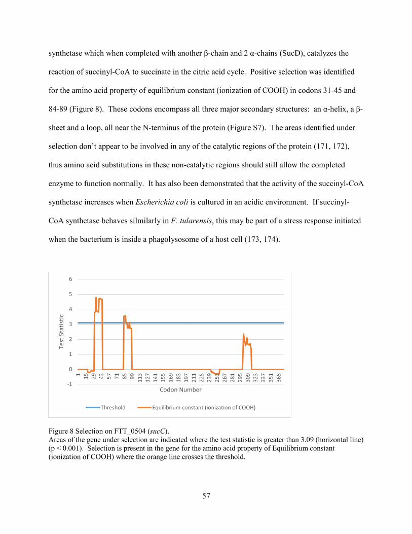
57
synthetase which when completed with another β-chain and 2 α-chains (SucD), catalyzes the
reaction of succinyl-CoA to succinate in the citric acid cycle. Positive selection was identified
for the amino acid property of equilibrium constant (ionization of COOH) in codons 31-45 and
84-89 (Figure 8). These codons encompass all three major secondary structures: an α-helix, a β-
sheet and a loop, all near the N-terminus of the protein (Figure S7). The areas identified under
selection don’t appear to be involved in any of the catalytic regions of the protein (171, 172),
thus amino acid substitutions in these non-catalytic regions should still allow the completed
enzyme to function normally. It has also been demonstrated that the activity of the succinyl-CoA
synthetase increases when Escherichia coli is cultured in an acidic environment. If succinyl-
CoA synthetase behaves silmilarly in F. tularensis, this may be part of a stress response initiated
when the bacterium is inside a phagolysosome of a host cell (173, 174).
Figure 8 Selection on FTT_0504 (sucC). Areas of the gene under selection are indicated where the test statistic is greater than 3.09 (horizontal line) (p < 0.001). Selection is present in the gene for the amino acid property of Equilibrium constant (ionization of COOH) where the orange line crosses the threshold.
-1
0
1
2
3
4
5
6
1 15 29 43 57 71 85 99 113
127
141
155
169
183
197
211
225
239
253
267
281
295
309
323
337
351
365
Test
Sta
tistic
Codon Number
Threshold Equilibrium constant (ionization of COOH)

58
The third and final gene under selection in the metabolism and biosynthesis group is
FTT_0936c which codes for the protein BioF. BioF is a 8-Amino-7-oxononanoate synthase
which is responsible for the first committed step in the biosynthesis of biotin (175). TreeSAAP
identified selection for 4 different amino acid properties: power to be at the N-terminal in
codons 8-22 and 198-210, power to be at the C-terminal in codons 8-22 and 198-202, coil
tendencies in codons 8-22 and 200-203, and compressibility at codons 8-22, and 198-209 (Figure
9). The secondary structures involved in the areas of selection include α-helices, part of a β-
sheet and loops (Figure S8). The active site for BioF is at residues 47 and 361 (176), well away
from the areas of selection in this gene. It’s worth reiterating that genes involved in metabolism
and biosynthesis aren’t considered virulence genes in the strictest sense, but they shouldn’t be
discounted either as they may prove to be effective drug targets. Such is the case with
plumbagin (5-hydroxy-2-methyl-1,4-naphthoquinone), a natural compound found to be an
effective herbicide through the inhibition of 8-Amino-7-oxononanoate synthase (BioF) (177).
Because plants, microbes, and some fungi synthesize their own biotin, while animals require
trace amounts of biotin in their diets, plumbagin may be a safe and effective herbicide by
disrupting the biotin biosynthesis pathway (176-178).
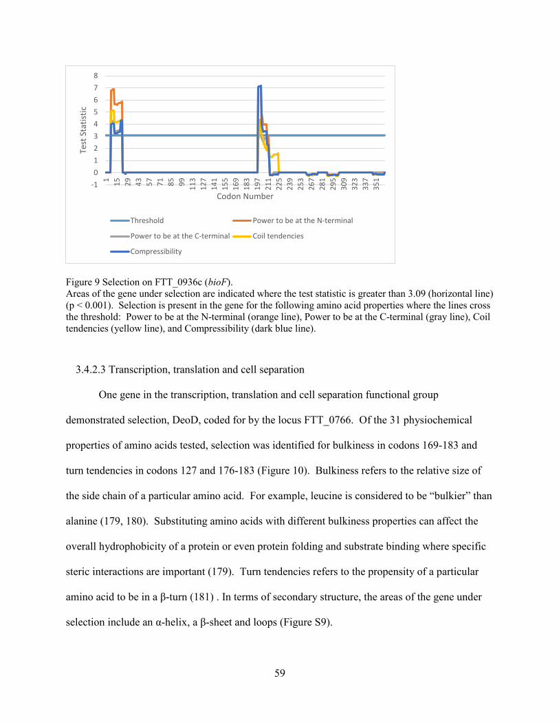
59
Figure 9 Selection on FTT_0936c (bioF). Areas of the gene under selection are indicated where the test statistic is greater than 3.09 (horizontal line) (p < 0.001). Selection is present in the gene for the following amino acid properties where the lines cross the threshold: Power to be at the N-terminal (orange line), Power to be at the C-terminal (gray line), Coil tendencies (yellow line), and Compressibility (dark blue line).
3.4.2.3 Transcription, translation and cell separation
One gene in the transcription, translation and cell separation functional group
demonstrated selection, DeoD, coded for by the locus FTT_0766. Of the 31 physiochemical
properties of amino acids tested, selection was identified for bulkiness in codons 169-183 and
turn tendencies in codons 127 and 176-183 (Figure 10). Bulkiness refers to the relative size of
the side chain of a particular amino acid. For example, leucine is considered to be “bulkier” than
alanine (179, 180). Substituting amino acids with different bulkiness properties can affect the
overall hydrophobicity of a protein or even protein folding and substrate binding where specific
steric interactions are important (179). Turn tendencies refers to the propensity of a particular
amino acid to be in a β-turn (181) . In terms of secondary structure, the areas of the gene under
selection include an α-helix, a β-sheet and loops (Figure S9).
-1012345678
1 15 29 43 57 71 85 99 113
127
141
155
169
183
197
211
225
239
253
267
281
295
309
323
337
351
Test
Sta
tistic
Codon Number
Threshold Power to be at the N-terminal
Power to be at the C-terminal Coil tendencies
Compressibility

60
Figure 10 Selection on FTT_0766 (deoD). Areas of the gene under selection are indicated where the test statistic is greater than 3.09 (horizontal line) (p < 0.001). Selection is present in the gene for the following amino acid properties where the lines cross the threshold: Bulkiness (orange line), and turn tendencies (gray line).
The product of the deoD gene is a purine nucleoside phophoylase (PNP) which catalyzes
the phosphorylation of purine ribonucleosides and 2’-deoxyribonucleosides as part of the purine
salvage pathway (182). The active site for DeoD is near the N-terminus of the protein while
selection was detected further downstream toward the C-terminus, indicating that selection isn’t
changing the specific function of this enzyme. However, enzymes that are part of the nucleotide
biosynthesis pathway, such as PNP, are a favorite target for antimicrobial therapy since they are
significantly different from the eukaryotic enzymes that perform the same function (183).
Understanding how selection operates on this gene, could lead to better, more effective
antimicrobial therapies.
3.4.2.4 Substrate binding and transport
The final functional group to have a gene implicated in selection is substrate binding and
transport. Selection was detected for two amino acid properties in the gene metQ (FTT_1125):
-1
0
1
2
3
4
5
6
7
8
1 10 19 28 37 46 55 64 73 82 91 100
109
118
127
136
145
154
163
172
181
190
199
208
217
226
Test
Sta
tistic
Codon Number
Threshold Bulkiness Turn Tendencies

61
composition and average number of surrounding residues, both in codons 105-119 (Figure 11).
These regions occur within α-helices (Figure S10). Composition refers to the individual amino
acid make up of a protein. Most amino acid residues in a protein cannot be replaced without a
change in function (184). The average number of surrounding residues refers to the number of
residues surrounding a particular amino acid within the effective distance of influence, and is an
important factor in changing the hydrophobicity index of the region (170).
Figure 11 Selection on FTT_1125 (metQ). Areas of the gene under selection are indicated where the test statistic is greater than 3.09 (horizontal line) (p < 0.001). Selection is present in the gene for the following amino acid properties where the lines cross the threshold: Composition (orange line), and Average number of surrounding residues (gray line).
MetQ is a D-Methionine-binding protein, which together with Met N (an ATPase) and
MetI (a D-methionine permease) make up the D-methionine ABC transporter (185). Similar to
RocE discussed earlier in the metabolism and biosynthesis section, which is an arginine
scavenger, this D-methionine transporter scavenges for D- methionine from its environment,
since F. tularensis cannot synthesize methionine. Once transported inside the cell, D-methionine
-1
0
1
2
3
4
5
6
7
8
1 19 37 55 73 91 109
127
145
163
181
199
217
235
253
271
289
307
325
343
361
379
397
415
433
451
469
Test
Sta
tistic
Codon Number
Threshold Composition Average number of surrounding residues

62
can be converted to L-methionine and used in protein synthesis (186). This is an important
strategy used by F. tularensis to survive intracellularly. The fact that MetQ is experiencing
selection, implies that F. tularensis is continuing to evolve strategies to acquire nutrients from its
environment to survive within a host cell.
3.4.2.5 Housekeeping genes
As a control to determine if the virulence genes of F. tularensis are experiencing more
selection than other genes in the genome, 10 housekeeping genes (recA, gyrB, groEL, dnaK,
rpoA1, rpoB, rpoD, rpoH, fopA, and sdhA) were subjected to the same tests of selection (HyPhy
and TreeSAAP) as the virulence genes described earlier. Since housekeeping genes are
responsible for the fundamental functions of the cell, these are typically highly conserved and are
not expected to be evolving at the same rate, or under similar selective pressures as the genes
responsible for virulence. Of the 10 housekeeping genes surveyed, only one gene, the molecular
chaperone GroEL, was shown to experience significant positive selection (category 7-8),
confirming the hypothesis that the virulence genes of F. tularensis are more likely to be
undergoing concerted selective pressures. The other nine housekeeping genes analyzed showed
no significant positive selection by either the HyPhy or TreeSAAP methods.
We initially expected that all of the housekeeping genes would not show significant
selection. However, while GroEL is classified as a housekeeping gene, its function as a
molecular chaperone includes responding to stress. In this way, GroEL may be functioning like
a virulence gene, experiencing positive selection in much the same way as the other virulence
genes.

63
3.5 Conclusions
Francisella tularensis virulence genes failed to recover statistically significant evidence
of positive selection using dN/dS ratios. However, subsequent analyses of 11 arbitrarily-chosen
virulence genes via TreeSAAP (152) identified 7 genes undergoing statistically significant
positive selection. The biological functions of the identified mutations are cautiously inferred.
However, since these genes appear to be under positive selection, they likely confer some
evolutionary advantage leading to an increase in the overall fitness of the organism. The genes
undergoing selection participate in a variety of functions, such as membrane transport, host
defense evasion, stress response, intracellular survival, and even certain metabolic and
biosynthetic pathways. Since the genes we identified as being under selection are virulence
genes, it can be inferred that the altered genes confer an adaptive benefit and increase the ability
of the organism to infect the host and/or evade host defenses. Although the number of
pseudogenes present in the genome F. tularensis indicate that it is a genome in decay (137), our
findings suggest that it is also undergoing an adaptive response to changes in its intracellular
environment by way of positive selection on its virulence genes.
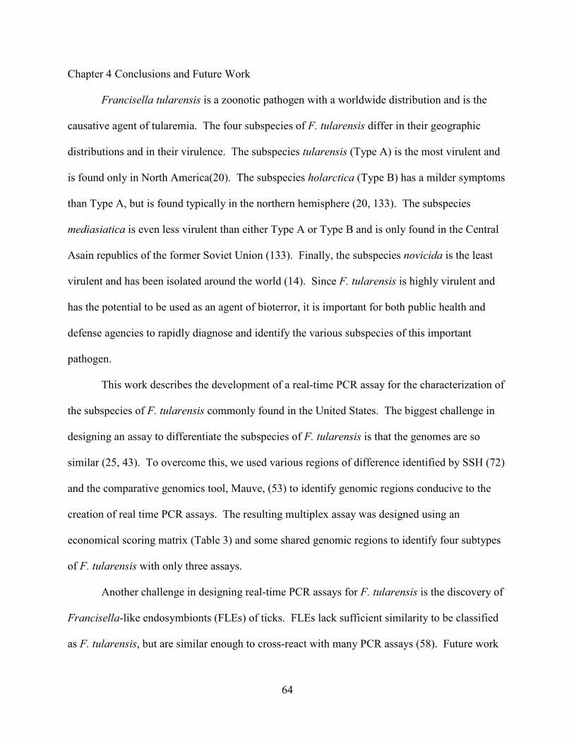
64
Chapter 4 Conclusions and Future Work
Francisella tularensis is a zoonotic pathogen with a worldwide distribution and is the
causative agent of tularemia. The four subspecies of F. tularensis differ in their geographic
distributions and in their virulence. The subspecies tularensis (Type A) is the most virulent and
is found only in North America(20). The subspecies holarctica (Type B) has a milder symptoms
than Type A, but is found typically in the northern hemisphere (20, 133). The subspecies
mediasiatica is even less virulent than either Type A or Type B and is only found in the Central
Asain republics of the former Soviet Union (133). Finally, the subspecies novicida is the least
virulent and has been isolated around the world (14). Since F. tularensis is highly virulent and
has the potential to be used as an agent of bioterror, it is important for both public health and
defense agencies to rapidly diagnose and identify the various subspecies of this important
pathogen.
This work describes the development of a real-time PCR assay for the characterization of
the subspecies of F. tularensis commonly found in the United States. The biggest challenge in
designing an assay to differentiate the subspecies of F. tularensis is that the genomes are so
similar (25, 43). To overcome this, we used various regions of difference identified by SSH (72)
and the comparative genomics tool, Mauve, (53) to identify genomic regions conducive to the
creation of real time PCR assays. The resulting multiplex assay was designed using an
economical scoring matrix (Table 3) and some shared genomic regions to identify four subtypes
of F. tularensis with only three assays.
Another challenge in designing real-time PCR assays for F. tularensis is the discovery of
Francisella-like endosymbionts (FLEs) of ticks. FLEs lack sufficient similarity to be classified
as F. tularensis, but are similar enough to cross-react with many PCR assays (58). Future work

65
could involve testing existing PCR assays, including the one described in Chapter 2 of this work
against FLEs and/or designing new assays or detection methods able to distinguish F. tulrarensis
from FLEs.
The completed multiplex assay was about 98% effective at identifying the subtypes of F.
tularensis compared to the MLVA. The assay did not cross react with other common laboratory
strains or near neighbors tested. The multiplex assay demonstrated a limit of detection of
approximately 1,136 organisms. While there are many varied assays and methods available to
identify the subtypes of F. tularensis, we believe that the multiplex assay presented here will be
another valuable tool for the identification and characterization of F. tularensis during outbreaks
of disease or events of bioterroism.
Whole genome comparisons among the subspecies of F. tularensis have revealed that the
pathogenic subspecies have evolved from the non-pathogenic subspecies (36). In addition, the
many pseudogenes present in F. tularensis, which are largely metabolic genes, suggest a genome
in decay furthering the pathoadaptation of F. tularensis to an intracellular lifestyle (10, 106, 111,
116, 137). We hypothesized that in addition to genome decay by the creation of pseudogenes,
positive natural selection in virulence genes is also a driving force in the continued evolution of
virulence in F. tularensis.
Analyses of 11 arbitrarily chosen virulent genes for positive selection via TreeSAAP
identified 7 genes experiencing statistically significant positive selection. In contrast, only one
of ten housekeeping genes showed positive selection. It can be inferred that those genes under
positive selection confer an adaptive benefit to the organism. These results suggest that F.
tularensis is undergoing an adaptive response, to its environment furthering pathoadaptation
through positive selection on its virulence genes.

66
Currently there is no automated pipeline for the analysis of selection with TreeSAAP on a
large or whole genome scale. For this reason, only 11 arbitrarily chosen virulence genes and 10
housekeeping genes were chosen for analysis. Future work could include the development of a
large scale pipeline for analyzing large sets of genes for positive selection. After such a pipeline
is developed, we recommend the reanalysis of the entire set of virulence genes, or even the
whole genome of F. tularensis. We suspect that such an analysis would yield further insights
into the evolution and pathoadaptation of this important pathogen.
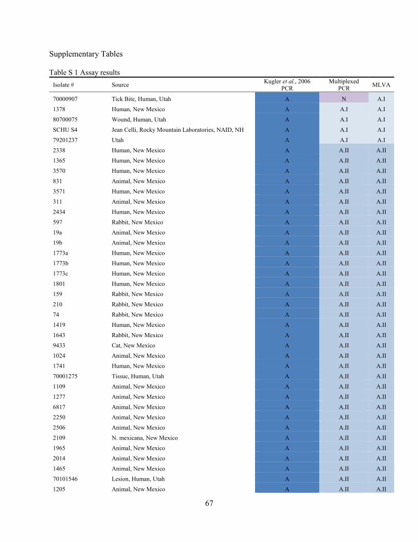
67
Supplementary Tables
Table S 1 Assay results Isolate # Source Kugler et al., 2006
PCR Multiplexed
PCR MLVA
70000907 Tick Bite, Human, Utah A N A.I
1378 Human, New Mexico A A.I A.I
80700075 Wound, Human, Utah A A.I A.I
SCHU S4 Jean Celli, Rocky Mountain Laboratories, NAID, NH A A.I A.I
79201237 Utah A A.I A.I
2338 Human, New Mexico A A.II A.II
1365 Human, New Mexico A A.II A.II
3570 Human, New Mexico A A.II A.II
831 Animal, New Mexico A A.II A.II
3571 Human, New Mexico A A.II A.II
311 Animal, New Mexico A A.II A.II
2434 Human, New Mexico A A.II A.II
597 Rabbit, New Mexico A A.II A.II
19a Animal, New Mexico A A.II A.II
19b Animal, New Mexico A A.II A.II
1773a Human, New Mexico A A.II A.II
1773b Human, New Mexico A A.II A.II
1773c Human, New Mexico A A.II A.II
1801 Human, New Mexico A A.II A.II
159 Rabbit, New Mexico A A.II A.II
210 Rabbit, New Mexico A A.II A.II
74 Rabbit, New Mexico A A.II A.II
1419 Human, New Mexico A A.II A.II
1643 Rabbit, New Mexico A A.II A.II
9433 Cat, New Mexico A A.II A.II
1024 Animal, New Mexico A A.II A.II
1741 Human, New Mexico A A.II A.II
70001275 Tissue, Human, Utah A A.II A.II
1109 Animal, New Mexico A A.II A.II
1277 Animal, New Mexico A A.II A.II
6817 Animal, New Mexico A A.II A.II
2250 Animal, New Mexico A A.II A.II
2506 Animal, New Mexico A A.II A.II
2109 N. mexicana, New Mexico A A.II A.II
1965 Animal, New Mexico A A.II A.II
2014 Animal, New Mexico A A.II A.II
1465 Animal, New Mexico A A.II A.II
70101546 Lesion, Human, Utah A A.II A.II
1205 Animal, New Mexico A A.II A.II

68
1385 Rabbit, New Mexico A A.II A.II
AS 1284 Rodent, New Mexico A A.II A.II
70102010 Axillary Lesion, Human, Utah A A.II A.II
70102009 Back Wound, Human, Utah A A.II A.II
804 Rabbit, New Mexico A A.II A.II
1103a Human, New Mexico A A.II A.II
1275 Animal, New Mexico A A.II A.II
6069 Animal, New Mexico A A.II A.II
14149 New Mexico A A.II A.II
1103b Human, New Mexico A A.II A.II
BA 4414 New Mexico A A.II A.II
80402860 right Arm Abcess, Human, Utah A A.II A.II
80402637 Skin, Human, Utah A A.II A.II
80606987 Blood, Feline, Utah A A.II A.II
80606984 Lymph node, Feline, Utah A A.II A.II
80606985 Spleen, Feline, Utah A A.II A.II
80700103 Hand drainage, Human, Utah A A.II A.II
80700051 Abscess, Human, Utah A A.II A.II
80800086 Neck lesion aspirate, Human, Utah A A.II A.II
80800071 Lymph node, Feline, Utah A A.II A.II
5906 Animal, New Mexico A A.II A.II
1513 Dog, New Mexico A A.II A.II
6733 New Mexico A A.II A.II
6734 New Mexico A A.II A.II
0018 7/88 Utah A A.II A.II
08902000 (11/16) Utah A A.II A.II
79000875 Utah A A.II A.II
79101487 Utah A A.II A.II
79101488 Utah A A.II A.II
79101615 Utah A A.II A.II
79102173 Utah A A.II A.II
79301108 Utah A A.II A.II
79400960 Utah A A.II A.II
880178-1 Utah A A.II A.II
880178-2 Utah A A.II A.II
880321 Utah A A.II A.II
93703 Utah A A.II A.II
LVS Sheep, Australia, Dugway Proving Ground B B B
1518 Human, New Mexico B B B
79901959 3rd finger wound, Human, Utah B B B
3459 Animal, New Mexico A B B
70001144 Eye Drainage, Human, Utah B B B
80501131 Pleural fluid, Human, Utah B B B

69
U112: CG69 Sheep, Australia, University of Victoria, Fran Nano N N N
U112: CG57 University of Victoria, Fran Nano N N N U112: GB2/pGB40 #7 University of Victoria, Fran Nano N N N
U112: SC119 University of Victoria, Fran Nano N N N
79201152 Utah A A.II NM
1815 Human, New Mexico A A.II NM
305 Animal, New Mexico A A.II NM
75 Rabbit, New Mexico A A.II NM
736 Rabbit, New Mexico A A.II NM
70102163 Blood, Human, Utah A A.II NM
AS 2058 Lapine, New Mexico A A.II NM
1276 Animal, New Mexico A A.II NM
AS 816 Feline, New Mexico A A.II NM
AS 713 Lapine, New Mexico A A.II NM
80502541 Groin, Human, Utah A A.II NM
80606986 Liver, Feline, Utah A A.II NM
80700132 Bite wound, Human, Utah B B NM
80700107 Face, Human, Utah A A.II NM
80700069 Lesion, Human, Utah A A.I NM
80800087 Wound, Human, Utah A A.II NM
80800082 Spleen, Feline, Utah A A.II NM
80800111 Finger wound, Human, Utah A A.II NM
AS 200801417 New Mexico A A.II NM
AS 200801507 New Mexico A A.II NM
U112: 1L University of Victoria, Fran Nano N N NM
U112: 68-11 University of Victoria, Fran Nano N N NM
U112: CG116 University of Victoria, Fran Nano N N NM
U112: CG62 University of Victoria, Fran Nano N N NM
U112: GB2 University of Victoria, Fran Nano N N NM
U112: GB5 University of Victoria, Fran Nano N N NM
U112: KM14 University of Victoria, Fran Nano N N NM
U112: KM14S University of Victoria, Fran Nano N N NM
1574 Utah A A.II NM
7/27 Utah A A.II NM
79001541 4/45 Utah A A.I NM
U112 Utah, Dugway N N NM
6168 Human, University of Victoria, Fran Nano N B N
A- tularensis Type A, A.I - tularensis Type A.I, A.II - tularensis Type A.II, B - holarctica, N - Novicida, NM - no match.

70
Supplementary Figures
Figure S 1 Mauve alignment of Francisella genomes The complete genomes of 7 Francisella isolates were obtained from GenBank and aligned using Mauve. The coloured blocks represent homologous sequences among the different strains. Blocks above the center line represent a ‘forward’ reading frame relative to the reference strain (lane 1) while blocks below the center line represent a ‘reverse’ reading frame. White spaces within the colored blocks represent areas of non-homology between strains. The search for unique genomic markers was focused within these regions of non-homology. (1) holarctica FTNF002-00, (2) holarctica OSU18, (3) holarctica LVS, (4) mediasiatica FSC147, (5) novicida U112, (6) tularensis Schu S4, (7) tularensis WY96-3418

71
Figure S 2 Representative MLVA analysis The peaks represent the detection of amplified PCR products from all 3 mixes using labeled primers. These peaks are compared to a database of known subspecies for identification.

72
(a)
(b)
(c)
(d)

73
Figure S 3 Sensitivities of singleplex assays Tenfold serial dilutions of F. tularensis chromosomal DNA were tested to determine individual assay sensitivities. (a) Type A.I, (b) Type A.II, (c) Type B (d) novicida. No Template Control (NTC) samples were negative for each sample processed. Calculated PCR efficiencies: (a) 91.5 %, (b) 88.0 %, (c) 90.6%, (d) 91. 9%.

74

75
Figure S 4 Predicted secondary structure of FTL_1134. Predicted secondary structure of FTL_1134 using the PSIPRED server at http://bioinf.cs.ucl.ac.uk/psipred/. The red boxes at codons 183-203, 271-285, 232-337 and 383-394 indicate the areas under selection as determined by TreeSAAP.

76

77
Figure S 5 Predicted secondary structure of FTT_0683 (pilD). Predicted secondary structure of FTT_0683 (pilD) using PSIPRED server at http://bioninf.cs.ucl.ac.uk/psipred/. The red boxes at codons 190-204 indicate the areas under selection as determined by TreeSAAP.

78

79
Figure S 6 Predicted secondary structure of FTT_0881 (rocE). Predicted secondary structure of FTT_0881 (rocE) using the PSIPRED server at http://bioninf.cs.ucl.ac.uk/psipred/. The red boxes at codons 109-116, 438 and 442 indicate the areas under selection as determined by TreeSAAP.

80
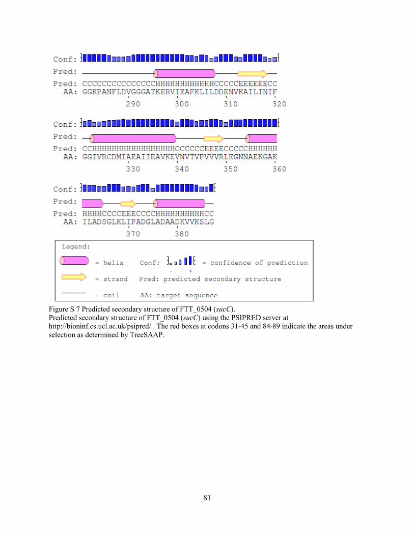
81
Figure S 7 Predicted secondary structure of FTT_0504 (sucC). Predicted secondary structure of FTT_0504 (sucC) using the PSIPRED server at http://bioninf.cs.ucl.ac.uk/psipred/. The red boxes at codons 31-45 and 84-89 indicate the areas under selection as determined by TreeSAAP.

82

83
Figure S 8 Predicted secondary structure of FTT_0936c (bioF). Predicted secondary structure of FTT_0936c (bioF) using the PSIPRED server at http://bioinf.cs.ucl.uk/psipred/. The red boxes at codons 8-22 and 198-210 indicate the areas under selection as determined by TreeSAAP.

84

85
Figure S 9 Predicted secondary strucutre of FTT_0766 (deoD). Predicted secondary strucutre of FTT_0766 (deoD) using the PSIPRED server at http://bioinf.cs.ucl.ac.uk/psipred/. The red boxes at codons 127 and 169-183 indicate the areas under selection as determined by TreeSAAP.

86

87

88
Figure S 10 Predicted secondary structure of FTT_1125 (metQ). Predicted secondary structure of FTT_1125 (metQ) using the PSIPRED server at http://bioninf.cs.ucl.uk/psipred/. The red boxes at codons 105-119 indicate the areas under selection as determined by TreeSAAP.

89
REFERENCES
1. Gunnell MK, Adams BJ, Robison RA. 2016. The genetic diversity and evolution of
Francisella tularensis with comments on detection by PCR. Current Issues in Molecular Biology 18:79-92.
2. Gunnell MK, Lovelace CD, Satterfield BA, Moore EA, O'Neill KL, Robison RA. 2012. A multiplex real-time PCR assay for the detection and differentiation of Francisella tularensis subspecies. Journal of Medical Microbiology 61:1525-1531.
3. McCoy G, Chapin C. 1912. Further observations on a plaguelike disease of rodents with a preliminary note on the causative agent Bacterium tularense. J Infect Dis 10:61-72.
4. Wherry WB, Lamb BH. 1914. Infection of man with Bacterium tularense. J Infect Dis 15:331-340.
5. Akimana C, Abu Kwaik Y. 2011. Francisella-arthropod vector interaction and its role in patho-adaptation to infect mammals. Frontiers in Microbiology 2: DOI: 10.3389/fmicb.2011.00034.
6. McCrumb FR. 1961. Aerosol infection of man with Pasteurella tularensis. Bacteriological Reviews 25:262-267.
7. Dennis DT, Inglesby TV, Henderson DA, Bartlett JG, Ascher MS, Eitzen E, Fine AD, Friedlander AM, Hauer J, Layton M, Lillibridge SR, McDade JE, Osterholm MT, O'Toole T, Parker G, Perl TM, Russell PK, Tonat K, Working Grp Civilian B. 2001. Tularemia as a biological weapon - Medical and public health management. Jama-Journal of the American Medical Association 285:2763-2773.
8. Salomonsson E. 2008. The role of the Type IV pili system in the virulence of Francisella tularensis. Doctoral. Umea University, Umea.
9. Olsufjev NG, Emelyanova OS, Dunaeva TN. 1959. Comparative study of strains of B. tularense in the Old and New World and their taxonomy. J Hyg Epdimiol Microbiol Immunol 3:139-149.
10. Larsson P, Elfsmark D, Svensson K, Wikstrom P, Forsman M, Brettin T, Keim P, Johansson A. 2009. Molecular evolutionary consequences of niche restriction in Francisella tularensis, a facultative intracellular pathogen. Plos Pathogens 5.
11. Kingry LC, Petersen JM. 2014. Comparative review of Francisella tularensis and Francisella novicida. Frontiers in Cellular and Infection Microbiology 4: DOI 10.3389/fcimb.2014.00035.
12. Garrity G. 2005. Bergey's Manual of Systematic Bacteriology, 2 ed, vol 2. Williams & Wilkins.
13. Larson CL, Wicht W, Jellison WL. 1955. A new organism resembling P. tularensis isolated from water. Public Health Reports 70:253-258.
14. Hollis DG, Weaver RE, Steigerwalt AG, Wenger JD, Moss CW, Brenner DJ. 1989. Francisella philomiragia comb. nov. (formerly Yersinia philomiragia) and Francisella tularensis biogroup novicida (formerly Francisella novicida) associated with human disease. Journal of Clinical Microbiology 27:1601-1608.
15. Huber B, Escudero R, Busse HJ, Seibold E, Scholz HC, Anda P, Kampfer P, Splettstoesser WD. 2010. Description of Francisella hispaniensis sp nov., isolated from human blood, reclassification of Francisella novicida (Larson et al. 1955) Olsufiev et al. 1959 as Francisella tularensis subsp novicida comb. nov and emended description of the

90
genus Francisella. International Journal of Systematic and Evolutionary Microbiology 60:1887-1896.
16. Johansson A, Celli J, Conlan W, Elkins KL, Forsman M, Keim PS, Larsson P, Manoil C, Nano FE, Petersen JM, Sjostedt A. 2010. Objections to the transfer of Francisella novicida to the subspecies rank of Francisella tularensis. International Journal of Systematic and Evolutionary Microbiology 60:1717-1718.
17. Busse HJ, Huber B, Anda P, Escudero R, Scholz HC, Seibold E, Splettstoesser WD, Kampfer P. 2010. Objections to the transfer of Francisella novicida to the subspecies rank of Francisella tularensis - response to Johansson et al. International Journal of Systematic and Evolutionary Microbiology 60:1718-1720.
18. Wayne LG, Brenner DJ, Colwell RR, Grimont PAD, Kandler O, Krichevsky MI, Moore LH, Moore WEC, Murray RGE, Stackebrandt E, Starr MP, Truper HG. 1987. Report of the ad-hoc-committee on reconciliation of approaches to bacterial systematics. International Journal of Systematic Bacteriology 37:463-464.
19. Staples JE, Kubota KA, Chalcraft LG, Mead PS, Petersen JM. 2006. Epidemiologic and molecular analysis of human tularemia, United States, 1964-2004. Emerging Infectious Diseases 12:1113-1118.
20. Johansson A, Farlow J, Larsson P, Dukerich M, Chambers E, Bystrom M, Fox J, Chu M, Forsman M, Sjostedt A, Keim P. 2004. Worldwide genetic relationships among Francisella tularensis isolates determined by multiple-locus variable-number tandem repeat analysis. Journal of Bacteriology 186:5808-5818.
21. Broekhuijsen M, Larsson N, Johansson A, Bystrom M, Eriksson U, Larsson E, Prior RG, Sjostedt A, Titball RW, Forsman M. 2003. Genome-wide DNA microarray analysis of Francisella tularensis strains demonstrates extensive genetic conservation within the species but identifies regions that are unique to the highly virulent F. tularensis subsp tularensis. Journal of Clinical Microbiology 41:2924-2931.
22. Whipp MJ, Davis JM, Lum G, de Boer J, Zhou Y, Bearden SW, Petersen JM, Chu MC, Hoggi G. 2003. Characterization of a novicida-like subspecies of Francisella tularensis isolated in Australia. Journal of Medical Microbiology 52:839-842.
23. Owen CR, Lackman DB, Jellison WL, Buker EO, Bell JF. 1964. Comparative studies of Francisella tularensis + Francisella novicida. Journal of Bacteriology 87:676-&.
24. Weiss DS, Brotcke A, Henry T, Margolis JJ, Chan K, Monack DM. 2007. In vivo negative selection screen identifies genes required for Francisella virulence. Proceedings of the National Academy of Sciences of the United States of America 104:6037-6042.
25. Champion MD, Zeng QD, Nix EB, Nano FE, Keim P, Kodira CD, Borowsky M, Young S, Koehrsen M, Engels R, Pearson M, Howarth C, Larson L, White J, Alvarado L, Forsman M, Bearden SW, Sjostedt A, Titball R, Michell SL, Birren B, Galagan J. 2009. Comparative genomic characterization of Francisella tularensis strains belonging to low and high virulence subspecies. Plos Pathogens 5.
26. Farlow J, Wagner DM, Dukerich M, Stanley M, Chu M, Kubota K, Petersen J, Keim P. 2005. Francisella tularensis in the United States. Emerging Infectious Diseases 11:1835-1841.
27. Oyston PCF. 2008. Francisella tularensis: unravelling the secrets of an intracellular pathogen. Journal of Medical Microbiology 57:921-930.

91
28. Olsufjev NG, Meshcheryakova IS. 1983. Subspecific taxonomy of Francisella tularensis McCoy and Chapin 1912. International Journal of Systematic Bacteriology 33:872-874.
29. Sandstrom G, Sjostedt A, Forsman M, Pavlovich NV, Mishankin BN. 1992. Characterization and classification of strains of Francisella tularensis isolated in the central Asain focus of the Soviet Union and in Japan. Journal of Clinical Microbiology 30:172-175.
30. Thomas R, Johansson A, Neeson B, Isherwood K, Sjostedt A, Ellis J, Titball RW. 2003. Discrimination of human pathogenic subspecies of Francisella tularensis by using restriction fragment length polymorphism. Journal of Clinical Microbiology 41:50-57.
31. Fujita O, Uda A, Hotta A, Okutani A, Inoue S, Tanabayashi K, Yamada A. 2008. Genetic diversity of Francisella tularensis subspecies holarctica strains isolated in Japan. Microbiology and Immunology 52:270-276.
32. Vivekananda J, Kiel JL. 2006. Anti-Francisella tularensis DNA aptamers detect tularemia antigen from different subspecies by aptamer-linked immobilized sorbent assay. Laboratory Investigation 86:610-618.
33. Larsson P, Oyston PCF, Chain P, Chu MC, Duffield M, Fuxelius HH, Garcia E, Halltorp G, Johansson D, Isherwood KE, Karp PD, Larsson E, Liu Y, Michell S, Prior J, Prior R, Malfatti S, Sjostedt A, Svensson K, Thompson N, Vergez L, Wagg JK, Wren BW, Lindler LE, Andersson SGE, Forsman M, Titball RW. 2005. The complete genome sequence of Francisella tularensis, the causative agent of tularemia. Nature Genetics 37:153-159.
34. Petrosino JF, Xiang Q, Karpathy SE, Jiang HY, Yerrapragada S, Liu YM, Gioia J, Hemphill L, Gonzalez A, Raghavan TM, Uzman A, Fox GE, Highlander S, Reichard M, Morton RJ, Clinkenbeard KD, Weinstock GM. 2006. Chromosome rearrangement and diversification of Francisella tularensis revealed by the type B (OSU18) genome sequence. Journal of Bacteriology 188:6977-6985.
35. Chaudhuri RR, Ren CP, Desmond L, Vincent GA, Silman NJ, Brehm JK, Elmore MJ, Hudson MJ, Forsman M, Isherwood KE, Gurycova D, Minton NP, Titball RW, Pallen MJ, Vipond R. 2007. Genome sequencing shows that European isolates of Francisella tularensis subspecies tularensis are almost identical to US laboratory strain Schu S4. Plos One 2.
36. Rohmer L, Fong C, Abmayr S, Wasnick M, Freeman TJL, Radey M, Guina T, Svensson K, Hayden HS, Jacobs M, Gallagher LA, Manoil C, Ernst RK, Drees B, Buckley D, Haugen E, Bovee D, Zhou Y, Chang J, Levy R, Lim R, Gillett W, Guenthener D, Kang A, Shaffer SA, Taylor G, Chen JZ, Gallis B, D'Argenio DA, Forsman M, Olson MV, Goodlett DR, Kaul R, Miller SI, Brittnacher MJ. 2007. Comparison of Francisella tularensis genomes reveals evolutionary events associated with the emergence of human pathogenic strains. Genome Biology 8.
37. Beckstrom-Sternberg SM, Auerbach RK, Godbole S, Pearson JV, Beckstrom-Sternberg JS, Deng ZM, Munk C, Kubota K, Zhou Y, Bruce D, Noronha J, Scheuermann RH, Wang AH, Wei XY, Wang JJ, Hao J, Wagner DM, Brettin TS, Brown N, Gilna P, Keim PS. 2007. Complete genomic characterization of a pathogenic A.II strain of Francisella tularensis subspecies tularensis. Plos One 2.
38. Barabote RD, Xie G, Brettin TS, Hinrichs SH, Fey PD, Jay JJ, Engle JL, Godbole SD, Noronha JM, Scheuermann RH, Zhou LW, Lion C, Dempsey MP. 2009.

92
Complete genome sequence of Francisella tularensis subspecies holarctica FTNF002-00. Plos One 4.
39. Nalbantoglu U, Sayood K, Dempsey MP, Iwen PC, Francesconi SC, Barabote RD, Xie G, Brettin TS, Hinrichs SH, Fey PD. 2010. Large direct repeats flank genomic rearrangements between a new clinical isolate of Francisella tularensis subsp tularensis A1 and Schu S4. Plos One 5.
40. Modise T, Ryder C, Mane SP, Bandara AB, Jensen RV, Inzana TJ. 2012. Genomic comparison between a virulent type A1 strain of Francisella tularensis and its attenuated O-antigen mutant. Journal of Bacteriology 194:2775-2776.
41. Svensson K, Sjodin A, Bystrom M, Granberg M, Brittnacher MJ, Rohmer L, Jacobs MA, Sims-Day EH, Levy R, Zhou Y, Hayden HS, Lim R, Chang J, Guenthener D, Kang A, Haugen E, Gillett W, Kaul R, Forsman M, Larsson P, Johansson A. 2012. Genome sequence of Francisella tularensis subspecies holarctica strain FSC200, isolated from a child with tularemia. Journal of Bacteriology 194:6965-6966.
42. La Scola B, Elkarkouri K, Li WJ, Wahab T, Fournous G, Rolain JM, Biswas S, Drancourt M, Robert C, Audic S, Lofdahl S, Raoult D. 2008. Rapid comparative genomic analysis for clinical microbiology: The Francisella tularensis paradigm. Genome Research 18:742-750.
43. Forsman M, Sandstrom G, Sjostedt A. 1994. Analysis of 16S ribosomal DNA sequences of Francisella strains and utilization for determination of the phylogeny of the genus and for identification of strains by PCR. International Journal of Systematic Bacteriology 44:38-46.
44. Pohanka M, Hubalek M, Neubauerova V, Macela A, Faldyna M, Bandouchova H, Pikula J. 2008. Current and emerging assays for Francisella tularensis detection: a review. Veterinarni Medicina 53:585-594.
45. Samrakandi MM, Zhang C, Zhang M, Nietfeldt J, Kim J, Iwen PC, Olson ME, Fey PD, Duhamel GE, Hinrichs SH, Cirillo JD, Benson AK. 2004. Genome diversity among regional populations of Francisella tularensis subspecies tularensis and Francisella tularensis subspecies holarctica isolated from the US. Fems Microbiology Letters 237:9-17.
46. Nano FE, Zhang N, Cowley SC, Klose KE, Cheung KKM, Roberts MJ, Ludu JS, Letendre GW, Meierovics AI, Stephens G, Elkins KL. 2004. A Francisella tularensis pathogenicity island required for intramacrophage growth. Journal of Bacteriology 186:6430-6436.
47. Nano FE, Schmerk C. 2007. The Francisella pathogenicity island. Francisella Tularensis: Biology, Pathogenicity, Epidemiology, and Biodefense 1105:122-137.
48. Rohmer L, Brittnacher M, Svensson K, Buckley D, Haugen E, Zhou Y, Chang J, Levy R, Hayden H, Forsman M, Olson M, Johansson A, Kaul R, Miller SI. 2006. Potential source of Francisella tularensis live vaccine strain attenuation determined by genome comparison. Infection and Immunity 74:6895-6906.
49. Green M, Choules G, Rogers D, Titball RW. 2005. Efficacy of the live attenuated Francisella tularensis vaccine (LVS) in a murine model of disease. Vaccine 23:2680-2686.
50. Dempsey MP, Dobson M, Zhang C, Zhang M, Lion C, Gutierrez-Martin CB, Iwen PC, Fey PD, Olson ME, Niemeyer D, Francesconi S, Crawford R, Stanley M, Rhodes J, Wagner DM, Vogler AJ, Birdsell D, Keim P, Johansson A, Hinrichs SH,
93
Benson AK. 2007. Genomic deletion marking an emerging subclone of Francisella tularensis subsp holarctica in France and the Iberian Peninsula. Applied and Environmental Microbiology 73:7465-7470.
51. Olsufjev NG, Meshcheryakova IS. 1982. Infraspecific taxonomy of tularemia agent Francisella tularensis McCoy et Chapin. Journal of Hygiene Epidemiology Microbiology and Immunology 26:291-299.
52. Nübel U, Reissbrodt R, Weller A, Grunow R, Porsch-Ozcurumez M, Tomaso H, Hofer E, Splettstoesser W, Finke EJ, Tschape H, Witte W. 2006. Population structure of Francisella tularensis. Journal of Bacteriology 188:5319-5324.
53. Darling ACE, Mau B, Blattner FR, Perna NT. 2004. Mauve: Multiple alignment of conserved genomic sequence with rearrangements. Genome Research 14:1394-1403.
54. Niebylski ML, Peacock MG, Fischer ER, Porcella SF, Schwan TG. 1997. Characterization of an endosymbiont infecting wood ticks, Dermacentor andersoni, as a member of the genus Francisella. Applied and Environmental Microbiology 63:3933-3940.
55. Scoles GA. 2004. Phylogenetic analysis of the Francisella-like endosymbionts of Dermacentor ticks. Journal of Medical Entomology 41:277-286.
56. Goethert HK, Telford SR. 2005. A new Francisella (Beggiatiales : Francisellaceae) inquiline within Dermacentor variabilis say (Acari : Ixodidae). Journal of Medical Entomology 42:502-505.
57. Dergousoff SJ, Chilton NB. 2012. Association of different genetic types of Francisella-like organisms with the Rocky Mountain Wood Tick (Dermacentor andersoni) and the American Dog Tick (Dermacentor variabilis) in localities near their northern distributional limits. Applied and Environmental Microbiology 78:965-971.
58. Szigeti A, Kreizinger Z, Hornok S, Abichu G, Gyuranecz M. 2014. Detection of Francisella-like endosymbiont in Hyalomma rufipes from Ethiopia. Ticks and Tick-Borne Diseases 5:818-820.
59. Kugeler KJ, Gurfield N, Creek JG, Mahoney KS, Versage JL, Petersen JM. 2005. Discrimination between Francisella tularensis and Francisella-like endosymbionts when screening ticks by PCR. Applied and Environmental Microbiology 71:7594-7597.
60. Sreter-Lancz Z, Szell Z, Sreter T, Marialigeti K. 2009. Detection of a novel Francisella in Dermacentor reticulatus: A need for careful evaluation of PCR-based identification of Francisella tularensis in Eurasian ticks. Vector-Borne and Zoonotic Diseases 9:123-126.
61. Foroshani NS, Karami A, Pourali F. 2013. Simultaneous and rapid detection of Salmonella typhi, Bacillus anthracis, and Yersinia pestis by using multiplex polymerase chain reaction (PCR). Iranian Red Crescent Medical Journal 15.
62. Sting R, Runge M, Eisenberg T, Braune S, Muller W, Otto P. 2013. Comparison of bacterial culture and polymerase chain reaction (PCR) for the detection of F. tularensis subsp holarctica in wild animals. Berliner Und Munchener Tierarztliche Wochenschrift 126:285-290.
63. Celebi B, Kilic S, Yesilyurt M, Acar B. 2014. Evaluation of a newly-developed ready-to-use commercial PCR kit for the molecular diagnosis of Francisella tularensis. Mikrobiyoloji Bulteni 48:135-142.
64. Splettstoesser WD, Tomaso H, Al Dahouk S, Neubauer H, Schuff-Werner P. 2005. Diagnostic procedures in tularaemia with special focus on molecular and immunological

94
techniques. Journal of Veterinary Medicine Series B-Infectious Diseases and Veterinary Public Health 52:249-261.
65. He J, Kraft AJ, Fan JA, Van Dyke M, Wang LH, Bose ME, Khanna M, Metallo JA, Henrickson KJ. 2009. Simultaneous detection of CDC category "A" DNA and RNA bioterrorism agents by use of multiplex PCR & RT-PCR enzyme hybridization assays. Viruses-Basel 1:441-459.
66. Kormilitsyna MI, Meshcheryakova IS, Mikhailova TV. 2013. Molecular and genetic characterization of Francisella tularensis strains of differing taxonomic status and virulence. Molecular Genetics Microbiology and Virology 28:110-114.
67. Varma-Basil M, El-Hajj H, Marras SAE, Hazbon MH, Mann JM, Connell ND, Kramer FR, Alland D. 2004. Molecular beacons for multiplex detection of four bacterial bioterrorism agents. Clinical Chemistry 50:1060-1063.
68. Skottman T, Piiparinen H, Hyytiainen H, Myllys V, Skurnik M, Nikkari S. 2007. Simultaneous real-time PCR detection of Bacillus anthracis, Francisella tularensis and Yersinia pestis. European Journal of Clinical Microbiology & Infectious Diseases 26:207-211.
69. Hsu HL, Huang HH, Liang CC, Lin HC, Liu WT, Lin FP, Kau JH, Sun KH. 2013. Suspension bead array of the single-stranded multiplex polymerase chain reaction amplicons for enhanced identification and quantification of multiple pathogens. Analytical Chemistry 85:5562-5568.
70. Bergval I, Sengstake S, Brankova N, Levterova V, Abadia E, Tadumaze N, Bablishvili N, Akhalaia M, Tuin K, Schuitema A, Panaiotov S, Bachiyska E, Kantardjiev T, de Zwaan R, Schurch A, van Soolingen D, van 't Hoog A, Cobelens F, Aspindzelashvili R, Sola C, Klatser P, Anthony R. 2012. Combined species identification, genotyping, and drug resistance detection of Mycobacterium tuberculosis cultures by MLPA on a bead-based array. Plos One 7.
71. Munro SB, Kuypers J, Jerome KR. 2013. Comparison of a multiplex real-time PCR assay with a multiplex Luminex assay for influenza virus detection. Journal of Clinical Microbiology 51:1124-1129.
72. Molins-Schneekloth CR, Belisle JT, Petersen JM. 2008. Genomic markers for differentiation of Francisella tularensis subsp tularensis A.I and A.II strains. Applied and Environmental Microbiology 74:336-341.
73. Molins CR, Carlson JK, Coombs J, Petersen JA. 2009. Identification of Francisella tularensis subsp tularensis A1 and A2 infections by real-time polymerase chain reaction. Diagnostic Microbiology and Infectious Disease 64:6-12.
74. Zasada AA, Forminska K, Zacharczuk K. 2013. Fast identification of Yersinia pestis, Bacillus anthracis, and Francisella tularensis based on conventional PCR. Polish Journal of Microbiology 62:453-455.
75. Sellek R, Jimenez O, Aizpurua C, Fernandez-Frutos B, De Leon P, Camacho M, Fernandez-Moreira D, Ybarra C, Cabria JC. 2008. Recovery of Francisella tularensis from soil samples by filtration and detection by real-time PCR and cELISA. Journal of Environmental Monitoring 10:362-369.
76. Pandya GA, Holmes MH, Petersen JM, Pradhan S, Karamycheva SA, Wolcott MJ, Molins C, Jones M, Schriefer ME, Fleischmann RD, Peterson SN. 2009. Whole genome single nucleotide polymorphism based phylogeny of Francisella tularensis and its application to the development of a strain typing assay. Bmc Microbiology 9.

95
77. Svensson K, Granberg M, Karlsson L, Neubauerova V, Forsman M, Johansson A. 2009. A real-time PCR array for hierarchical identification of Francisella isolates. Plos One 4.
78. Woubit A, Yehualaeshet T, Habtemariam T, Samuel T. 2012. Novel genomic tools for specific and real-time detection of biothreat and frequently encountered foodborne pathogens. Journal of Food Protection 75:660-670.
79. Abril C, Nimmervoll H, Pilo P, Brodard I, Korczak B, Seiler M, Miserez R, Frey J. 2008. Rapid diagnosis and quantification of Francisella tularensis in organs of naturally infected common squirrel monkeys (Saimiri sciureus). Veterinary Microbiology 127:203-208.
80. Yang S, Rothman RE, Hardick J, Kuroki M, Hardick A, Doshi V, Ramachandran P, Gaydos CA. 2008. Rapid polymerase chain reaction-based screening assay for bacterial biothreat agents. Academic Emergency Medicine 15:388-392.
81. Angelakis E, Roux V, Raoult D, Rolain JM. 2009. Real-time PCR strategy and detection of bacterial agents of lymphadenitis. European Journal of Clinical Microbiology & Infectious Diseases 28:1363-1368.
82. Simsek H, Taner M, Karadenizli A, Ertek M, Vahaboglu H. 2012. Identification of Francisella tularensis by both culture and real-time TaqMan PCR methods from environmental water specimens in outbreak areas where tularemia cases were not previously reported. European Journal of Clinical Microbiology & Infectious Diseases 31:2353-2357.
83. Birdsell DN, Vogler AJ, Buchhagen J, Clare A, Kaufman E, Naumann A, Driebe E, Wagner DM, Keim PS. 2014. TaqMan real-time PCR assays for single-nucleotide polymorphisms which identify Francisella tularensis and its subspecies and subpopulations. Plos One 9.
84. Matero P, Hemmila H, Tomaso H, Piiparinen H, Rantakokko-Jalava K, Nuotio L, Nikkari S. 2011. Rapid field detection assays for Bacillus anthracis, Brucella spp., Francisella tularensis and Yersinia pestis. Clinical Microbiology and Infection 17:34-43.
85. Seiner DR, Colburn HA, Baird C, Bartholomew RA, Straub T, Victry K, Hutchison JR, Valentine N, Bruckner-Lea CJ. 2013. Evaluation of the FilmArray (R) system for detection of Bacillus anthracis, Francisella tularensis and Yersinia pestis. Journal of Applied Microbiology 114:992-1000.
86. Rachwal PA, Rose HL, Cox V, Lukaszewski RA, Murch AL, Weller SA. 2012. The potential of TaqMan array cards for detection of multiple biological agents by real-time PCR. Plos One 7.
87. Weller SA, Cox V, Essex-Lopresti A, Hartley MG, Parsons TM, Rachwal PA, Stapleton HL, Lukaszewski RA. 2012. Evaluation of two multiplex real-time PCR screening capabilities for the detection of Bacillus anthracis, Francisella tularensis and Yersinia pestis in blood samples generated from murine infection models. Journal of Medical Microbiology 61:1546-1555.
88. Jacob D, Sauer U, Housley R, Washington C, Sannes-Lowery K, Ecker DJ, Sampath R, Grunow R. 2012. Rapid and high-throughput detection of highly pathogenic bacteria by Ibis PLEX-ID technology. Plos One 7.
89. Jeng K, Hardick J, Rothman R, Yang S, Won H, Peterson S, Hsieh YH, Masek BJ, Carroll KC, Gaydos CA. 2013. Reverse transcription-PCR-electrospray ionization mass

96
spectrometry for rapid detection of biothreat and common respiratory pathogens. Journal of Clinical Microbiology 51:3300-3307.
90. Whitehouse CA, Kesterson KE, Duncan DD, Eshoo MW, Wolcott M. 2012. Identification and characterization of Francisella species from natural warm springs in Utah, USA. Letters in Applied Microbiology 54:313-324.
91. Duncan DD, Vogler AJ, Wolcott MJ, Li F, Sarovich DS, Birdsell DN, Watson LM, Hall TA, Sampath R, Housley R, Blyn LB, Hofstadler SA, Ecker DJ, Keim P, Wagner DM, Eshoo MW. 2013. Identification and typing of Francisella tularensis with a highly automated genotyping assay. Letters in Applied Microbiology 56:128-134.
92. Euler M, Wang YJ, Otto P, Tomaso H, Escudero R, Anda P, Hufert FT, Weidmann M. 2012. Recombinase polymerase amplification assay for rapid detection of Francisella tularensis. Journal of Clinical Microbiology 50:2234-2238.
93. Euler M, Wang YJ, Heidenreich D, Patel P, Strohmeier O, Hakenberg S, Niedrig M, Hufert FT, Weidmann M. 2013. Development of a panel of recombinase polymerase amplification assays for detection of biothreat agents. Journal of Clinical Microbiology 51:1110-1117.
94. del Rio JS, Adly NY, Acero-Sanchez JL, Henry OYF, O'Sullivan CK. 2014. Electrochemical detection of Francisella tularensis genomic DNA using solid-phase recombinase polymerase amplification. Biosensors & Bioelectronics 54:674-678.
95. Jacob D, Wahab T, Edvinsson B, Peterzon A, Boskani T, Farhadi L, Barduhn A, Grunow R, Sandstrom G. 2011. Identification and subtyping of Francisella by pyrosequencing and signature matching of 16S rDNA fragments. Letters in Applied Microbiology 53:592-595.
96. Turingan RS, Thomann HU, Zolotova A, Tan E, Selden RF. 2013. Rapid focused sequencing: A multiplexed assay for simultaneous detection and strain typing of Bacillus anthracis, Francisella tularensis, and Yersinia pestis. Plos One 8.
97. Svensson K, Larsson P, Johansson D, Bystrom M, Forsman M, Johansson A. 2005. Evolution of subspecies of Francisella tularensis. Journal of Bacteriology 187:3903-3908.
98. Birdsell DN, Johansson A, Ohrman C, Kaufman E, Molins C, Pearson T, Gyuranecz M, Naumann A, Vogler AJ, Myrtennas K, Larsson P, Forsman M, Sjodin A, Gillece JD, Schupp J, Petersen JM, Keim P, Wagner DM. 2014. Francisella tularensis subsp tularensis group A.I, United States. Emerging Infectious Diseases 20:861-865.
99. Dempsey MP, Nietfeldt J, Ravel J, Hinrichs S, Crawford R, Benson AK. 2006. Paired-end sequence mapping detects extensive genomic rearrangement and translocation during divergence of Francisella tularensis subsp tularensis and Francisella tularensis subsp holarctica populations. Journal of Bacteriology 188:5904-5914.
100. Keim P, Johansson A, Wagner DM. 2007. Molecular epidemiology, evolution, and ecology of Francisella. Francisella Tularensis: Biology, Pathogenicity, Epidemiology, and Biodefense 1105:30-66.
101. Larsson P, Svensson K, Karlsson L, Guala D, Granberg M, Forsman M, Johansson A. 2007. Canonical insertion-deletion markers for rapid DNA typing of Francisella tularensis. Emerging Infectious Diseases 13:1725-1732.
102. Vogler AJ, Birdsell D, Price LB, Bowers JR, Beckstrom-Sternberg SM, Auerbach RK, Beckstrom-Sternberg JS, Johansson A, Clare A, Buchhagen JL, Petersen JM,

97
Pearson T, Vaissaire J, Dempsey MP, Foxall P, Engelthaler DM, Wagner DM, Keim P. 2009. Phylogeography of Francisella tularensis: Global expansion of a highly fit clone. Journal of Bacteriology 191:2474-2484.
103. Sjödin A, Svensson K, Ohrman C, Ahlinder J, Lindgren P, Duodu S, Johansson A, Colquhoun DJ, Larsson P, Forsman M. 2012. Genome characterisation of the genus Francisella reveals insight into similar evolutionary paths in pathogens of mammals and fish. Bmc Genomics 13.
104. Zeytun A, Malfatti SA, Vergez LM, Shin M, Garcia E, Chain PSG. 2012. Complete genome sequence of Francisella philomiragia ATCC 25017. Journal of Bacteriology 194:3266-3266.
105. Wenger JD, Hollis DG, Weaver RE, Baker CN, Brown GR, Brenner DJ, Broome CV. 1989. Infection caused by Francisella philomiragia (formerly Yersinia philomiragia - a newly recognized human pathogen. Annals of Internal Medicine 110:888-892.
106. Bliven KA, Maurelli AT. 2012. Antivirulence genes: insights into pathogen evolution through gene loss. Infection and Immunity 80:4061-4070.
107. Turner PE, Williams E, Okeke C, Cooper VS, Duffy S, Wertz JE. 2014. Antibiotic resistance correlates with transmission in plasmid evolution. Evolution 68:3368-3380.
108. Dunlop PSM, Ciavola M, Rizzo L, McDowell DA, Byrne JA. 2015. Effect of photocatalysis on the transfer of antibiotic resistance genes in urban wastewater. Catalysis Today 240:55-60.
109. Ying JC, Wang HF, Bao BK, Zhang Y, Zhang JF, Zhang C, Li AF, Lu JW, Li PZ, Ying J, Liu Q, Xu T, Yi HG, Li JS, Zhou L, Zhou TL, Xu ZY, Ni LY, Bao QY. 2015. Molecular variation and horizontal gene transfer of the homocysteine methyltransferase gene mmuM and its distribution in clinical pathogens. International Journal of Biological Sciences 11:11-21.
110. Gestin B, Valade E, Thibault F, Schneider D, Maurin M. 2010. Phenotypic and genetic characterization of macrolide resistance in Francisella tularensis subsp. holarctica biovar I. Journal of Antimicrobial Chemotherapy 65:2359-2367.
111. Siddaramappa S, Challacombe JF, Petersen JM, Pillai S, Kuske CR. 2012. Genetic diversity within the genus Francisella as revealed by comparative analyses of the genomes of two North American isolates from environmental sources. Bmc Genomics 13.
112. Sutera V, Levert M, Burmeister WP, Schneider D, Maurin M. 2014. Evolution toward high-level fluoroquinolone resistance in Francisella species. Journal of Antimicrobial Chemotherapy 69:101-110.
113. Gallagher LA, McKevitt M, Ramage ER, Manoil C. 2008. Genetic dissection of the Francisella novicida restriction barrier. Journal of Bacteriology 190:7830-7837.
114. Schunder E, Rydzewski K, Grunow R, Heuner K. 2013. First indication for a functional CRISPR/Cas system in Francisella tularensis. International Journal of Medical Microbiology 303:51-60.
115. Sampson TR, Weiss DS. 2013. Degeneration of a CRISPR/Cas system and its regulatory target during the evolution of a pathogen. Rna Biology 10:1618-1622.
116. Hager AJ, Bolton DL, Pelletier MR, Brittnacher MJ, Gallagher LA, Kaul R, Skerrett SJ, Miller SI, Guina T. 2006. Type IV pili-mediated secretion modulates Francisella virulence. Molecular Microbiology 62:227-237.

98
117. Siddaramappa S, Challacombe JF, Petersen JM, Pillai S, Hogg G, Kuske CR. 2011. Common ancestry and novel genetic traits of Francisella novicida-like isolates from North America and Australia as revealed by comparative genomic analyses. Applied and Environmental Microbiology 77:5110-5122.
118. Dennis DT, Inglesby TV, Henderson DA, Bartlett JG, Ascher MS, Eitzen E, Fine AD, Friedlander AM, Hauer J, Layton M, Lillibridge SR, McDade JE, Osterholm MT, O'Toole T, Parker G, Perl TM, Russell PK, Tonat K. 2001. Tularemia as a biological weapon: medical and public health management. JAMA (Journal of the American Medical Association) 285:2763-2773.
119. Hollis DG, Weaver RE, Steigerwalt AG, Wenger JD, Moss CW, Brenner DJ. 1989. Francisella philomiragia comb nov (formerly Yersinia philomiragia) and Francisella tularensis biogroup novicia (formerly Francisella novicida) associated with human disease. Journal of Clinical Microbiology 27:1601-1608.
120. Gustafsson K. 1989. Growth and survival of 4 strains of Francisella tularensis in a rich medium preconditioned with Acanthamobea palestiensis. Canadian Journal of Microbiology 35:1100-1104.
121. Abd H, Johansson T, Golovliov I, Sandstrom G, Forsman M. 2003. Survival and growth of Francisella tularensis in Acanthamoeba castellanii. Applied and Environmental Microbiology 69:600-606.
122. El-Etr SH, Margolis JJ, Monack D, Robison RA, Cohen M, Moore E, Rasley A. 2009. Francisella tularensis Type A strains cause the rapid encystment of Acanthamoeba castellanii and survive in amoebal cysts for three weeks postinfection. Applied and Environmental Microbiology 75:7488-7500.
123. Del Blanco NG, Dobson ME, Vela AI, De La Puente VA, Gutierrez CB, Hadfield TL, Kuhnert P, Frey J, Dominguez L, Ferri EFR. 2002. Genotyping of Francisella tularensis strains by pulsed-field gel electrophoresis, amplified fragment length polymorphism fingerprinting, and 16S rRNA gene sequencing. Journal of Clinical Microbiology 40:2964-2972.
124. Johansson A, Ibrahim A, Goransson I, Eriksson U, Gurycova D, Clarridge JE, Sjostedt A. 2000. Evaluation of PCR-based methods for discrimination of Francisella species and subspecies and development of a specific PCR that distinguishes the two major subspecies of Francisella tularensis. Journal of Clinical Microbiology 38:4180-4185.
125. Versage JL, Severin DDM, Chu MC, Petersen JM. 2003. Development of a multitarget real-time TaqMan PCR assay for enhanced detection of Francisella tularensis in complex specimens. Journal of Clinical Microbiology 41:5492-5499.
126. Kugeler KJ, Pappert R, Zhou Y, Petersen JM. 2006. Real-time PCR for Francisella tularensis types A and B. Emerging Infectious Diseases 12:1799-1801.
127. Tomaso H, Reisinger EC, Al Dahouk S, Frangoulidis D, Rakin A, Landt O, Neubauer H. 2003. Rapid detection of Yersinia pestis with multiplex real-time PCR assays using fluorescent hybridisation probes. Fems Immunology and Medical Microbiology 38:117-126.
128. Stewart A, Satterfield B, Cohen M, O'Neill K, Robison R. 2008. A quadruplex real-time PCR assay for the detection of Yersinia pestis and its plasmids. Journal of Medical Microbiology 57:324-331.
99
129. Jones CL, Napier BA, Sampson TR, Llewellyn AC, Schroeder MR, Weiss DS. 2012. Subversion of host recognition and defense systems by Francisella spp. Microbiology and Molecular Biology Reviews 76:383-404.
130. Sjostedt A. 2007. Tularemia: history, epidemiology, pathogen physiology, and clinical manifestations. Francisella Tularensis: Biology, Pathogenicity, Epidemiology, and Biodefense 1105:1-29.
131. Oyston PCF, Quarry JE. 2005. Tularemia vaccine: past, present and future. Antonie Van Leeuwenhoek International Journal of General and Molecular Microbiology 87:277-281.
132. Anonymous. 2005. Bergey's Manual of Systematic Bacteriology, 2 ed, vol 2. Williams & Wilkins.
133. Broekhuijsen M, Larsson P, Johansson A, Bystrom M, Eriksson U, Larsson E, Prior R, Sjostedt A, Titball RW, Forsman M. 2003. Genome-wide DNA microarray analysis of Francisella tularensis strains demonstrates extensive genetic conservation within the species but identifies regions that are unique to the highly virulent F. tularensis subsp. tularensis. Journal of Clinical Microbiology 41:2924-2931.
134. Kaper JB. 2005. Pathogenic Escherichia coli. International Journal of Medical Microbiology 295:355-356.
135. Barker JR, Chong A, Wehrly TD, Yu JJ, Rodriguez SA, Liu JR, Celli J, Arulanandam BP, Klose KE. 2009. The pathogenicity island encodes a secretion system that is required for phagosome escape and virulence. Molecular Microbiology 74:1459-1470.
136. Sridhar S, Sharma A, Kongshaug H, Nilsen F, Jonassen I. 2012. Whole genome sequencing of the fish pathogen Francisella noatunensis subsp orientalis Toba04 gives novel insights into Francisella evolution and pathogenecity. Bmc Genomics 13.
137. Santic M, Al-Khodor S, Abu Kwaik Y. 2010. Cell biology and molecular ecology of Francisella tularensis. Cellular Microbiology 12:129-139.
138. Ward PN, Holden MTG, Leigh JA, Lennard N, Bignell A, Barron A, Clark L, Quail MA, Woodward J, Barrell BG, Egan SA, Field TR, Maskell D, Kehoe M, Dowson CG, Chanter N, Whatmore AM, Bentley SD, Parkhill J. 2009. Evidence for niche adaptation in the genome of the bovine pathogen Streptococcus uberis. Bmc Genomics 10.
139. Feng Y, Chen Z, Liu SL. 2011. Gene decay in Shigella as an incipient stage of host-adaptation. Plos One 6.
140. Hain T, Chatterjee SS, Ghaia R, Kuenne CT, Billion A, Steinweg C, Domann E, Karst U, Jansch L, Wehland J, Eisenreich W, Bacherc A, Joseph B, Schar J, Kreft J, Klumpp J, Loessner MJ, Dorscht J, Neuhaus K, Fuchs TM, Scherer S, Doumith M, Jacquet C, Martin P, Cossart P, Rusniock C, Glaser P, Buchrieser C, Goebel W, Chakraborty T. 2007. Pathogenomics of Listeria spp. International Journal of Medical Microbiology 297:541-557.
141. Harris SR, Robinson C, Steward KF, Webb KS, Paillot R, Parkhill J, Holden MTG, Waller AS. 2015. Genome specialization and decay of the strangles pathogen, Streptococcus equi, is driven by persistent infection. Genome Research 25:1360-1371.
142. Angiuoli SV, Gussman A, Klimke W, Cochrane G, Field D, Garrity G, Kodira CD, Kyrpides N, Madupu R, Markowitz V, Tatusova T, Thomson N, White O. 2008.

100
Toward an online repository of standard operating procedures (SOPs) for (meta) genomic annotation. Omics-a Journal of Integrative Biology 12:137-141.
143. Su J, Yang J, Zhao D, Kawula TH, Banas JA, Zhang J-R. 2007. Genome-wide identification of Francisella tularensis virulence determinants. Infection and Immunity 75:3089-3101.
144. Larkin MA, Blackshields G, Brown NP, Chenna R, McGettigan PA, McWilliam H, Valentin F, Wallace IM, Wilm A, Lopez R, Thompson JD, Gibson TJ, Higgins DG. 2007. Clustal W and clustal X version 2.0. Bioinformatics 23:2947-2948.
145. Nei M, Kumar S. 2000. Molecular evolution and phylogenetics. Oxford University Press, New York.
146. Tamura K, Peterson D, Peterson N, Stecher G, Nei M, Kumar S. 2011. MEGA5: molecular evolutionary genetics analysis using maximum likelihood, evolutionary distance, and maximum parsimony methods. Molecular Biology and Evolution 28:2731-2739.
147. Muse SV, Gaut BS. 1994. A likelihood approach for comparing synonymous and nonsynonymous nucleotide substitution rates, with application to the chloroplast genome. Molecular Biology and Evolution 11:715-724.
148. Hasegawa M, Kishino H, Yano TA. 1985. Dating of the human ape splitting by a molecular clock of mintochondrial-DNA. Journal of Molecular Evolution 22:160-174.
149. Pond SLK, Frost SDW. 2005. Not so different after all: a comparison of methods for detecting amino acid sites under selection. Molecular Biology and Evolution 22:1208-1222.
150. Suzuki Y, Gojobori T. 1999. A method for detecting positive selection at single amino acid sites. Molecular Biology and Evolution 16:1315-1328.
151. Pond SLK, Frost SDW, Muse SV. 2005. HyPhy: hypothesis testing using phylogenies. Bioinformatics 21:676-679.
152. Woolley S, Johnson J, Smith MJ, Crandall KA, McClellan DA. 2003. TreeSAAP: selection on amino acid properties using phylogenetic trees. Bioinformatics 19:671-672.
153. Jones DT. 1999. Protein secondary structure prediction based on position-specific scoring matrices. Journal of Molecular Biology 292:195-202.
154. McDonald JH, Kreitman M. 1991. Adaptive protein evolution at the adh locus in Drosophila. Nature 351:652-654.
155. Fay JC. 2011. Weighing the evidence for adaptation at the molecular level. Trends in Genetics 27:343-349.
156. Messer PW, Petrov DA. 2013. Frequent adaptation and the McDonald-Kreitman test. Proceedings of the National Academy of Sciences of the United States of America 110:8615-8620.
157. Crandall KA, Kelsey CR, Imamichi H, Lane HC, Salzman NP. 1999. Parallel evolution of drug resistance in HIV: failure of nonsynonymous/synonymous substitution rate ratio to detect selection. Molecular Biology and Evolution 16:372-382.
158. Sharp PM. 1997. In search of molecular darwinism. Nature 385:111-112. 159. Taylor SD, de la Cruz KD, Porter ML, Whiting MF. 2005. Characterization of the
long-wavelength opsin from Mecoptera and Siphonaptera: Does a flea see? Molecular Biology and Evolution 22:1165-1174.

101
160. McClellan D, Ellison D. 2010. Assessing and improving the accuracy of detecting protein adaptation with the TreeSAAP analytical software. International Journal of Bioinformatics Research and Applications 6:120-133.
161. Prahhakaran M, Ponnuswamy PK. 1979. The spatial distribution of physical, chemical, energetic and conformational properties of amino acid residues in globular proteins. Journal of Theoretical Biology 80:485-504.
162. Strom MS, Lory S. 1993. Structure-function and biogenesis of the type-IV pili. Annual Review of Microbiology 47:565-596.
163. Strom MS, Nunn DN, Lory S. 1993. A single bifunctional enzyme, PilD, catalyzes cleavage and N-methylation of proteins belonging to the type-IV pilin family. Proceedings of the National Academy of Sciences of the United States of America 90:2404-2408.
164. Lory S, Strom MS. 1997. Structure-function relationship of type-IV prepilin peptidase of Pseudomonas aeruginosa - A review. Gene 192:117-121.
165. Hensel M, Shea JE, Gleeson C, Jones MD, Dalton E, Holden DW. 1995. Simultaneous identification of bacterial virulence genes by negative selection. Science 269:400-403.
166. Chiang SL, Mekalanos JJ, Holden DW. 1999. In vivo genetic analysis of bacterial virulence. Annual Review of Microbiology 53:129-154.
167. Casadevall A, Pirofski LA. 1999. Host-pathogen interactions: redefining the basic concepts of virulence and pathogenicity. Infection and Immunity 67:3703-3713.
168. Steele S, Brunton J, Ziehr B, Taft-Benz S, Moorman N, Kawula T. 2013. Francisella tularensis harvests nutrients derived via ATG5-independent autophagy to support intracellular growth. Plos Pathogens 9.
169. Kone BC, Kuncewicz T, Zhang WZ, Yu ZY. 2003. Protein interactions with nitric oxide synthases: controlling the right time, the right place, and the right amount of nitric oxide. American Journal of Physiology-Renal Physiology 285:F178-F190.
170. Ponnuswamy PK, Prabhakaran M, Manavalan P. 1980. Hydrophobic packing and spatial arrangement of amino-acid-residues in globular-proteins. Biochimica Et Biophysica Acta 623:301-316.
171. Fraser ME, James MNG, Bridger WA, Wolodko WT. 1999. A detailed structural description of Escherichia coli succinyl-CoA synthetase. Journal of Molecular Biology 285:1633-1653.
172. Wolodko WT, Fraser ME, James MNG, Bridger WA. 1994. The crystal-structure of cussinyl-CoA synthetase from Escherichia coli at 2.5 angstrom resolution. Journal of Biological Chemistry 269:10883-10890.
173. Stancik LM, Stancik DM, Schmidt B, Barnhart DM, Yoncheva YN, Slonczewski JL. 2002. pH-dependent expression of periplasmic proteins and amino acid catabolism in Escherichia coli. Journal of Bacteriology 184:4246-4258.
174. Walshaw DL, Wilkinson A, Mundy M, Smith M, Poole PS. 1997. Regulation of the TCA cycle and the general amino acid permease by overflow metabolism in Rhizobium leguminosarum. Microbiology-Uk 143:2209-2221.
175. Alexeev D, Alexeeva M, Baxter RL, Campopiano DJ, Webster SP, Sawyer L. 1998. The crystal structure of 8-amino-7-oxononanoate synthase: A bacterial PLP-dependent, acyl-CoA-condensing enzyme. Journal of Molecular Biology 284:401-419.

102
176. Kerbarh O, Campopiano DJ, Baxter RL. 2006. Mechanism of alpha-oxoamine synthases: identification of the intermediate Claisen product in the 8-amino-7-oxononanoate synthase reaction. Chemical Communications doi:10.1039/b511837a:60-62.
177. Choi JS, Park NJ, Lim HK, Ko YK, Kim YS, Ryu SY, Hwang IT. 2012. Plumbagin as a new natural herbicide candidate for Sicyon angulatus control agent with the target 8-amino-7-oxononanoate synthase. Pesticide Biochemistry and Physiology 103:166-172.
178. Nudelman A, Marcovici-Mizrahi D, Nudelman A, Flint D, Wittenbach V. 2004. Inhibitors of biotin biosynthesis as potential herbicides. Tetrahedron 60:1731-1748.
179. Dhanasekaran M, Negi S, Imanishi M, Suzuki M, Sugiura Y. 2008. Effects of bulkiness and hydrophobicity of an aliphatic amino acid in the recognition helix of the GAGA zinc finger on the stability of the hydrophobic core and DNA binding affinity. Biochemistry 47:11717-11724.
180. Sengupta D, Kundu S. 2012. Do topological parameters of amino acids within protein contact networks depend on their physico-chemical properties? Physica a-Statistical Mechanics and Its Applications 391:4266-4278.
181. Chou PY, Fasman GD. 1978. Empirical predictions of protein conformation. Annual Review of Biochemistry 47:251-276.
182. Luo W, Liu YP, Zhu XC, Zhao WJ, Huang L, Cai J, Xu ZN, Cen PL. 2011. Cloning and characterization of purine nucleoside phosphorylase in Escherichia coli and subsequent ribavirin biosynthesis using immobilized recombinant cells. Enzyme and Microbial Technology 48:438-444.
183. Grenha R, Levdikov VM, Fogg MJ, Blagova EV, Brannigan JA, Wilkinson AJ, Wilson KS. 2005. Structure of purine nucleoside phosphorylase (DeoD) from Bacillus anthracis. Acta Crystallographica Section F-Structural Biology and Crystallization Communications 61:459-462.
184. Hormoz S. 2013. Amino acid composition of proteins reduces deleterious impact of mutations. Scientific Reports 3.
185. Merlin C, Gardiner G, Durand S, Masters M. 2002. The Escherichia coli metD locus encodes an ABC transporter which includes Abc (MetN), YaeE (MetI), and YaeC (MetQ). Journal of Bacteriology 184:5513-5517.
186. Santic M, Abu Kwaik Y. 2013. Nutritional virulence of Francisella tularensis. Frontiers in Cellular and Infection Microbiology 3.
Recommended