UNIVERSITY OF MEDICINE AND PHARMACY "Gr T. Popa "Iasi
Faculty of Medicine
Department of Biophysics and Nuclear Medicine
SUMMARY OF THE PHD THESIS
THE ROLE OF NUCLEAR MEDICINE INVESTIGATIONS IN
DIAGNOSIS, TREATMENT AND MONITORING THE PROSTATE
CANCER
Scientific coordinator
Prof. Dr. Valeriu RUSU PhD student
Daniela RUSU
IAȘI 2011
2
THE ROLE OF NUCLEAR MEDICINE INVESTIGATIONS IN
DIAGNOSIS, TREATMENT AND MONITORING THE PROSTATE CANCER

3
ACKNOWLEDGEMENTS
To Professor Valeriu Rusu, the dean of the medical school of my generation, my guide on the full of
mystery path of nuclear medicine, my model of self diligence, hard work and creative power, for the confidence he entrusted to me and for all I have joyfully and gratefully learned from him during the
past 21 years.
To my husband, Daniel Rusu, who constantly encouraged me to achieve my full potential, for his
sacrifices so that I could complete my PhD.
To my son, Enoh Rusu, who remained alone in Romania and realized how important was for me to go
to France.
To my daughter, Ioana Rusu, who accepted I have to work a lot for my PhD.
To my parents, Constantin and Maria Petraru, who pray for me and who taught me to finish up everything I started.
To my grandfather, Gheorghe Stămăteanu - blessed be his memory - and to my grandmother,
Domnica Stămăteanu, who always encouraged me and prayed for me.
To my colleague, Irena Raileanu, who shared with me her experience in going through doctoral steps
and who was always ready to offer me, with generosity, practical advice.
To Mrs. Dr. Maria Rusu, who kindly and enthusiatically taught me nuclear medicine.
To Mrs. Professor Cipriana Stefanescu, for her encouragement for me.
To my friend, Dr. Irina Pelin, for helping me with the graphical part of the thesis.
To my Romanian colleague and brother, Dr. Costin Ungureanu, for his patience and kindness when
sharing with me his knowledge of nuclear medicine.
To my colleagues from Montbéliard, Dr. Sebastian Klingelschmitt, Dr. Marcel Bertin and Dr. Boris
Rudenko, because they accepted me in their team.
To Professor Hatem Boulahdour, the head of the Nuclear Medicine Department at "Jean Minjoz"
University Hospital in Besançon, for his patience and generosity in teaching me, especially Nuclear
Neurology.
To God, “for in Him we live and move and have our being”.

4
CONTENTS
INTRODUCTION
GENERAL PART
I. THE PRESENT STAGE OF THE KNOWLEDGE ON THE MECHANISMS OF
CARCINOGENESIS IN PROSTATE NEOPLASM I.1. MOLECULAR EPIDEMIOLOGY OF PROSTATE
NEOPLASM
I.1.1. Alterations in DNA methylation • I.1.1.1. GSTP1
I.1.2. Androgen signalling cascade
I.2. MOLECULAR GENETICS OF PROSTATE CANCER I.2.1. Oncogenes and tumor suppressor genes
I.2.2. Gene amplification
I.2.3. Initial molecular changes in the progression of prostate carcinoma
I.2.4. Chromosomal instability and telomeres
I.2.5. Androgens receptors and prostate cancer development
I.3. PROGRESS IN IDENTIFYING GENES INVOLVED IN PROSTATE ADENOCARCINOMA I.3.1. Polymorphisms associated with increased risk for prostate cancer
I.3.2. Polymorphisms associated with advanced prostate cancer
I.3.3. Somatic chromosomal alterations in prostate cancer progression I.3.4. Tumour suppressor genes and the loss of heterozygosis
I.3.5. Oncogenes
I.3.6. Genes that suppresses invasion and metastasis in prostate cancer I.4. FAMILY PROSTATE CANCER
1.4.1. Identification of locus: studies of genetic linkage 1.4.2. Evaluation of genes candidate for the hereditary prostate cancer: association studies
1.4.3. Family aggregation of prostate carcinoma with other cancers
1.5. MOLECULAR MECHANISMS IN THE INVASION AND METASTASES IN PROSTATE
NEOPLASM
I.6. THE ROLE OF ESTROGEN IN PROSTATE CARCINOGENESIS
1.7. THE ROLE OF ANGIOGENESIS IN THE EVOLUTION OF PROSTATE CANCER 1.8. DOES PROSTATE CANCER PRESENT SEVERAL SUBTYPES?
1.9. CONCLUSIONS
II. THE ROLE OF RADIOISOTOPE INVESTIGATIONS IN THE DIAGNOSIS AND STAGING OF
THE PROSTATE CANCER
II.1. PROSTATE CANCER SCREENING II.2. RISK ASSESSMENT IN PROSTATE CANCER
II.3. PROSTATE CANCER DETECTION
II.3.1. Symptomatic diagnosis II.3.2. Diagnostic tests
• II.3.2.1. Digital rectal examination • II.3.2.2. PSA and its derivatives • II.3.2.3. Cross-rectal
ultrasound II.3. 3. Prostatic biopsy
II.4. CLINICAL, HISTOLOGICAL AND IMAGING STAGING OF PROSTATE NEOPLASM
II. 5. MOLECULAR STAGING OF PROSTATE CANCER II.5.1. Serum biomarkers
• II.5.1.1. Prostate specific antigen (PSA) • II.5.1.2. Specific membrane antigen prostate (PSMA) •
II.5.1.3. Prostatic acid phosphatise II.5.2. Tissue biomarkers
• II.5.2.1. p53 • II.5.2.2. bcl-2 • II.5.2.3. P27 - kinas inhibiting Cycling-dependent • II.5.2.4. E-caderina
• II.5.2.5. PTEN (Phostaze and tensing homologue deleted on chromosome 10) • II.5.2.6. • II.5.2.7 Androgen receptor. c-myc and PSCA (Prostate Stem Cell Antigen) • II.5.2.8. Thymosin B15 • II.5.2.9.

5
Maspin
II.5.3. Conclusions
II.6. IMAGING STRATEGIES USEFUL IN DIAGNOSIS AND STAGING OF THE PROSTATE
CANCER, OTHER THAN RADIOISOTOPES II.6.1. Ultrasound
• II.6.1.1. Classic ultrasound and Colour Doppler • II.6.1.2. Cross-rectal ultrasound elastography •
II.6.2.3. Fluorocarbon micro bubble ultrasound II.6.2. Computed Tomography
II.6.3. MRI (Magnetic Resonance Imaging)
• II.6.3.1. Magnetic resonance spectroscopic imaging (IRMS) II.6.4. Standard radiography
II.7. NUCLEAR MEDICINE INVESTIGATIONS IN THE DIAGNOSIS AND STAGING OF
PROSTATE MALIGNACIES II.7.1. Bone scintigraphy
II.7.2. Radioimunoscintigraphy
II.7.3. PET and PET-CT
II.7.4. Limfoscintigraphy
II.8.Conclusions
III. TREATMENT OF PROSTATE CANCER. THE ROLE OF RADIONUCLIDE THERAPY III.1. CURRENT TREATMENTS OTHER THAN RADIONUCLIDES
III.1.1. Newly diagnosed prostate cancer
• III. 1.1. 1. Active monitoring. Watchful waiting • III.1.1.2. Radical prostatectomy • III.1.1.3. Radiotherapy • III.1.1.4. Hormone therapy • III.1.1.5 New therapies used in clinical trials
III.1.2. Treatment of prostate cancer recurrence
• III.1.2.1. Treatment of biochemical recurrence after treatment with curative intent • III.1.2.2. Hormonoresistant prostate cancer treatment
III.2. RADIONUCLIDE TREATMENT III.2.1. History of radionuclide therapy in bone metastases
III.2.2. 89Sr
III.2.3. Rhenium 186 (186 Re)
III.2.4. Rhenium 188 (188 Re)
III.2.5. 153 samarium (153 Sm)
III.2.6. Combined therapy III.2.7. Recommendations for the use of radionuclides
III.2.8. Perspectives in radionuclide therapy
III.2.9. Conclusions III.3. PROGRESS IN THE PROSTATE CANCER TREATMENT
III.3.1. Vaccines (dendritic cell vaccines)
III.3.2. Gene therapy III.3.3. New chemotherapy
III.3.4. Analogues of radiomarked bombesin
III.3.5 New targets IV. THE ROLE OF RADIOISOTOPE INVESTIGATIONS IN MONITORING THE EVOLUTION
OF PROSTATE CARCINOMA
IV.1. MONITORING THE EVOLUTION AFTER THE TREATMENT WITH CURATIVE INTENT IV.1.1. Monitoring PSA
IV.1.2. Digital rectal examination
IV.1.3. Cross-rectal ultrasound and biopsy IV.1.4. Bone scintigraphy
IV.1.5. CT or MRI
IV.1.6. PET / CT IV.2. MONITORING THE EVOLUTION AFTER HORMONOTHERAPY
IV.2.1. Monitoring PSA
IV.2.2. Creatinine, Hb and monitoring the liver function IV.2.3. Alkaline phosphatise

6
IV.2.4. Monitoring of testosterone
IV.2. 5. Monitoring of metabolic complications IV.2.6.Bone scintigraphy, ultrasound and radiography
IV.3. EVALUATING THE TREATMENT IN HORMONORESISTANT PROSTATE CANCER
IV.3.1. PSA
PERSONAL CONTRIBUTION V. STUDY OBJECTIVES
VI. INTRODUCING THE FOUR GROUPS OF PATIENTS
VI.1. A COMPARISON BETWEEN TOTAL PSA, Gleason SCORE AND THE RESULTS OF BONESCAN FOR DIFFERENT AGE GROUPS
VI.1.1. Introduction
VI.1.2. Material and methods VI.1.3. The study group
VI.1.4. Results and discussion
VI.I.5. Conclusions
VI.2. COMPARISON OF SENSITIVITY AND SPECIFICITY OF BONE SCINTIGRAPHY
DIPHOSPHONATES WITH PET-CT 18F-NaF
VI.2.1. Introduction VI.2.2. The purpose of the study
VI.2.3. Material and method
VI.2.4. The study group VI.2.5. Results
VI.2.6. Discussion
VI.2.7. Conclusions VI.3. EVALUATION OF EFFICIENCY AND TOXICITY OF QUADRAMET TREATMENT IN
PROSTATE CANCER TREATMENT VI.3.1. Introduction
VI.3.2. Material and methods
VI.3.3. The group study
VI.3.4. Results and discussion
VI.3.5. Conclusions
VI.4. THE ROLE OF BONE SCINTIGRAPHY IN MANAGING THE PATIENTS WITH PROSTATE CANCER
VI.4.1. Introduction
VI.4.2. The purpose of the study VI.4.3. Material and method
VI.4.4. The study group
VI.4.5. Results and discussion • VI.4.5.1. The age of the patients at diagnosis • VI.4.5.2. Proportion of patients with metastases at
diagnosis • VI.4.5.3. • VI.4.5.4 Circumstances and diagnostic. Digital rectal examination results
(known for 77 patients) • VI.4.5.5. The correlation between digital rectal examination and Gleason score • VI.4.5.6. Patients with prostate cancer associated with other malignancies • VI.4.5.7. Stage at
diagnosis (for patients recommended for scintigraphy) • VI.4.5.8. Total PSA at diagnosis • VI.4.5.9.
Correlation between Gleason biopsy / surgical Gleason • VI.4.5.10. Biochemical recurrence • VI.4.5.11. Patients with previous negative biopsies • V.4.5.12. PC in family (probably) • VI.4.5.13.
Number of patients deceased and the cause of death • VI.4.5.14. The lot of patients with metastases.
VI.4.6. Conclusions VII. CONCLUSIONS
ANNEX 1. ABBREVIATIONS
ANNEX 2. PAPERS PUBLISHED OR SUBMITTED IN RELATION TO PhD THESIS REFERENCES
NB: The numbering of the chapters, subchapters, figures and tables, as well as the selected references correspond to that from the thesis

7
INTRODUCTION
I have chosen the theme of the thesis because prostate cancer is one of the most important medical
problems, being the most frequent neoplasia in male oncological pathology and the second death cancer
cause following lung cancer.
Molecular imaging techniques have focused on improving the sensitivity and the specificity in
detecting the cancer by looking for the specific characteristics of the disease’s biology. The evolution
of these techniques have determined a new role for imaging in the diagnosis and treatment of prostate cancer.
Current research on prostate cancer diagnosis faces the following clinical dilemmas: 1) identification of
specific markers which could differentiate between aggressive and mild prostate tumors; 2) identification of specific tests which could allow an evaluation for the results of the biopsy in order to
exclude unnecessary biopsies; 3) seeking for methods of imaging to accurately determine the areas of
intraprostatic tumor in view of taking effectively biopsy samples; 4) the treatment of prostate cancer
resistant at castration, which often leads to bone metastases, the only major cause of death in prostate
adenorcarcinoma .
What is the role of nuclear medicine in investigating and treating this cancer? This is the
question I began with seven years ago.
Nuclear Medicine is the specialty I have chosen 14 years ago following the exam of residency in February 1997. I had two reasons in choosing this specialty: it opened unexpected
perspectives in imaging - the field which I was heading to - and the second reason, a personal one, it
brought the memory of a pleasant atmosphere, as though enveloped in peace, that of the biophysics courses taken in first year of college, the courses taught by Professor Valeriu Rusu, the Head of the
Nuclear Medicine lab, the courses which introduced me to the world of medicine and gave me the motivation to persevere.
Two months after starting the residency, I went to the United States to accompany my husband who
was offered a scholarship to a theological seminary. I tried then to get familiar from books with physics
and biophysics of Nuclear Medicine. Two years later, I returned to Romania and I resumed the
residency. Two years I have spent to raise up Ioana, born less than a year after returning from the
United States. In 2002, I came back to Nuclear Medicine Lab, where, under the guidance of the
distinguished Professor Valeriu RUSU and of Mrs. Maria RUSU I have learned joyfully - in an
atmosphere fitted to studying, having access to the extensive library of the laboratory - the
interpretation of scintigraphic examinations. In December 2004, being in the last year of residency, after passing an exam for admission
to PhD, I became a PhD student at UMF "Gr T. Popa "Iasi. In 2005 I received the qualification as a
physician and my desire was to work in the lab where I got trained, in the place I felt like being my second family. In order to remain hooked up with Nuclear Medicine, I decided to work as a volunteer
one or two days a week in the Nuclear Medicine Lab. Meanwhile, I continued to prepare myself for the
exams and do the papers for the PhD.
Mr. Professor Valeriu Rusu told me even from the beginning of my PhD that an internship in a Nuclear
Medicine Lab in a country with a good tradition in this area would be very useful. The opportunity
showed up in March 2009 when a Romanian colleague from France sent an e-mail to all nuclear
medicine doctors across the country, announcing that a job will be available in the fall that year at the
Nuclear Medicine Lab in Besançon, France. Therefore, I had the chance to work in a lab equipped
with a SPECT gamma camera, a SPECT / CT gamma camera, a PET / CT and a semiconductor gamma camera for nuclear cardiology.
At Besançon and Montbéliard - where is the second laboratory of Nuclear Medicine where I worked
due to contract work - I found a rich casuistry of patients who had bone scintigraphy and were treated with QUADRAMET. In addition, beginning with June 2010, a number of patients- relatively small -
had PET / CT 18F-NaF examination at Montbéliard, an additional examination for bone scintigraphy.
At the moment, these are the nuclidic examinations mostly used in clinical practice; they are accompanied by PET / CT 18F-Colina, but this radiotracer is available especially in Centres for
fighting cancer.

8
The year 2011 is recognized as the year of Marie Curie, a tribute to Nobel prize awarded in 1903 in
physics for her studies on radioactivity – a prize awarded as well to her husband, Pierre Curie, and to
Henry Becquerel who "accidentally" discovered natural radioactivity in 1896 - and Nobel prize
awarded in 1911 in chemistry for the studies on radium. Marie Curie is actually the one who coined the
term for "radioactivity" and the history of nuclear medicine is built on the discovery of radioactivity. Several decades later, the daughter of Marie Curie, Irene, along with her husband, Frederic Joliot Curie,
will discover artificial radioactivity, a moment in time considered as a milestone in nuclear medicine.
They will be awarded with the Nobel Prize in chemistry in 1935.
In the near future, Nuclear Medicine, the first speciality to ever use computers in medicine on a daily
basis, will likely have a role in treating patients equal to surgery: it would not be merely "molecular
diagnosis" cancer, but molecular treatment and monitoring at the molecular level of the treatment’s
efficiency (Wagner, 2006, ref. 175).
GENERAL SECTION
9
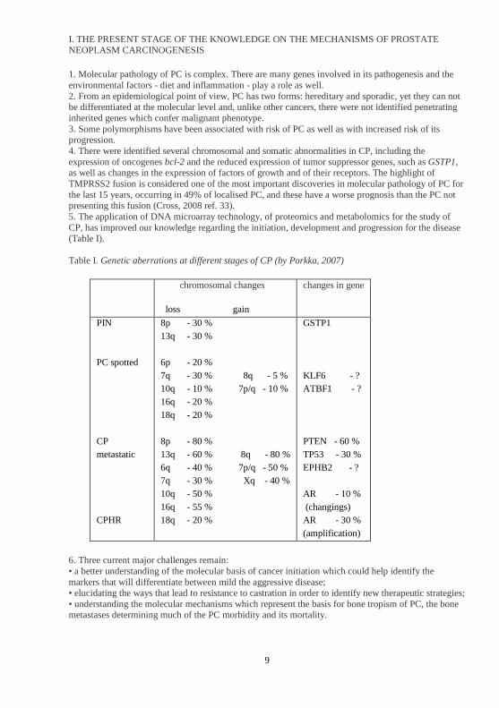
I. THE PRESENT STAGE OF THE KNOWLEDGE ON THE MECHANISMS OF PROSTATE
NEOPLASM CARCINOGENESIS
1. Molecular pathology of PC is complex. There are many genes involved in its pathogenesis and the
environmental factors - diet and inflammation - play a role as well.
2. From an epidemiological point of view, PC has two forms: hereditary and sporadic, yet they can not be differentiated at the molecular level and, unlike other cancers, there were not identified penetrating
inherited genes which confer malignant phenotype.
3. Some polymorphisms have been associated with risk of PC as well as with increased risk of its progression.
4. There were identified several chromosomal and somatic abnormalities in CP, including the
expression of oncogenes bcl-2 and the reduced expression of tumor suppressor genes, such as GSTP1, as well as changes in the expression of factors of growth and of their receptors. The highlight of
TMPRSS2 fusion is considered one of the most important discoveries in molecular pathology of PC for
the last 15 years, occurring in 49% of localised PC, and these have a worse prognosis than the PC not presenting this fusion (Cross, 2008 ref. 33).
5. The application of DNA microarray technology, of proteomics and metabolomics for the study of
CP, has improved our knowledge regarding the initiation, development and progression for the disease (Table I).
Table I. Genetic aberrations at different stages of CP (by Porkka, 2007)
chromosomal changes
loss gain
changes in gene
PIN
PC spotted
CP
metastatic
CPHR
8p - 30 %
13q - 30 %
6p - 20 %
7q - 30 % 8q - 5 %
10q - 10 % 7p/q - 10 %
16q - 20 %
18q - 20 %
8p - 80 %
13q - 60 % 8q - 80 %
6q - 40 % 7p/q - 50 %
7q - 30 % Xq - 40 %
10q - 50 %
16q - 55 %
18q - 20 %
GSTP1
KLF6 - ?
ATBF1 - ?
PTEN - 60 %
TP53 - 30 %
EPHB2 - ?
AR - 10 %
(changings)
AR - 30 %
(amplification)
6. Three current major challenges remain:
• a better understanding of the molecular basis of cancer initiation which could help identify the
markers that will differentiate between mild the aggressive disease; • elucidating the ways that lead to resistance to castration in order to identify new therapeutic strategies;
• understanding the molecular mechanisms which represent the basis for bone tropism of PC, the bone metastases determining much of the PC morbidity and its mortality.

10
II. THE ROLE OF NUCLEAR MEDICINE INVESTIGATIONS IN DIAGNOSING AND STAGING
OF THE PROSTATE CANCER
II.1. PROSTATE CANCER SCREENING
The results of the two large studies, in the U.S. and in Europe, published in 2009, evaluating the effect of screening on mortality in CP are contradictory.
Currently screening in CP remains a source of uncertainty and controversy. Early detection of PC is not
clearly recommended, nor contraindicated. In this case, the patient’s informed decision plays an important role in screening (Vedel, 2011, ref. 173; Evans, 2010, ref. 44 Perrin, 2008, ref. 118).
II.5. MOLECULAR STAGING OF PROSTATE CANCER
PC shows different biological behaviours. Preoperative serum PSA, Gleason score and the stage are the
variables currently most used to assess the prognosis, the recurrence and the metastatic potential. As a result of early detection in PC, the patients show up with increasingly more intracapsular disease.
However, a significant percentage of these patients have relapse after prostatectomy. The purpose of
molecular staging for prostate cancer is to identify the genes involved in the relevant ways for the
pathogenesis of prostate cancer and their use as prognostic markers.
II.6. IMAGING STRATEGY USEFUL IN DIAGNOSIS AND STAGING OF PROSTATE CANCER,
OTHER THAN RADIOISOTOPES
II.6.1. Ultrasound
Cross-rectal biopsy eco guided is the standard diagnostic test in the localisation of the tumor. (Fig. 1)
a) b) c)
Fig. 1. 80 years old patient, diagnosed with prostate cancer, Gleason score 10; images crossing the
middle region of the prostate. a) Classic ultrasound in gray scale reveals a hypo ecogene area in the left middle sided area (arrows). b) Real-time elastography reveals reduced elastic tissue at the level of
hypo ecogene area of classic ultrasound (arrows). c) Colour Doppler shows increased vascularisation
inside and around the tumor mass (arrows) (after Halpern, 2006, ref. 63).
II.6.2. Computed tomography has a role in evaluating the disease’s extension (Fig. 3).
Fig. 3. Cross-section CT: suprarenal metastasis (arrow). PC hardly metastasize at the lungs, liver, or suprarenal pleura (after Kundra, 2007, ref. 90).
II.6.3. Magnetic resonance imaging has a role in tumor localization (Fig. 4).

11
Fig. 4. Endorectal MRI examination, cross section. Prostate tumor (Ca) located in the left peripheral
zone (after Carroll, 2006, ref. 27).
II.6.3.1 Spectroscopic magnetic resonance imaging allows the assessment of prostatic metabolites
coline and citrate. Compared with normal peripheral zone, there are significantly higher levels of coline and significantly lower levels of citrate in the areas with cancer (Fig. 5).
Fig. 5. Prostate cancer detected in the left peripheral region. A. T2-weighted MRI cross section and three-dimensional MRSI spectrum. B. Corresponding three-dimensional MRSI spectrum indicating the
presence of an apparently aggressive tumors (the pick of the coline is very high and that of the citrate
very low) in the left peripheral zone. C. RM DWI (diffusion of MRI) reveals prostate tumor in the same place as T2 MRI and IRMS. D. Representative spectrum taken from the region of healthy prostate
tissue and tumoral. PPM, parts per million (after Carroll, 2006, ref. 27)
II.7. USEFUL RADIOISOTOPES NINVESTIGATIONS IN DIAGNOSIS AND STANDING OF PROSTATIC MALIGNANCIES
II.7.1. Bone scintigraphy: it does remain the standard imaging method for identifying bone metastases (Fig. 7).

12
II.7.2. Radioimunoscintigraphy
Unlike anatomical imaging, radioimunoscintigraphy detects signals from radiomarked antibodies which
recognize the prostatic tissue.
A. B. C.
Fig. 8. Merged SPECT-CT images. A. Radioimunoscintigraphy. B. Computed tomography. C.
Merged images. Periaortic node (PAN). Bo: activity at the colon level. A: aorta, IVC: inferior cave
vena. (After Keane, 2006, ref. 82).
II.7.3. PET-CT
In the last 15 years PET-CT has become one of the most innovative and important applications in oncology imaging (Rusu, 2006, ref. 129).
• 18 F-FDG indicates a more important capture in tumors with high Gleason score; there is a good
correlation between PSA level and FDG caption (Fig. 9) (Jadvar, 2011, ref. 75).
Fig. 9. 67 year old man with prostate cancer confirmed at biopsy (Gleason 8), with PSA of 14.6 ng / ml.
PET / CT 18F-FDG shows an intense hypermetabolism in the right prostate lobe (SUV 7.7) (after Jadvar H, 2011, ref. 75).
Fig. 7. Whole body bone
scintigraphy. Multiple metastases in
spine, costal grid, pelvis and bilateral femur. (Archive of Nuclear
Medicine Laboratory - Hospital "St.
Spiridon" Iasi.)

13
• 11 C-acetate. The acetate participates in the synthesis of the cytoplasmic lipid, which is probably
increased in tumors. In primary tumor detection it is more sensitive than 18F-FDG. • 11 C-Colina. 11 C-colina PET is a sensitive and accurate method in preoperative staging of pelvic
lymph nodes in prostate cancer (fig. 11).
Fig. 11. PET-CT 18-F Colina. Patient with prostate cancer treated by radical prostatectomy, in biochemical relapse. Abnormal accumulation of radiopharmaceutical in the right internal iliac lymph
node (arrows) (After Jadvar, 2011, ref. 75).
• 18 F-Fluoride seems to be more sensitive for detecting bone metastases if compared to 99mTc-MDP
bone scintigraphy.
II.7.4. Lymphoscintigraphy
Currently there is no non-invasive means to identify with certainty the patients with node invasion. The
main means of identifying lymph node metastases remains surgical staging lymphadenectomy.
Lymphoscintigraphy is used to identify the lymph node sentinel which will be subsequently submitted
to biopsy.
II.8.Conclusions
Ideally, imaging could accomplish in PC:
▪ diagnosis, localization and characterization (mild vs. lethal) of primary tumor ▪ determination of extra capsular extension
▪ guidance and evaluation of local therapy of the disease limited to prostate
▪ staging loco regional lymph nodes ▪ detecting the recurrent or metastatic disease
▪ guidance in radiotherapy
▪ prognosis of tumor response to therapy and systemic salvage
▪ monitoring of the active surveillance and defining a trigger for definitive therapy
▪ prognosis of the time until to hormone refractory tumor stage and of overall survival (Jadvar, 2011,
ref. 75). Recent development of imaging techniques, especially PET and MRI, could lead to
significant improvements in detecting and staging located PC. Diffused MRI could improve the tumor
detection - including by guiding the targeted biopsy, especially in the cases with patients who had previously negative biopsies - staging, determining the aggressiveness of the tumor and monitoring the
post-treatment evolution (Kim, 2011, ref. 86).
14
III. THE TREATMENT OF PROSTATE METASTATSIS. THE ROLE OF RADIONUCLIDE
THERAPY
III.1. CURRENT TREATMENTS OTHER THAN RADIONUCLIDES
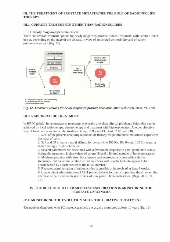
III.1.1. Newly diagnosed prostate cancer
There are several treatment options for newly diagnosed prostate cancer, treatments with curative intent
or not, depending on the stage of the disease, in view of associated co morbidity and of patient preferences as well (fig. 12).
Fig. 12. Treatment options for newly diagnosed prostate neoplasm (after Wilkinson, 2008, ref. 179)
III.2. RADIONUCLIDE TREATMENT
In HRPC painful bone metastases represents one of the prevalent clinical problems. Pain relief can be
achieved by local radiotherapy, chemotherapy and treatment with biphosphonates. Another effective
way of treatment is radionuclidic treatment (Bagi, 2005, ref.13, Henk, 2007, ref. 68). 1. 50% of the patients receiving radionuclide therapy for painful bone metastases experience
decrease of pain.
2. 32P and 89 Sr has a natural affinity for bone, while 186 Re, 188 Re and 153 Sm requires their binding to diphosphonates.
3. Several parameters are associated with a favourable response to pain: good OMS status
during the treatment, higher values of serum Hb and a limited number of bone metastases. 4. Myelosuppression with thrombocytopenia and neutropenia occurs with a similar
frequency, but the administration of radionuclides with shorter half-life appear to be
accompanied by a faster return to the initial number. 5. Repeated administration of radionuclides is possible at intervals of at least 6 weeks.
6. Concomitant administration of CHT proved to be effective in improving the effect on the
decrease of pain and on the occurrence of new painful bone metastases. (Bagi, 2005, ref.
13).
IV. THE ROLE OF NUCLEAR MEDICINE EXPLORATION IN MONITORING THE
PROSTATE CARCINOMA
IV.1. MONITORING THE EVOLUTION AFTER THE CURATIVE TREATMENT
The patients diagnosed with PC treated curatively are usually monitored at least 10 years (fig. 13).

15
Fig. 13. Algorithm for managing the patients after curative therapy or under active monitoring (after
Wilkinson, 2008, ref.179)
IV.2. MONITORING THE EVOLUTION AFTER HORMONOTHERAPY
Recurrence of cancer after castration
Its exact definition is controversial. Prostate cancer resistant to castration (CRPC) is different from hormone-resistant prostate cancer (HRPC). CRPC is still hormone-sensitive, responding to secondary
hormonal manipulation (suppression of ant androgens, estrogens, corticosteroids). HRPC is resistant to
all hormonal treatments (Fig. 14).
Fig. 14. Algorithm for managing the patients with systemic disease under hormonotherapy or
watchful waiting (after Wilkinson, 2008, ref.179)

16
PERSONAL CONTRIBUTION
V. THE OBJECTIVES OF THE STUDY
For the study I formulated the following hypothesis:
A) Between the results of bone scintigraphy, the total PSA value and the Gleason histological score
there may be a direct correlation.
B) Bone scintigraphy is an important element in the management of prostate cancer, in staging and
monitoring the evolution.
C) Imaging PET / CT 18NaF can be an effective method for detection of bone metastases in prostate
cancer.
D) Metabolic therapy with 153Sm can be an effective alternative for pain treatment in bone metastatic
prostate cancer.
In order to support the hypotheses stated above, I had the following objectives:
1. Correlation of total PSA value, of Gleason histological score and of bone scan results for
different age groups.
2. Comparison of sensitivity and specificity of two types of nuclear medicine exams - bone
scan with diphosphonates and MET / CT 18F-NaF - in detecting the bone metastasis of prostate cancer.
3. Assessing the efficiency and the toxicity of the QUADRAMET treatment in patients with painful bone metastases from prostate cancer.
4. The role of bone scintigraphy in managing the prostate cancer in the context of other imaging and laboratory investigations.
I have analyzed 4 lots of patients, one prospective (lot 2), and three retrospective:
Oncology Clinic from the Polyclinic no. 1 - Lot 1: 180 patients with bone scintigraphy recommended for the evaluation of bone metastases in the
initial assessment or in the development of prostate cancer. Bone scintigraphies were performed in the
Laboratory of Nuclear Medicine of the University Hospital "St. Spiridon "Iasi, in 2004-2009. For PSA values and histological results I checked out the files of the patients from the archive of the
Department of Oncology and Radiotherapy of "St. Spiridon " Hospital Iasi as well as the archive of
the.
- Lot 2: 13 patients with bone scintigraphy and PET / CT 18F-NaF, for the evaluation of prostate
cancer bone dissemination. Scintigraphic acquisitions were made in the Laboratory of Nuclear Medicine Hospital in Montbéliard, France, from June to December 2010.
- Lot 3: 57 patients treated palliatively with 153Sm EDTMP for bone metastatic prostate cancer, from 2001 to 2011, in the Nuclear Medicine Department of the University Hospital of Besançon, France.
- Lot 4: 178 patients diagnosed with prostate cancer who had bone scintigraphy for initial staging, restaging or for therapy evaluation. For this lot I monitored the patient's age at diagnosis, the percentage
of patients with metastases at diagnosis, the circumstances at diagnosis, the results for digital rectal

17
examination and the correlation of these results with Gleason score, the associated malignancies, the
clinical or pathological stage at diagnosis, the PSA value at diagnosis, the concordance surgery Gleason score - biopsy Gleason score, the time from diagnosis to RCB depending on the initial local treatment,
the number of patients with previous negative biopsies, the number of the deceased patients and the
cause of death. Of the 178 patients, 37 had bone metastases; they were considered a lot of study for which I examined
the age at diagnosis, the PSA at diagnosis, the general time from diagnosis to metastases, the time from
diagnosis to metastasis depending on the initial treatment, the correlation between the initial clinical or pathological stage and the occurrence of metastases, the correlation Gleason score – metastasis and the
correlation between the results of the digital rectal examination and the metastasis.
A special group have been the patients with metastases at diagnosis. Another distinct group is represented by the patients likely having family PC.
VI. INTRODUCING THE FOUR LOTS OF PATIENTS
VI.1. A COMPARISON BETWEEN TOTAL PSA, GLEASON SCORE AND THE RESULTS
OF BONESCAN FOR DIFFERENT AGE GROUPS
VI.1.1. Introduction Currently, bone scan performed with 99mTc - methylene diphosphonate (99mTc-MDP) is the standard
imaging method used to detect bone metastases. PSAt is the biomarker used in screening, diagnosis and
monitoring the evolution of prostate cancer. Researches have shown a positive relationship between the PSAt value and the presence of bone metastases (Lai, 2009, ref. 91; Chow, 2005, ref. 30). The purpose
of this study was to compare the value of PSAt with the pathologic Gleason score and with the results
of bone scintigraphy for different age groups (Albersten, 1998, ref. 5)
VI.1.2. Material and methods Bone scintigraphy was performed by using a double-head gamma camera (Axis - Philips, or Siemens).
Image acquisition was performed 2-3 hours after the intravenous injection of 15-20 mCi (740 MBq) of
99mTc-MDP depending on the body weight. Scintigraphic examination consisted of whole body planar images to which were added static images focused on areas of interest or tomoscintigraphic images in
order to have a higher resolution.
VI.1.3. The study group
Between 1 January 2004 to March 31 2009, 180 patients diagnosed with prostate cancer performed bone scintigraphy in our Department of Nuclear Medicine. Bone scintigraphy has been recommended
for initial pre-treatment staging or in the case of increase value of PSAt during disease progression or
for bone pain, regardless of the PSAt. For 86 patients the PSAt was known, that representing the value of PSAt at diagnosis (in the case of
newly diagnosed patients) or the maximum value of PSAt (in the case of patients evaluated during the
treatment). Out of the 86 patients, the Gleason score is known for 55. The patients were divided into three age groups: ▪ ≤ 60 years; ▪ 61-70 years; ▪> 70 years. The 86
patients were included, also, depending on the value of PSAt in 5 groups: ▪ 4 - 10 ng / ml, ▪ 11 - 20 ng /
ml, ▪ 21-50 ng / ml, ▪ 51-99 ng / ml, ▪ ≥ 100 ng / ml. In this study group there were not patients with PSAt values ≤ 3 ng / ml.
The 55 patients with known Gleason score were divided into three groups: ▪ <7, ▪ = 7, ▪> 7.
The scintigraphic results allowed for inclusion of the patients into 3 groups: ▪ without bone metastases; ▪ without bone metastases; ▪ with a probability (low, intermediate or high) of bone metastases.

18
Depending on the number of metastases, the 33 patients with metastatic bone scintigraphy were
classified in the four groups of Soloway: ▪ I degree - less than 6 metastases (a lesion of an entire vertebral body is considered as two metastases); ▪ II degree - between 6 and 20 metastases; ▪ II degree:
more than 20 metastases, but less than a super scan (fig. 16); ▪ IV degree: super scan (diffuse, intense,
symmetric radiotracer fixation, with the absence of the kidney shadows).
The results of our study can be summarized as following (Table II and III):
1. Among the patients with PSAt higher than 20 ng / ml, themselves considered at high risk
for bone metastases according to CCAFU recommendations in 2007, 21 (32.81%) out of 74 patients do not show bone metastases.
2. For PSAt higher than 50 ng / ml: ▪ All the 6 patients under the age of 60 years show
metastases (5 patients) or high probability of metastasis (1 patient); ▪ 10 (58%) of the 17
patients from the group of 61-70 years old show metastases, 1 (5.88%) show a low probability and 1 (5.88%) show intermediate probability. ▪ 15 (62.5%) out of 24 patients
aged over 70 years show metastases, and 1 (4%) low probability. ▪ 13 (27.65%) out of 47
patients from this group do not have metastases (7 aged 61-70 and 6 aged over 70 years).
3. 6 (21.42%) out of 28 patients with PSAt> 100 ng / ml do not show metastases.
4. Out of the 8 patients with PSAt = 11-20 ng / ml, with intermediate risk of metastasis, 1
patient has bone metastases, I degree Soloway (fig.14).
5. Out of the 11 patients with PSAt = 4-10 ng / ml, 1 patient has intermediate probability. 6. 10 (43.4%) out of 23 patients with Gleason score <7, themselves considered with low risk
for developing metastasis, show up secondary bone lesions (6 patients - 26%) or low
probability of metastasis (4 patients - 17.4%) (Table II, Fig. 19).
7. 7 (50%) of 14 patients with Gleason score = 7 show scintigraphic bone metastases.
8. 10 (56%) out of the 18 patients with Gleason score> 7 show metastases (8 patients - 44%)
or the probability of metastasis (2 patients - 12%).
9. There is no direct link between Gleason score and the presence of metastases for the
groups with intermediate risk (14 patients) and high risk (18 patients). It is likely that this
incongruity is due to the small number of patients.
Fig. 16. Patient, MI, aged 56 years.
Prostate cancer with multiple bone metastases in the axial skeleton and
apendicular, III degree Soloway.
Treatment: Bonefos. PSA: 22.5 ng / ml (Archive of Nuclear Medicine
Laboratory, "St. Spiridon" Hospital
Iasi).

19
TABEL II. Corelaţie rezultate scintigrafice - Scor Gleason
Number of cases
Gleason score
Metastases present
Probability of metastasis
Metastases absent
23 <7 6 4 13
14 =7 7 - 7
18 >7 8 2 8
Fig. 19. Correlation scintigraphic results - Gleason score. Graphical representation of data in Table
II.
3. The two patients with metastases in the group 21-50 shows metastasis PSAt = grade II Soloway.
4. Of the 30 patients with metastases and PSAt> 50 ng / ml, 8 represents the level II, 16 grade III (Fig. 15) and 4 grade IV Soloway (fig.16).
TABLE III. Correlation scan result - total PSA - age
(M - metastases, P - probability)
number
cases
PSA
(ng/ml)
age
(years)
M.
present
Grad
Soloway
M.
absent
P.
low
P.
interme-
diary
P.
high
No. of
cases
(partly)
14 4-10 50-60
61-70
>70
-
-
-
4
6
3
-
-
-
1
-
-
-
-
-
5
6
3

20
VI.I.5. Conclusions Our study confirms that the probability of bone metastases in an increased PSAt is inversely
proportional to the age, probably due to benign prostatic hyperplasia common in elderly patients.
There is not a direct correlation between Gleason score and bone scan results at intermediate and high risk groups of bone metastases.
VI.2. COMPARISON OF SENSITIVITY AND SPECIFICITY OF BONE SCINTIGRAPHY
DIPHOSPHONATES WITH PET-CT 18F-NaF
VI.2.1. Introduction
Evaluation of bone metastases from PC is now being made by bone scintigraphy with diphosphonates,
as the first exploratory wethod. Rebirth of scintigraphic examination with 18F-NaF, mainly due to technological progress of hybrid PET-CT, allows also the evaluation of metastases in the entire
skeleton.
VI.2.2. The purpose of the study
Our study compares the sensitivity and specificity of bone scintigraphy HMDP and PET-CT 18F-NaF in detecting bone metastases of PC.
VI.2.3. Materials and methods
8 11-20 50-60
61-70
>70
-
-
1
I
1
5
1
-
-
-
-
-
-
-
-
-
1
5
2
17 21-50 50-60
61-70
>70
1
1
-
II
II
1
4
5
-
3
-
-
-
-
1
-
1
3
8
6
18 51-99 50-60
61-70
>70
1
2
5
III
1III
1IV
3II
2IV
-
2
5
-
1
1
-
-
-
1
-
-
2
5
11
29 >/=100 50-60
61-70
>70
4
8
10
1I
1II
2III
1I
1II
6III
3II
6III
1IV
-
3
3
-
-
-
-
1
-
-
-
-
4
12
13

21
Bone scintigraphy was performed using a dual-head gamma detection cameras (Axis - Philips),
equipped with a large rectangular detector with a high resolution and low energy collimator, the peak energy of Tc - 140 keV + / - 20%, at 3 hours after the intravenous injection of 99mTc-HMDP 10 MBq
per kilogram of body mass. There were made aquisitions for the whole body, as well as SPECT focused
on different regions with anomalies on planar whole body images.
PET-CT examination was performed with a PET camera (Siemens) and the images were subsequently
fussioned with the CT images. There were made whole body acquisitions. The images were recorded one hour after an injection of 2 MBq/kg per kg of body weight 18F-NaF.
The two tests were conducted within an average time of 11.7 days, with the extremes from 5 days to 56
days, in a nuclear medicine department at Montbéliard Hospital, France.
VI.2.4. The study group
13 patients diagnosed with PC have been investigated with a double nuclear medicine exploration, bone
scintigraphy with 99mTc-HMDP and PET-CT with 18F-NaF during June to December 2010. The average age of the patients was 72 years, with the extremes from 62 to 86 years. The indication of
radioisotope exploration was represented by the evaluation of the extension of the newly diagnosed
neoplasia or by the detection of the bone extension during the evolution of PC in the case of increasing PSA values.
VI.2.5. Results
Comparing the results obtained by PET / CT using 18F-NaF with those from the bone scintigraphy
(OS) in detecting the bone metastases, I identified the following cases:
• Examination PET / CT negative and scintigraphy examination negative: PET / CT - / SO-; • Examination PET / CT positive and scintigrapy examination positive: PET / CT + / SO +;
• Examination PET / CT positive and scintigrapy examination negative: PET / CT + / SO -; • Examination PET / CT negative and scintigraphy examination positive: PET / CT-- / SO +;
• Probability of metastases, both in PET / CT study and bone scintigraphy.
• Probability of metastases at PET / CT examination and normal examination at bone scintigraphy.
▪ PET / CT - / SO-
Only one patient was part of this group without having images of metastases, neither on scintigraphic images nor on those of PET / CT (Fig. 20).
Fig. 20. TEP 18F-NaF - MIP incidence (maximum intensity projection) multiple. The examination
reveals degenerative phenomena in the spine and in the large joints, especially in the left knees and

22
ankles, and in the bilateral rizartroza thumb, without any site suspecte of secondary bone
dissemination. (Laboratory of Nuclear Medicine Archive - Hospital of Montbéliard, France)
• PET / CT + / SO +
This group was made up of 6 patients. Note that in all cases the lesions visible on bone scan were found in the PET-CT study. Only in one case a single lesion was found in both studies (one lesion at the level
of ischiopubic ramus). In the other 5 cases, the PET-CT study showed more lesions than the
conventional bone scintigraphy. Thus, in the case of a 76 years old patient, having the Gleason score 6 and the PSA 10.10 ng / ml, having as the recommandation the initial assessment of the tumoral
extension, PET-CT showed a site on the left parietal bone site, besides the costal site noticed on the
bone scintigraphy (fig. 21).
a) b)
c) d)
Fig. 21. Bone scintigraphy (06/04/2010): a) the whole body - anterior and posterior incidence and
pelvis centered images. b) Tomoscintigraphy centered on thorax); PET / CT (June 9, 2010): c) PET
whole body, multiple incidences d ) images PET fussioned with CT images, centered on the skull). Suspect of the posterior arch of the 8th right ribs highlighted on bone scan. PET examination finds this anomaly and, moreover, hightlights a hote area on the left parietal. (Laboratory of Nuclear Medicine
Archive - Montbéliard Hospital).
• PET / CT + / SO - In the group with abnormal fixation for the radiotrasor only for the PET / CT study, therefore with
normal scintigram, two patients were included. One of them, a man of 73 years old, having biochemical
relapse with no significant change of de radiopharmaceutical fixation, either for bone scintigraphy or for PET-FDG examination, presents three sites of metastases at PET-CT examination with 18F-NaF.

23
• PET / CT - / SO + There were no patients in this group to have been shown suspicious abnormalities in bone scintigraphy,
while their PET / CT examination was normal.
• Probability of metastases in both studies
Two patients were diagnosed by scintigraphy with the probability of bone dissemination scintigraphy in both studies. One of them has shown at bone scintigraphy the probability of bone dissemination on the
right sacroiliac bone, in its upper part; the same issue is found on the examination with PET / CT. In
addition, the latter examination shows the probability of metastasis for the posterior arch of the second dorsal vertebrae.
The second patient from this group shows a small left scapular site, an anomaly found in both
radioisotope exploration, suggesting the probability of dissemination at this level.
• The probability of metastases in the PET / CT study and normal examination in bone
scintigraphy
2 patients have been part of this group. The first one, a man of 73 years, shows no abnormalities of
fixing the radiotracer to the bone scintigraphy, but PET-CT hightlights a hypermetabolic retro-orbital
site, at the right temporal bone level, whose etiology remains to be determined.
VI.2.6. Discussion:
• 18F-Fluoride is a PET radiotracer with bone tropism emitting positrons which is assessing the
osteoblast activity (Beheshti, 2009, ref. 16). It was first described 40 years ago, yet it has been extensively investigated with respect to bone metastases only in recent years due to the improving of
PET / CT devices (Cook, 2010, ref. 32, Rusu, 2007, ref. 131) . • 18F-fluoride captation reflects the blood flow and bone remodeling. 18F-Fluoride is greedily and
early accumulated in the cortical bone in the case of bone response to a metastasis. In 2008 it was
authorized on the French market, including for the evaluation of bone metastases of PC (Huchet, 2009,
ref. 72).
• The assessment of 18 F-fluoride kinetics using PET quantitative methods allows for the
characterization of lesions and monitoring of the response to therapy. Although the mechanism of 18F-fluoride capture corresponds to osteoblastic activity, this tracer is also sensitive in detecting the lytic
metastases and those of bone marrow by identifying osteoblastic changes accompanying them, even
when they are minimal (Even-Sapir, 2007, ref . 46). • Positron emission tomography is a noninvasive functional imaging technique that allows highlighting
regional metabolic processes. PET is coupled with a imaging morphological method. Because of the
fact that the functional changes from the tumoral processes precede the morphologic changes, PET imaging provides a new dimension to classical imaging (Boujelbene, 2011, ref. 23).
• The number of the evocative lesions of bone metastases hightlighted by PET / CT examination is
superior to that detected by bone scintigraphy, so PET / CT has an important role in monitoring the PC, especially in the detection of its recurrence and of bone metastases (Bouchelouche, 2009, ref. 22).
• In the case of our study, two patients had PET / CT positive and normal scitigraphy, and 5 out of the 6
patients having both tests positive had more lesions at the PET / CT exam in comparision with SO. In one of the two cases of bone dissemination probability, a probability assessed both by SO and PET /
CT, the PET / CT exam revealed a new hypermetabolic site in comparision to SO. 2 patients in our
study had negative SO and a probability of bone dissemination at PET / CT. It can thus be inferred that the sensitivity of PET / CT method is superior to bone scintigraphy.
• The specificity of PET / CT is much improved by the presence of CT, with its essential role in
attenuating the correction, leading to greater accuracy by reducing artifacts (Even-Sapir, 2006, ref. 7). Moreover, CT helps to locate the lesions and allows for their morphological characterization and for a
better differentiation of metastasis in benign lesions. In our study, CT has differentiated intramedullary
location of costal sites without cortical disruption, that being characteristic to bone metastases, in contrast to fractures (Fig. 23). However, NaF is fixed like diphosphonates in the areas of hyperemia and
of important osteogenesis, including at the level of inflammatory or infectious sites, osteoarthritis,

24
post-traumatic, as well as in various bone diseases (Paget, metabolic bone diseases, osteonecrosis).
• The criteria for interpretation are similar to those used in the interpretation of bone scintigraphy. There are osteolytic processes that can not be detected. The degree of captation does not differentiate
the malignant lesions from the benign ones. PET / CT 18F-NaF could be negative in intense sclerotic
lesions , which is probably reflecting the effect of treatment (Beheshti, 2008, ref. 17). • PET / CT examination is faster than bone scintigraphy with diphosphonates (Grant, 2008, ref. 61).
Dosimetry is similar for the two exams (Segal, 2010, ref. 140).
• 18F-fluoride could provide a more sensitive "conventional" bone scintigraphy. PET with 18F-fluoride is superior to FDG in the evaluation of tumors which do not capture FDG, although "early disease"
FDG has clear advantages over 18F-fluoride (Langsteger, 2006, ref. 96).
• Unlike PET / CT Colina, which is recomanded in non invasive restaging of PC, in the case of increse of PSA after radical treatment, the use of NaF in clinical practice requires yet to be confirmed (Picchio,
2011, ref. 119).
VI.2.7. Conclusions
1. PET / CT 18F-NaF is more sensitive and more specific than bone scintigraphy in detecting bone
metastases, including bone tomoscintigraphy. 2. For similar dosimetry, 18F NaF PET provides a faster study then bone scintigraphy with
diphosphonates.
VI.3. ASSESSING THE EFICIENCY AND THE TOXICITY OF QUADRAMET TREATMENT
BONE METASTATIC PROSTATE CANCER
VI.3.1. Introduction Radionuclide therapy is a palliative treatment method used for patients with pain caused by bone
metastases, especially when bone dissemination is multiple.
VI.3.2. Material and methods
The purpose of this retrospective study is to evaluate the effectiveness and the toxicity of Quadramet
treatment in the case of the patients diagnosed with bone metastatic prostate cancer. For each patient, before administering the metabolic treatment, it was performed bone scintigraphy in
about 3 hours after intravenous injection of 10 MBq of 99mTc-HMDP per kilogram of body mass. The
exam consisted of aquisitions of the whole body, and in each case, centered static images or tomoscintigraphic, depending on the anomalies of fixation for the radiotrasor on the images of the
whole body.
Radionuclide treatment consisted of an intravenous dose of 37 MBq of 153 Sm-EDTMP (Ethylene diamine tetramethylene phosphonate) per kilogram body weight. Quadramet administration was
preceded and followed by a perfusion of 500 ml of physiological serum. The patient was hospitalized in
our laboratory for 5-6 hours. At 48-96 hours after Quadramet administration, a whole body scan test was performed to confirm the fixation of the radiopharmaceutical at the bone metastases level,
highlighted by bone scintigraphy with diphosphonates.
Clinical examination performed before administering Quadramet, as well as in 6 weeks post theraphy, was to assess the pain locations and their intensity, using visual analog scale method and to
evaluate the OMS life quality status. After therapy, it was monitored the presence of the phenomenon
of "flare". It was also noted the treatment for before and after antalgic therapy. By biological evaluation prior to therapy it was monitored the renal function and the ionograme.
PSA - tumor marker in PC - was measured before the treatment and in 6 weeks after the treatment.
CBC was analyzed pre and postoperatively, to assess metabolic haematotoxicity treatment. The access to the Axigate database, where I found the history of patients’s desease, to the Sirilog
database, where are shown the examinations carried out by the patients, as well as to the patients
records within the Nuclear Medicine Laboratory at Besancon allowed me to evaluate the effectiveness of the treatment by analyzing the OMS status of life quality, the evolution of pain using visual analog

25
scale and the change in antalgic treatment. I monitored, also, the presence of "flare" phenomena and its
predictive effect on the outcome of the therapy, as well as the evolution of PSA. Haematological toxicity of Quadramet was demonstrated by the results of CBC.
VI.3.3. The study group
From May 1991 until May 2011, 57 patients diagnosed with prostate cancer with painful bone metastases have received radiopharmaceutical treatment in the Nuclear Medicine Department of
Besançon. Out of these, 5 patients had repeated the treatment with Quadramet, and 3 had three cures of
Quadramet. The metabolic treatment was not administered in cases of the patients who received chemotherapy or external radiotherapy in the last 6 weeks, nether in the case of those treated with
diphosphonates in the last 3 months.
The time from PC diagnosis to the treatment with 153 Sm was known for 42 patients. The average of this time is 5.45 years, with extremes between a few months from diagnosis for patients who had
metastases at diagnosis up to 28 years after diagnosis (only 4 patients with a period over 10 years) for
patients who developed late metastases.
IV.3.4. Results and discussion
153 Sm-EDTMP is a therapeutic agent consisting of a radioisotope - 153 Sm - and a chelate tetraphosphonate - EDTMP. The β particle of the radiotrasor (average energy of 233 kev) has a path of
3.1 mm in soft tissues and 1.7 mm in the bone marrow which limits the irradiation of bone marrow and
other adjacent tissues. Physical half-life time is 46.3 hours (Chow, 2005, ref. 30). The radioisotope emits a gamma radiation of 103 kev (29%), which allows the imaging (Lam, 2008, ref. 93). 153 Sm-
EDTMP capture is similar to diphosphonate captation in bone scintigraphy (Fig. 30).
a) b)
a. Antalgic effectiveness of Quadramet
In our study, the effect of radionuclide therapy was evaluated primarily clinical - assessing the decrease, the persistence or worsening the pain at 6 weeks after Quadramet. Secondly, the antalgic
effect was assessed with the help of the visual analogue scale. The antalgic effectiveness of vectorial
internal radiotherapy was also assessed for the lot of patients with repeated administration. ● Clinical assessement – the characterization of bone pain after six weeks from Quadramet
administration Out of the 51 treatments, 38 (74.51%) were followed by a positive response to Quadramet, 15 (29.41%)
recording a total attenuation of pain, and 23 (45.09%) decrease of pain: significant decrease (8 patients
- 15.68%), moderate (6 patients - 11.76%) or partial (9 patients - 17.64%).
Fig. 30. a) Whole body bone
scintigraphy (23/05/2006). Multiple
secondary dissemination in the axial skeleton and apendicular
b) Whole body scan two days after
Quadramet (22/06/2006). Hiperfixantes sites overlapped of
those from the bone scintigraphy
(Archive of Nuclear Medicine Laboratory - University Hospital,
Besançon, France).

26
The absence of pain decrease was recorded in the case of 10 treatments (19.61%) and worsening the
pain was reported in 3 treatments (5.88%), with a total of 13 ineffective treatments out of 51 (25.49%) (Table IV).
TABLE IV. The distribution of the number of treatments depending on the characterization of pain
development after administrating 153 Sm. The evaluation has taken place at 6 weeks after
radionuclide treatment.
* In this category are includes also the patients who have experienced loss of pain up to 5 weeks after
the treatment, followed by their recurrence.
Characterization of pain post Quadramet Numberof treatments
Loss of pain 15
Important improvemet 8
Moderate improvement 6
Partial improvement* 9
Absence of pain improvement 10
Pain worsening 3
● Assessing the evolution of pain using visual analog scale 19 patients have self-assessed the pain before and after the treatment with Quadramet EVA analogue-
visual scale. (The patient receives a ruler gradated from 0 to 10, 0 degree meaning no pain and 10
degree meaning unbearable pain; on this scale the patient indicates manualy the intensity of the pain he is complaining.) 14 patients (73.68 %) mentioned a decrease of pain by an average of 3.05 points (out
of 10), 2 patients (10.52%) perceived the same degree of pain and 3 (15.78%) accused an increased
pain with an average of 1.66 points out of 10 (fig. 35).
● Antalgic effectiveness of Quadramet at repeated administration
If the case of the three patients, each having three radionuclidic palliative treatments, the therapy was
always effective. Among patients treated twice, 4 showed improvement at one of the treatments, but therapy was ineffective at its repetition (3 cases) or - in one case – the therapy was initially ineffective
and subsequently effective. The therapy was effective at the administration of two doses in the case of a
patient.
Fig. 35. Graphical representation of the
distribution of the patients according to self-
assessement of pain with visual analog scale.

27
In conclusion, Quadramet was effective in 74.5% of the cases (51 treatments) in regard to decrease or
loss of pain. 73.68% of 19 patients mentioned - using visual analogue scale – the decrease of pain with an average of 3.05 points (10 points). Quadramet allowed for the reduced dosage or interupting the
treatment for 40.90% of the patients (from a total of 22). The antalgic effectiveness of Quadramet in
our study is comparable to that found in other studies, 70-80% (Klingelschmitt, 2002, ref. 87; Dolezaj, 2007, ref. 39; Liepe, 2005, ref. 102; Liepe, 2007 , ref. 103, Lam, 2007, ref. 92).
b) Change in pain relief medication
9 patients (40.90%) have reduced or interupted the posology of the antilalgic treatment, thus demonstrating the efficacy of Quadramet treatment. The treatment has remained unchanged in the case
of 9 patients (40.90%) and the posology was increased in the case of 4 patients (18.2%) (Table VI).
TABLE VI. The distribution of the number of patients depending on Qudramet effect over the
antalgic medication.
The effect of Quadramet on antalgic theraphy Number of patients
Interruption of teraphy 2
Reduction of dosage 7
Tratment unchanged 9
Increase of dosage 4
In the study of Liepe, 13% of the patients abandoned the antalgic therapy and have shown no pain
(Liepe, 2007, ref. 103).
c) The evaluation of OMS quality of life score
Out of the 23 patients with known OMS score, assessed before and after the treatment with Quadramet,
3 (13.04%) are showing the improvement of the score by 1 level, 17 (73.91%) do not change the score, and 3 patients (13.04 %) are showing a degradation of OMS score by an average of 1.33 (Table VII and
Fig. 36).
TABLE VII. The distribution of the number of patients depending on the evolution of OMS quality
of life score.
The evolution of OMS quality of life score Number of patients
improvement 3
same score 17
worsening 3
Most patients - 17 out of 23 (73.91%) have the same OMS quality of life score, while 3 patients have
improvement and 3 have degradation of OMS score. The result can be explained by the fact that this scale which is presenting values from 0 to 5 is less sensitive (Klingelschmitt, 2002, ref. 87), so then it is
required a significant improvement of life quality to move from one level to another. In addition, our
assessment was performed only 6 weeks after the treatment, therefore not allowing the assessement for the treatment’s effect on a long range.

28
Fig. 36. Graphical representation of the distribution of the number of patients depending on the
evolution of OMS life quality score (data in Table VII).
d) "flare" phenomenon
Out of the 22 patients for whom it was noted the presence or the absence of "flare" phenomenon - a temporary increase of pain after metabolic therapy - 12 patients (54.54%) manifested this phenomenon,
and 9 (40.90%) did not. 1 patient (4.54%) presented the "flare" phenomenon during an effective
treatment but did not during the subsequent ineffective treatment. In our study, there is no direct correlation between the presence of "flare" phenomenon and the
response to Quadramet.
f) Haematological toxicity of Quadramet therapy
Out of the 29 patients, 11 (37.93%) do not show up haematological toxicity, 11 (37.93%) show a reduced toxicity, and 5 patients show moderate toxicity (grade II). 1 patient has tricitopeny (unknown
grade), and 1 patient passes from grade II to grade III anemia. None of the patients had grade IV
toxicity (Table IX). In total, 62% of patients experienced grade II haematotoxicity or lower grade.
TABLE IX. The distribution of the patients depending on the evaluation of hematologic toxicity.
Assessing the hematologic
toxicity
Number of patients
Absent 11
Reduced (degree 0 sau I) 11
Moderate (degree II) 5
Sartor (ref. 135) observed a toxicity of grade two or lower for the leukocyte and for the trombocytes in
the case of 92%, respectively of 97% of patients who received 1 mCi of 153 Sm lexidronam per kg of
body weight. Grade 3 of leukopenia was observed for less than 7% of patients, regardless of the number of administration of 153Sm (Sartor, ref. 136).
VI.3.5. Conclusions:

29
1. Systematic radionuclide radiotherapy has obvious advantages:
▪ an important antalgic effect; in our study 74.5% of the patients experienced disappearance or relief of pain;
▪ 9 of 22 patients (40.90%) have reduced or interrupted the antalgic therapy.
▪ simultaneous treatment of all secondary bone sites, while the selective absorption in bone metastases limits the irradiation of normal tissues;
▪ a single intravenous injection to a patient who requires only a few hours of hospitalization;
▪ the use of early radionuclides in managing the disease could be a complementary therapy or may delay the use of other palliative methods, such as external radiotherapy, chemotherapy, hormone
therapy, bisphosphonates and analgesics
▪ metabolic therapy may have not just only a palliative effect, but also a tumoricid or tomorostatic effect. Early use of radionuclides in pain therapy may limit cancer progression by inhibiting the
development of oligometastasis (Hillegonds, 2007, ref. 70).
▪ in addition to the important effect of painkiller, radionuclide therapy allows for improving the mobilization for many patients, reducing the dependence on painkillers and offering a better quality of
life (Lam, 2008, ref. 93).
▪ there are several parameters associated with a favorable response to pain: good OMS status during the
treatment, higher values of serum Hb and a limited number of bone metastases.
▪ simultaneous administration of CHT was proved to be effective in improving the effect on pain relief
and on the appearance of new metastatic bone pain (stick, 2005, ref. 13). ▪ Repeated treatments and combining radionuclidic treatment with other therapies, such as
bisphosphonates, chemotherapy and / or external radiotherapy are possible. In addition, combined
therapy could provide a more effective pain relief caused by bone metastases (Anderson, 2007, ref. 6). ▪ Particularly, the association of docetaxel - the only therapy proven to have an effect on prolonging life
in the case of patients with metastatic HRCP, the standard therapy in the case of these patients - with
153 Sm-EDTMP can be performed at normal, repeated doses (Morris, 2009, ref. 109). 2. Haematological toxicity of Quadramet is reduced or moderate.
▪ In our study, 62% of patients experienced grade II of haematotoxicity or lower. ▪ Repeated administration of 153 Sm, as well as previous radiotherapy or chemotherapy, does not result
in an increased myelotoxicity. Haematological toxicity is reversible in about 8 weeks.
▪ Repeated administration of radionuclides is possible, at intervals of at least 6 weeks.
▪ Patients with metastatic bone disease can survive for a long time and can be safely treated with
multiple combined therapies (Heron, 2008, ref. 69).
▪ Bianki (2009, ref. 20) proposes a method of quantification for Quadramet fixing depending on the radiotrasor captation on the bonescan, therefore improving the dosimetry and optimizing the Quadramet
administration.
VI.4. THE ROLE OF BONE SCINTIGRAPHY IN MANAGING THE PATIENTS WITH
PROSTATE CANCER
VI.4.1. Introduction
Bone scintigraphy with diphosphonic labeled 99mTc is the method of choice for evaluating bone
dissemination at diagnosis, during disease progression (the case of biochemical relapse), as well as to
evaluate the therapeutic effect.
VI.4.2. The purpose of the study
Assessing the role of bone scintigraphy in managing the patients with prostate cancer and its correlation
with different other parameters used in diagnosing and monitoring the developement of PC.
VI.4.3. Material and method
Bone scintigraphy was performed using a dual head gamma camera for detection (GE and Philips) and consisted of early aquisitions - early phase- in the case of symptomatic patients, 3-minute planar
acquisition centered on painful areas, and of late acquisitions - bone phase – the whole body and in

30
most cases, SPECT focused on the areas with abnormalities of the radiotrasor caption seen on the
whole body images. Axigate database access allowed me to know the history of the patients, and the access to Sirilog, which
groups the explorations performed by the patients, allowed me to compare the bonescan results with
results of other image explorations.
Therefore, I monitored the age of the patient age at diagnosis, the percentage of patients with metastases at diagnosis, the diagnosis circumstances, the results for the rectal touch examination and
the correlation of these results with Gleason score, associated malignancies, clinical or pathological
stage at diagnosis, PSA value at diagnosis, the concordance of operational Gleason score – the biopsy Gleason score, the time from diagnosis to RCB depending on the initial local treatment, the number of
patients with previous negative biopsies, the number of patients deceased and the cause of death.
VI.4.4. The study group Between November 2009 and October 2010, there were 178 patients who performed bone scan in the
Nuclear Medicine Laboratory of Besançon College Hospital (some had repeated bonescan), most of
them being recomended by the Radiotherapy and Oncology services. I mention the fact that the lot of 178 patients are only the patients whose history I found in the Axigate
database.
Out of the 178 patients, 37 showed bone metastases; they have formed a study group for which I have analyzed the age at diagnosis, the PSA at diagnosis, the general time from diagnosis to metastases, the
time from diagnosis to metastasis depending on the initial treatment, the correlation between the initial
clinical or pathological stage and the occurence of metastases, the Gleason score correlation – the metastasis and the correlation between the results for rectal touch examination and the metastasis.
A special group have been the patients with metastases at diagnosis. Another distinct group is represented by the patients with a probability of PC in family.
VI.4.5. Results and discussion
Currently, most used radiotracers for bone scan are diphosphonates labeled with 99mTc (Rusu, 2003,
ref. 132).
VI.4.5.1. Age of patients at diagnosis
The average age of the 178 patients sent to the Nuclear Medicine Laboratory of Besançon within a year
(November 2009-October 2010) is 67.64, with extremes between 41 and 92 years (Figure 47).

31
Fig. 47. Graphical representation of the distribution of the patients according to their age at prostate
cancer diagnosis (data contained in Table XI). It is observed that the distribution of the number of patients follows the Gaussian curve demonstrating
the homogeneity of the lot. Greene (2005, ref. 60) finds an age at diagnosis of 65 years.
VI.4.5.2. The proportion of patients with metastases at diagnosis
9 out of 178 patients showed bone metastases at diagnosis (5%) and 4 patients (2.24%) showed a
probability of metastases at diagnosis.
In the post era of PSA, 5% of patients show metastases at diagnosis, according to Jadvar (2009, ref. 76).
VI.4.5.3. Circumstances of diagnosis
To evaluate the effect of screening on mortality in PC, two major studies were conducted in the United
States (PLCO) and in Europe (ERSPC); in 2009 there were published their results. The results of these two studies are contradictory.
Currently screening in PC remains a source of uncertainty and controversity. Early detection in PC is
not clearly recommended nor contraindicated. In this case, the constient decision of the patient plays an
important role in screening (Vedel, 2011, ref. 173; Evans, 2010, ref. 44 Perrin, 2008, ref. 118).
♦ Screening Algorithm In the situation of patients who decide to benefit from screening, according to the American Cancer
Society (Wolf, 2010, ref. 180):
• the screening is recommended with PSA or with PSA and TR.
• the screening will be canceled for men with PSA 2.5 ng / ml.
• for PSA <2.5 ng / ml, the screening will be performed in two years.
• in the case of patients with common risk, the biopsy is recommended for a PSA of 4 ng / ml.
• for PSA values from 2.5 to 4 ng / ml, it is calculate the individual risk especially for high level of PC, in view for the biopsy recommendation. The factors which increase risk of PC include the African-
American population, the family history of PC, advanced age and an abnormal TR. A previous negative
biopsy decreases the risk for prostate neoplasia (fig. 49).
European Association of Urology recommends an assessment of PSA at the age of 40 (baseline PSA),
depending on which it will be decided the frequency of the following tests. For values of PSA <1ng/ml, 8 years would be enoght (Heindenreich, 2011, ref. 67).
Fig. 49. Screening algorithm, according to the recommendations of the American Cancer Society
(Wolf, 2010, ref. 180).

32
In our study, the circumstances of diagnosis for the 92 patients (whose circumstance diagnosis were known) were:
▪ early detection / individual detection: 55 patients. In France, there is no PC screening performed for
the population yet early detection is encouraged through PSA dosing and rectal digital examination. ▪ clinical manifestations: 29 patients. The most frequent clinical manifestations were: abnormal
micturition, dysuria, prostatitis, acute urinary retention. A patient showed up for a leg edema and the
CT scanning showed a large prostate tumor accompanied by retroperitoneal and groin lymph nodes (the latter causing the clinical edema).
▪ detection during the assesement of other diseases: 3 patients.
▪ the patients do not know the circumstances of diagnosis: 5 patients (Fig. 50).
Fig. 50. The percentages of patients depending on the circumstances of diagnosis.
VI.4.5.4. The results of rectal digital examination (known for 77 patients) In our study, 26 out of 77 patients (33.76%) showed a normal rectal digital examination and 51 patients
(66.24%) showed a pathological rectal digital examination (Fig. 51).
Paradoxically, in a study done by Ankerst et al (2009, ref. 9), it was noticed that 70% of the patients with abnormal TR, within a period of 1 year, have presented a normal TR, even the patients with PC.
On the other hand, other studies argue that the probability of diagnosing PC at biopsy is higher for
patients with positive TR and associating PSA ≥ 3ng/ml with abnormal TR leads to detecting a significantly higher number of PC with Gleason score> 7 (Gooselar, 2008, ref. 58).
34%
66%
Fig. 51. Graphical representation of the distribution on the number of patients
depending on the rectal digital results.
early detection clinical manifestations balance for other deseases the patient does not know
Normal TR Pathologic TR

33
VI.4.5.5. The correlation between rectal digital examination and Gleason score
♦ The question where I have started is: Is there a correlation between clinical data (TR) and
histological data (Gleason score)?
I analyzed separately the lot of patients with normal TR and the lot with pathological TR, correlated
with biopsy or operator Gleason score (the latter known only for patients who have had prostatectomy).
The data was grouped in Figure 56. I obtained the following results:
▪ normal TR: 25 patients with known Gleason score - bioptic (13 cases) or surgical (12 cases); for a
patient Gleason score not known. ▪ pathological TR: 50 patients with known Gleason Score - bioptic or surgical; for a patient Gleason
score not known.
Fig. 56. Graphical representation for comparing the results of TR and of bioptic or surgical Gleason
score.
I found the following results:
1.In the case of the patients with normal TR, 52% had a Gleason score <7; only 24% of the patients with pathological TR have the same score.
2.40% of the patients with normal TR and 50% of those with pathological TR have 7 Gleason score.
3.26% of the patients with pathological TR have a Gleason score> 7, while only 8% of those with normal TR have the same score.
In conclusion, there is a direct correlation between the value of Gleason score (histological result)
and TR results (clinical evaluation).
VI.4.5.6. Patients with prostate cancer associated with other malignancies.
In our group I found (Table XXIV):
TABLE XXIV. Distribution of the number of patients with malignancies associated to prostate
cancer
Associated neoplasia type
number of
patients
percentage

34
▪ 27 patients have shown two or more malignancies: 23 patients with two cancers, 2 patients with three
cancers and two with four cancers.
▪ bladder cancer: 8 out of 33 (24.24%); renal cancer 5 out of 33 (15.15%); thyroid, colon and lung cancer: 3 of each out of 33 (9.09%, respectively).
▪ two patients with rectal cancer and two with melanoma; one patient each with hepatocellular
carcinoma, colangiocarcinom, vocal cords, neurinom, myeloma, gastric and CML.
I observed:
▪ PC is most frequently associated - in our study - with urinary tract cancers. Bladder and renal neoplasms represent 39.39% of cancers associated with PC.
▪ Thyroid, colon and lung cancers show a significant association (i.e. 9.09% of the cases).
bladder cancer
8 24,24 %
kidney cancer
5 15,15 %
thyroid cancer
3 9,09 %
colon cancer
3 9,09 %
lung cancer 3 9,09 %
rectal cancer 2 6,06 %
melanoma 2 6,06 %
hepatocellular carcinoma
1 3,03 %
colangiocarcinom 1 3,03 %
vocal cord cancer
1 3,03 %
LMC 1 3,03 %
neurinom 1 3,03 %
myeloma
1 3,03 %
gastric cancer
1 3,03 %
Total 33 100 %

35
VI.4.5.7. The stage at diagnosis (for patients recomanded for scintigraphy)
In the lot of the 178 patients I have found persons who presented the following clinical or pathological stages at diagnosis:
T1 - 17 patients; T2 - 29 patients; T3 - 13 patients; T4 - 0.
PT2 - 23 patients; PT3 - 37 patients; PT4 - 1 patient.
I observed:
▪ 49.15% out of the 59 patients with known T-score were diagnosed with clinical T2 score, while only
22.03% were diagnosed with clinical T3. A large percentage of patients - 28.81% showed T1 stage at diagnosis.
▪ Out of the 62 patients with known pT score (i.e. patients who have been treated with radical
prostatectomy), 98% were classified in classes PT2 (39%) and PT3 (59%). Only 1 patient has presented PT4 stage.
VI.4.5.8. PSA at diagnosis
Out of the 155 patients with known PSA at diagnosis, 5 patients had a PSA <4 ng / ml (3.22%), 66 had
a PSA of 4-10 ng / ml (42.58%) , 54 a PSA of 10-30 (34.83%) and 30 patients had a PSA> 30 ng / ml (19.35%) (fig. 59).
Patients considered formerly as belonging to "gray zone" (4-10 ng / ml) represent the largest percentage
(42.58%) in our study, followed by the PSA group with values of 10-30 ng / ml (34.83 %). These two groups represent 77.41% of diagnosed cases. 3.22% are patients with PSA <4. Only 19.35% are
patients with PSA> 30 ng / ml.
VI.4.5.9. Bioptic Gleason / surgical Gleason concordance (both values are known for the cases of 16
patients who received PR)
Out of the 16 patients with bioptic and surgical Gleason score known, the values in 8 cases were
consistent and in 8 cases inconsistent.
VI.4.5.10. Biochemical recurrence
I have monitored the time from diagnosis to the biochemical relapse, according to the initial local
treatment (PR, RT, brachytherapie, HT)
▪ PR (Figure 60) - 16 out of the 44 patients who have been treated with radical prostatectomy showed after surgery a
detectable PSA. Of these patients, 3 had positive surgical margins, 6 extracapsular invasion, 2 invasion
of seminal vesicles, and 2 patients showed positive surgical margins as well as extracapsular invasion.
- 5 patients had a postoperative PSA 0.2 and one had 0.19.
- RCB was observed on an average of 3.43 years after PR.
Fig. 59. The percentage of the patients depending on the PSA value (ng / ml) at diagnosis

36
Fig. 60. Graphical representation of the distribution for the number of the patients depending on the
time from diagnosis to biochemical relapse after radical prostatectomy. ▪ RT (figure 61)
-the average time until biochemical relapse in the case of 17 patients who received external
radiotherapy is 3.2 years.
Fig. 61. Graphical representation of the distribution of the patients depending on the time from
diagnosis to biochemical relapse after external radiotherapy.
VI.4.5.11. Patients with previous negative biopsies
15 patients from the lot (8.42%) are known to have had historicaly at least one negative puncture
biopsy. Of these, 3 had two series of negative biopsies and one patient had three negative biopsies.
V.4.5.12. PC in family (probably)
In the lot of the 178 patients I found 7 patients with the probability of PC in family. I observed:
▪ The average age for diagnosis of neoplasia is 63.85 years, compared to 67.64 years for the total lot.
▪ 6 of the 7 patients had siblings diagnosed with PC (3 patients one brother, 2 patients two brothers and one patient three brothers having the same neoplasia). Only in the case of a patient, his father was
diagnosed with PC.
▪ A young patient (56 years) with metastases at diagnosis has a sister with breast cancer (probably he has also PC in family), and one patient introduced in this sublot has a daughter diagnosed with breast
cancer. It has been confirmed the incidence of both cancers in some families.
VI.4.5.14. The group of the patients with metastases
♦ The age at diagnosis

37
The average age at diagnosis was 69.97 years, compared to 67.94 years for general lot
♦ PSA at diagnosis (known for 31 patients) (fig. 66)
Fig. 66. Graphical representation of the distribution for the patients with metastases depending on
initial PSA value.
I noticed that among the 31 patients with metastases having a known PSA at diagnosis, 8 (25.8%) had PSA values of 4-10 ng / ml.
All 6 patients with PSA> 100 ng / ml (from the lot of 178 patients) showed metastases at diagnosis.
♦ The time from diagnosis to metastasis
9 of the patients showed bone metastases at diagnosis (4 of the 178 patients showed a probability of
metastases at diagnosis), 4 in the first year, 10 in 2nd to 5th year, 8 in 6th to 10th year, 2 after 10 years and in the case of 4 patients the time from diagnosis to metastasis is unknown.
♦ Correlation Gleason score - metastases (for 33 patients) 77.77% of the patients with metastases had a biopsy score ≥ 7; of the 7 patients with known operatory
Gleason score, none had a score <7. It is thus noticed that, in the case of the patients with metastases,
high Gleason score correlates with bad prognosis.
♦ Correlation of rectal digital examination - metastases
For 12 patients with metastasis it is known the TR result: 11 pathological (91.67%), 1 normal (8.33%).
An initial negative TR could be a less aggressive cancer. However, the number for the lot is very small.
♦ The time from biochemical relapse to metastases (known for 9 patients treated with RT and 7 patients treated with PR)
The patients who have been treated with PR present an average of 2.85 years from biochemical relapse
to the appearance of metastases, 3 patients showing metastases in less than 2 years from RCB, 3 patients within 2 to 5 years and 1 patient after 5 years (the last one after 11 years from RCB).
The patients who have been treated with RT have an average of 3.11 years from RCB to metastases,
two men show metastases in less than 2 years from RCB and 7 patients – between 2 to 5 years after RCB.
VI.4.6. Conclusions
● 5% of the patients show up with bone metastases at diagnosis.
● 59.78% of the patients were diagnosed by screening (early detection), and 31.52% showed clinical manifestations.
● 43% of the patients have a PSA between 4-10 ng / ml at diagnosis, the formerly "gray"
zone. ● Rectal digital examination was negative for 33.76% of the patients with prostatic
neoplasia.
● There is a direct correlation between clinical data (rectal digital examination) and histological data (Gleason score).
● 8.42% of the patients showed previous negative biopsies.

38
● 49.15% of the patients were diagnosed with clinical T2 score, while only 22.03% were
diagnosed with clinical T3. 28.81% showed T1 stage at diagnosis. ● Most of the patients who received radical prostatectomy - 98% - were classified in stage
pT2 (39%) and pT3 (59%).
● In our study, prostate cancer was most often associated with cancers of the urinary tract (39.39%), 8 of the 33 malignancies associated with PC being represented by bladder cancer
(24.24%) and 5 (15.15%) of kidney cancer.
● 24.32% of the patients (37 patients) with metastases in our lot already showed metastases at diagnosis. 40.54% developed metastases during the first five years post diagnostic and
27.02% after 5 years. In the case of 4 patients (10.81%) it is unknown the time from
diagnosis to metastasis. ● In the case of patients with metastases, high Gleason score correlates with bad prognosis.
● Bone scan with diphosphonates remains the standard examination in detecting bone
metastases, commonly found it in prostate cancer. ● Bone scan is useful in staging, restaging and monitoring the evolution of prostate cancer
as well as in assessing the response to therapy.
VII. CONCLUSIONS
1. The field of Nuclear Oncology has now - due to the advances in Nuclear Medicine - an important role in diagnosis and treatment of malignant tumors.
2. Prostate cancer is the most frequently diagnosed malignancy in male oncological pathology and is ranking second in cancer deaths. Therefore, I have chosen the theme of my thesis, The role of nuclear
medicine investigations in diagnosis, treatment and monitoring the evolution in prostate cancer.
3. In the general part of the thesis, I have presented an overview of current data on the subject of the
thesis, structured in four chapters, as it follows: ♦ I. Current status of understanding the mechanisms of carcinogenesis in prostate neoplasm ♦ II. The role of the radioisotope investigations in diagnosing
and staging prostate cancer ♦ III. The treatment of prostate neoplasia. The role of radionuclidic
therapy ♦ IV. The role of radioisotopes exploration in monitoring the the evolution of prostate carcinoma, containing 14 figures and 1 table.
4. The personal contribution for the thesis includes ♦ Hyphotesis for work ♦ A comparison between
total PSA, Gleason score and the results of bone scintigraphy for different age groups ♦ Comparative
study of bone scintigraphy - PET / CT in detecting bone metastases in prostate cancer ♦ A study of
effectivenes and toxicity for QUADRAMET treatment in metastatic prostate cancer at bone level ♦
The role of bone scintigraphy in managing the patients with prostate cancer, containing 53 figures
and 37 tables.
5. Subsequently to these studies, I will mention the following:
● The probability of bone metastases for a high PSAt is inversely proportional to the age, probably due to benign prostatic hyperplasia commonly to elderly patients.
● I did not find a plain direct correlation between Gleason score and scintigraphic results, especially for the groups with intermediate and increased risk of metastasis.
● PET / CT 18F-NaF is more sensitive and more specific in detecting bone metastases than bone scintigraphy, including tomoscintigraphy.
● 153 Sm-EDTMP has a major antalgic effect. 74.5% of the patients treated with this

39
radiopharmaceutical for bone pain caused by metastatic prostate cancer experienced loss or relief of
pain.
● Transient hematotoxicty associated with radionuclide therapy is reduced or moderate.
● 5% of the patients diagnosed with prostate cancer show bone metastases at diagnosis.
● Most patients with prostate cancer are diagnosed by early detection, but 31.52% are diagnosed due to clinical manifestations.
● 43% of patients show at diagnosis a PSA between 4-10 ng / ml, and 3% a PSA <4 ng / ml.
● Rectal digital examination was negative for 33.76% of the patients with prostatic neoplasia.
● In our study there is a direct correlation between clinical data (rectal digital examination) and
histological data (Gleason score).
● 8.42% of the patients have previously showd negative biopsies.
● 78% of patients were diagnosed in clinical stage T1-T2.
● Prostate cancer was most often associated with urinary tract cancer (39.39% of the cases of multiple
malignancies).
● Currently, bone scintigraphy represents an extremely valuable tool in staging, restaging and assessing
the response to the therapy for the patients diagnosed with prostate cancer.

40
ANNEX 1
ABBREVIATIONS
ADT- androgen deprivation therapy
AR - androgen receptor
BAC - complete androgen blockade BPH - benign prostatic hyperplasia
BMD - bone mineral density
CGH - comparative genomic hybridization CHT - chemotherapy
PS - prostate cancer
CT - computed tomography CPRC - castration resistant prostate cancer
CPHR-hormone-refractory prostate cancer DAI - intermittent androgen deprivation
DES - Diethylstilbestrol
DHT – Dihydrotestosterone
DNA - deoxyribonucleic acid
ER - estrogen receptor
ERSPC – The European Randomised Study of Screening The Prostate Cancer
FISH - Fluorescence in situ hibridization
FSH - follicle stimulating hormone HIFU - high intensity focused ultrasound
HGPIN - high grade prostate intraepithelial
neoplazia HMDP –hydroxymethyl diphosphonate
HT - hormone therapy
IRM - magnetic resonance imaging IRMS - magnetic resonance spectroscopic
imaging
In - Indium LD – lincage disequilibrium
LH - luteinizing hormone
LMC - chronic myeloid leukemia LOD - logarithm of the relative probability
LOH - loss of heterozigozity
LHRH - luteinizing hormone-releasing-hormone
LUTS - lower urinary tract symptoms
Mbq - megabequerels MRI - magnetic resonance imaging
NaF - sodium fluoride
OS - overall survival
PET - Positron Emission Tomography PIA - proliferative inflammatory atrophy
PIN - prostatic intraepithelial neoplasia
PLCO - Prostate, Lung, Colorectal and Ovarian Cancer Screening
PSA - prostate specific antigen
PSAD – PSA density PSADT – time of PSD doubling
PSMA - prostate specific membrane antigen PSAV – PSA velocity
RCB - biochemical relapse
Ra – Radium
Re - Renium
RT - radiotherapy
RT-PCR - reverse transcription polymerase chain reaction
Sm – Samarium
SPECT/CT – Single Photon Emission
Tomography/Computed Tomography
Sr - Strontium
SO - bone scintigraphy Te - Technetium
TR – rectal digital examination
TSGs - tumor suppressor genes TRUS - transrectal ultrasound
41
ANNEX 2. PAPERS PUBLISHED OR SUBMITTED TO RELATED TO PhD THESIS
1. Daniela Rusu, V. Rusu, Cipriana Stefanescu, Maria Rusu, Irena Raileanu, Ana Maria Statescu. [A comparison between total PSA value, Gleason score and bone scintigraphy results for different age
groups]. Rev Med Chir Soc Med Nat Iasi 114: 476-483, 2010.
2. Daniela Rusu, V. Rusu, Irena Raileanu Cipriana Stefanescu. [PSA as a marker in the evolution of
prostate cancer]. Rev Med Chir Soc Med Nat Iasi 114: 792-797, 2010.
3. Daniela Rusu, V. Rusu, Cipriana Stefanescu, Maria Rusu, Irena Raileanu, Ana-Maria. A Comparison
Between PSA value, Gleason Score and the results for the Bone Scintiscan results for different age
groups - summary (P297) published in Congress volume EANM (European Association of Nuclear Medicine), October 12. 2009, Barcelona.

42
SELECTIVE BIBLIOGRAPHY
(selected references are only those cited in the summary)
5. Albersten PC, Hanley JA, Gleason DE, Barry MJ. Competing risk analysis of men aged 55 to 74
years at diagnosis managed conservatively for clinically localized prostate cancer. JAMA 1998; 280(11): 975-980.
6. Andersen J, Sternberg C. Adapting treatment for prostate cancer according to risk of disease
progression. Clin Rev Oncol Hematol. 2008, 68:23-31. 7. Anderson P, Nuňez R. Samarium lexidronam (153 Sm-EDTMP): skeletal radiation for osteoblastic
bone metastases and osteosarcoma. Exper Rev Anticancer Ther. 2007; 7(11): 1517-1527.
9. Ankerst DP, Miyamoto R, Nair PV, Pollock BH, Thompson IM, Parekh DJ. Yearly Prostate Specific Antigen and Digital Rectal Examination Fluctuations in a Screened Population. J Urol. 2009; 181(5):
2071-2076.
13. Bagi CM. Targeting of therapeutic agents to bone to treat metastatic cancer. Adv Drug Del Rev.
2005; 57:995-1010.
16. Beheshti M, Langsteger W, Fogelman Ignac. Prostate Cancer: Role of SPECT and PET in Imaging
Bone Metastases. Semin Nucl Med 2009; 39(6): 396-407. 17. Beheshti M, Vali R, Waldenberger P, Fitz F, Nader M, Loidl W et al. Detection of bone metastases
in patients with prostate cancer by (18)F fluorocholine and (18)F fluoride PET-CT: a comparative
study. Eur J Nucl Med Mol Imaging. 2008; 35:1766-1774. 20. Bianchi L, Baroli A, Marzoli L, Verusio C, Chiesa C, Pozzi L. Prospective dosimetry with 99 mTc-
MDP in metabolic radiotherapy of bone metastases with 153 Sm-EDTMP. Eur J Nucl Med Mol
Imaging. 2009; 36(1); 122-129. 22. Bouchelouche K, Capala J, Oehr P. PET/CT and Radioimmunotherapy of Prostate Cancer. Curr
Opin Oncol. 2009; 21(5):469-474. 23. Boujelbene N, Prior JO, Boubaker A, Azia D, Schaffer M, Gez E et al. Intérêt de la tomographie
par émission de positons couplée à la scanographie (TEP/TDM) dans les cancers urologiques. Cancer
Radiother. 2011; 15(4):307-315.
27. Carroll PR, Coakley FV, Kurhanewicz J. Magnetic Resonance Imaging and Spectroscopy of
Prostate Cancer. Rev Urol. 2006; 8(1): 4-10.
30. Chow E, Moore MJ, Wu J et al. Prostate Cancer. In: Jasmin C, Capanna R, Coleman RE et al editors. Textbook of Bone Metastases. John Wiley & Sons, Ltd, Chichester, England, 2005, 483-504.
33. Cross S. Simon, Rehman Ishtiaq, Hamdy C. Freddie, Sethuraman Chitra, Goepel R. John, Maltby
Etna. TMPRSS2 fusions in prostate cancer. Diagn Histopat. 2008; 14(3):134-138. 39. Dolezaj J, Vizda J, Odrazka K. Prospective evaluation of samarium-153-EDTMP radionuclide
treatment for bone metastases in patients with hormone-refractory prostate cancer. Urol Int. 2007;
78(1): 50-57. 44. Evans R, Joseph-Williams N, Edwards A, Newcombe RG, Wright P, Kinnersley P. Supporting
informed decision making for prostate specifique antigen (PSA) testing on web: an online randomized
controlled trial. J Med Internet Res. 2010; 12(3):e27. 46. Even-Sapir E, Mishani E, Flusser G, Metser U. 18F-fluoride Positron Emission Tomography and
Positron Emission Tomography/Computed Tomography. Semin Nucl Med. 2007; 37(6): 462-469.
58. Gosselaar C, Roobol MJ, Roemeling S, Schröder. The Role of Digital Rectal Examination in Subsequent Screening Visits in the European Randomized Study of Screening for Prostate Cancer
(ERSPC), Rotterdam. Eur Urol. 2008; 54(3): 581-588.
60. Greene KL, Cowan JE, Cooperberg MR, Meng MV, DuChane J, Carroll PR. Who is the average patient presenting with prostate cancer? Urol. 2005; 66(5): 76-82.
63. Halpern EJ. Contrast-Enhanced Ultrasound Imaging of Prostate Cancer. Rev Urol. 2006; 8(1): 29-
37. 67. Heindenreich A, Bellmunt J, Bolla M, Joniau S, Mason M, Matveev V. EAU Guidelines on Prostate
Cancer. Part 1: Screening, Diagnosis and Treatment of Clinically Localised Disease. Eur Urol. 2011;
59: 61-71.

43
68. Henk G vand der Poel. Radionuclide Treatment in Metastasized Prostate Cancer. EAU – EAU
Update Series. 2007; (5): 113-125. 69. Heron DE, Brufsky A, Beriwal S, Kurman M. Myelotoxicity of samarium Sm153 lexidronam in
patients receiving prior treatment with chemotherapy or radiotherapy. Ann Oncol. 2008; 19(9): 1639-
1643. 70. Hillegonds DJ, Franklin S, Shelton DK, Vijayakumar S, Vijayakumar V. The management of
Painful Bone Metastases with an Emphasis on Radionuclide Therapy. J Natl Med Assoc. 2007; 99(7):
785-794. 72. Huchet V, Kerrou K, Balogova S, Nataf V, Montravers F, Talbot JN. Tomographie par émission de
positons et cancer de la prostate. Med Nucl. 2008, 32:409-417.
75. Jadvar, H. Prostate Cancer: PET with 18 F-FDG, 18F- or 11C-Acetate, and 18F- or 11C-Choline. J Nucl Med. 2011; 52(1):81-89.
76. Jadvar H. Molecular imaging of prostate cancer with 18F-fluorodeoxyglucose PET. Nat Rev Urol.
2009; 6(6): 317-323. 82. Keane TE, Rosner IL, Wingo MS, McLeod DG. The Emergency of Radioimmunoscintigraphy for
Prostate Cancer. Rev Urol 2006; 8(1): 20-28.
86. Kim CK, Park BK, Kim B. Diffusion Weighted MRI at 3 T for the Evaluation of Prostate Cancer.
AJR 2010, 194:1461-1469.
87. Klingelschmitt S, Boulahdour H, Blagosklonov O, Rudenko B, Aubry R, Bidet AC, Bosset JF,
Cardot JC. Traitement des métastases osseuses douloureuses par Quadramet: Expérience rétrospective chez 77 patients sur 2 ans. Med Nucl. 2002. 26(9): 461-468.
90. Kundra V, Silverman PM, Matin SF, Choi H. Imaging in Oncology form the University of Texas
M. D. Anderson Cancer Center: Diagnosis, Staging, and Surveillance of Prostate cancer. AJR. 2007; 189: 830-844.
91. Lai MHY, Luk WH, Chan JCS. Predicting bone scan findings using sPSA in patients newly
diagnosed of prostate cancer: Feasibility in Asian population. doi:10.1016/j.urolonc.2009.05.007. 92. Lam MG, de Klerk JM, van Rijk PP, Zonnenberg BA. Bone seeking radiopharmaceuticals for
palliation of pain in cancer patients with osseuses metastases. Anticancer Agents Med Chem. 2007; 7(4): 381-397.
93. Lam MGDH, Dahmane A, Stevens WHM, van Rijk PP, de Klerk HMH, Zonnenberg BA.
Combined use of zoledronic acid and 153 Sm-EDTMP in hormone-regractory prostate cancer patients
with bone metastases. Eur J Nucl Med Mol Imaging. 2008; 35: 756-765.
96. Langsteger W, Heinisch M, Fogelman I. The role of fluorodeoxiglucose, 18 F
dihydroxyphenylalanine, 18 F-choline and 18F-fluoride in bone imaging with empahsis on prostate and bret. Semin Nucl Med. 2006; 36:73-92.
102. Liepe K, Runge B, Kotzerke J. The benefit of bone-seeking radiopharmaceuticals in the treatment
of metastatic bone pain. J Cancer Res Clin Oncol. 2005; 131(1): 60-66. 103. Liepe K, Kotzerke J. A comparative study of 188Re-HEDP, 186Re-HEDP, 153Sm-EDTMP and
89Sr in the treatment of painful skeletal metastases. Nucl Med Commun. 2007; 28(8): 623-630.
109. Morris MJ, Pandit-Taskar N, Carrasquillo J, Divgi CR, Slovin S, Kelly WK et al. Phase I Study of Samarium-153 Lexidronam With Docetaxel in Castration-Resistant Metastatic Prostate Cancer. J Clin
Oncol. 2009; 27(15): 2436-2442.
116. Onik G. Percutaneous Image-Guided Prostate Cancer Treatment: Cryoablation as a Successful Example. Tech Vasc Interventional Rad. 2007; 10:149-158.
118. Perrin P. Dépistage du cancer de la prostate: les arguments «contre». Med Nucl. 2008; 32(1): 41-
45. 119. Picchio M, Giovannini E, Messa C. The role of PET/computed tomography scan in the
management of prostate cancer. Curr Opin Urol. 2011; 21(3): 230-236.
121. Porkka KP, Visakorpi T. Somatic Alterations in Prostate Cancer Progression. In Prostate Cancer – Biology, Genetics and the New Therapeutics, ed. a 2a, Ed. Chung LWK, Isaacs WB, Simons JW.
129. Rusu V, Răileanu I. PET-CT scan: a major progress toward fused, metabolic and functional
images. Rev Med Chir Soc Med Nat Iasi. 2006; 110(3): 650-656. 131. Rusu V, Ștefănescu C. PET și PET-CT: Principiu fizic și aplicații medicale. Journalul de
Chirurgie. 2007; 3(3): 92-101.

44
132. Rusu V și colab. Aplicații ale izotopilor radioactivi în biologie și medicină. În Lucrări pracțice și
demonstrații de Biofizică și Fizică medicală. Edit. „Gr. T. Popa”, Iași, 2003, 353-379. 136. Sartor O, Reid RH, Bushnell DL, Quick DP, Ell PJ. Safety and efficacy of repeat administration of
samarium Sm-153 lexidronam to patients with metastatic bone pain. Cancer. 2007; 109(3): 637-643.
140. Segall G, Delbeke D, Stabin MG, Even-Sapir E, Fair J, Sajdak R. SNM Practice Guideline for Sodium 18F-Fluoride PET/CT Bone Scans 1.0. JNM. 2010; 51(11): 1813-1820.
153. Stigbrand T, Carlsson J, Adams GP. Developmental Trends in Targeted Radionuclide Therapy:
Biological Aspects. În Targeted Radionuclide Tumor Therapy – Biological Aspects, Springer, 2008, p. 387-396.
173. Vedel I, Puts MTE, MOnette M, Monette J, Bergman H. The decision-making process in prostate
cancer screening in primary care with a prostate-specifique antigen: A systematic review. J Geriatr Oncol, 2011; 2(3): 161-176.
175. Wagner HN. A Personal History of Nuclear Medicine. Editura Springer-Verlag, Londra, 2006,
244-252. 179. Wilkinson AN, Brundage MD, Siemens R. Approach to primary care follow-up of patients with
prostate cancer. Can Fam Physician. 2008; 54:204-210.
180. Wolf AMD, Wender RC, Etzioni RB, Thompson IM, DʼAmico, Volk RJ et al. American Cancer
Society Guideline for the Early Detection of Prostate Cancer: Update 2010. CA Cancer J Clin. 2010; 60: 70-98.
Recommended