THE ROLE OF WNT8 IN POSTERIOR MESODERM FORMATION
A Thesis
by
CATHRYN RENEE KELTON
Submitted to the Office of Graduate Studies of Texas A&M University
in partial fulfillment of the requirements for the degree of
MASTER OF SCIENCE
December 2008
Major Subject: Biology
THE ROLE OF WNT8 IN POSTERIOR MESODERM FORMATION
A Thesis
by
CATHRYN RENEE KELTON
Submitted to the Office of Graduate Studies of Texas A&M University
in partial fulfillment of the requirements for the degree of
MASTER OF SCIENCE
Approved by:
Chair of Committee, Arne C. Lekven Committee Members, Ginger Carney Vlad Panin Brian Perkins Head of Department, Thomas McKnight
December 2008
Major Subject: Biology
iii
ABSTRACT
The Role of Wnt8 in Posterior Mesoderm Formation.
(December 2008)
Cathryn Renee Kelton, B.S., Emory and Henry College
Chair of Advisory Committee: Dr. Arne Lekven
The formation of vertebrate mesoderm relies on the integration of positional
information provided by several intercellular signaling pathways, including the Wnt and
Bone Morphogenic Protein (Bmp) pathways. Zygotic Wnt signaling has been shown in
multiple vertebrate systems to perform two functions: to restrict the size of the dorsal
mesoderm structure known as the organizer, and to promote the development of
posterior mesoderm that populates the trunk and tail. Importantly, the organizer is a
source of secreted Bmp antagonists that regulate Bmp-dependent ventral and posterior
mesoderm patterning. Because the organizer impacts Bmp signaling activity, it is not
clear whether functions attributed to zygotic Wnt signaling are in fact indirectly due to
reduced Bmp activity.
The objective of this thesis is to test the hypothesis that zygotic Wnt signaling
plays two critical functions: to restrict the size of the organizer and to promote posterior
mesoderm development in a Bmp-independent manner. To test this hypothesis, we
characterized in depth the phenotypic defects of zebrafish embryos lacking Wnt8, the
central ligand involved in zygotic Wnt-dependent mesoderm patterning. To identify
Bmp-independent functions of Wnt8 signaling, we used double loss-of-function
iv
conditions to elevate Bmp signaling in embryos lacking Wnt8 function. Embryos were
analyzed for the expression of a comprehensive set of mesoderm markers indicative of
cell fates found in all spatial positions of the embryo.
Our results show that, in addition to posterior mesoderm precursors being
drastically reduced in Wnt8 morphants, anterior fates are disrupted as well. We found
that increasing Bmp signaling largely has no effect on the Wnt8 morphant phenotype.
However, slight rescue was observed in pronephric, heart tube, and vasculature
precursors. We believe these results support the hypothesis that Wnt signaling maintains
mesoderm progenitor cell populations, while Bmp signaling patterns mesoderm cell
fates. Accordingly, Wnt8 signaling will appear to be epistatic to Bmp signaling during
vertebrate axis patterning.

v
DEDICATION
To Mike for his love, Paw for his wisdom, Granny for her cards, Mom for her
support, Dad for fixing things, and Christy for being gangsta
vi
ACKNOWLEDGEMENTS
They say it takes a village to raise a child, if that is the case; it takes a city to
raise a graduate student. There are many people who helped me along the way, and for
that I am very grateful. First, I would like to thank my family for their support and love
through this process. Without them, I would not be where I am today. To Lacy,
Krithika, Silvana, Amy, Leah, Kari, Sarah, and Anand, my life is richer with you as
friends, you each have a special place in my heart. Thank you for your support and the
many good times we shared together. The bond of true friendship never breaks over long
distances, to Susan, Coris, Kathi, Abby, and Alan, thank you for your continuing
friendship over the years and miles, I miss you.
I would like to thank Dr. Kay Goldman for all of her hard work, help, and candy
throughout my time in graduate school. A special thanks should be extended to Rhonda
for maintaining the fish rooms, and to members of the Riley and Perkins labs for sharing
reagents, equipment, and conversation. For wonderful advice and guidance, I would like
to acknowledge Dr. Rita Moyes. Thank you to the members of the Lekven lab for
helping me grow as a scientist. Lastly, to Mike, I love you more than Lilly loves bacon.
vii
NOMENCLATURE
APC Adenomatous Polyposis Coli
Bmp Bone Morphogenic Protein
Cmlc2 Cardio Myosin Light Chain 2
Chd Chordin
Dvl Dishevelled
Eve1 Even-Skipped 1
Fsta Follistatin a
GSK3β Glycogen Synthase Kinase 3β
MHB Midbrain Hindbrain Boundary
MO Morpholino
MO4 Morpholino for Wnt8
Morphant Embryo that has been injected with morpholino(s)
viii
TABLE OF CONTENTS
Page
ABSTRACT .............................................................................................................. iii
DEDICATION .......................................................................................................... v
ACKNOWLEDGEMENTS ...................................................................................... vi
NOMENCLATURE .................................................................................................. vii
TABLE OF CONTENTS .......................................................................................... viii
LIST OF FIGURES ................................................................................................... x
LIST OF TABLES .................................................................................................... xi
CHAPTER
I INTRODUCTION ................................................................................ 1
Wnt signaling ................................................................................. 1 Bmp signaling ................................................................................ 2 Mesoderm formation results in two domains with progenitors that differentiate in spatially restricted ways .............. 3 Embryonic axis patterning is regulated by zygotic Wnt and Bmp signaling .................................................................. 7 Wnt signaling regulates dorsoventral patterning through the organizer ..................................................................... 7 Wnt8 is expressed in ventrolateral mesoderm ................................ 8 Summary and focus of research ..................................................... 9 II MATERIALS AND METHODS ......................................................... 11
Fish maintenance and strains .......................................................... 11 Injections and morpholinos ............................................................ 11 In situ hybridizations and probes ................................................... 11 III CLASSIFYING THE WNT8 PHENOTYPE ....................................... 12
Wnt8 is required to promote the development of posterior mesoderm fates ............................................................... 12 Wnt8 is required for cardiac progenitor specification .................... 17
ix
CHAPTER ........................................................................................................ Page Wnt8 promotes anterior paraxial mesoderm specification ............. 18 Summary and conclusions .............................................................. 19 IV IDENTIFYING BMP-INDEPENDENT FUNCTIONS OF WNT8 .... 22
Introduction .................................................................................... 22 Chd knockdown partially suppresses the wnt8MO4 phenotype ....... 22 Paraxial mesoderm is comprised in wnt8MO4;chdMO embryos ....... 25 Intermediate mesoderm patterning is rescued in wnt8MO4;chdMO embryos ................................................................ 26 Cardiac mesoderm is not rescued in wnt8MO4;chdMO embryos ...... 29 Summary and conclusions .............................................................. 29 V SUMMARY AND CONCLUSIONS ................................................... 32
REFERENCES .......................................................................................................... 37
VITA ......................................................................................................................... 43
x
LIST OF FIGURES
Page Figure 1 Canonical Wnt Pathway .................................................................... 2 Figure 2 Bmp Signaling ................................................................................... 4 Figure 3 Fate Map of Mesoderm and Endoderm Precursors ........................... 5 Figure 4 Fate Map of Ventral Lateral Domains ............................................... 6 Figure 5 Absence of Wnt8 Causes a Reduction in Paraxial and Presomitic Mesoderm ................................................................. 13 Figure 6 Expression of Follistatin a at 5-6 Somites ......................................... 14
Figure 7 Wnt8 Morphants Show a Disruption in Blood Precursors ................ 15
Figure 8 Pax2.1 Expression in Wnt8 Morphants ............................................. 17
Figure 9 Loss of Wnt8 Causes Reduction in Heart and Vasculature Precursors ...................................................................... 18 Figure 10 Hgg1 and Follistatin Expression in Wnt8 Morphants ....................... 20
Figure 11 24 hour Phenotype of Chd/MO4 Morphants ..................................... 24
Figure 12 Posterior Markers in ChdMO4 Morphants ........................................ 26
Figure 13 Pax2.1 Expression in Chd/MO4 Morphants ..................................... 27
Figure 14 Fli1 Expression in Chd/MO4 Morphants .......................................... 28
Figure 15 Cmlc2 Expression in Chd/MO4 Morphants ...................................... 29
xi
LIST OF TABLES
Page
Table 1 Phenotypes of single Wnt8MO and ChdMO and double Wnt8 + Chd injected embryos ........................................................... 23
1
CHAPTER I
INTRODUCTION
Wnt signaling
The Wnt/Wingless/Int family of proteins is a large family of secreted proteins
that control embryonic patterning and cell-fate decisions in development (Eastman et al.,
1999). Wnt signaling can stimulate two downstream pathways, the canonical and the
non-canonical pathways. The focus of this study is the canonical Wnt/β-catenin
pathway, which is named for the downstream effector β-catenin. In the absence of Wnt
signaling, β-catenin is sequestered from entering the nucleus and activating TCF/LEF
transcription factors by a complex made up of Axin, glycogen synthase kinase 3β
(GSK3β), CK1, and adenomatous polyposis coli (APC). This complex causes β-catenin
to become phosphorylated, ubiquitinated, and destroyed (Clevers, 2006).
In the presence of Wnt signaling, Wnts bind to their receptor Frizzled (Fz), a
seven-pass transmembrane protein (Bhanot et al., 1996). After a Wnt protein binds to Fz,
the receptor then interacts with the coreceptor LRP5/6 in vertebrates or Arrow in
Drosophila, and activates the signaling cascade. Fz causes Dishevelled (Dvl) to become
activated, and Axin is recruited to the LRP5/6 receptor thus breaking apart the β-catenin
destruction complex. Once the complex is destroyed; β-catenin is free to enter the
____________ This thesis follows the style of Developmental Biology.
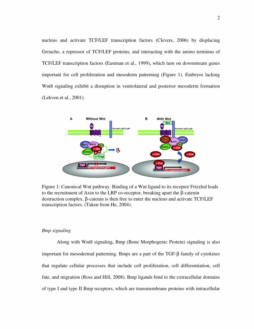
2
nucleus and activate TCF/LEF transcription factors (Clevers, 2006) by displacing
Groucho, a repressor of TCF/LEF proteins, and interacting with the amino terminus of
TCF/LEF transcription factors (Eastman et al., 1999), which turn on downstream genes
important for cell proliferation and mesoderm patterning (Figure 1). Embryos lacking
Wnt8 signaling exhibit a disruption in ventrolateral and posterior mesoderm formation
(Lekven et al., 2001).
Figure 1: Canonical Wnt pathway. Binding of a Wnt ligand to its receptor Frizzled leads to the recruitment of Axin to the LRP co-receptor, breaking apart the β-catenin destruction complex. β-catenin is then free to enter the nucleus and activate TCF/LEF transcription factors. (Taken from He, 2004).
Bmp signaling
Along with Wnt8 signaling, Bmp (Bone Morphogenic Protein) signaling is also
important for mesodermal patterning. Bmps are a part of the TGF-β family of cytokines
that regulate cellular processes that include cell proliferation, cell differentiation, cell
fate, and migration (Ross and Hill, 2008). Bmp ligands bind to the extracellular domains
of type I and type II Bmp receptors, which are transmembrane proteins with intracellular
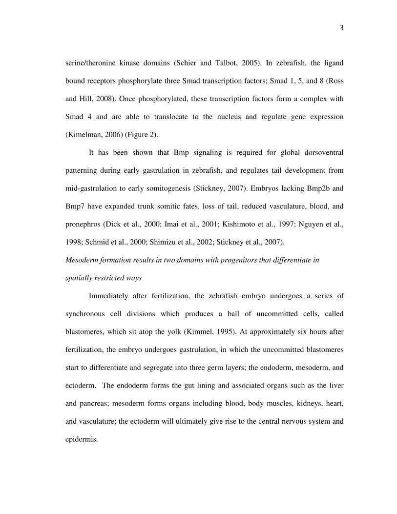
3
serine/theronine kinase domains (Schier and Talbot, 2005). In zebrafish, the ligand
bound receptors phosphorylate three Smad transcription factors; Smad 1, 5, and 8 (Ross
and Hill, 2008). Once phosphorylated, these transcription factors form a complex with
Smad 4 and are able to translocate to the nucleus and regulate gene expression
(Kimelman, 2006) (Figure 2).
It has been shown that Bmp signaling is required for global dorsoventral
patterning during early gastrulation in zebrafish, and regulates tail development from
mid-gastrulation to early somitogenesis (Stickney, 2007). Embryos lacking Bmp2b and
Bmp7 have expanded trunk somitic fates, loss of tail, reduced vasculature, blood, and
pronephros (Dick et al., 2000; Imai et al., 2001; Kishimoto et al., 1997; Nguyen et al.,
1998; Schmid et al., 2000; Shimizu et al., 2002; Stickney et al., 2007).
Mesoderm formation results in two domains with progenitors that differentiate in
spatially restricted ways
Immediately after fertilization, the zebrafish embryo undergoes a series of
synchronous cell divisions which produces a ball of uncommitted cells, called
blastomeres, which sit atop the yolk (Kimmel, 1995). At approximately six hours after
fertilization, the embryo undergoes gastrulation, in which the uncommitted blastomeres
start to differentiate and segregate into three germ layers; the endoderm, mesoderm, and
ectoderm. The endoderm forms the gut lining and associated organs such as the liver
and pancreas; mesoderm forms organs including blood, body muscles, kidneys, heart,
and vasculature; the ectoderm will ultimately give rise to the central nervous system and
epidermis.
4
In the zebrafish, the precursors for mesoderm and endoderm comprise a mixed
population of cells at the embryonic margin at the onset of gastrulation. Because of this
cellular arrangement, it is often referred to as “mesendoderm” prior to segregation of the
germ layers (Dougan, 2003).
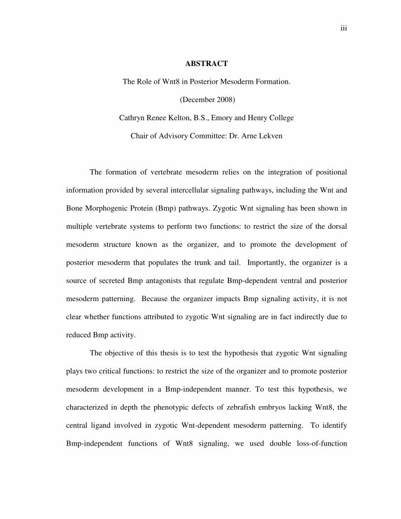
Figure 2: Bmp signaling. A Bmp ligand binds to Type I and Type II Bmp receptors which phosphorylate Smads 1, 5, and 8. The phosphorylated Smad proteins form a complex with Smad4, enter the nucleus and regulate target genes (adapted from Kimelman, 2006©).
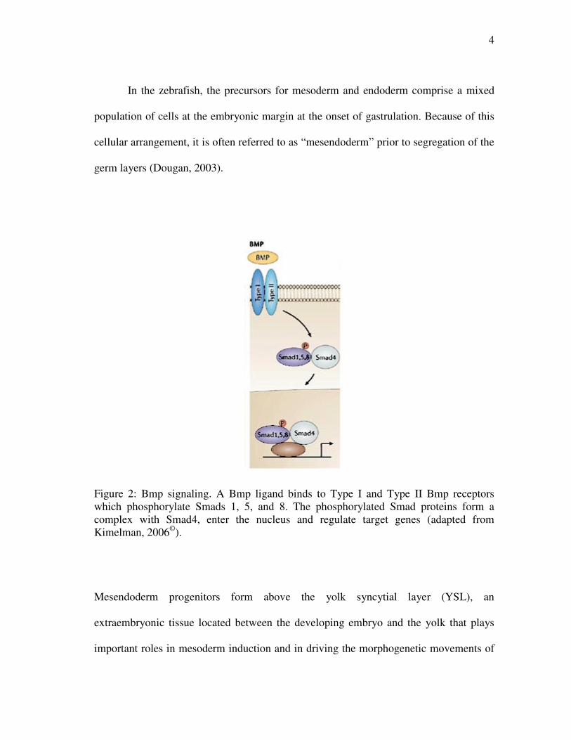
Mesendoderm progenitors form above the yolk syncytial layer (YSL), an
extraembryonic tissue located between the developing embryo and the yolk that plays
important roles in mesoderm induction and in driving the morphogenetic movements of
5
epiboly during gastrulation (Figure 3) (Kimmel and Law, 1985; Solnica-Krezel and
Driever 1994; Kimelman et al., 2000; Kimelman and Griffin, 2000 Kimmel; Chen et al.,
2006).
Figure 3: Fate map of mesoderm and endoderm precursors. Mesoderm precursors (red) are distinct from endoderm precursors (green) in Xenopus embryos, while in zebrafish both groups of precursors are mixed along the margin. (Adapted from Kimelman, 2006©).
Mesoderm induction in zebrafish occurs in response to signals secreted by the
YSL (Kimelman, 2006). Upon induction, the mesoderm is divided into two gross
domains referred to as dorsal and ventrolateral domains (Figure 4A). The dorsal domain
will differentiate into axial structures such as the notochord. The ventrolateral domain
comprises progenitors for mesoderm structures that form away from the dorsal axis, such
as blood, kidney and body muscles (Kimmel et al., 1990). The dorsal mesoderm domain
contributes to a visible thickening of the zebrafish embryonic margin, called the
embryonic shield, at the onset of gastrulation. The shield corresponds to the zebrafish
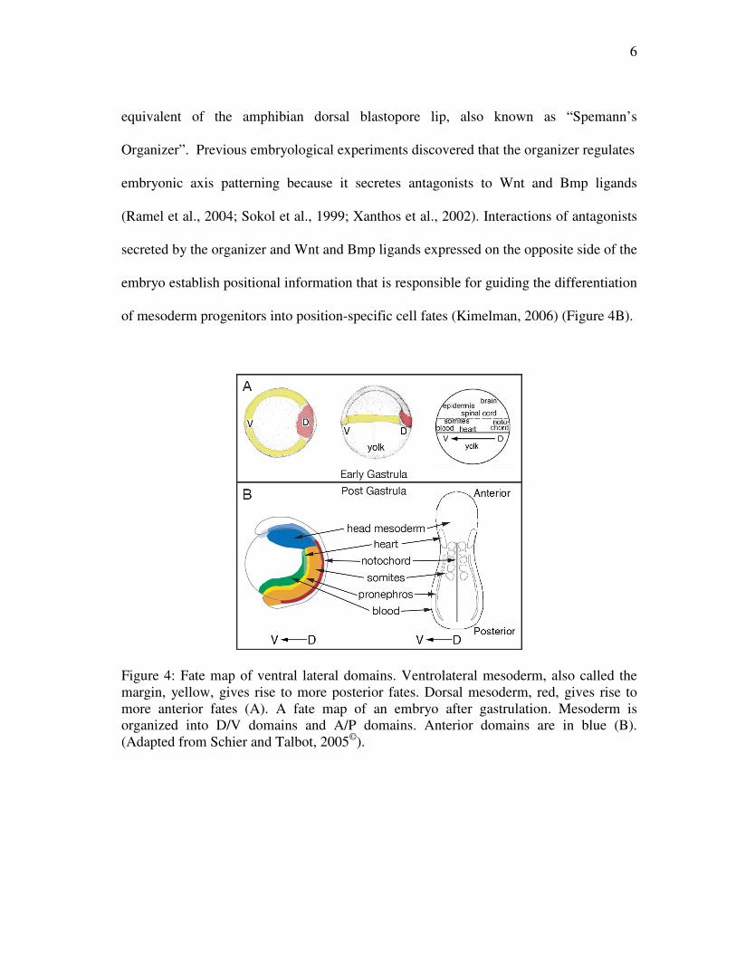
6
equivalent of the amphibian dorsal blastopore lip, also known as “Spemann’s
Organizer”. Previous embryological experiments discovered that the organizer regulates
embryonic axis patterning because it secretes antagonists to Wnt and Bmp ligands
(Ramel et al., 2004; Sokol et al., 1999; Xanthos et al., 2002). Interactions of antagonists
secreted by the organizer and Wnt and Bmp ligands expressed on the opposite side of the
embryo establish positional information that is responsible for guiding the differentiation
of mesoderm progenitors into position-specific cell fates (Kimelman, 2006) (Figure 4B).
Figure 4: Fate map of ventral lateral domains. Ventrolateral mesoderm, also called the margin, yellow, gives rise to more posterior fates. Dorsal mesoderm, red, gives rise to more anterior fates (A). A fate map of an embryo after gastrulation. Mesoderm is organized into D/V domains and A/P domains. Anterior domains are in blue (B). (Adapted from Schier and Talbot, 2005©).
7
Embryonic axis patterning is regulated by zygotic Wnt and Bmp signaling
Early studies on the organizer showed that cell fate specification in the
anteroposterior axis is intertwined with patterning of the dorsoventral axis. For example,
in zebrafish, the absence of Wnt8 signaling results in an expanded organizer
(dorsoventral patterning defect) and a shortened body axis (anteroposterior patterning
defect) (Hoppler et al., 1996; Lekven et al, 2001). Likewise, zebrafish embryos lacking
Bmp2b and Bmp7 have expanded trunk somites, reduced vasculature, blood, and
pronephros (dorsoventral patterning defects) and loss of tail (anteroposterior patterning
defect) (Dick et al., 2000; Imai et al., 2001; Kishimoto et al., 1997; Nguyen et al., 1998;
Schmid et al., 2000; Shimizu et al., 2002; Stickney et al., 2007). Thus, Wnt and Bmp
signaling are essential to both dorsoventral and anteroposterior axis patterning.
Wnt signaling regulates dorsoventral patterning through the organizer
The organizer secretes both Wnt and Bmp antagonists. These antagonists create a
gradient of Wnt and Bmp signaling that is higher in the ventral domain of the embryo
and lower in dorsal domain. Wnt antagonists secreted by the organizer can be classified
into two families based on how they perform. The first group consists of the secreted
Frizzled-related proteins (sFRPs) which include frzb-1, sFRP-2 and crescent. These
proteins inhibit Wnt signaling by binding to the Wnt ligands and preventing
ligand/receptor interactions. The second group includes the Dkks, which interact with
the LRP5/6 coreceptor to prevent Wnt binding and activation (reviewed in De Robertis
et al., 2000). Overexpression of Wnt antagonists produces embryos that have a
dorsalized phenotype such as enlarged eyes, enlarged head, expansion of the organizer,
8
and a shortened tail (Glinka et al., 1998; Hoppler et al., 1996; Leyns et al., 1997).
Chordin, Noggin, and Follistatin are all Bmp antagonists which, like the Wnt
antagonists, produce a dorsalized phenotype when overexpressed. Thus, Wnt signaling
restricts organizer size and thereby limits the expression of Wnt and Bmp antagonists
from the organizer. As a consequence, this function may impact both dorsoventral and
anteroposterior embryo axis patterning. While these observations show that
anteroposterior and dorsoventral patterning are coordinately regulated, whether
anteroposterior and dorsoventral fate specification are an output of separable molecular
mechanisms has not been determined.
Wnt8 is expressed in ventrolateral mesoderm
Wnt8 is expressed in the ventrolateral embryonic margin of zebrafish (Kelly et
al., 1995). Wnt8 signaling is required to maintain high levels of vent, vox and ved
expression in the ventrolateral margin during gastrulation (Ramel and Lekven, 2004).
Vent, Vox, and Ved act as repressors to prevent the expression of dorsal genes in the
ventral region of the embryo (Melby et al., 2000; Imai et al., 2001; Shimizu et al., 2002;
Ramel et al., 2004; and Ramel et al., 2005). Thus, Wnt8 regulates dorsoventral
patterning through Vent, Vox and Ved-dependent organizer regulation. This leaves open
the question of whether Wnt8 signaling has a direct role in specifying anteroposterior
mesoderm fates.
There is evidence that in addition to regulating dorsoventral fates, Wnt8 plays a
direct role in patterning anteroposterior fates. It has been found that sp5l, a gene that
functions in tail development, is downstream of wnt8 (Thorpe et al., 2005).
9
Interestingly, expression of the posterior genes cdxla and cdx4 was reduced in wnt8
mutants, but not Bmp mutants (Shimizu et al., 2005). This evidence suggests that Wnt8
has a separate anterior-posterior patterning function that is independent of Bmp-
dependent dorsoventral patterning.
Summary and focus of research
How dorsoventral and anteroposterior patterning is controlled by Wnt8 signaling
in the zebrafish embryos is poorly understood. It is known that Wnt8 and Bmp play a
role in patterning dorsoventral mesoderm. Mutants deficient in wnt8 and bmp lack
various structures that arise from dorsoventral mesoderm. Interestingly, there is evidence
that these genes have different functions when it comes to anteroposterior patterning.
Zebrafish embryos that lack wnt8 signaling show a dramatic loss of posterior
mesodermal structures. To gain a better understanding of Wnt8 function, various
mesodermal markers were analyzed by in situ hybridization in wild type and Wnt8 loss-
of-function embryos produced by morpholino antisense oligonucleotide (MO) gene
knockdown.
The research presented here was designed to test the hypothesis that Wnt8
signaling regulates Bmp-dependent and Bmp-independent patterning. Embryos lacking
Wnt8 have expanded organizers that secrete elevated levels of Bmp antagonists that
reduce Bmp signaling. Thus, wnt8 mutants have reduced Wnt8 signaling as well as
reduced Bmp signaling. As a consequence, to reveal Wnt8-specific fate specification, it
is necessary to restore Bmp signaling activity within wnt8 loss-of-function embryos.
10
Mesoderm fates that fail to be specified under these conditions must require Wnt8
signaling but not Bmp.
We have taken a double loss-of-function approach to restore Bmp signaling
activity in embryos lacking Wnt8. The Bmp antagonist Chordin (chd) is produced from
the organizer. We used morpholino antisense oligonucleotides to simultaneously reduce
both Wnt8 and Chordin expression, and then we analyzed the effect on multiple
mesoderm markers. Our results show that, in addition to posterior mesoderm precursors
being drastically reduced in Wnt8 morphants, anterior fates are disrupted as well. We
found that increasing Bmp signaling largely has no effect on the Wnt8 morphant
phenotype. However, slight rescue was observed in pronephric, heart tube, and
vasculature precursors. These results support the hypothesis that Wnt signaling
maintains mesoderm progenitor cell populations, while Bmp signaling patterns
mesoderm cell fates. Accordingly, Wnt8 signaling will appear to be epistatic to Bmp
signaling during vertebrate axis patterning.
11
CHAPTER II
MATERIALS AND METHODS
Fish maintenance and strains
Fish were maintained as described in (Westerfield, 2000). Fish used in this study
were AB x TL. To generate AB x TL fish, AB wild-type fish were crossed with TL
wild-type fish. Progeny were raised and crossed to produce embryos used in this study.
Injections and morpholinos
A combination of four morpholinos (MOs; Genetools, LLC) was used to block
splicing of wnt8 pre-mRNA. The sequence of each morpholino has been previously
described (Ramel et al., 2005). The chordin (chd) MO has been previously described
(Nasevicius and Ekker, 2000). MOs were diluted in 1X Danieau’s buffer and injected
into one to four cell stage wild-type embryos. To generate wnt8MO;chdMO embryos, each
MO was individually injected into the same wild-type embryo. In all injections, the
volume of MO injected per embryo was approximately 3 nL.
In situ hybridizations and probes
In situ hybridizations were essentially preformed as described in (Jowett, 2001).
The probes used were: cardiac myosin light chain-2 (cmlc2; Huang et al., 2003), even-
skipped-1 (eve1; Joly et al., 1993), fli1 (Brown et al., 2000), follistatin a (Bauer et al.,
1998), gata1 (Dietrich et al., 1995), hgg1 (Vogel and Gerster, 1997), myf5 (Rescan,
2001), myoD (Weinberg et al., 1996), neurogenin-1 (Blader et al., 1997), pax2.1
(Abdelilah et al., 1996), and T-box24 (tbx24; Nikaido et al., 2002).
12
CHAPTER III
CLASSIFYING THE WNT8 PHENOTYPE
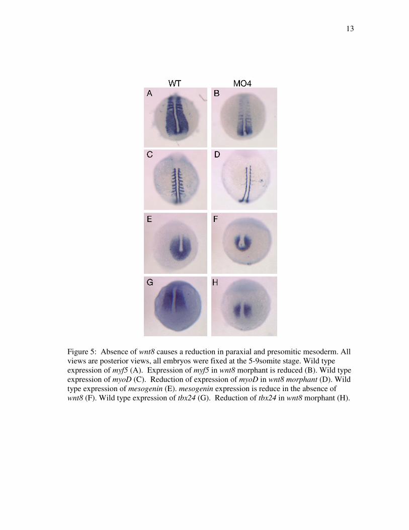
Wnt8 is required to promote the development of posterior mesoderm fates
To better understand the role Wnt8 plays in patterning, wild-type embryos were
injected with wnt8 morpholinos, fixed at the 5 to 9 somite stage (approximately 12 to 13
hours after fertilization) and analyzed by in situ hybridization to detect several
mesodermal markers. The morpholinos used are a cocktail of four morpholinos designed
to block splicing of wnt8 pre-mRNAs (Ramel et al., 2005; embryos injected with this
morpholino cocktail are referred to as wnt8MO4 embryos). Because the wnt8 phenotype
is characterized by a severe lack of posterior mesoderm, we began by examining the
expression of four paraxial and presomitic mesoderm markers: myf5, myoD, mesogenin,
and tbx24. In all cases, the expression domains of these markers were decreased in
length and width in the majority of the morphants (Figure 5 A-H). This result suggests
that Wnt8 is required for the maintenance or specification of presomitic mesoderm
progenitors.
The reduction in somitic progenitors observed would be predicted to result in
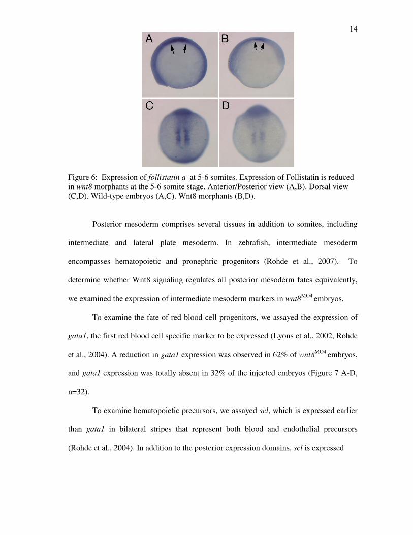
smaller somites. At the 5 to 6 somite stage, follistatin a (fsta) expression marks anterior
somites. Consistent with the above results, the expression domain of fsta was reduced in
50% of wnt8MO4 embryos, (Figure 6 A-D, n=24), and was barely visible or undetectable
in 41% of the embryos. The results of these in situs indicate that Wnt8 is required for
normal presomitic and somitic mesoderm development.
13
Figure 5: Absence of wnt8 causes a reduction in paraxial and presomitic mesoderm. All views are posterior views, all embryos were fixed at the 5-9somite stage. Wild type expression of myf5 (A). Expression of myf5 in wnt8 morphant is reduced (B). Wild type expression of myoD (C). Reduction of expression of myoD in wnt8 morphant (D). Wild type expression of mesogenin (E). mesogenin expression is reduce in the absence of wnt8 (F). Wild type expression of tbx24 (G). Reduction of tbx24 in wnt8 morphant (H).
14
Figure 6: Expression of follistatin a at 5-6 somites. Expression of Follistatin is reduced in wnt8 morphants at the 5-6 somite stage. Anterior/Posterior view (A,B). Dorsal view (C,D). Wild-type embryos (A,C). Wnt8 morphants (B,D).
Posterior mesoderm comprises several tissues in addition to somites, including
intermediate and lateral plate mesoderm. In zebrafish, intermediate mesoderm
encompasses hematopoietic and pronephric progenitors (Rohde et al., 2007). To
determine whether Wnt8 signaling regulates all posterior mesoderm fates equivalently,
we examined the expression of intermediate mesoderm markers in wnt8MO4 embryos.
To examine the fate of red blood cell progenitors, we assayed the expression of
gata1, the first red blood cell specific marker to be expressed (Lyons et al., 2002, Rohde
et al., 2004). A reduction in gata1 expression was observed in 62% of wnt8MO4 embryos,
and gata1 expression was totally absent in 32% of the injected embryos (Figure 7 A-D,
n=32).
To examine hematopoietic precursors, we assayed scl, which is expressed earlier
than gata1 in bilateral stripes that represent both blood and endothelial precursors
(Rohde et al., 2004). In addition to the posterior expression domains, scl is expressed
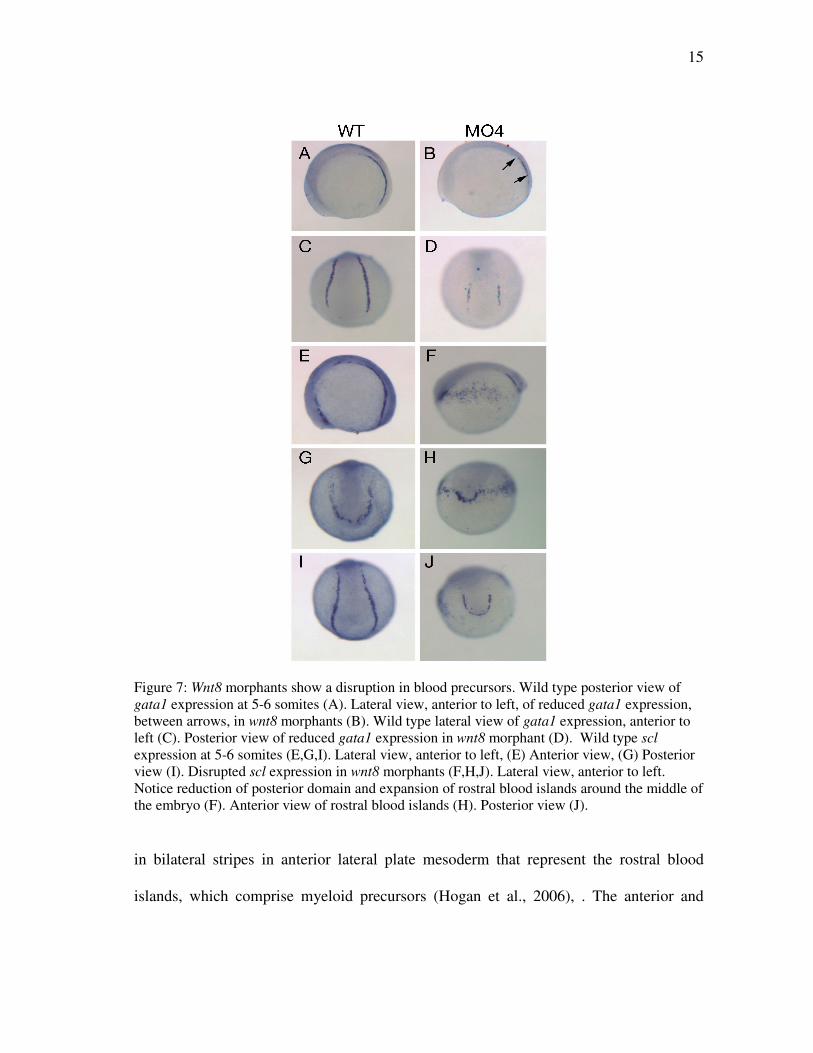
15
Figure 7: Wnt8 morphants show a disruption in blood precursors. Wild type posterior view of gata1 expression at 5-6 somites (A). Lateral view, anterior to left, of reduced gata1 expression, between arrows, in wnt8 morphants (B). Wild type lateral view of gata1 expression, anterior to left (C). Posterior view of reduced gata1 expression in wnt8 morphant (D). Wild type scl expression at 5-6 somites (E,G,I). Lateral view, anterior to left, (E) Anterior view, (G) Posterior view (I). Disrupted scl expression in wnt8 morphants (F,H,J). Lateral view, anterior to left. Notice reduction of posterior domain and expansion of rostral blood islands around the middle of the embryo (F). Anterior view of rostral blood islands (H). Posterior view (J).
in bilateral stripes in anterior lateral plate mesoderm that represent the rostral blood
islands, which comprise myeloid precursors (Hogan et al., 2006), . The anterior and
16
posterior expression domains of scl are separated by a gap and do not touch. Consistent
with the reduction of gata1 expression, a reduction in the posterior expression domain of
scl was observed in wnt8MO4 embryos. In contrast, the expression marking the rostral
blood islands, normally restricted to the anterior end of the embryo, expanded almost to
the posterior pole of wnt8MO4 embryos. This phenotype was observed in 67% of the
morphants (Figure 7 E-J, n=28). Thus, wnt8MO4 embryos display reduced paraxial and
intermediate mesoderm progenitors, and this is accompanied by a significant expansion
of anterior lateral plate mesoderm progenitors.
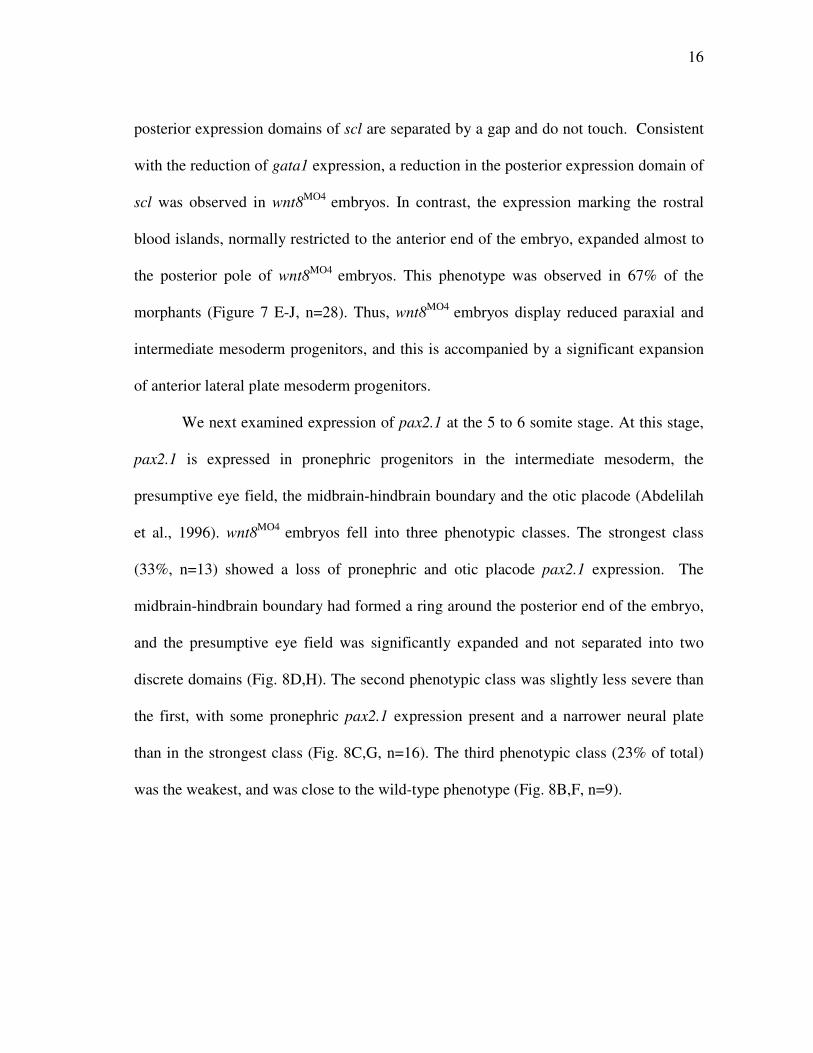
We next examined expression of pax2.1 at the 5 to 6 somite stage. At this stage,
pax2.1 is expressed in pronephric progenitors in the intermediate mesoderm, the
presumptive eye field, the midbrain-hindbrain boundary and the otic placode (Abdelilah
et al., 1996). wnt8MO4 embryos fell into three phenotypic classes. The strongest class
(33%, n=13) showed a loss of pronephric and otic placode pax2.1 expression. The
midbrain-hindbrain boundary had formed a ring around the posterior end of the embryo,
and the presumptive eye field was significantly expanded and not separated into two
discrete domains (Fig. 8D,H). The second phenotypic class was slightly less severe than
the first, with some pronephric pax2.1 expression present and a narrower neural plate
than in the strongest class (Fig. 8C,G, n=16). The third phenotypic class (23% of total)
was the weakest, and was close to the wild-type phenotype (Fig. 8B,F, n=9).
17
Figure 8: Pax2.1 expression in Wnt8 morphants. Lateral view of wild type embryo (A). Lateral view of Wnt8 morphants (B,C,D). Dorsal view of wild type embryo (E). Dorsal view of Wnt8 morphants (F,G,H). Wnt8 is required for cardiac progenitor specification
The previous results showed that Wnt8 promotes posterior mesoderm
development and antagonizes anterior lateral plate mesoderm and neurectoderm
specification. The cardiogenic mesoderm is situated between anterior and posterior
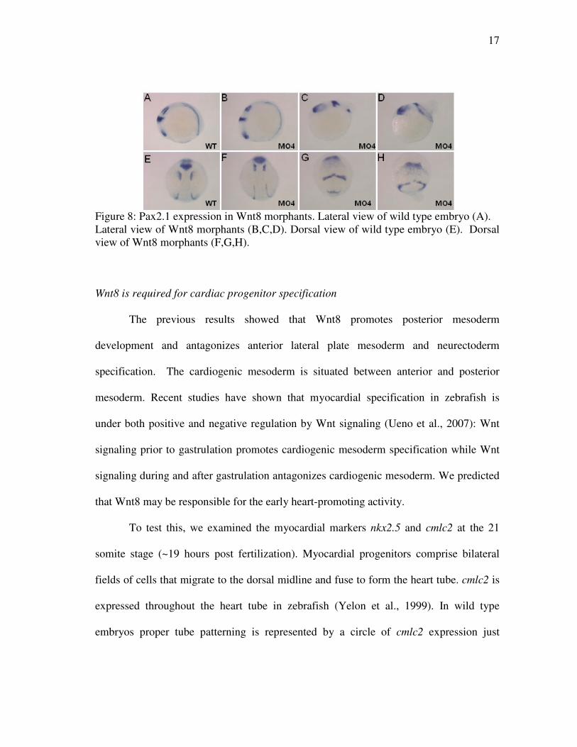
mesoderm. Recent studies have shown that myocardial specification in zebrafish is
under both positive and negative regulation by Wnt signaling (Ueno et al., 2007): Wnt
signaling prior to gastrulation promotes cardiogenic mesoderm specification while Wnt
signaling during and after gastrulation antagonizes cardiogenic mesoderm. We predicted
that Wnt8 may be responsible for the early heart-promoting activity.
To test this, we examined the myocardial markers nkx2.5 and cmlc2 at the 21
somite stage (~19 hours post fertilization). Myocardial progenitors comprise bilateral
fields of cells that migrate to the dorsal midline and fuse to form the heart tube. cmlc2 is
expressed throughout the heart tube in zebrafish (Yelon et al., 1999). In wild type
embryos proper tube patterning is represented by a circle of cmlc2 expression just
18
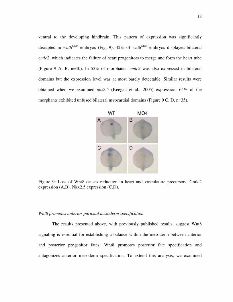
ventral to the developing hindbrain. This pattern of expression was significantly
disrupted in wnt8MO4 embryos (Fig. 9). 42% of wnt8MO4 embryos displayed bilateral
cmlc2, which indicates the failure of heart progenitors to merge and form the heart tube
(Figure 9 A, B, n=40). In 53% of morphants, cmlc2 was also expressed in bilateral
domains but the expression level was at most barely detectable. Similar results were
obtained when we examined nkx2.5 (Keegan et al., 2005) expression: 64% of the
morphants exhibited unfused bilateral myocardial domains (Figure 9 C, D, n=35).
Figure 9: Loss of Wnt8 causes reduction in heart and vasculature precursors. Cmlc2 expression (A,B). Nkx2.5 expression (C,D).
Wnt8 promotes anterior paraxial mesoderm specification
The results presented above, with previously published results, suggest Wnt8
signaling is essential for establishing a balance within the mesoderm between anterior
and posterior progenitor fates: Wnt8 promotes posterior fate specification and
antagonizes anterior mesoderm specification. To extend this analysis, we examined
19
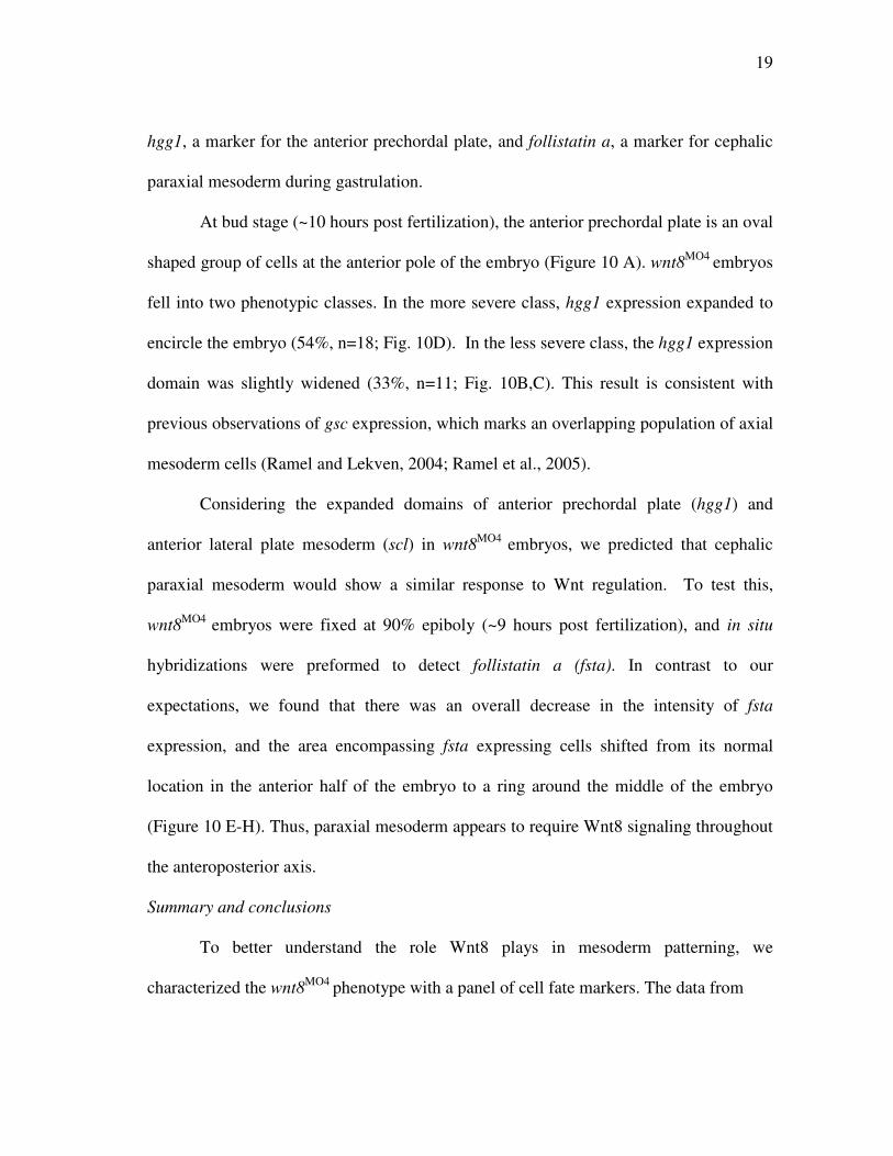
hgg1, a marker for the anterior prechordal plate, and follistatin a, a marker for cephalic
paraxial mesoderm during gastrulation.
At bud stage (~10 hours post fertilization), the anterior prechordal plate is an oval
shaped group of cells at the anterior pole of the embryo (Figure 10 A). wnt8MO4 embryos
fell into two phenotypic classes. In the more severe class, hgg1 expression expanded to
encircle the embryo (54%, n=18; Fig. 10D). In the less severe class, the hgg1 expression
domain was slightly widened (33%, n=11; Fig. 10B,C). This result is consistent with
previous observations of gsc expression, which marks an overlapping population of axial
mesoderm cells (Ramel and Lekven, 2004; Ramel et al., 2005).
Considering the expanded domains of anterior prechordal plate (hgg1) and
anterior lateral plate mesoderm (scl) in wnt8MO4 embryos, we predicted that cephalic
paraxial mesoderm would show a similar response to Wnt regulation. To test this,
wnt8MO4 embryos were fixed at 90% epiboly (~9 hours post fertilization), and in situ
hybridizations were preformed to detect follistatin a (fsta). In contrast to our
expectations, we found that there was an overall decrease in the intensity of fsta
expression, and the area encompassing fsta expressing cells shifted from its normal
location in the anterior half of the embryo to a ring around the middle of the embryo
(Figure 10 E-H). Thus, paraxial mesoderm appears to require Wnt8 signaling throughout
the anteroposterior axis.
Summary and conclusions
To better understand the role Wnt8 plays in mesoderm patterning, we
characterized the wnt8MO4 phenotype with a panel of cell fate markers. The data from
20
Figure 10: Hgg1 and follistatin expression in Wnt8 morphants. Hgg1 (A,B,C,D), Follistatin (E,F), lateral view (G,H). . these in situ hybridization experiments suggest that Wnt8 signaling is required for
specification of paraxial mesoderm progenitors throughout the anteroposterior axis.
Further, intermediate mesoderm fates in the posterior embryo are positively regulated by
Wnt8 signaling. Of anterior mesoderm fates, axial mesoderm and lateral plate
mesoderm progenitors are antagonized by Wnt8 signaling, thus these populations expand
in the absence of Wnt8 signaling.
21
These results lead to several unanswered questions. What is happening to the
mesoderm progenitors that would normally be fated to contribute to the posterior
embryo? There is clearly a reduction, but is it because fewer posterior mesoderm
progenitors are specified, because the cells die early, or because they undergo fewer
rounds of cell division? Because the dorsoventral axis is established before the
anteroposterior axis, are the patterning defects we see in wnt8MO4 embryos due to an
earlier defect in dorsoventral patterning? Clearly any of these possibilities would result
in fewer cells contributing to the posterior mesoderm. In the next chapter, I describe
experiments that test whether reduced Bmp signaling lies behind the reduction in
posterior mesoderm specification in wnt8MO4 embryos.
22
CHAPTER IV
IDENTIFYING BMP-INDEPENDENT FUNCTIONS OF WNT8
Introduction
It can be suggested that the reason there is an anterior/posterior patterning defect
in Wnt8 morphants is because there is an early dorsoventral patterning defect. That is,
embryos lacking Wnt8 have expanded organizers that secrete elevated levels of Bmp
antagonists, such as Chordin, that reduce Bmp signaling levels. Thus, the full extent of
the effects of wnt8 loss of function may be masked by Bmp-dependent effects. To
ascertain if reduced Bmp signaling contributes to the wnt8 loss-of-function phenotype,
we simultaneously knocked down Wnt8 and the Bmp antagonist Chordin with
morpholino antisense oligos. Reducing Chordin protein levels should promote elevated
Bmp activity in the context of Wnt8 loss of function. If the phenotype of the double
morphants looks like a wild-type embryo, then it would suggest that increased Bmp
signaling can compensate for the loss of Wnt8, and a defect in early Bmp signaling
might explain at least part of the Wnt8 loss-of-function phenotype.
Chd knockdown partially suppresses the wntMO4 phenotype
To begin, we injected wild type embryos with morpholinos targeting wnt8 and
chordin, and then examined morphological phenotypes at 24 hours post fertilization.
wnt8MO4 embryos fall into phenotypic classes that can be categorized according to a
classification scheme devised for Bmp mutants (Kishimoto et al., 1997). According to
this scheme, C4 and C5 represent the most severe phenotypes and are characterized by
23
an absent yolk extension, severe axis truncation and severe head disorganization. C3
embryos also fail to make a yolk extension but show only a mildly disorganized head
and only moderate axis truncation. C2 embryos form a partial yolk extension, head
development is relatively normal, and the tail is only slightly shortened but lacks the
ventral tail fin. C1 embryos lack the ventral tail fin but are otherwise wild-type. wnt8MO4
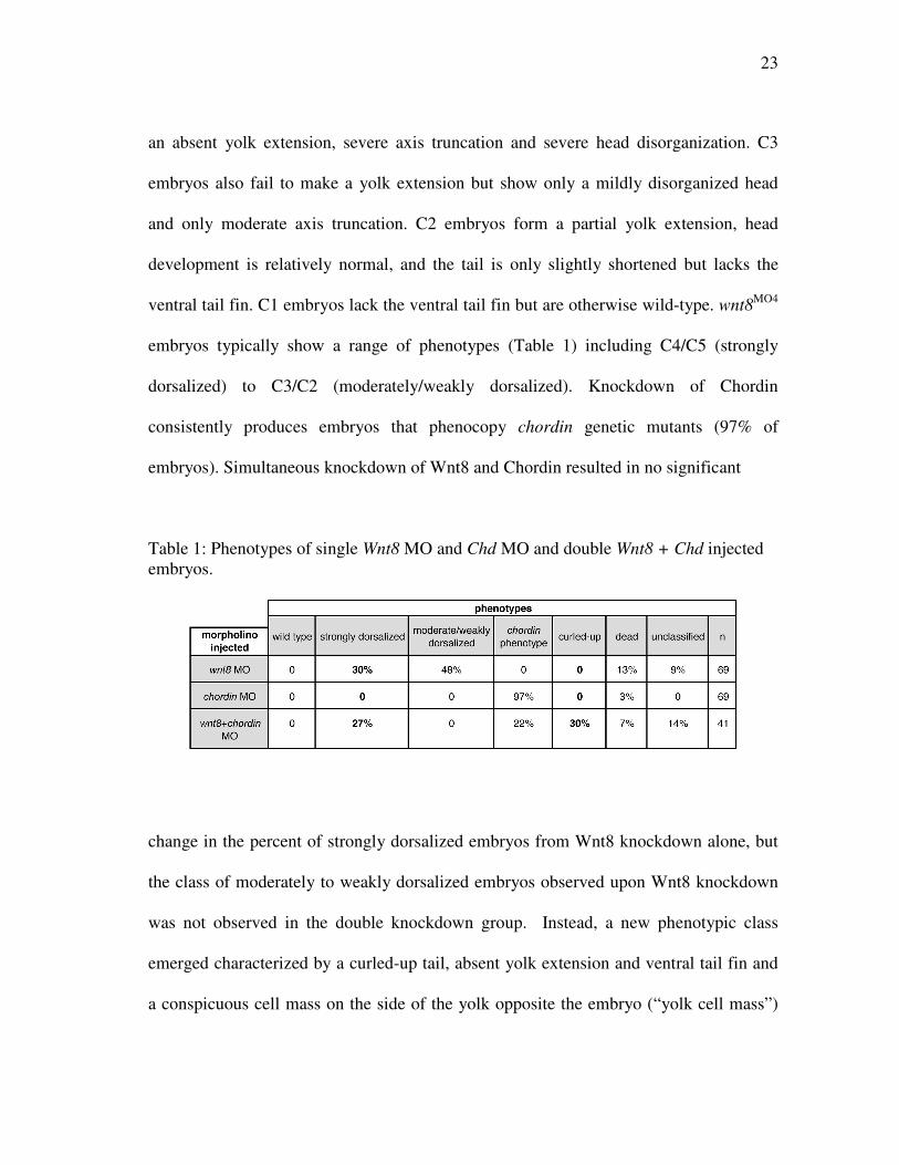
embryos typically show a range of phenotypes (Table 1) including C4/C5 (strongly
dorsalized) to C3/C2 (moderately/weakly dorsalized). Knockdown of Chordin
consistently produces embryos that phenocopy chordin genetic mutants (97% of
embryos). Simultaneous knockdown of Wnt8 and Chordin resulted in no significant
Table 1: Phenotypes of single Wnt8 MO and Chd MO and double Wnt8 + Chd injected embryos.
change in the percent of strongly dorsalized embryos from Wnt8 knockdown alone, but
the class of moderately to weakly dorsalized embryos observed upon Wnt8 knockdown
was not observed in the double knockdown group. Instead, a new phenotypic class
emerged characterized by a curled-up tail, absent yolk extension and ventral tail fin and
a conspicuous cell mass on the side of the yolk opposite the embryo (“yolk cell mass”)
24
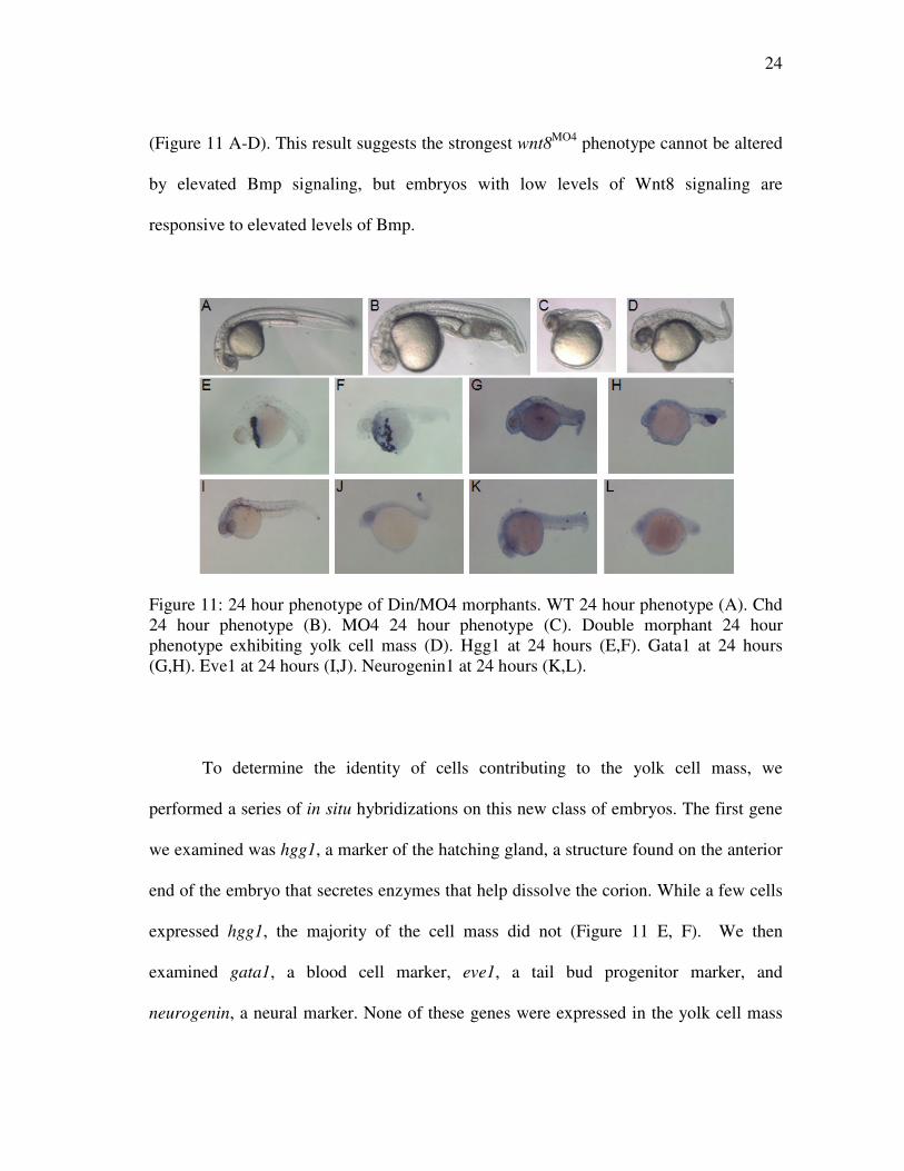
(Figure 11 A-D). This result suggests the strongest wnt8MO4 phenotype cannot be altered
by elevated Bmp signaling, but embryos with low levels of Wnt8 signaling are
responsive to elevated levels of Bmp.
Figure 11: 24 hour phenotype of Din/MO4 morphants. WT 24 hour phenotype (A). Chd 24 hour phenotype (B). MO4 24 hour phenotype (C). Double morphant 24 hour phenotype exhibiting yolk cell mass (D). Hgg1 at 24 hours (E,F). Gata1 at 24 hours (G,H). Eve1 at 24 hours (I,J). Neurogenin1 at 24 hours (K,L).
To determine the identity of cells contributing to the yolk cell mass, we
performed a series of in situ hybridizations on this new class of embryos. The first gene
we examined was hgg1, a marker of the hatching gland, a structure found on the anterior
end of the embryo that secretes enzymes that help dissolve the corion. While a few cells
expressed hgg1, the majority of the cell mass did not (Figure 11 E, F). We then
examined gata1, a blood cell marker, eve1, a tail bud progenitor marker, and
neurogenin, a neural marker. None of these genes were expressed in the yolk cell mass
25
(Figure 11 G-L). While the identity of these cells remains undetermined, they may be
similar to a smaller population of apoptotic cells found ventral to the yolk extension of
chordin mutant embryos (Hammerschmidt et al., 1996).
Paraxial mesoderm is compromised in wnt8MO4;chdMO embryos
We fixed wnt8MO4;chdMO embryos between the 5 and 9 somites stages and
performed in situ hybridizations using the posterior paraxial mesoderm markers, myf5,
myoD, and tbx24 to ascertain the effects of increased Bmp in a wnt8 loss of function
background. All three genes are markers of presomitic mesoderm but are not expressed
in tailbud progenitors. Thus, their expression at the 5-6 somite stage marks paraxial
mesoderm that will contribute to the trunk somites (Holley and Takeda, 2002). All three
genes show reduced expression in wnt8MO4 and chdMO embryos at the 5-6 somite stage.
In wnt8MO4 embryos, this may be explained by a role for Wnt8 in promoting paraxial
mesoderm formation in general (Yamaguchi, 2001). In chdMO embryos, reduced
expression of presomitic mesoderm markers may reflect the diversion of trunk paraxial
mesoderm progenitors toward a tail fate (Szeto and Kimelman, 2006), since cells that
express myf5, myoD and tbx24 at the 5-6 somite stage will contribute to trunk somites,
not tail somites.
myf5 expression was reduced in the wnt8 morphants, but still showed some
somite expression. It was also reduced in the chd morphants; however somite expression
had been lost. tbx24 expression was similarly reduced in wnt8MO4, chdMO and
wnt8MO4;chdMO embryos (Figure 12). myoD is expressed in somites and adaxial cells.
wnt8MO4 embryos showed decreased myoD expression, but still had somite expression,

26
while the chdMO embryos showed no somite expression and light expression in the
adaxial stripes (Figure 12). The double morphants showed an additive phenotype: no
somite expression as in chdMO embryos but short adaxial stripes as in wnt8MO4 (Figure
12E-H).
�
Figure 12: Posterior markers in Chd/MO4 morphants. All views are posterior. Myf5 expression (A,B,C,D). MyoD expression (E,F,G,H). Tbx24 expression (I,J,K,L). Intermediate mesoderm patterning is rescued in wnt8MO4;chdMO embryos
We next examined whether Chordin knockdown could rescue intermediate
mesoderm fate specification in wnt8 morphants. As shown in Chapter III, pax2.1-
expressing intermediate mesoderm is either absent or almost absent in wnt8MO4 embryos
(Figure 8 , Figure 13). Because pax2.1 is also expressed in the neural plate and otic
vesicles at the 5-6 somite stage, we could also use its expression to evaluate the extent of
neural induction in the knockdown conditions.
Chd MO4 Chd/MO4 WT

27
Embryos deficient in Wnt8 showed a loss of pronephric expression and otic
placode, the midbrain hindbrain boundary (MHB) thinned and expanded into a ring
encircling the embryo (Figure 13), and the forebrain expression was disorganized
(Figure 13 A,B,F,G). In chdMO embryos, pax2.1 expression domains are present but are
shifted in the anterior embryo toward the dorsal midline. This shift reflects suppressed
neural induction by elevated Bmp (in the neural plate) and the consequence of reduced
trunk paraxial mesoderm that separates intermediate mesoderm from the dorsal midline.
In contrast, the pronephric region of the embryos had widened around the posterior end
(Figure 13 C,H), reflecting increased numbers of tail mesoderm progenitors.
Figure 13: pax2.1 expression in Chd/MO4 morphants. Lateral view (A,B,C,D,E). Dorsal/Posterior view (F,G,H,I,J). Wild type (A,F). wnt8MO4 (B,G). chdMO (C,H). Double morphants (D,E,I,J). Arrows show very slight return of pronephric expression.
wnt8MO4;chdMO embryos could be classified into two phenotypic groups. The
weakly affected group resembled very closely the chdMO phenotype: a relatively normal
anterior end, slightly reduced otic placode expression, a slightly narrower midbrain-
hindbrain boundary (MHB), and diminished expression in the pronephric region.
Chd/MO4 Chd MO4 WT
28
Interestingly, wnt8MO4;chdMO embryos display a MHB width midway between wild-type
and the wnt8MO4 phenotype, but the distribution of pax2.1+ pronephric progenitors
resembled that of chdMO embryos, although expression was significantly reduced (n=27).
Thus, Chordin knockdown only slightly suppressed the expanded neural induction of
Wnt8 knockdown, but rescued the patterning of intermediate mesoderm progenitors
(Figure 13 D,E,I,J).
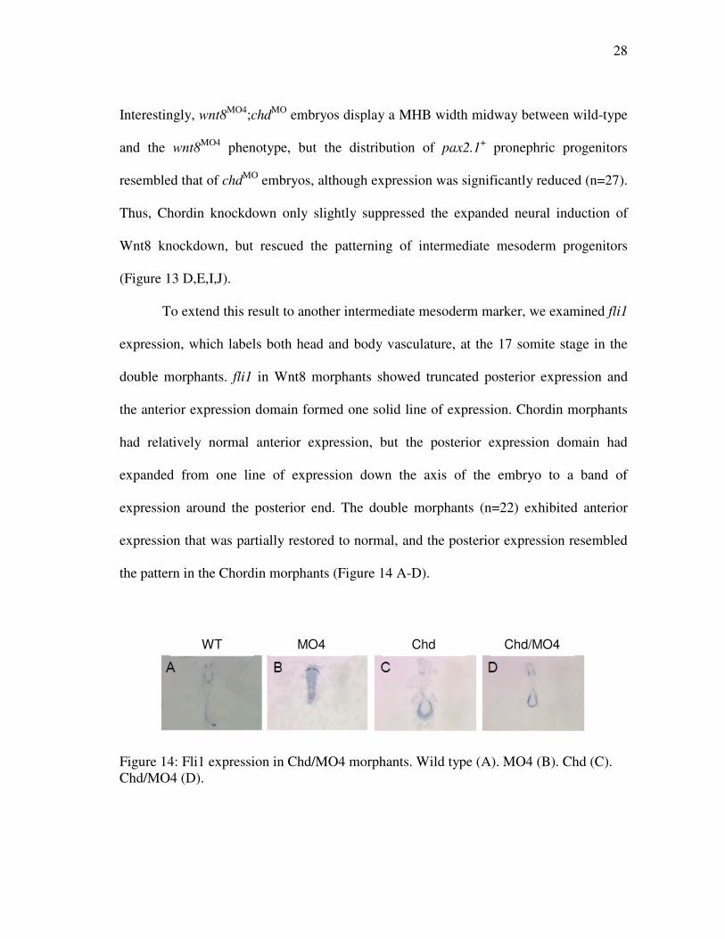
To extend this result to another intermediate mesoderm marker, we examined fli1
expression, which labels both head and body vasculature, at the 17 somite stage in the
double morphants. fli1 in Wnt8 morphants showed truncated posterior expression and
the anterior expression domain formed one solid line of expression. Chordin morphants
had relatively normal anterior expression, but the posterior expression domain had
expanded from one line of expression down the axis of the embryo to a band of
expression around the posterior end. The double morphants (n=22) exhibited anterior
expression that was partially restored to normal, and the posterior expression resembled
the pattern in the Chordin morphants (Figure 14 A-D).
Figure 14: Fli1 expression in Chd/MO4 morphants. Wild type (A). MO4 (B). Chd (C). Chd/MO4 (D).
Chd Chd/MO4 MO4 WT
29
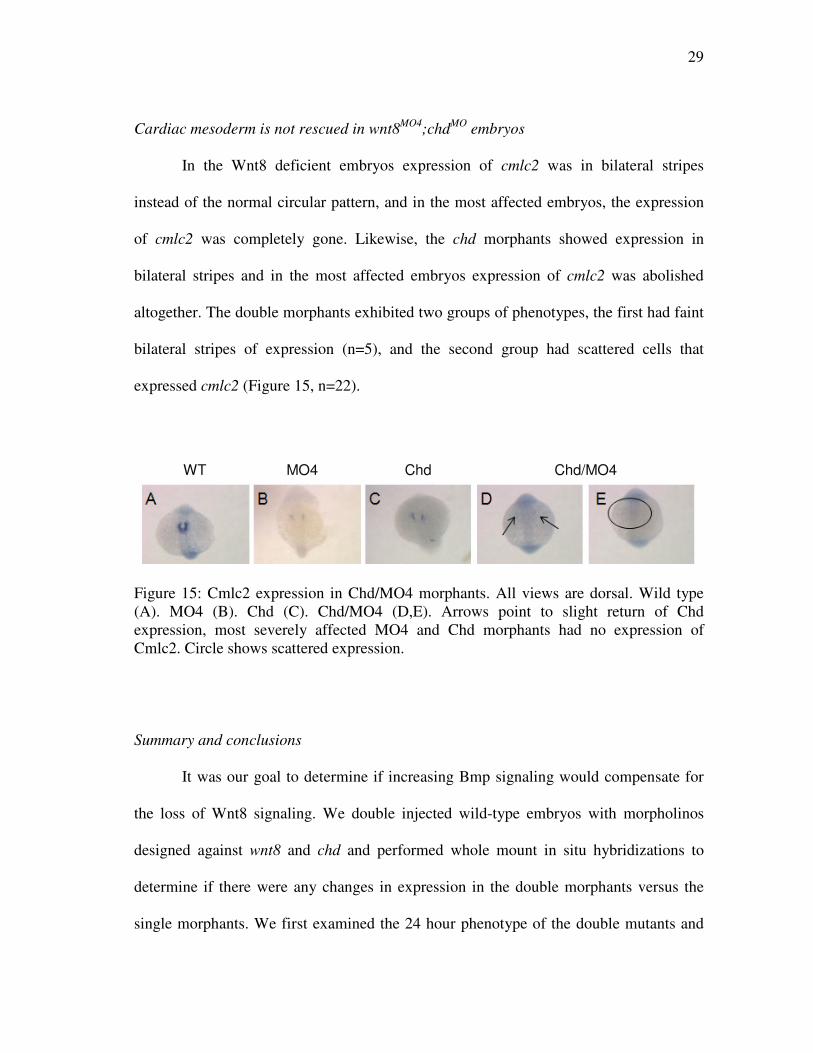
Cardiac mesoderm is not rescued in wnt8MO4;chdMO embryos
In the Wnt8 deficient embryos expression of cmlc2 was in bilateral stripes
instead of the normal circular pattern, and in the most affected embryos, the expression
of cmlc2 was completely gone. Likewise, the chd morphants showed expression in
bilateral stripes and in the most affected embryos expression of cmlc2 was abolished
altogether. The double morphants exhibited two groups of phenotypes, the first had faint
bilateral stripes of expression (n=5), and the second group had scattered cells that
expressed cmlc2 (Figure 15, n=22).
Figure 15: Cmlc2 expression in Chd/MO4 morphants. All views are dorsal. Wild type (A). MO4 (B). Chd (C). Chd/MO4 (D,E). Arrows point to slight return of Chd expression, most severely affected MO4 and Chd morphants had no expression of Cmlc2. Circle shows scattered expression.
Summary and conclusions
It was our goal to determine if increasing Bmp signaling would compensate for
the loss of Wnt8 signaling. We double injected wild-type embryos with morpholinos
designed against wnt8 and chd and performed whole mount in situ hybridizations to
determine if there were any changes in expression in the double morphants versus the
single morphants. We first examined the 24 hour phenotype of the double mutants and
Chd Chd/MO4 MO4 WT
30
noticed that a peculiar cell mass developed on the bottom of the yolk. We tried to
determine the nature of the cell mass by performing in situ hybridizations using the
probes hgg1, gata1, eve1, and neurogenin1. While a few cells in the cell mass turned out
to be part of the hatching gland, we did not find a marker that would label the entire or
majority of the cells in the cell mass. These findings lead us to believe that the yolk cell
mass probably consists of undifferentiated cells.
Next we tested the fates of anterior and posterior cell populations when we
increased Bmp signaling by knocking down its antagonist chordin and knocked down
Wnt8 signaling at the same time. We first examined the posterior markers myf5, myoD,
and tbx24 and saw in each case that increasing Bmp signaling did not restore the wnt8
phenotype. The case of myf5 and myoD the double morphants both have lost somite
expression like that of the Din morphants, and the adaxial expression is still truncated
like that of the Wnt8 morphants. We would have expected somite expression and longer
adaxial expression if increasing Bmp would have restored some or all of the wnt8
phenotype. When we examined tbx24 expression the double morphants’ expression
looked very similar to both the individual morphants’ expression. If Bmp signaling could
have restored tbx24 expression in a Wnt8 morphant, we would expect to see a larger area
of expression in the double morphants.
We then examined pax2.1, cmlc2, and fli1, which mark anterior cell populations,
and in the case of pax2.1, posterior cells as well. Interestingly, pax2.1 expression in the
pronephros in Wnt8 morphants is completely absent. In the double morphants, we saw
very light pronephric expression. The only anterior cell population that still expressed
31
Pax2.1 was the MHB which had thinned and lengthened horizontally. Because of the
light pronephric staining we believe that increasing Bmp signaling can slightly reverse
the loss of pronephric cells due to a reduction in wnt8 signaling. We then looked at
cmlc2 and fli1 markers for heart precursors and vasculature respectively.
In both instances, the double morphants showed signs that the wnt8 phenotype is
being reversed. In the single morphants, the heart tube is expressed as two bilateral lines
instead of a nice round structure, in the most affected embryos; heart tube expression is
completely absent. In the double morphants we saw two groups of embryos. The first
group exhibited slight expression of cmlc2 in bilateral stripes, indicating that the increase
in Bmp signaling can reverse the phenotype due to a loss of wnt8. Interestingly, in the
second group, cmlc2 expression was discovered to be in scattered cells with no clear
pattern. Whether this case represents embryos that did not get as much of the Din MO as
the other ones, resulting in lesser amount of Bmp signaling so that a small amount of
cells are being fated to become heart tube, is not clear and needs to be studied more.
The last marker we examined was fli1. The MO4 injected embryos showed a loss
of posterior and a disruption in anterior vasculature, while the Din morphants had
relatively normal anterior expression, and expanded posterior expression. The double
morphants showed a partial restoration of the anterior expression, and a gain of posterior
expression. This leads us to believe that increasing Bmp signaling in the absence of wnt8
can partially restore the wnt8 loss of function phenotype.
32
CHAPTER V
SUMMARY AND CONCLUSIONS
It was the goal of this research to better characterize the wnt8 phenotype and to
investigate if increasing Bmp signaling could restore the wnt8 phenotype to wild type.
To accomplish these goals we first injected wild type embryos with a morpholino
designed to knock down wnt8 signaling. We fixed embryos at various time points and
examined several probes by whole mount in situ hybridization. Embryos deficient in
wnt8 signaling lack the posterior region, because of this we first looked at various
posterior markers, myf5, myoD, mesogenin, tbx24, follistatin, and gata1, to examine if
expression was reduced or totally absent. In all cases the markers were either severely
reduced, or completely absent. It was apparent that wnt8 is essential for proper
expression of paraxial mesoderm, presomitic mesoderm, and blood precursors. We next
examined expression of pax2.1 which not only marks posterior pronephric mesoderm,
but also structures derived from anterior mesoderm. In addition to pax2.1, we
investigated expression of scl, which is a hematopoietic marker that has anterior and
posterior expression.
Not surprisingly, we saw a decrease in the posterior expression of pax2.1, but
surprisingly we saw a disruption in anterior expression as well. The otic placode was
either reduced or completely missing; the forebrain expression had expanded from a
tight formation to an unorganized field, and the MHB had thinned and had formed a ring
that circled what was left of the posterior end. We saw similar results in the scl in situs
33
as well. The anterior portion of expression was disrupted, and the posterior expression
was absent. The disruption in the anterior region of the wnt8 morphants prompted us to
examine other anterior mesoderm markers to determine if other anterior cell populations
were disrupted as well. We first examined the heart and vasculature makers, cmlc2,
nkx2.5, and fli1. We saw improper heart tube formation, and a disruption in vasculature
formation, indicating that not only is wnt8 important for posterior mesoderm, it is also
important for cardiac mesoderm. The last two anterior markers we examined were hgg1
and follistatin. They mark anterior prechordal plate and at an early stage presumptive
head mesoderm respectively. In data that was consistent with our previous results, we
saw a disruption in these anterior mesoderm precursors, leading us to conclude that not
only is wnt8 important for forming posterior mesoderm, it is important for proper
pattering of anterior mesoderm as well.
It is possible that the reduction in posterior fates is due to the cells being fated
properly but dying early, or the cells might be undergoing fewer rounds of cell division.
To answer these questions, TUNEL and BrdU assays can be performed to detect dying
and proliferating cells. If the cells were being fated to become anterior precursors, we
would expect to see an increase in anterior fates, which we did not. The anterior results
were interesting and need further investigation to understand what is happening to these
cells. One way we propose to understand what happens to posterior cells and why there
are anterior patterning problems is to perform a series of fate mapping experiments to
learn what happens to posterior mesoderm cells in Wnt8 morphants. Exactly how wnt8
controls posterior mesoderm development is still unknown. It is known that fgf8 and
34
fgf24 play important roles in patterning the posterior mesoderm, embryos deficient in
both genes look remarkably similar to wnt8 mutants (Draper et al, 2003). It also should
be noted that the Fgf pathway and the Wnt pathway share a downstream effector,
GSK3β (Dailey et al, 2005). Finding the relationship between Wnt and Fgf signaling
pathways will help in understanding how wnt8 can pattern both dorsoventral and
anteroposterior patterning.
It is known that Bmp signaling plays a role in dorsoventral patterning (Stickney,
2007), and mutants lacking in Bmp signaling show a reduction in posterior fates (Dick et
al., 2000; Imai et al., 2001; Kishimoto et al., 1997; Nguyen et al., 1998; Schmid et al.,
2000; Shimizu et al., 2002; Stickney et al., 2007). We next asked the question can over
expressing Bmp signaling compensate for the loss of Wnt signaling. To answer this
question we injected wild type embryos with MOs designed against Wnt8 and Din to
create single controls. We then injected both MOs into the same embryos, fixed at
various time points, and performed in situ hybridizations using various markers to
ascertain any changes in different mesodermal precursors. We first examined the double
morphants at 24 hours after fertilization to examine morphology changes. We saw an
array of phenotypes; however, the majority of embryos exhibited a cell mass on the
bottom of the yolk. After performing in situ hybridizations using markers for hgg1,
eve1, gata1, and neurogenin1 we failed to type the cell mass. This data lead us to believe
that the cell mass consists mainly of undifferentiated cells.
We next examined expression of the posterior markers: myf5, myoD, and tbx24.
We did not see a rescue of the wnt8 phenotype in any of the double morphants. We then
35
examined expression of pax2.1 which has anterior and posterior expression. The
posterior pronephric expression is completely lost in Wnt8 morphants, but not in Din
morphants. Interestingly, we saw a partial rescue of pronephric precursors when we
examined pax2.1 expression in the double morphants. There was a slight return of
pronephric expression in the double morphants. This result was exciting because it
indicated that increasing Bmp signaling in the absence of Wnt8 can slightly reverse the
wnt8 phenotype.
We then examined expression of cmlc2 and fli1 which are markers for the heart
tube and vasculature respectively. In both the individual morphants we saw a complete
loss of cmlc2 expression in the most severely affected embryos, while the normally
round heart tube structure had failed to form normally, instead was in two bilateral
stripes in the lesser affected embryos. The embryos injected with both MOs showed one
of two phenotypes. In the first we saw light bilateral expression, indicating a slight
restoration of heart tube precursors. In the second group, the cmlc2 expressing cells were
scattered around the embryo. Lastly, we examined expression of fli1, a marker for
vasculature precursors. Again, we witnessed partial restoration of the wnt8 phenotype in
the double morphants. The anterior region which was disrupted in the Wnt8 morphants
was relatively normal in the double injected embryos, and the posterior region, which
had mostly disappeared in the Wnt8 morphants, had returned, but had the phenotype of a
Din morphant.
Taken together we believe this data supports the theory that in general Wnts are
involved in maintaining mesodermal states, and Bmps are responsible for patterning the
36
mesoderm (Kimelman, 2006). It has been found that Bmp functions during late blastula
stage and early gastrula stage to pattern more dorsal mesoderm, and it functions during
later gastrula stages to pattern more ventral mesoderm (Pyati et al, 2005; Tucker et al,
2008). Because it is thought Wnts maintain mesoderm and Bmps pattern mesoderm, this
could be why we do not see a better rescue of mesodermal fates by increased Bmp
signaling in Wnt8 morphants. If wnt8 is not there to maintain the mesoderm, Bmp
cannot pattern mesoderm that is not there. While our research sheds some light on how
wnt8 functions to regulate mesoderm precursors, there are still many more questions that
need to be answered. Further research needs to be performed to fully understand how
wnt8 regulates posterior mesoderm development.
37
REFERENCES
Abdelilah, S., Mountcastle-Shah, E., Harvey, M., Solnica-Krezel, L., Schier, A.F., Stemple D.L., Malicki, J., Neuhauss, S.C., Zwartkruis, F., Stainier, D.Y., Rangini, Z., Driever, W., 1996. Mutations affecting neural survival in the zebrafish danio rerio. Development 123, 217-227.
Arce, L., Yokoyama, N.N., and Waterman, M.L., 2006. Diversity of lef/tcf action in
development and disease. Oncogene 25(57), 7492-7504. Bauer, H., Meier, A., Hild, M., Stachel, S., Economides, A., Hazelett, D., Harland, R.M.,
and Hammerschmidt, M., 1998. Follistatin and Noggin are exluded from the zebrafish organizer. Dev Biol 204, 488-507.
Bhanot, P., Brink, M., Samos, C.H., Hsieh, J.C., Wang, Y., Macke, J.P., Andrew, D.,
Nathans, J., and Nusse, R., 1996. A new member of the frizzled family from drosophila functions as a wingless receptor. Nature 382, 225-230.
Blader, P., Fischer, N., Gradewchl, G., Guillemut, F., and Strahle, U., 1997. The activity
of neurogenin1 is controlled by local cues in the zebrafish embryo. Development 124, 4557-4569.
Brown, L.A., Rodaway, A.R.F., Schilling, T.F., Jowett, T., Ingham, P.W., Patient, R.K.,
and Sharroks, A.D., 2000. Insights into early vasculogenesis revealed by expression of the ets domain transcription factor fli-1 in wild type and mutant zebrafish embryos. Mech Dev 90(2) 237-252.
Chen, G.D., Chou, C.M., Hwang, S.P., Wang, F.F., Chen, Y.C., Hung, C.C., Chen, J.Y.,
and Huang, C.J., 2006. Requirement of nuclear localization and transcriptional activity of p53 for its targeting to the yolk syncytial layer (ysl) nuclei in zebrafish embryo and its use for apoptosis assay. Biochem Biophys Res Commun 344(1), 272-282.
Clevers, H., 2006. Wnt/β-catenin signaling in development and disease. Cell 127, 469-
480. Dailey, L., Ambrosetti, D., Mansukhani, A., Basilico, C., 2005. Mechanisms underlying
differential responses to fgf signaling. Cytokine Growth Factor Rev 16, 233-247. Dick, A., Hild, M., Bauer, H., Imai, Y., Maifeld, H., Schier, A.F., Talbot, W.S.,
Bouwmeester, T., and Hammerschmidt, M., 2000. Essential role of bmp7
38
(snailhouse) and its prodomain in dorsoventral patterning of the zebrafish embryo. Development 127, 343-354.
De Robertis, E.M., Larrain, J., Oelgeschlager, M., Wessely, O., 2000. The establishment
of spemann’s organizer and patterning of the vertebrate embryo. Nat Rev Genet 1 (3), 171-181.
Detrich, H.W. III, Kieran, M.W., Chan, F.Y., Barone, L.M., Yee, K. Rundstadler, J.A.,
Pratt, S., Ransom, D., and Zon, L.I., 1995. Intraembryonic hematopoietic cell migration during vertebrate development. Proc. Natl. Acad. Sci. U.S.A. 92, 10713-10717.
Dougan, S.T., Warga, R.M., Kane, D.A., Schier, A.F., and Talbot, W.S., 2003. The role
of the zebrafish nodal-related genes squint and cyclops in patterning of mesendoderm. Development 130(9), 1837-1851.
Draper, B.W., Stock, D.W., and Kimmel, C.B., 2003. Zebrafish fgf24 functions with
fgf8 to promote posterior mesodermal development. Development 130, 4639-4654.
Eastman, Q., and Grosschedl, R., 1999. Regulation of lef-1/tcf transcription factors by
wnt and other signals. Curr Opin Cell Biol 11, 233-240. Genetools LLC Glinka, A., Wu, W., Delius, H., Monaghan, A.P., Blumenstock, C., Niehrs, C., 1998.
Dickkopf-1 is a member of a new family of secreted proteins and functions in head induction. Nature 391 (6665), 357-362.
Hammerschmidt, M., Pelegri, F., Mullins, M.C., Kane, D.A., Brand, M., van Eeden, F.J.,
Furutani-Seiki, M., Granato, M., Haffter, P., Heisenberg, C.P., Jiang, Y.J., Kelsh, R.N., Odenthal, J., Warga, R.M., Nusslein-Volhard, C., 1996. Mutations affecting morphogenesis during gastrulation and tail formation in the zebrafish, danio rerio. Development 123, 143-151.
He, X., Semenov, M., Tamai, K., and Zeng, X., 2004. LDL receptor-related proteins 5
and 6 in wnt/b-catenin signaling: arrows point the way. Development 131, 1663-1677.
Holley, S.A., and Takeda, H., 2002. Catching a wave: the oscillator and wavefront that
create the zebrafish somite. Semin Cell Dev Biol 13(6), 481-488. Hoppler, S., Brown, J.D., and Moon, R.T., 1996. Expression of a dominant-negative wnt
blocks induction of myoD in xenopus embryos. Genes Dev 10 (21), 2805-2817.
39
Huang, C.J., Tu, C.T., Hsiao, C.D., Hsieh, F.J., and Tsai, H.J., 2003. Germ-line
transmission of a myocardium specific gfp transgene reveals critical regulatory elements in the cardiac myosin light chain 2 promoter of zebrafish. Dev Dyn 228 (1), 30-40.
Imai, Y., Gates, M.A., Melby, A.E., Kimelman, D., Schier, A.F., and Talbot, W.S.,
2001. The homeobox genes vox and ved are redundant repressors of dorsal fates in zebrafish. Development 128, 2407-2420.
Joly, J.S., Joly, C., Schulte-Merker, S., Boulekbache, H., and Condamine, H., 1993. The
ventral and posterior expression of the zebrafish homeobox gene eve1 is perturbed in dorsalized and mutant embryos. Development 119, 1261-1275.
Jowett, T., 2001. Double in situ hybridization techniques in zebrafish. Methods 23(4),
345-358. Keegan, B.R., Feldman, J.L., Begemann, G., Ingham, P.W., and Yelon, D., 2005.
Retinoic acid signaling restricts the cardiac progenitor pool. Science 307, 247-249.
Kimmel, C.B., Ballard, W.W., Kimmel, S.R., Ullmann, B., and Schilling, T.F., 1995.
Stages of embryonic development of the zebrafish. Dev Dyn 203(3), 253-310. Kimmel, C.B., and Law, R.D., 1985. Cell lineage of zebrafish blastomeres. II. Formation
of the yolk syncytial layer. Dev Biol 108(1), 86-93. Kimmel, C.B., Warga, R.M., and Schilling, T.F., 1990. Origin and organization of the
zebrafish fate map. Development 108(4), 581-594. Kimelman, D., 2006. Mesoderm induction: from caps to chips. Nature Rev Gen 7, 360-
372. Kimelman, D., and Griffin, K.J.P., 2000. Vertebrate mesendoderm induction and
patterning. Curr Opin Gen Dev 10, 350-356. Kishimoto, Y., Lee, K.H., Zon, L., Hammerschmidt, M., Schulte-Merker, S., 1997. The
molecular nature of zebrafish swirl: bmp2 function is essential during early dorsoventral patterning. Development 124(22), 4457-4466.
Lekven, A.C., Thorpe, C.J., Waxman, J.S., and Moon, R.T., 2001. Zebrafish wnt8
encodes two wnt8 proteins on a bicistronic transcript and is required for mesoderm and neurectoderm patterning. Dev Cell 1(1), 103-114.
40
Leyns, L., Bouemeester, T., Kim, S.H., Piccolo, S., and De Robertis, E.M., 1997. Frzb-1 is a secreted antagonist of wnt signaling expressed in the spemann organizer. Cell 88(6), 747-756.
Melby, A.E., Beach, C., Mullins, M., and Kimelman, D., 2000. Patterning the early
zebrafish by the opposing actions of bozozok and vox/vent. Dev Biol 224, 275-285.
Nasevicius, A., and Ekker, S.C., 2000. Effective targeted gene ‘knockdown’ in
zebrafish. Nat Genet 26(2), 216-220. Nguyen, V.H., Schmid, B., Trout, J., Connors, S.A., Ekker, M., and Mullins, M.C.,
1998. Ventral and lateral regions of the zebrafish gastrula, including the neural crest progenitors, are established by a bmp2b/swirl pathway of genes. Dev Biol 199(1), 93-110.
Nikaido, M., Kawakami, A., Sawada, A., Furutani-Seiki, M., Takeda, H., and Araki, K.,
2002. Tbx24, encoding a t-box protein, is mutated in the zebrafish somite-segmentation mutant fused somites. Nat Genet 31(2), 195-199.
Pyati, U.J., Webb, A.E., and Kimelman, D., 2005. Transgenic zebrafish reveal stage-
specific roles for bmp signaling in ventral and posterior mesoderm development. Development 132, 2333-2343.
Ramel, M.C., and Lekven, A.C., 2004. Repression of the vertebrate organizer by wnt8 is
mediated by vent and vox. Development 131, 3991-4000. Ramel, M.C., Buckles, G.R., Baker, K.D., and Lekven, A.C., 2005. Wnt8 and bmp2b co-
regulate non-axial mesoderm patterning during zebrafish gastrulation. Dev Biol 287, 237-248.
Rescan, P.Y., 2001. Regulation and functions of myogenic regulatory factors in lower vertebrates. Biochem Physiol B Biochem Mol Biol 130(1), 1-12.
Rohde, L.A., Oates, A.C., and Ho, R.K., 2004. A crucial interaction between embryonic
red blood cell progenitors and paraxial mesoderm revealed in spadetail embryos. Dev Cell 7, 251-262.
Ross, S., and Hill, C.S., 2008. How the smads regulate transcription. Int J Biochem Cell
Biol 40(3), 383-408. Schier, A.F., and Talbot, W.S., 2005. Molecular genetics of axis formation in zebrafish.
Annu Rev Genet 39, 561-613.
41
Schmid, B., Furthauer, M., Connors, S.A., Trout, J., Thisse, B., Thisse, C., and Mullins, M.C., 2000. Equivalent genetic roles for bmp7/snailhouse and bmp2b/swirl in dorsoventral pattern formation. Development 127(5), 957-967.
Shimizu, T., Bae, Y.K., Muraoka, O., and Hibi, M., 2005. Interaction of wnt and caudal-
related genes in zebrafish posterior body formation. Dev Biol 279, 125-141. Shimizu, T., Vamanaka, Y., Ncjima, H., Yabe, T., Hibi, M., and Hirano, T., 2002. A
novel repressor-type homeobox gene, ved, is involved in dharma/bozozok mediated dorsal organizer formation in zebrafish. Mech Dev 118, 125-138.
Sokol, S.Y., 1999. Wnt signaling and dorso-ventral axis specification in vertebrates.
Curr Opin Genet Dev 9(4), 405-410. Solnica-Krezel, L., and Driever, W., 1994. Microtubule arrays of the zebrafish yolk cell:
organization and function during epiboly. Development 120(9), 2443-2455. Stickney, H., Imai, Y., Draper, B., Moens, C., and Talbot, W.S., 2007. Zebrafish bmp4
functions during late gastrulation to specify ventroposterior cell fates. Dev Biol 310(1), 71-84.
Szeto, D.P., and Kimelman, D., 2006. The regulation of mesodermal progenitor cell
commitment to somitogenesis subdivides the zebrafish body musculature into distinct domains. Genes Dev 20(14), 1923-1932.
Thorpe, C., Weidinger, G., and Moon, R., 2005. Wnt/β-catenin regulation of the sp1-
related transcription factor sp5l promotes tail development in zebrafish. Development 132, 1763-1772.
Tucker, J.A., Mintzer, K.A., and Mullins, M.C., 2008. The BMP signaling gradient
patterns dorsoventral tissues in a temporally progressive manner along the anteroposterior axis. Dev Cell 14(1), 108-119.
Vogel, A.M., and Gerster, T., 1997. Expression of a zebrafish cathepsin l gene in
anterior mesendoderm and hatching gland. Dev Genes Evol 206(7), 477-479. Weinberg, E.S., Allende, M.L., Kelly,C.S., Abdelhamid, A., Murakami, T., Andermann,
P., Doerre, O.G., Grunwald, D.J., and Riggleman, B., 1996. Developmental regulation of zebrafish myoD in wild-type, no-tail, and spadetail embryos. Development 122, 271-280.
Westerfield, M., 2000. The Zebrafish Book. A Guide for the Laboratory Use of Zebrafish (Danio rerio), 4th ed. University of Oregon Press, Eugene.
42
Xanthos, J.B., Kofron, M., Tao, Q., Schaible, K., Wylie, C., and Heasman, J., 2002. The roles of three signaling pathways in the formation and function of the spemann organizer. Development 129(17), 4027-4043.
Yamaguchi, T.P., 2001. Heads or tails: wnts and anterior-posterior patterning. Curr Biol 11(17), 713-724.
Yelon, D., Horne, S.A., and Stainier, D.Y.R., 1999. Restricted expression of cardia
myosin genes reveals regulated aspects of heart tube assembly in zebrafish. Dev Biol 214, 23-37.
43
VITA
Name: Cathryn Renee Kelton
Address: Department of Biology, Texas A&M University 3258 TAMU College Station, TX 77843-3258 Email Address: [email protected] Education: B.S., Biology, Emory and Henry College, 2003 M.S., Biology, Texas A&M University, 2008
Recommended