The Skeletal SystemThe Skeletal System
Parts of the skeletal system
Bones (skeleton)
Joints
Cartilages
Ligaments
Divided into two divisions
Axial skeleton – longitudinal axis
Appendicular skeleton – limbs and girdles
Functions of BonesFunctions of Bones
Support of the body
Protection of soft organs – esp. with skull, ribs, thorax
Movement due to attached skeletal muscles via tendons
Storage of minerals and fats – Ca, P
Blood cell formation - hematopoiesis
Bones of the Human BodyBones of the Human Body
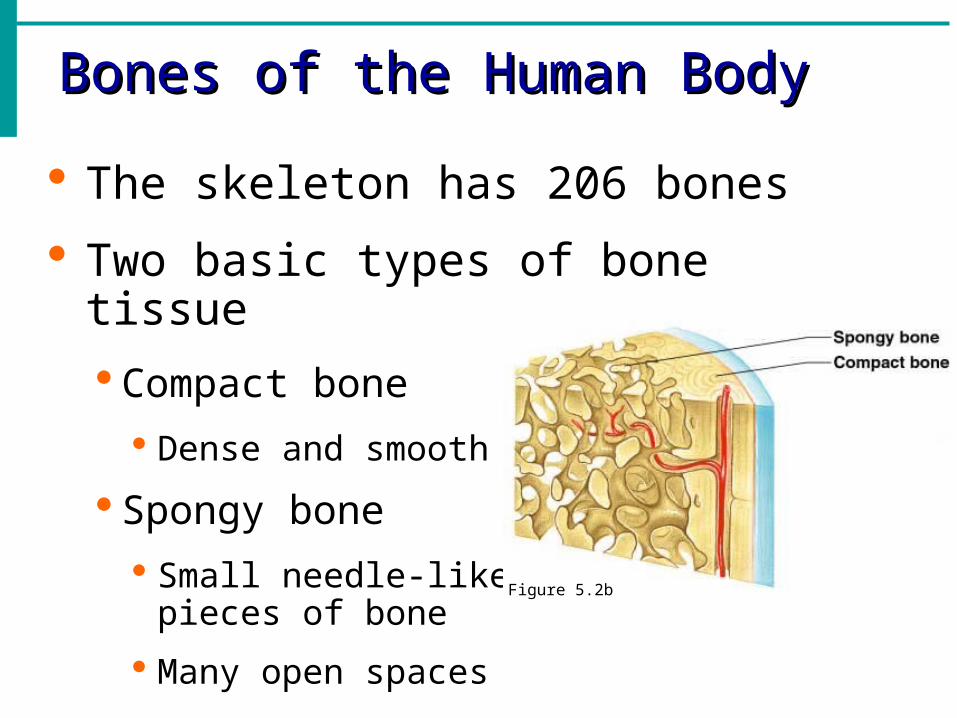
The skeleton has 206 bones
Two basic types of bone tissue
Compact bone
Dense and smooth
Spongy bone
Small needle-like pieces of bone
Many open spacesFigure 5.2b
Classification of BonesClassification of Bones
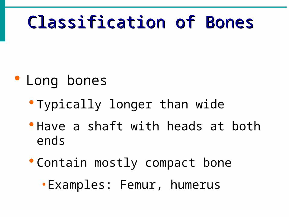
Long bones
Typically longer than wide
Have a shaft with heads at both ends
Contain mostly compact bone
• Examples: Femur, humerus
Classification of BonesClassification of Bones
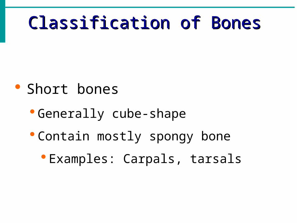
Short bones
Generally cube-shape
Contain mostly spongy bone
Examples: Carpals, tarsals
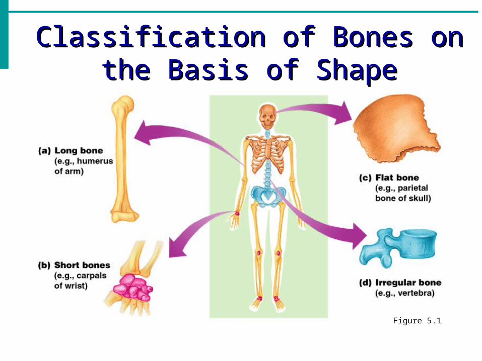
Classification of Bones on the Classification of Bones on the Basis of ShapeBasis of Shape
Figure 5.1
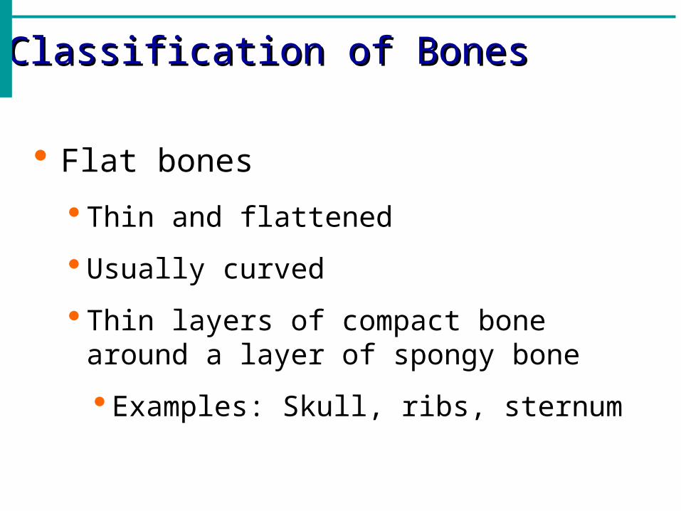
Classification of BonesClassification of Bones
Flat bones
Thin and flattened
Usually curved
Thin layers of compact bone around a layer of spongy bone
Examples: Skull, ribs, sternum
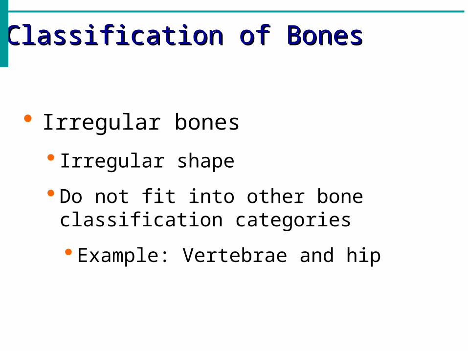
Classification of BonesClassification of Bones
Irregular bones
Irregular shape
Do not fit into other bone classification categories
Example: Vertebrae and hip
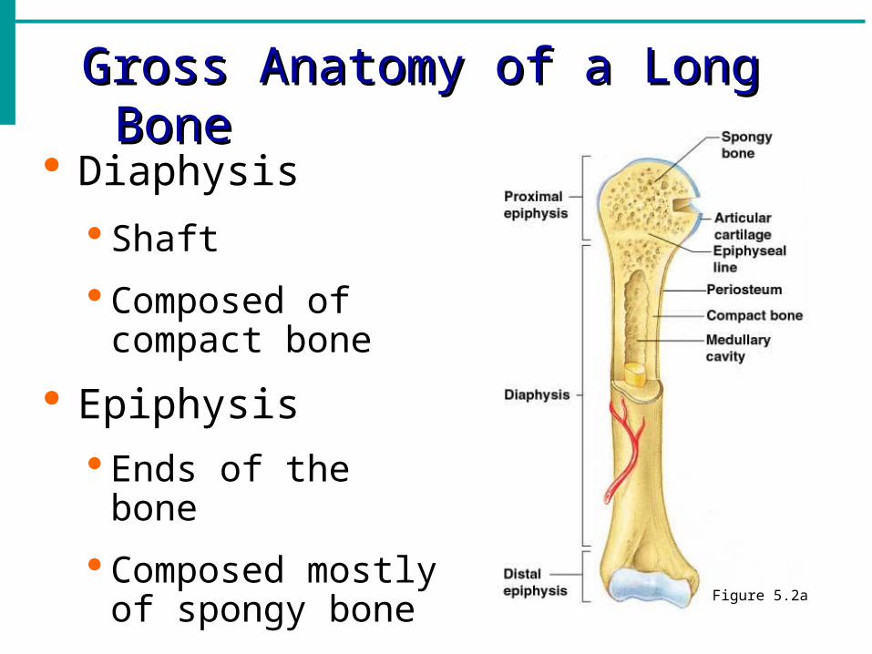
Gross Anatomy of a Long BoneGross Anatomy of a Long Bone
Diaphysis
Shaft
Composed of compact bone
Epiphysis
Ends of the bone
Composed mostly of spongy bone
Figure 5.2a
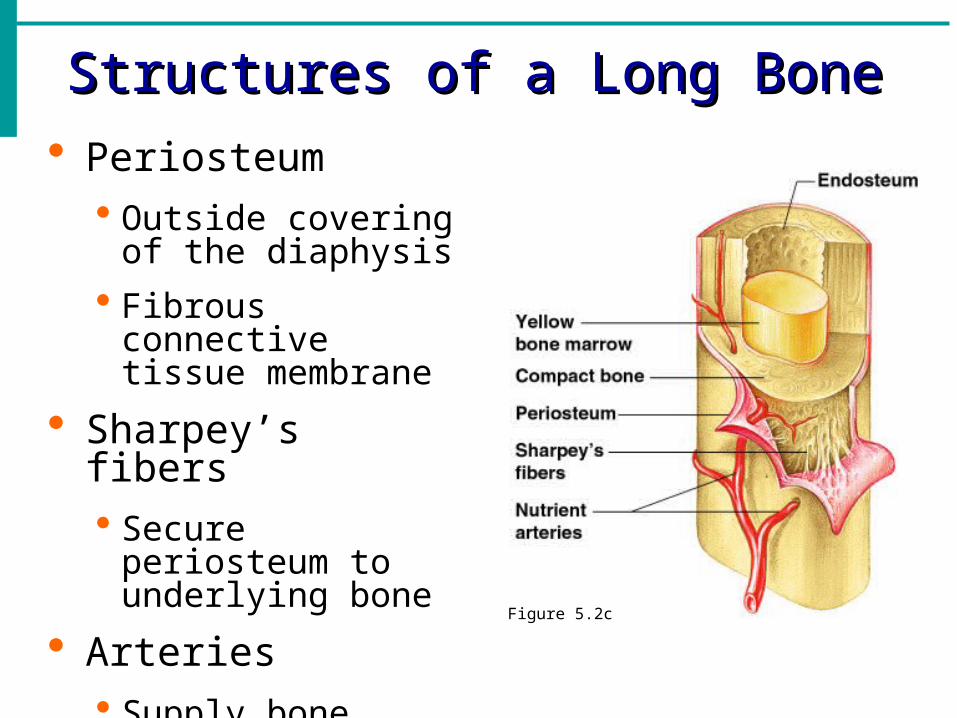
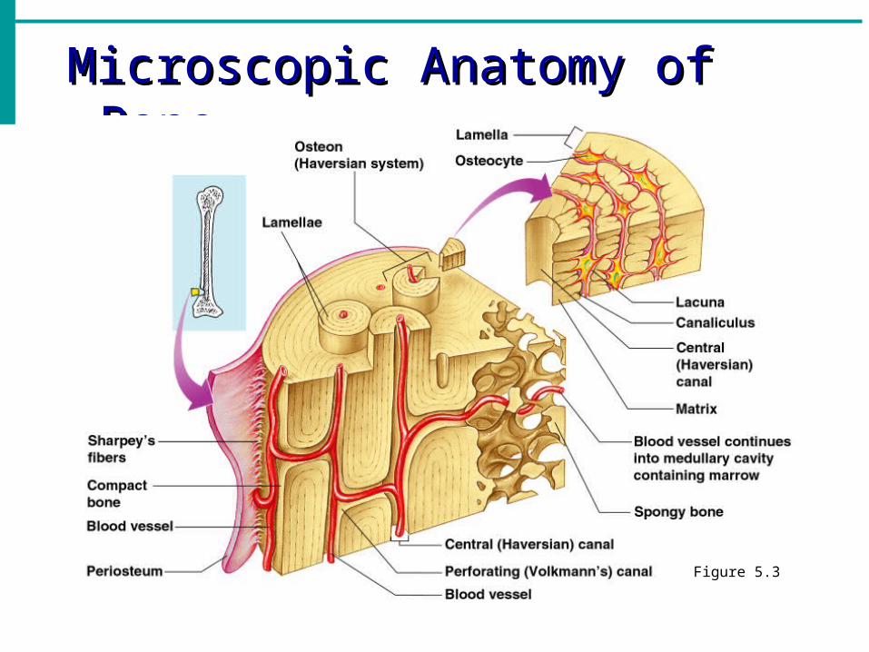
Structures of a Long BoneStructures of a Long Bone Periosteum
Outside covering of the diaphysis
Fibrous connective tissue membrane
Sharpey’s fibers Secure periosteum to
underlying bone
Arteries Supply bone cells
with nutrientsFigure 5.2c
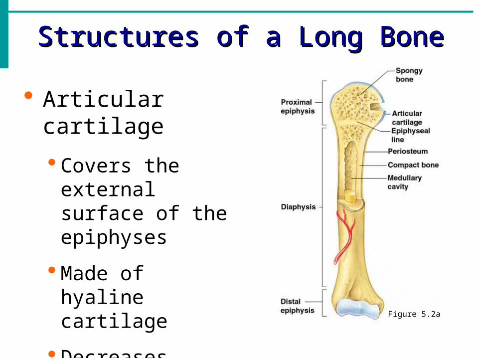
Structures of a Long BoneStructures of a Long Bone
Articular cartilage
Covers the external surface of the epiphyses
Made of hyaline cartilage
Decreases friction at joint surfaces Figure 5.2a
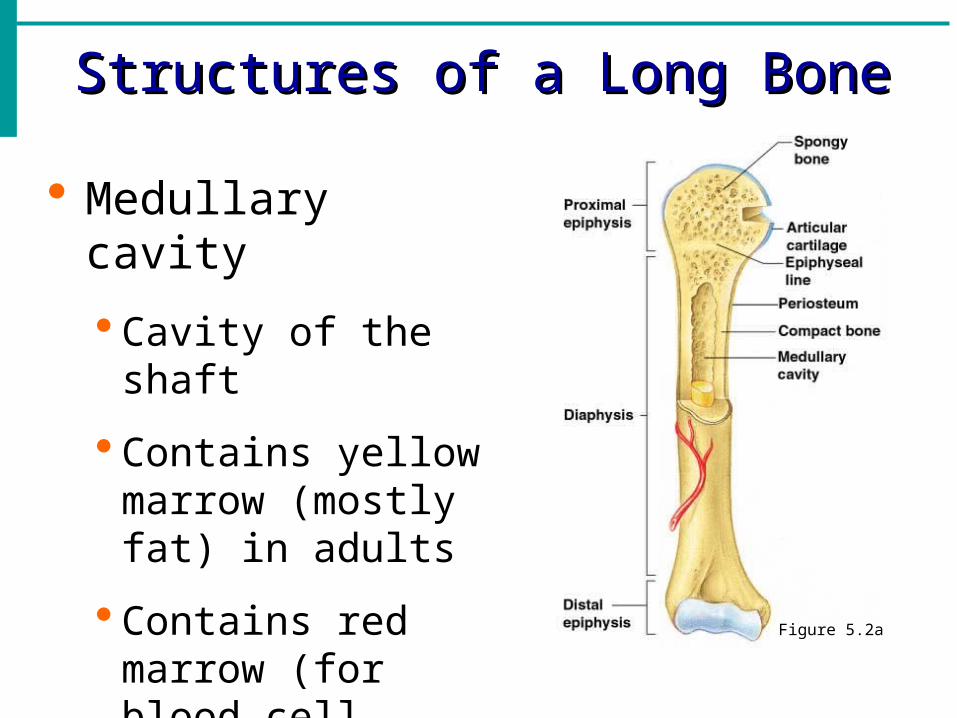
Structures of a Long BoneStructures of a Long Bone
Medullary cavity
Cavity of the shaft
Contains yellow marrow (mostly fat) in adults
Contains red marrow (for blood cell formation) in infants Figure 5.2a
Bone MarkingsBone Markings
Surface features of bones
Sites of attachments for muscles, tendons, and ligaments
Passages for nerves and blood vessels
Categories of bone markings
Projections and processes – grow out from the bone surface
Depressions or cavities – indentations
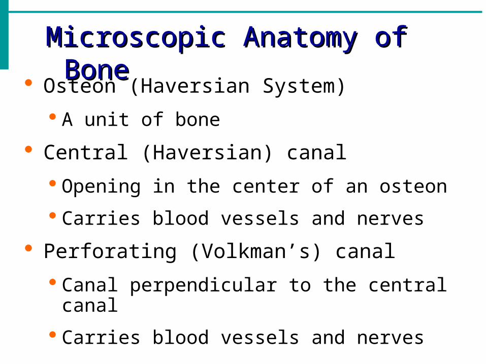
Microscopic Anatomy of BoneMicroscopic Anatomy of Bone
Osteon (Haversian System)
A unit of bone
Central (Haversian) canal
Opening in the center of an osteon
Carries blood vessels and nerves
Perforating (Volkman’s) canal
Canal perpendicular to the central canal
Carries blood vessels and nerves
Microscopic Anatomy of BoneMicroscopic Anatomy of Bone
Figure 5.3
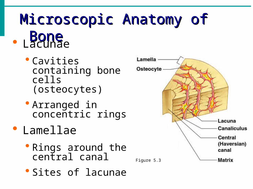
Microscopic Anatomy of BoneMicroscopic Anatomy of Bone
Lacunae Cavities containing
bone cells (osteocytes)
Arranged in concentric rings
Lamellae Rings around the
central canal
Sites of lacunae Figure 5.3
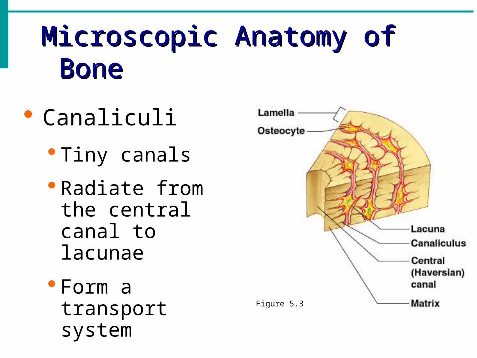
Microscopic Anatomy of BoneMicroscopic Anatomy of Bone
Canaliculi
Tiny canals
Radiate from the central canal to lacunae
Form a transport system
Figure 5.3
Changes in the Human SkeletonChanges in the Human Skeleton
In embryos, the skeleton is primarily hyaline cartilage
During development, much of this cartilage is replaced by bone
Cartilage remains in isolated areas
Bridge of the nose
Parts of ribs
Joints
Bone GrowthBone Growth
Epiphyseal plates allow for growth of long bone during childhood
New cartilage is continuously formed
Older cartilage becomes ossified
Cartilage is broken down
Bone replaces cartilage
Bone GrowthBone Growth
Bones are remodeled and lengthened until growth stops
Bones change shape somewhat
Bones grow in width
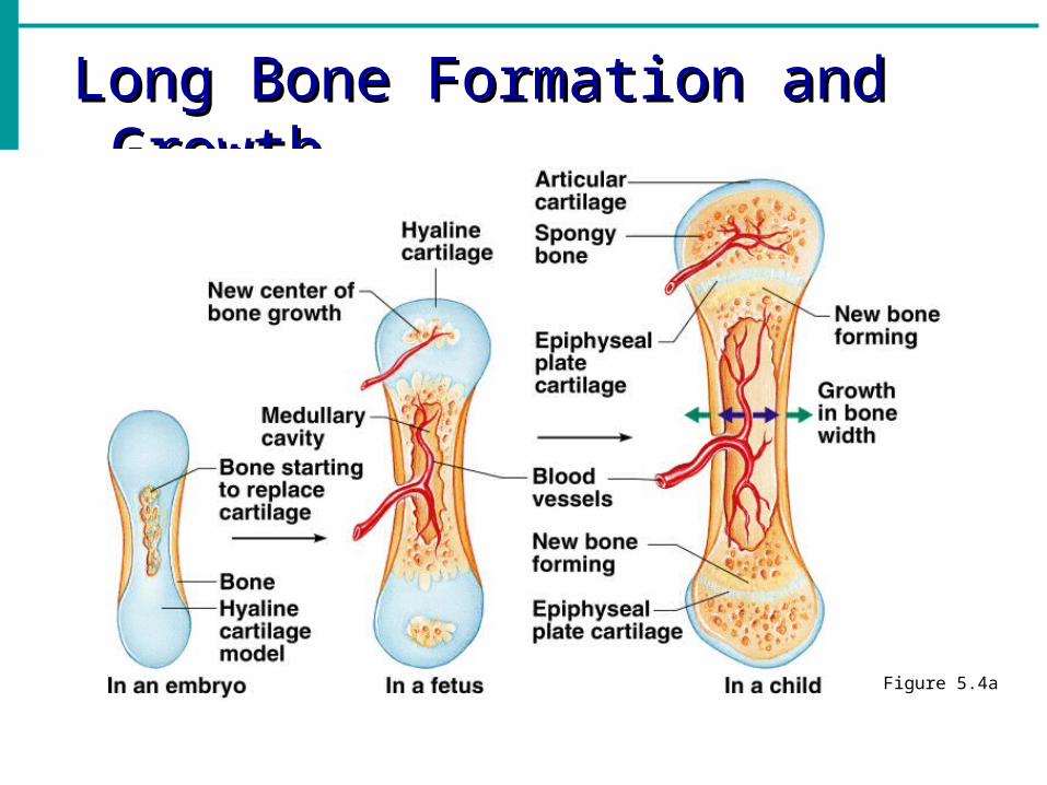
Long Bone Formation and GrowthLong Bone Formation and Growth
Figure 5.4a
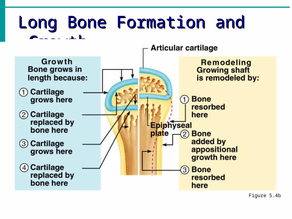
Long Bone Formation and GrowthLong Bone Formation and Growth
Figure 5.4b
Types of Bone CellsTypes of Bone Cells Osteocytes
Mature bone cells
Osteoblasts Bone-forming cells
Osteoclasts Bone-destroying cells
Break down bone matrix for remodeling and release of calcium
Bone remodeling is a process by both osteoblasts and osteoclasts
Recommended