NERVOUS SYSTEMANATOMY
to
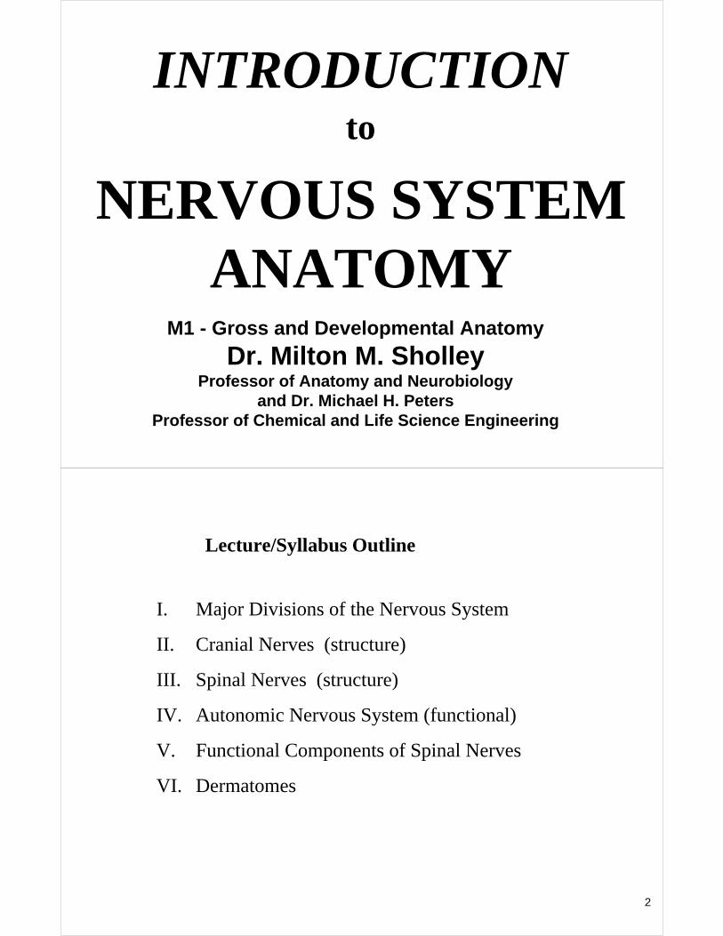
INTRODUCTION
M1 - Gross and Developmental Anatomy
Dr. Milton M. SholleyProfessor of Anatomy and Neurobiology
and Dr. Michael H. PetersProfessor of Chemical and Life Science Engineering
2
I. Major Divisions of the Nervous System
II. Cranial Nerves (structure)
III. Spinal Nerves (structure)
IV. Autonomic Nervous System (functional)
V. Functional Components of Spinal Nerves
VI. Dermatomes
Lecture/Syllabus Outline
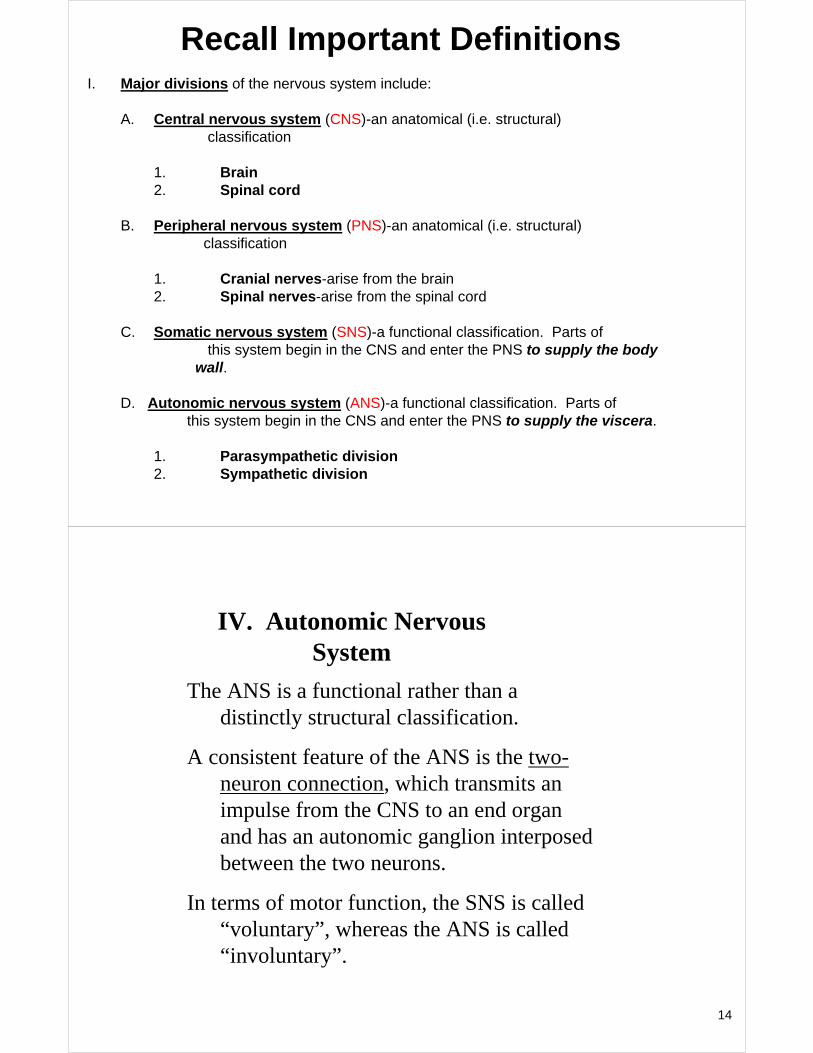
I. Major divisions of the nervous system include:
A. Central nervous system (CNS)-an anatomical (i.e. structural) classification
1. Brain2. Spinal cord
B. Peripheral nervous system (PNS)-an anatomical (i.e. structural) classification
1. Cranial nerves-arise from the brain2. Spinal nerves-arise from the spinal cord
C. Somatic nervous system (SNS)-a functional classification. Parts of this system begin in the CNS and enter the PNS to supply the body
wall (“soma”).D. Autonomic nervous system (ANS)-a functional classification. Parts of
this system begin in the CNS and enter the PNS to supply the viscera.
1. Parasympathetic division2. Sympathetic division
Important Definitions
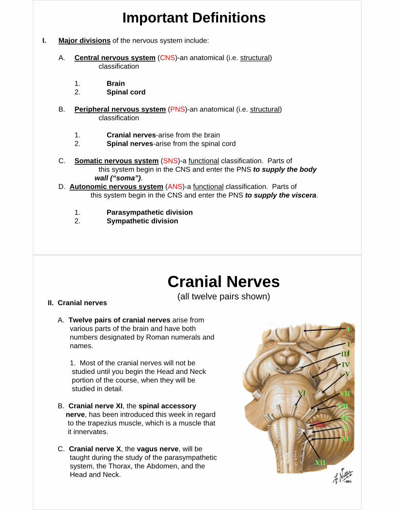
II. Cranial nerves
A. Twelve pairs of cranial nerves arise from various parts of the brain and have both numbers designated by Roman numerals and names.
1. Most of the cranial nerves will not be studied until you begin the Head and Neck portion of the course, when they will be studied in detail.
B. Cranial nerve XI, the spinal accessory nerve, has been introduced this week in regard to the trapezius muscle, which is a muscle that it innervates.
C. Cranial nerve X, the vagus nerve, will be taught during the study of the parasympathetic system, the Thorax, the Abdomen, and the Head and Neck.
I
IIIII
IVV
VI VII
VIIIIXX
XI
XII
Cranial Nerves(all twelve pairs shown)
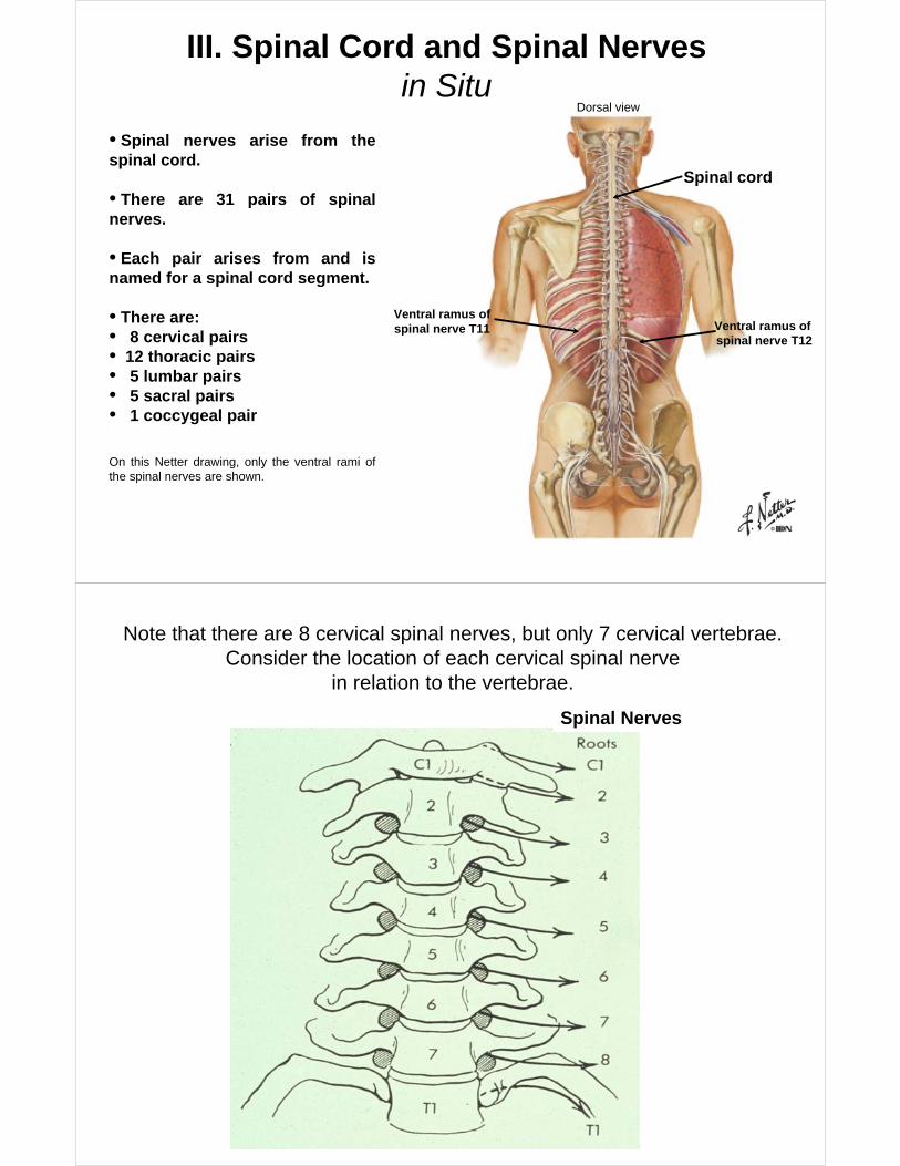
III. Spinal Cord and Spinal Nervesin Situ
Spinal cord
Ventral ramus ofspinal nerve T12
Ventral ramus of spinal nerve T11
Dorsal view
• Spinal nerves arise from the spinal cord.
• There are 31 pairs of spinal nerves.
• Each pair arises from and is named for a spinal cord segment.
• There are:• 8 cervical pairs• 12 thoracic pairs• 5 lumbar pairs• 5 sacral pairs• 1 coccygeal pair
On this Netter drawing, only the ventral rami of the spinal nerves are shown.
Spinal Nerves
Note that there are 8 cervical spinal nerves, but only 7 cervical vertebrae.Consider the location of each cervical spinal nerve
in relation to the vertebrae.
7
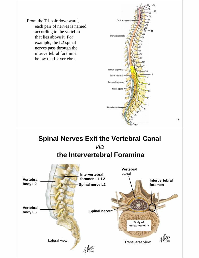
From the T1 pair downward, each pair of nerves is named according to the vertebra that lies above it. For example, the L2 spinal nerves pass through the intervertebral foramina below the L2 vertebra.
Spinal Nerves Exit the Vertebral Canalvia
the Intervertebral Foramina
Vertebralbody L2
Vertebralbody L5
Intervertebralforamen L1-L2
Spinal nerve L2
Lateral view Transverse view
Body of lumbar vertebra
Intervertebralforamen
Spinal nerve
Vertebralcanal
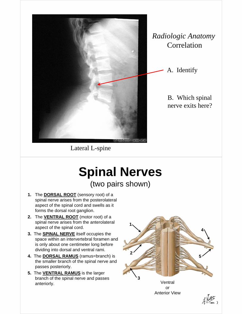
A. Identify
B. Which spinalnerve exits here?
Radiologic AnatomyCorrelation
Lateral L-spine
10
Spinal Nerves(two pairs shown)
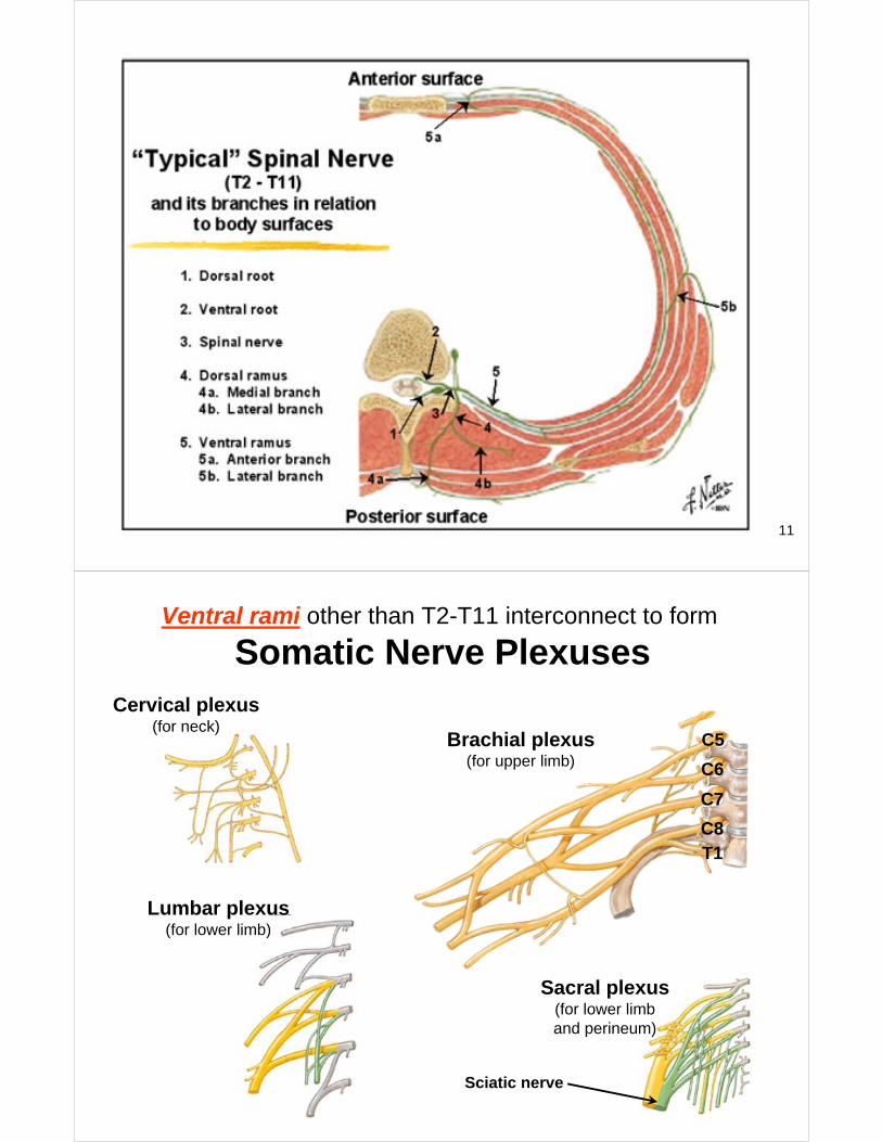
1. The DORSAL ROOT (sensory root) of a spinal nerve arises from the posterolateralaspect of the spinal cord and swells as it forms the dorsal root ganglion.
2. The VENTRAL ROOT (motor root) of a spinal nerve arises from the anterolateralaspect of the spinal cord.
3. The SPINAL NERVE itself occupies the space within an intervertebral foramen and is only about one centimeter long before dividing into dorsal and ventral rami.
4. The DORSAL RAMUS (ramus=branch) is the smaller branch of the spinal nerve and passes posteriorly.
5. The VENTRAL RAMUS is the larger branch of the spinal nerve and passes anteriorly.
1
2
3
4
5
Ventralor
Anterior View
11
Ventral rami other than T2-T11 interconnect to form
Somatic Nerve Plexuses
Brachial plexus(for upper limb)
C5
T1
C6
C7
C8
Cervical plexus(for neck)
Lumbar plexus(for lower limb)
Sacral plexus(for lower limband perineum)
Sciatic nerve
I. Major divisions of the nervous system include:
A. Central nervous system (CNS)-an anatomical (i.e. structural) classification
1. Brain2. Spinal cord
B. Peripheral nervous system (PNS)-an anatomical (i.e. structural) classification
1. Cranial nerves-arise from the brain2. Spinal nerves-arise from the spinal cord
C. Somatic nervous system (SNS)-a functional classification. Parts of this system begin in the CNS and enter the PNS to supply the body
wall.
D. Autonomic nervous system (ANS)-a functional classification. Parts of this system begin in the CNS and enter the PNS to supply the viscera.
1. Parasympathetic division2. Sympathetic division
Recall Important Definitions
14
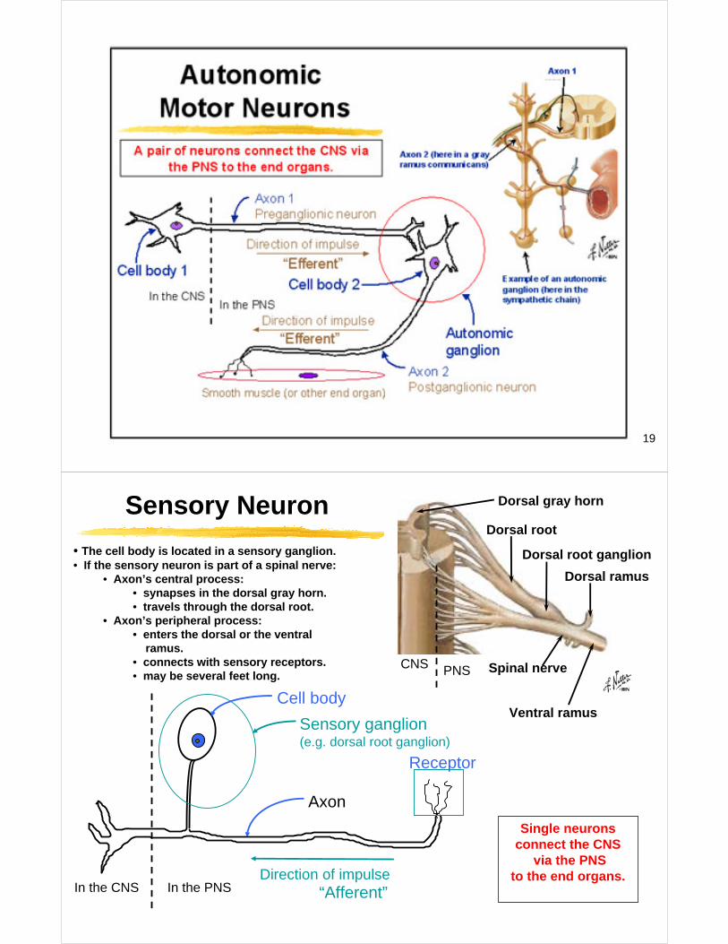
IV. Autonomic Nervous System
The ANS is a functional rather than a distinctly structural classification.
A consistent feature of the ANS is the two-neuron connection, which transmits an impulse from the CNS to an end organ and has an autonomic ganglion interposed between the two neurons.
In terms of motor function, the SNS is called “voluntary”, whereas the ANS is called “involuntary”.
15
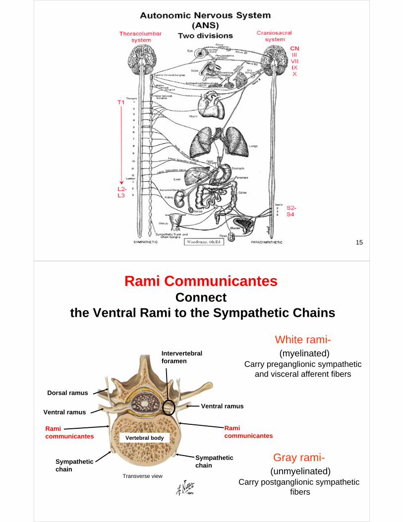
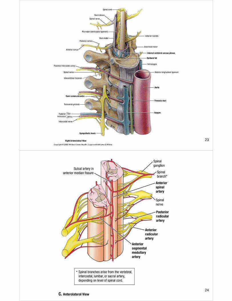
Rami CommunicantesConnect
the Ventral Rami to the Sympathetic Chains
Transverse view
Vertebral body
Intervertebralforamen
Dorsal ramus
Ventral ramus
Ramicommunicantes
Sympatheticchain
Ramicommunicantes
Sympatheticchain
Ventral ramus
White rami-(myelinated)
Carry preganglionic sympathetic and visceral afferent fibers
Gray rami-(unmyelinated)
Carry postganglionic sympathetic fibers
17
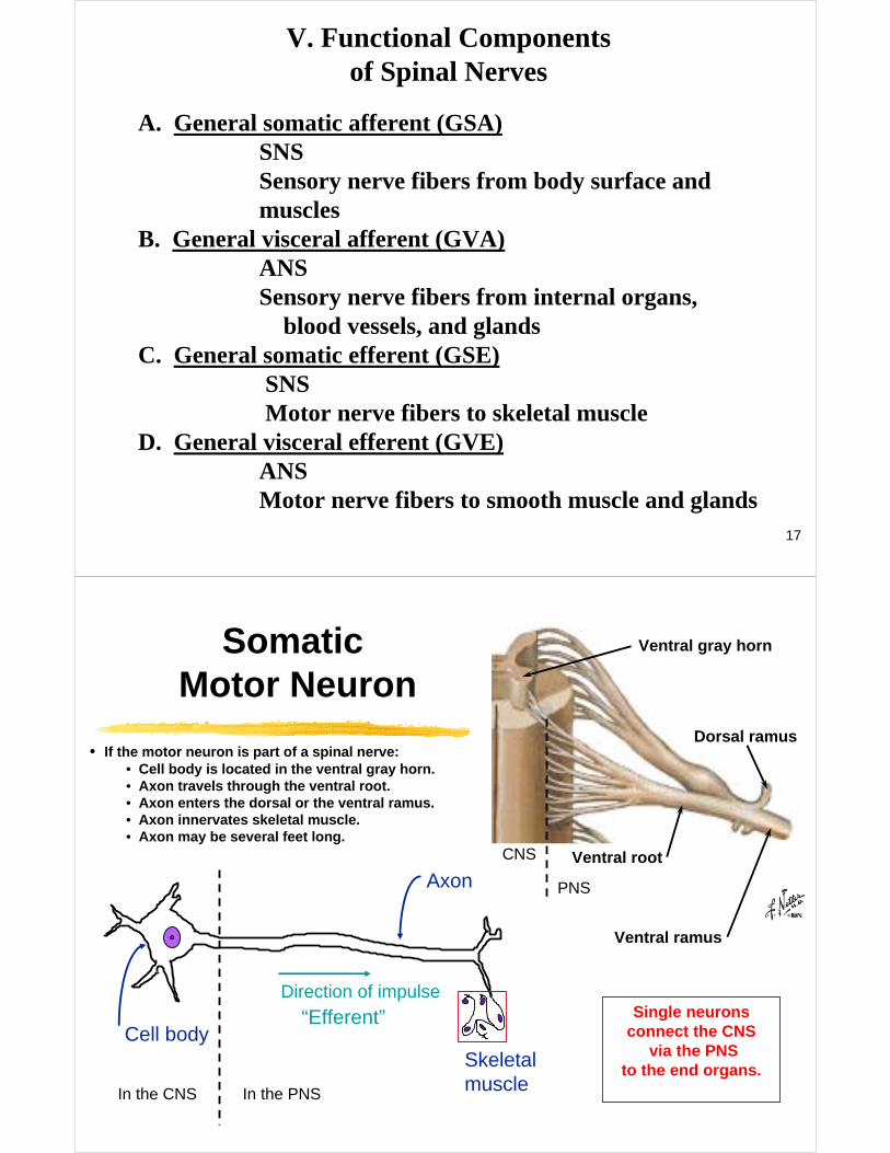
A. General somatic afferent (GSA)SNSSensory nerve fibers from body surface and muscles
B. General visceral afferent (GVA)ANSSensory nerve fibers from internal organs,
blood vessels, and glandsC. General somatic efferent (GSE)
SNSMotor nerve fibers to skeletal muscle
D. General visceral efferent (GVE)ANSMotor nerve fibers to smooth muscle and glands
V. Functional Componentsof Spinal Nerves
SomaticMotor Neuron
• If the motor neuron is part of a spinal nerve:• Cell body is located in the ventral gray horn.• Axon travels through the ventral root.• Axon enters the dorsal or the ventral ramus.• Axon innervates skeletal muscle.• Axon may be several feet long.
Skeletalmuscle
Direction of impulse
“Efferent”
In the CNS In the PNS
Cell body
Axon
Ventral gray horn
Ventral root
Ventral ramus
Dorsal ramus
CNS
PNS
Single neuronsconnect the CNS
via the PNSto the end organs.
19
Axon
Direction of impulse“Afferent”
Receptor
In the CNS In the PNS
Cell body
Sensory ganglion(e.g. dorsal root ganglion)
Spinal nerve
Dorsal gray horn
Dorsal root
Ventral ramus
Dorsal ramus
Dorsal root ganglion
CNS PNS
Sensory Neuron
• The cell body is located in a sensory ganglion.• If the sensory neuron is part of a spinal nerve:
• Axon’s central process:• synapses in the dorsal gray horn.• travels through the dorsal root.
• Axon’s peripheral process: • enters the dorsal or the ventral
ramus.• connects with sensory receptors.• may be several feet long.
Single neuronsconnect the CNS
via the PNSto the end organs.
21
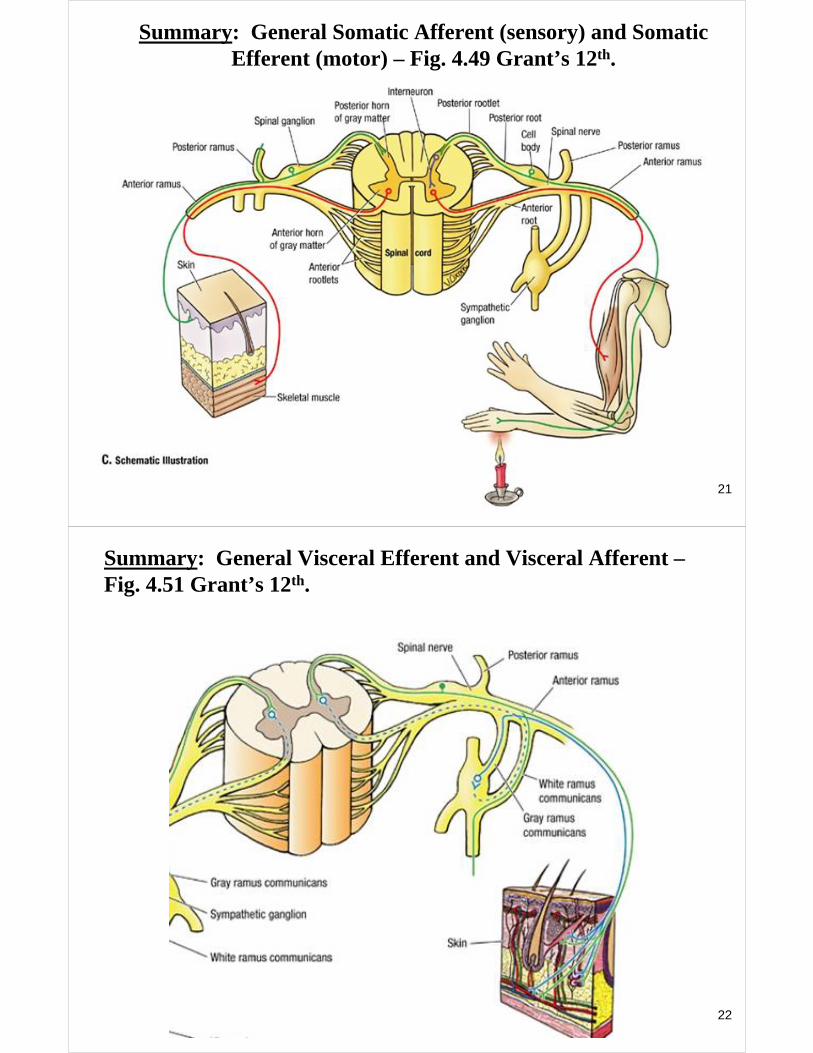
Summary: General Somatic Afferent (sensory) and Somatic Efferent (motor) – Fig. 4.49 Grant’s 12th.
22
Summary: General Visceral Efferent and Visceral Afferent –Fig. 4.51 Grant’s 12th.
23
24
25
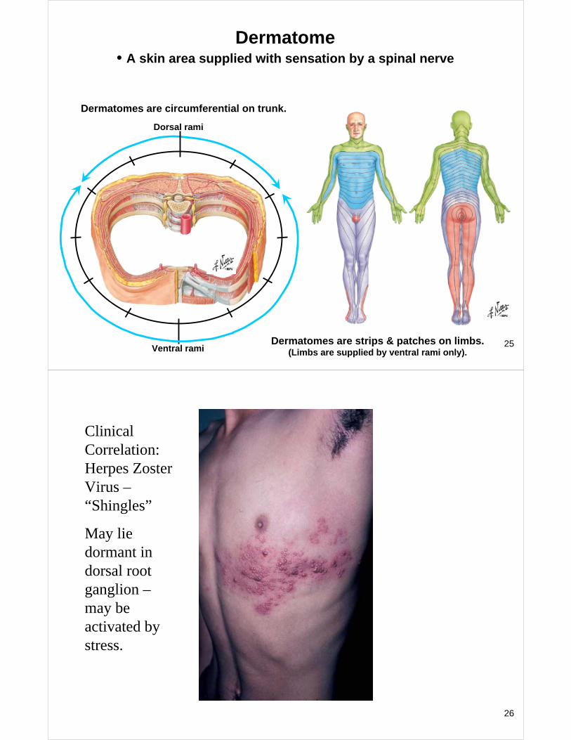
Dermatome• A skin area supplied with sensation by a spinal nerve
Dermatomes are circumferential on trunk.
Dermatomes are strips & patches on limbs.(Limbs are supplied by ventral rami only).
Dorsal rami
Ventral rami
26
Clinical Correlation: Herpes Zoster Virus –“Shingles”
May lie dormant in dorsal root ganglion –may be activated by stress.
27
Segmental Innervation of Muscles
Each skeletal muscle is innervated by somatic motorfibers from specific spinal nerves.
1. Because of the formation of somatic nerve plexuses, the segmental innervation of muscles cannot be recognized by dissection.
2. The segmental innervation of muscles has been discerned by clinical studies and this information is important to the anatomist, neurologist, and neurosurgeon.
3. The named peripheral nerve innervation of musclescan be recognized by dissection.
28
Laminectomy:
Recommended