607
Turk J Biol36 (2012) 607-621© TÜBİTAKdoi:10.3906/biy-1112-6
Utilization of gold nanostructures in biomedical applications
Gamze TAN1, Mehmet Ali ONUR2, Necdet SAĞLAM3
1Department of Biology, Institute of Science, Hacettepe University, 06800 Ankara − TURKEY2Department of Biology, Faculty of Science, Hacettepe University, 06800 Ankara − TURKEY
3Department of Nanotechnology and Nanomedicine, Institute of Science, Hacettepe University, 06800 Ankara − TURKEY
Received: 05.12.2011 ● Accepted: 21.05.2012
Abstract: Every living organism on earth owes its viability to its different sizes of nanostructures and the interaction of these structures at the nano size. Nanotechnology gives us an opportunity to understand nanoscale processes in living organisms and interfere with and manipulate them. Today, biocompatible nanosized structures are designed by applying developments in nanotechnology to biomedicine; thus, therapeutic agents are available to reach diseased tissues and even cells. However, it is essential that nanomaterials that would be used for therapeutic aims be targetable to diseased areas and have low toxicity. In addition, these nanomaterials must have high biocirculation and pharmacokinetic properties. Compared to conventional methods, gold nanoparticles (AuNPs) have the appropriate physical, chemical, mechanical, optical, and electronic properties for the design of nanobiomaterials that exhibit high selectivity, specificity, and sensitivity in the early detection, diagnosis, and treatment of diseases. Recently, gold has been used in prominent drug and gene carrier platforms because it binds various therapeutic agents and biomolecules in a stable way to create biocompatible complex structures. In addition, it has nontoxic nuclei and surface properties such as charge and hydrophobicity, which are adjustable in a monolayer. In the near infrared region, AuNPs are effective probes for in vivo and in vitro imaging with their high plasmon resonance absorption and scattering. In addition, their ability to rapidly convert optical energy to heat energy enables the ablation of invasive cancer tissues photothermally, even at a low power. This review sheds light on the synthesis, surface functionality, and potential applications of colloidal AuNPs in biomedicine.
Key words: Gold nanoparticles, colloidal gold, nanotechnology, nanostructures, nanobiomaterials, biomedicine, cancer, photothermal therapy
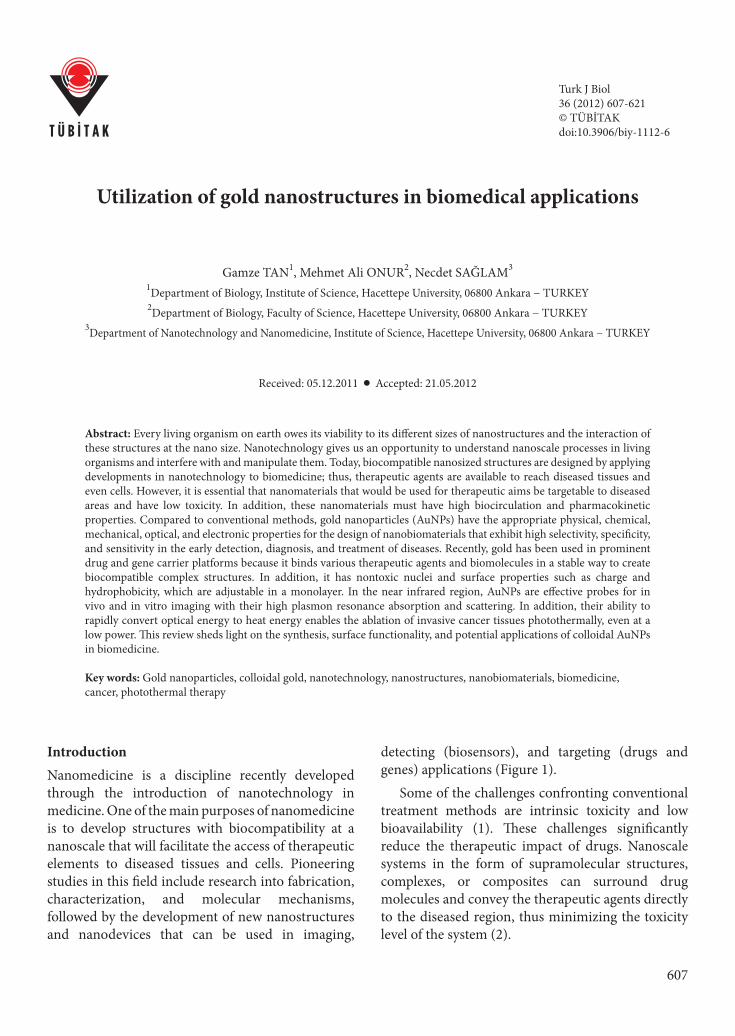
IntroductionNanomedicine is a discipline recently developed through the introduction of nanotechnology in medicine. One of the main purposes of nanomedicine is to develop structures with biocompatibility at a nanoscale that will facilitate the access of therapeutic elements to diseased tissues and cells. Pioneering studies in this field include research into fabrication, characterization, and molecular mechanisms, followed by the development of new nanostructures and nanodevices that can be used in imaging,
detecting (biosensors), and targeting (drugs and genes) applications (Figure 1).
Some of the challenges confronting conventional treatment methods are intrinsic toxicity and low bioavailability (1). These challenges significantly reduce the therapeutic impact of drugs. Nanoscale systems in the form of supramolecular structures, complexes, or composites can surround drug molecules and convey the therapeutic agents directly to the diseased region, thus minimizing the toxicity level of the system (2).
Utilization of gold nanostructures in biomedical applications
608
Nanotechnological drug delivery systems carry drugs to the target region in the body. Delivery can be made through active or passive routing (3). Therapeutic chemicals carried to the diseased region will not make contact with healthy tissues and thus will not damage healthy tissues (3,4). Drug delivery systems based on nanotechnology differ from conventional drug delivery systems in certain ways. Such differences arise from the size of the delivery molecules in the solutions used. Due to their size, i.e. a billionth or 10−9 of a meter, these particles exhibit new physicochemical features. In delivery systems at micro levels, targeting is limited by the physical characteristics of the capillary beds. This new sizing
at nano levels makes the chemical to be targeted in the delivery system even freer and renders the selection of targeted tissue more specifiable with surface characteristics. By these means, the delivery of therapeutic agents into specific cells and the diagnosis and cure of diseases occurs at the cellular level (5). However, in addition to biocirculatory and pharmacokinetic properties of these materials used as the drug delivery system, the location and time of release of the carried drug molecules are extremely important. It is also expected that these materials will cause no toxicity and be easily discharged from the system.
Nuclear targeting(oligonucleotide, peptides (NLS, TaT, etc.))
Drug delivery(paclitaxel TNF-α, MTX)
Cancer treatment(phototherapy, radio/photo sensitizer)
Abnormal cell
Ex vivo, in vivo imaging(OCT, PAT, Raman)
Figure 1. Biomedical applications of gold nanoparticles. This figure inspired by ref. 12 and 17.

G. TAN, M. A. ONUR, N. SAĞLAM
609
Why nanogold?
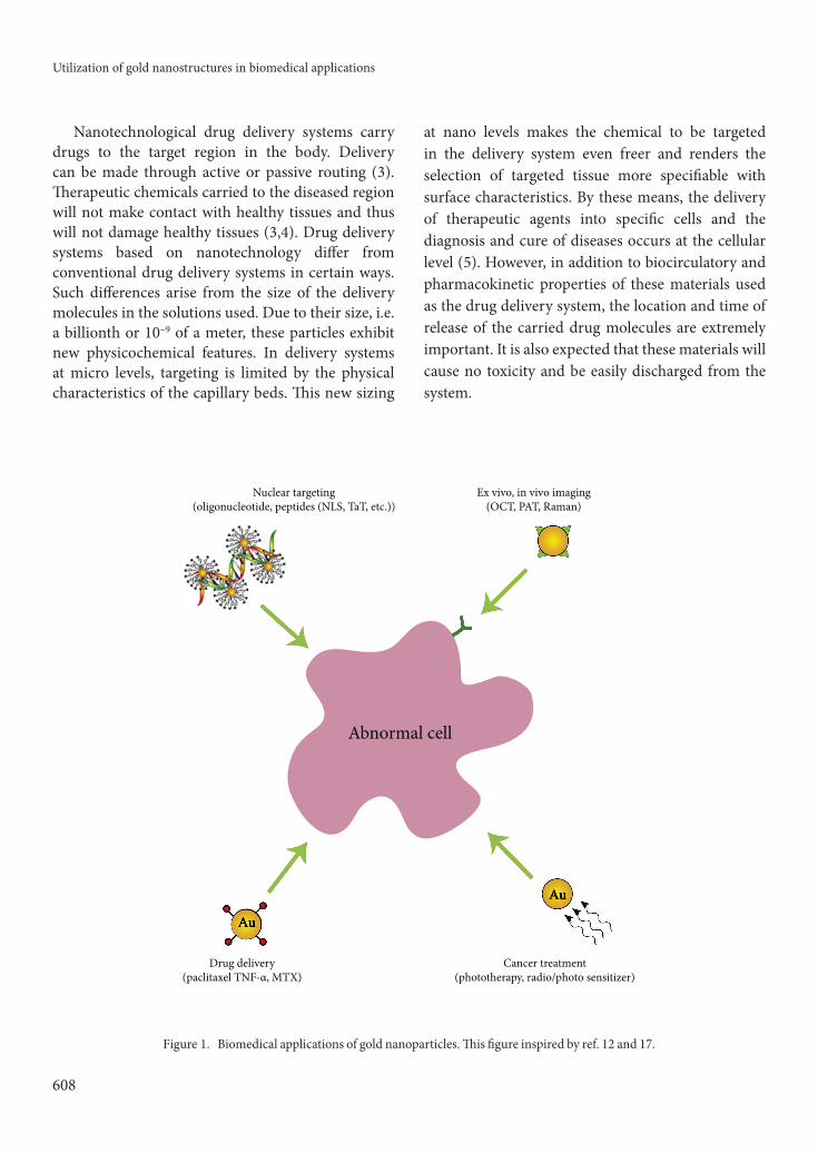
Although there are many nanotechnological structures used in drug targeting and controlled release, gold nanoparticles (AuNPs) hold a special place. Gold is a noble metal and has nontoxic characteristics (6). It is stable to oxidation compared to other metals (7) and is not easily affected by an acidic environment (8). AuNPs can be readily modified with ligands containing functional groups such as thiols, phosphines, and amines to anchor the ligands, and additional moieties such as oligonucleotides, proteins, and antibodies can be used to impart even greater functionality (9). When directed into the cell, AuNPs cause very low cytotoxic effects without damaging healthy cells (10,11) depending on the capping agent used in the synthesis procedure. AuNPs enable the adjusting of surface features such as charge and hydrophobia without interaction with any other covering agent (12).
AuNPs interact with the thiol (-SH) group, which is easily linked with biomolecules, and provide selectivity in nuclear targeting (gene carrying, antisense treatment) applications. The targeted nanoparticles release their therapeutic payloads in the target tissues and cells through intracellular
interaction [e.g., pH (13), glutathione (14)] or external stimuli [e.g., light (15)] (Figure 2).
Due to surface modification, in vivo pharmacokinetics, biopropagation, and the circulation period of biomolecules based on AuNPs will be enhanced (18). Ligands attached to the nanoparticle surface interact with membrane receptors and enable nanoparticles to penetrate from the cell membrane into the cytoplasm. For this reason, the ligand number on the nanoparticle surface is very important during the penetration of nanoparticles from the cell membrane. Murray et al. (1998) prepared dodecanethiolate-stabilized AuNPs with core sizes from 1.5 to 5.2 nm, and at this range the Au core contained an average of approximately 110−4800 Au atoms and from 53 to nearly 520 alkanethiolate chains in the monolayer (19). Gibson et al. succeeded in coupling approximately 70 molecules of paclitaxel, a chemotherapeutic drug, to a AuNP with 2-nm core sizes (20).
A plasmon is a collective oscillation of free electrons in the conduction band of metals stimulated by incident light. It is an optical fact resulting from the interaction between electromagnetic waves and metal surface electrons. When a metal absorbs light
Ligand exchange
Nucleus
Lower pH
Figure 2. The therapeutic agents carried by AuNPs are released through low pH, glutathione (GSH), or stimulation with radiation (16).
Utilization of gold nanostructures in biomedical applications
610
at a resonant wavelength, it causes the electron cloud to vibrate and spread the energy. This vibration spreads just like the propagation of an acoustic vibration harmonically generated by a stringed instrument (plasmon resonance). For metals, plasmon resonance is seen in the infrared region; for AuNPs, it is seen in both the visible (the interval that may be perceived by eye) and infrared regions (21). In addition, AuNPs create strong light absorption and scattering at plasmon wavelength (22,23). This strong light scattering enables AuNPs to show high specificity and sensitivity in diagnostic and sensing applications. Yet another advantage of AuNPs is their ease of synthesis; monodisperse AuNPs can be formed in various shapes including nanosphere, nanorod, and nanocube with core sizes ranging from 2 to 150 nm (24−26).
Photothermal features of AuNPs
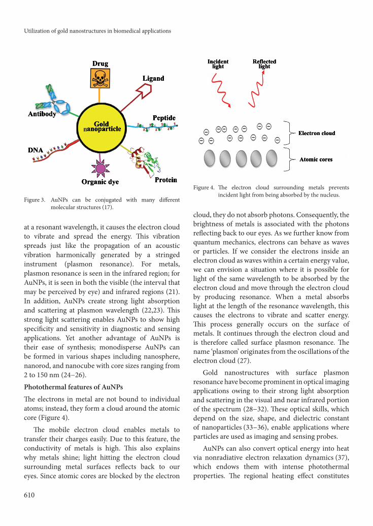
The electrons in metal are not bound to individual atoms; instead, they form a cloud around the atomic core (Figure 4).
The mobile electron cloud enables metals to transfer their charges easily. Due to this feature, the conductivity of metals is high. This also explains why metals shine; light hitting the electron cloud surrounding metal surfaces reflects back to our eyes. Since atomic cores are blocked by the electron
cloud, they do not absorb photons. Consequently, the brightness of metals is associated with the photons reflecting back to our eyes. As we further know from quantum mechanics, electrons can behave as waves or particles. If we consider the electrons inside an electron cloud as waves within a certain energy value, we can envision a situation where it is possible for light of the same wavelength to be absorbed by the electron cloud and move through the electron cloud by producing resonance. When a metal absorbs light at the length of the resonance wavelength, this causes the electrons to vibrate and scatter energy. This process generally occurs on the surface of metals. It continues through the electron cloud and is therefore called surface plasmon resonance. The name ‘plasmon’ originates from the oscillations of the electron cloud (27).
Gold nanostructures with surface plasmon resonance have become prominent in optical imaging applications owing to their strong light absorption and scattering in the visual and near infrared portion of the spectrum (28−32). These optical skills, which depend on the size, shape, and dielectric constant of nanoparticles (33−36), enable applications where particles are used as imaging and sensing probes.
AuNPs can also convert optical energy into heat via nonradiative electron relaxation dynamics (37), which endows them with intense photothermal properties. The regional heating effect constitutes
Figure 3. AuNPs can be conjugated with many different molecular structures (17).
Figure 4. The electron cloud surrounding metals prevents incident light from being absorbed by the nucleus.
G. TAN, M. A. ONUR, N. SAĞLAM
611
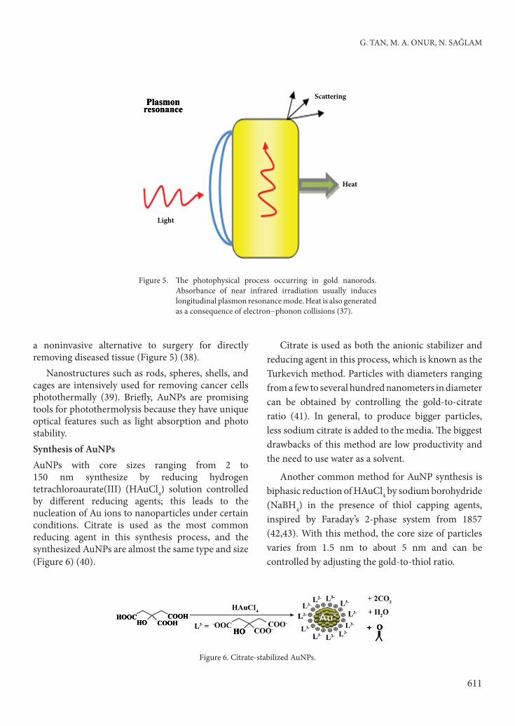
a noninvasive alternative to surgery for directly removing diseased tissue (Figure 5) (38).
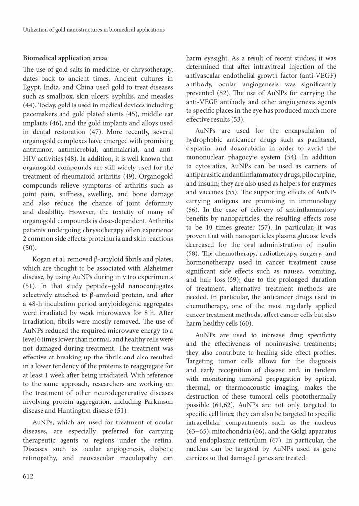
Nanostructures such as rods, spheres, shells, and cages are intensively used for removing cancer cells photothermally (39). Briefly, AuNPs are promising tools for photothermolysis because they have unique optical features such as light absorption and photo stability. Synthesis of AuNPs AuNPs with core sizes ranging from 2 to 150 nm synthesize by reducing hydrogen tetrachloroaurate(III) (HAuCl4) solution controlled by different reducing agents; this leads to the nucleation of Au ions to nanoparticles under certain conditions. Citrate is used as the most common reducing agent in this synthesis process, and the synthesized AuNPs are almost the same type and size (Figure 6) (40).
Citrate is used as both the anionic stabilizer and reducing agent in this process, which is known as the Turkevich method. Particles with diameters ranging from a few to several hundred nanometers in diameter can be obtained by controlling the gold-to-citrate ratio (41). In general, to produce bigger particles, less sodium citrate is added to the media. The biggest drawbacks of this method are low productivity and the need to use water as a solvent.
Another common method for AuNP synthesis is biphasic reduction of HAuCl4 by sodium borohydride (NaBH4) in the presence of thiol capping agents, inspired by Faraday’s 2-phase system from 1857 (42,43). With this method, the core size of particles varies from 1.5 nm to about 5 nm and can be controlled by adjusting the gold-to-thiol ratio.
Scattering
Heat
Light
Figure 5. The photophysical process occurring in gold nanorods. Absorbance of near infrared irradiation usually induces longitudinal plasmon resonance mode. Heat is also generated as a consequence of electron−phonon collisions (37).
Figure 6. Citrate-stabilized AuNPs.
Utilization of gold nanostructures in biomedical applications
612
Biomedical application areas
The use of gold salts in medicine, or chrysotherapy, dates back to ancient times. Ancient cultures in Egypt, India, and China used gold to treat diseases such as smallpox, skin ulcers, syphilis, and measles (44). Today, gold is used in medical devices including pacemakers and gold plated stents (45), middle ear implants (46), and the gold implants and alloys used in dental restoration (47). More recently, several organogold complexes have emerged with promising antitumor, antimicrobial, antimalarial, and anti-HIV activities (48). In addition, it is well known that organogold compounds are still widely used for the treatment of rheumatoid arthritis (49). Organogold compounds relieve symptoms of arthritis such as joint pain, stiffness, swelling, and bone damage and also reduce the chance of joint deformity and disability. However, the toxicity of many of organogold compounds is dose-dependent. Arthritis patients undergoing chrysotherapy often experience 2 common side effects: proteinuria and skin reactions (50).
Kogan et al. removed β-amyloid fibrils and plates, which are thought to be associated with Alzheimer disease, by using AuNPs during in vitro experiments (51). In that study peptide−gold nanoconjugates selectively attached to β-amyloid protein, and after a 48-h incubation period amyloidogenic aggregates were irradiated by weak microwaves for 8 h. After irradiation, fibrils were mostly removed. The use of AuNPs reduced the required microwave energy to a level 6 times lower than normal, and healthy cells were not damaged during treatment. The treatment was effective at breaking up the fibrils and also resulted in a lower tendency of the proteins to reaggregate for at least 1 week after being irradiated. With reference to the same approach, researchers are working on the treatment of other neurodegenerative diseases involving protein aggregation, including Parkinson disease and Huntington disease (51).
AuNPs, which are used for treatment of ocular diseases, are especially preferred for carrying therapeutic agents to regions under the retina. Diseases such as ocular angiogenesis, diabetic retinopathy, and neovascular maculopathy can
harm eyesight. As a result of recent studies, it was determined that after intravitreal injection of the antivascular endothelial growth factor (anti-VEGF) antibody, ocular angiogenesis was significantly prevented (52). The use of AuNPs for carrying the anti-VEGF antibody and other angiogenesis agents to specific places in the eye has produced much more effective results (53).
AuNPs are used for the encapsulation of hydrophobic anticancer drugs such as paclitaxel, cisplatin, and doxorubicin in order to avoid the mononuclear phagocyte system (54). In addition to cytostatics, AuNPs can be used as carriers of antiparasitic and antiinflammatory drugs, pilocarpine, and insulin; they are also used as helpers for enzymes and vaccines (55). The supporting effects of AuNP-carrying antigens are promising in immunology (56). In the case of delivery of antiinflammatory benefits by nanoparticles, the resulting effects rose to be 10 times greater (57). In particular, it was proven that with nanoparticles plasma glucose levels decreased for the oral administration of insulin (58). The chemotherapy, radiotherapy, surgery, and hormonotherapy used in cancer treatment cause significant side effects such as nausea, vomiting, and hair loss (59); due to the prolonged duration of treatment, alternative treatment methods are needed. In particular, the anticancer drugs used in chemotherapy, one of the most regularly applied cancer treatment methods, affect cancer cells but also harm healthy cells (60).
AuNPs are used to increase drug specificity and the effectiveness of noninvasive treatments; they also contribute to healing side effect profiles. Targeting tumor cells allows for the diagnosis and early recognition of disease and, in tandem with monitoring tumoral propagation by optical, thermal, or thermoacoustic imaging, makes the destruction of these tumoral cells photothermally possible (61,62). AuNPs are not only targeted to specific cell lines; they can also be targeted to specific intracellular compartments such as the nucleus
(63−65), mitochondria (66), and the Golgi apparatus and endoplasmic reticulum (67). In particular, the nucleus can be targeted by AuNPs used as gene carriers so that damaged genes are treated.
G. TAN, M. A. ONUR, N. SAĞLAM
613
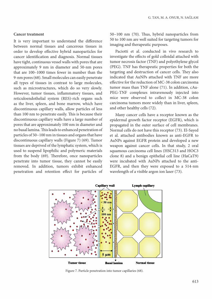
Cancer treatmentIt is very important to understand the difference between normal tissues and cancerous tissues in order to develop effective hybrid nanoparticles for cancer identification and diagnosis. Normal tissues have tight, continuous vessel walls with pores that are approximately 9 nm in diameter and 50-nm pores that are 100–1000 times fewer in number than the 9-nm pores (68). Small molecules can easily penetrate all types of tissues in contrast to large molecules, such as microstructures, which do so very slowly. However, tumor tissues, inflammatory tissues, and reticuloendothelial system (RES)-rich organs such as the liver, spleen, and bone marrow, which have discontinuous capillary walls, allow particles of less than 100 nm to penetrate easily. This is because their discontinuous capillary walls have a large number of pores that are approximately 100 nm in diameter and no basal lamina. This leads to enhanced penetration of particles of 50−100 nm in tissues and organs that have discontinuous capillary walls (Figure 7) (69). Tumor tissues are deprived of the lymphatic system, which is used to suspend lipophilic and polymeric materials from the body (69). Therefore, once nanoparticles penetrate into tumor tissue, they cannot be easily removed. In addition, tumors exhibit enhanced penetration and retention effect for particles of
50−100 nm (70). Thus, hybrid nanoparticles from 50 to 100 nm are well suited for targeting tumors for imaging and therapeutic purposes.
Paciotti et al. conducted in vivo research to investigate the effects of gold colloidal attached with tumor necrosis factor (TNF) and polyethylene glycol (PEG). TNF has therapeutic properties for both the targeting and destruction of cancer cells. They also indicated that AuNPs attached with TNF are more effective for the reduction of MC-38 colon carcinoma tumor mass than TNF alone (71). In addition, cAu-PEG-TNF complexes intravenously injected into mice were observed to collect in MC-38 colon carcinoma tumors more widely than in liver, spleen, and other healthy cells (72).
Many cancer cells have a receptor known as the epidermal growth factor receptor (EGFR), which is propagated in the outer surface of cell membranes. Normal cells do not have this receptor (73). El-Sayed et al. attached antibodies known as anti-EGFR to AuNPs against EGFR protein and developed a new weapon against cancer cells. In that study, 2 oral squamous carcinoma cell lines (HSC313 and HOC3 clone 8) and a benign epithelial cell line (HaCaT9) were incubated with AuNPs attached to the anti-EGFR, and then they were exposed to a 514-nm wavelength of a visible argon ion laser (73).
Figure 7. Particle penetration into tumor capillaries (68).
Utilization of gold nanostructures in biomedical applications
614
As a result of that study, it was determined that the energy level required to destroy malignant cells after the incubation of AuNPs with anti-EGFR was very low (approximately one-half) compared to benign cells (73). In addition, under the appropriate laser radiation conditions (19−32 W/cm2), tumor cells were destroyed without harming healthy cells through the use of AuNPs. In light of these results, it is clear that AuNPs have become prominent as a new type of selective photothermal agent under continuous-wave laser radiation at low power.Nuclear targeting and gene deliveryTargeted admission into cells is an increasingly important research area. In intracellular targeting, the nucleus in particular is a desirable target because the genetic information of the cell and transcription machinery resides there. Diagnoses of disease phenotype, identification of potential drug candidates, and treatment of disease by novel methods such as antisense therapy would be enhanced greatly by the efficient transport of materials to the living cell nuclei (74).
Targeted nuclear delivery is a challenging task, as any nuclear probe must satisfy the following minimum requirements (75) and be able to:
1. cross the cellular membrane directly or bind to specific plasma membrane receptors, as in receptor-mediated endocytosis (RME),
2. escape endosomal/lysosomal pathways,3. possess a nuclear localization signal (NLS) to
interact with the nuclear pore complex,4. be small enough to penetrate cells and cross
the nuclear membrane (<100 nm for uptake by RME and <30 nm for import through nuclear pores), and
5. have low toxicity.In biology, the most efficient nuclear targeting
is accomplished by viruses, which commonly utilize different peptides for crossing each cellular membrane barrier.
In the pioneering work in this field, Feldherr and Akin studied the nuclear translocation of AuNPs carrying peptides from the SV40 large T antigen; they used microinjection or chemically modified cells, thus bypassing cellular membrane entry (76).
Zhao et al. derivatized supermagnetic nanoparticles carrying HIV TAT peptides (77). TAT peptides are generic translocation peptides, and when conjugated to magnetic nanoparticles, effective nuclear targeting was achieved for the investigated HeLa cell line.
Tkachenko et al. conducted a study to identify the most capable peptides for accomplishing each of the tasks stated above and then combined these peptides on a single nanometer-sized platform. They chose AuNPs of 20 nm in diameter as the platform. The gold particle was modified with a shell of bovine serum albumin (BSA) conjugated to various cellular targeting peptides (78). AuNPs attached to different peptide series were explored to achieve nuclear targeting in intact HepG2 cells. They chose HepG2 cells instead of the HeLa cell lines, which were used in many studies due to the difficulty of membrane translocation. In that study, BSA-AuNP-peptides were incubated with the HepG2 cell line for 2 h, and then a color video microscope and differential interference contrast microscopy combination was used to monitor the trajectories of the particles inside cells.
As a result of this nucleus localization study, it was observed that AuNPs conjugated with peptide #1 (CGGGPKKKRKVGG), which was derived from SV40 large T antigen and had a nucleus localization signal that was internalized via RME; however, it was unable to escape the endosome and target the nucleus. Particles conjugated with peptide #2 (CGGFSTSLRARKA), which was derived from adenoviral fiber protein and had a nucleus localization signal, could not enter HepG2 cells. The particles conjugated with peptide #3 (CKKKKKKSEDEYPYVPN), which originated from adenoviral protein and contained RME, entered the cell (B) but remained trapped in endosomes and did not reach the nucleus. The particles carrying peptide #4 (CKKKKKKKSEDEYPYVP-NFSTSLRARKA), which was obtained from adenoviral fiber protein formed with a long single peptide chain including both RME and NLS, managed to target the nucleus; however, nanoparticles carrying peptides #2 and #3 had a greater propensity for nuclear targeting than
G. TAN, M. A. ONUR, N. SAĞLAM
615
any other single peptide explored (78). In view of these results, the nanoparticle complex must present both RME and NLS peptides to enter intact HepG2 cells and achieve nuclear localization (78). The origin of the higher nuclear targeting efficiency in nanoparticles carrying 2 short peptides versus 1 long sequence could be structural or spatial. Thus, when 2 short peptides are attached to a single nanoparticle, it is likely that the individual targeting signals are more accessible to cellular receptors (78).
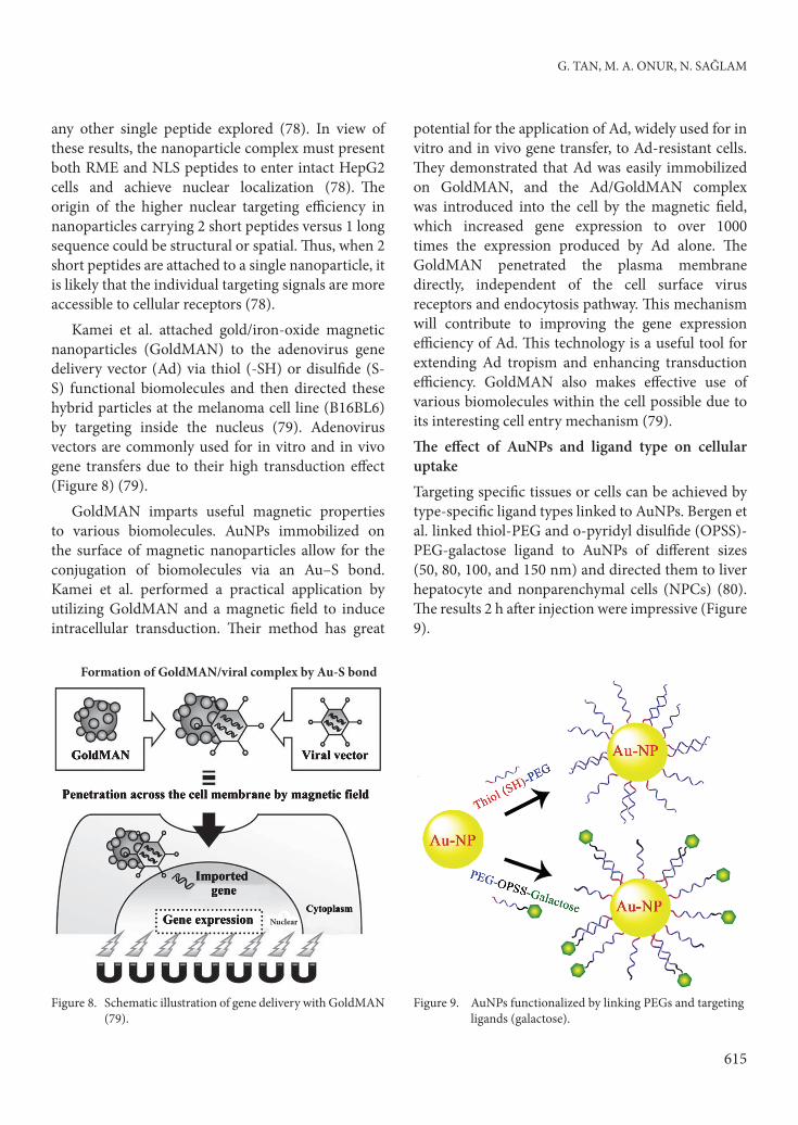
Kamei et al. attached gold/iron-oxide magnetic nanoparticles (GoldMAN) to the adenovirus gene delivery vector (Ad) via thiol (-SH) or disulfide (S-S) functional biomolecules and then directed these hybrid particles at the melanoma cell line (B16BL6) by targeting inside the nucleus (79). Adenovirus vectors are commonly used for in vitro and in vivo gene transfers due to their high transduction effect (Figure 8) (79).
GoldMAN imparts useful magnetic properties to various biomolecules. AuNPs immobilized on the surface of magnetic nanoparticles allow for the conjugation of biomolecules via an Au–S bond. Kamei et al. performed a practical application by utilizing GoldMAN and a magnetic field to induce intracellular transduction. Their method has great
potential for the application of Ad, widely used for in vitro and in vivo gene transfer, to Ad-resistant cells. They demonstrated that Ad was easily immobilized on GoldMAN, and the Ad/GoldMAN complex was introduced into the cell by the magnetic field, which increased gene expression to over 1000 times the expression produced by Ad alone. The GoldMAN penetrated the plasma membrane directly, independent of the cell surface virus receptors and endocytosis pathway. This mechanism will contribute to improving the gene expression efficiency of Ad. This technology is a useful tool for extending Ad tropism and enhancing transduction efficiency. GoldMAN also makes effective use of various biomolecules within the cell possible due to its interesting cell entry mechanism (79).The effect of AuNPs and ligand type on cellular uptakeTargeting specific tissues or cells can be achieved by type-specific ligand types linked to AuNPs. Bergen et al. linked thiol-PEG and o-pyridyl disulfide (OPSS)-PEG-galactose ligand to AuNPs of different sizes (50, 80, 100, and 150 nm) and directed them to liver hepatocyte and nonparenchymal cells (NPCs) (80). The results 2 h after injection were impressive (Figure 9).
Nuclear
Formation of GoldMAN/viral complex by Au-S bond
Figure 8. Schematic illustration of gene delivery with GoldMAN (79).
Figure 9. AuNPs functionalized by linking PEGs and targeting ligands (galactose).
Utilization of gold nanostructures in biomedical applications
616
In AuNP ligand nanoconjugation, AuNP-PEG-galactose complexes exhibited more specificity than AuNP-PEG or AuNPs alone. In particular, 50-nm AuNPs and AuNP-PEG-galactose particles were internalized by liver hepatocyte and NPC at the highest ratio (80).
Latest developments in AuNP design
Designing controlled drug delivery platforms that allow for management of location and timing of drug release under physiological conditions remains a challenge. In particular, when nanoparticles carry or encapsulate toxic pharmaceutical payloads, zero premature release and stimuli-responsive controlled release of the precious and often toxic pharmaceutical cargo are 2 important prerequisites that impact therapeutic efficacy and cytotoxicity (81).
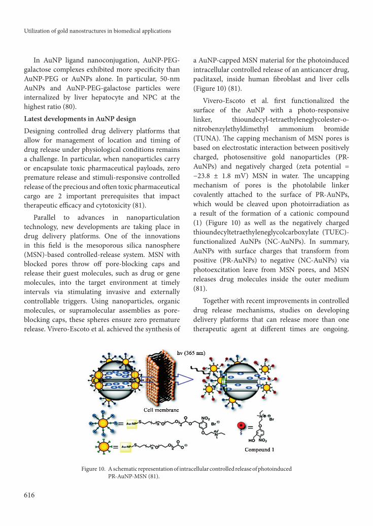
Parallel to advances in nanoparticulation technology, new developments are taking place in drug delivery platforms. One of the innovations in this field is the mesoporous silica nanosphere (MSN)-based controlled-release system. MSN with blocked pores throw off pore-blocking caps and release their guest molecules, such as drug or gene molecules, into the target environment at timely intervals via stimulating invasive and externally controllable triggers. Using nanoparticles, organic molecules, or supramolecular assemblies as pore-blocking caps, these spheres ensure zero premature release. Vivero-Escoto et al. achieved the synthesis of
a AuNP-capped MSN material for the photoinduced intracellular controlled release of an anticancer drug, paclitaxel, inside human fibroblast and liver cells (Figure 10) (81).
Vivero-Escoto et al. first functionalized the surface of the AuNP with a photo-responsive linker, thioundecyl-tetraethyleneglycolester-o-nitrobenzylethyldimethyl ammonium bromide (TUNA). The capping mechanism of MSN pores is based on electrostatic interaction between positively charged, photosensitive gold nanoparticles (PR-AuNPs) and negatively charged (zeta potential = −23.8 ± 1.8 mV) MSN in water. The uncapping mechanism of pores is the photolabile linker covalently attached to the surface of PR-AuNPs, which would be cleaved upon photoirradiation as a result of the formation of a cationic compound (1) (Figure 10) as well as the negatively charged thioundecyltetraethyleneglycolcarboxylate (TUEC)-functionalized AuNPs (NC-AuNPs). In summary, AuNPs with surface charges that transform from positive (PR-AuNPs) to negative (NC-AuNPs) via photoexcitation leave from MSN pores, and MSN releases drug molecules inside the outer medium (81).
Together with recent improvements in controlled drug release mechanisms, studies on developing delivery platforms that can release more than one therapeutic agent at different times are ongoing.
Figure 10. A schematic representation of intracellular controlled release of photoinduced PR-AuNP-MSN (81).
G. TAN, M. A. ONUR, N. SAĞLAM
617
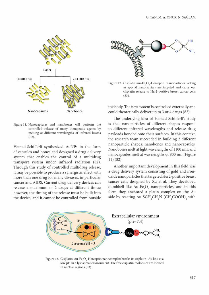
Hamad-Schifferli synthesized AuNPs in the form of capsules and bones and designed a drug delivery system that enables the control of a multidrug transport system under infrared radiation (82). Through this study of controlled multidrug release, it may be possible to produce a synergistic effect with more than one drug for many diseases, in particular cancer and AIDS. Current drug delivery devices can release a maximum of 2 drugs at different times; however, the timing of the release must be built into the device, and it cannot be controlled from outside
the body. The new system is controlled externally and could theoretically deliver up to 3 or 4 drugs (82).
The underlying idea of Hamad-Schifferli’s study is that nanoparticles of different shapes respond to different infrared wavelengths and release drug payloads bonded onto their surfaces. In this context, the research team succeeded in building 2 different nanoparticle shapes: nanobones and nanocapsules. Nanobones melt at light wavelengths of 1100 nm, and nanocapsules melt at wavelengths of 800 nm (Figure 11) (82).
Another important development in this field was a drug delivery system consisting of gold and iron-oxide nanoparticles that targeted Her2-positive breast cancer cells designed by Xu et al. They developed dumbbell-like Au-Fe3O4 nanoparticles, and in this form they anchored a platin complex on the Au side by reacting Au-SCH2CH2N (CH2COOH)2 with
Figure 11. Nanocapsules and nanobones will perform the controlled release of many therapeutic agents by melting at different wavelengths of infrared beams (82).
Figure 12. Cisplatin-Au-Fe3O4-Herceptin nanoparticles acting as special nanocarriers are targeted and carry out cisplatin release to Her2-positive breast cancer cells (83).
Extracellular environment(ph=7.4)
Figure 13. Cisplatin-Au-Fe3O4-Herceptin nanocomplex breaks its cisplatin−Au link at a low pH in a lysosomal environment. The free cisplatin molecules are located in nuclear regions (83).
Utilization of gold nanostructures in biomedical applications
618
cisplatin and the Her2-specific monoclonal antibody Herceptin (targeting agent); this was linked to the iron-oxide side through PEG3000-CONH-Herceptin (Figure 12) (83).
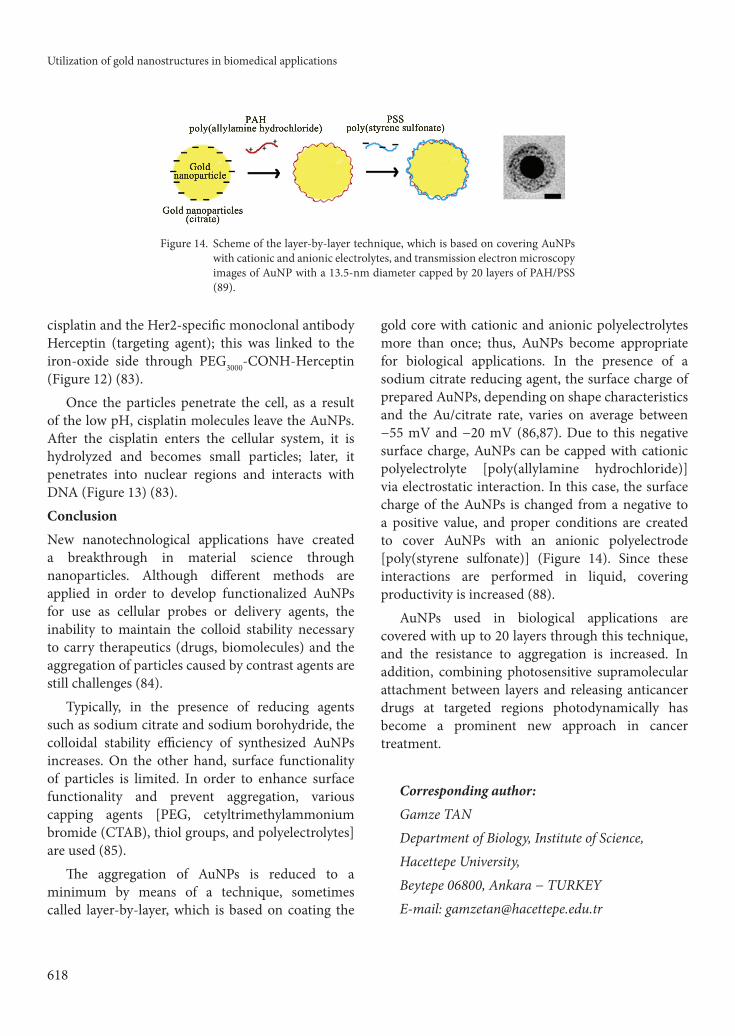
Once the particles penetrate the cell, as a result of the low pH, cisplatin molecules leave the AuNPs. After the cisplatin enters the cellular system, it is hydrolyzed and becomes small particles; later, it penetrates into nuclear regions and interacts with DNA (Figure 13) (83).ConclusionNew nanotechnological applications have created a breakthrough in material science through nanoparticles. Although different methods are applied in order to develop functionalized AuNPs for use as cellular probes or delivery agents, the inability to maintain the colloid stability necessary to carry therapeutics (drugs, biomolecules) and the aggregation of particles caused by contrast agents are still challenges (84).
Typically, in the presence of reducing agents such as sodium citrate and sodium borohydride, the colloidal stability efficiency of synthesized AuNPs increases. On the other hand, surface functionality of particles is limited. In order to enhance surface functionality and prevent aggregation, various capping agents [PEG, cetyltrimethylammonium bromide (CTAB), thiol groups, and polyelectrolytes] are used (85).
The aggregation of AuNPs is reduced to a minimum by means of a technique, sometimes called layer-by-layer, which is based on coating the
gold core with cationic and anionic polyelectrolytes more than once; thus, AuNPs become appropriate for biological applications. In the presence of a sodium citrate reducing agent, the surface charge of prepared AuNPs, depending on shape characteristics and the Au/citrate rate, varies on average between −55 mV and −20 mV (86,87). Due to this negative surface charge, AuNPs can be capped with cationic polyelectrolyte [poly(allylamine hydrochloride)] via electrostatic interaction. In this case, the surface charge of the AuNPs is changed from a negative to a positive value, and proper conditions are created to cover AuNPs with an anionic polyelectrode [poly(styrene sulfonate)] (Figure 14). Since these interactions are performed in liquid, covering productivity is increased (88).
AuNPs used in biological applications are covered with up to 20 layers through this technique, and the resistance to aggregation is increased. In addition, combining photosensitive supramolecular attachment between layers and releasing anticancer drugs at targeted regions photodynamically has become a prominent new approach in cancer treatment.
Corresponding author: Gamze TAN Department of Biology, Institute of Science, Hacettepe University, Beytepe 06800, Ankara − TURKEYE-mail: [email protected]
Figure 14. Scheme of the layer-by-layer technique, which is based on covering AuNPs with cationic and anionic electrolytes, and transmission electron microscopy images of AuNP with a 13.5-nm diameter capped by 20 layers of PAH/PSS (89).

G. TAN, M. A. ONUR, N. SAĞLAM
619
References
1. Chen PC, Mwakwari SC, Oyelere AK. Gold nanoparticles: from nanomedicine to nanosensing. Nanotechnol Sci Appl 1: 45−66, 2008.
2. Sahoo SK, Labhasetwar V. Nanotech approaches to drug delivery and imaging. Drug Discov Today 8: 1112−20, 2003.
3. Sahoo SK, Parveen S, Panda JJ. The present and future of nanotechnology in human health care. Nanomed Nanotechnol Biol Med 3: 20−31, 2007.
4. Prato M, Kostarelos K, Bianco A. Functionalized carbon nanotubes in drug design and discovery. Acc Chem Res 41: 60−8, 2007.
5. Mandal D, Maran A, Yaszemski M et al. Cellular uptake of gold nanoparticles directly cross-linked with carrier peptides by osteosarcoma cells. J Mater Sci Mater Med 20: 347−50, 2009.
6. Connor EE, Mwamuka J, Gole A et al. Gold nanoparticles are taken up by human cells but do not cause acute cytotoxicity. Small 1: 325−7, 2005.
7. Murphy CJ, Gole AM, Hunyadi SE et al. One-dimensional colloidal gold and silver nanostructures. Inorg Chem 45: 7544−54, 2006.
8. Lu AH, Salabas EL, Schüth F. Magnetic nanoparticles: synthesis, protection, functionalization, and application. Angew Chem Int Ed 46: 1222−44, 2007.
9. Giljohann GA, Seferos DS, Daniel WL et al. Gold nanoparticles for biology and medicine. Angew Chem Int Ed 49: 3280–94, 2010.
10. Lasagna-Reeves C, Gonzalez-Romero D, Barria MA et al. Bioaccumulation and toxicity of gold nanoparticles after repeated administration in mice. Biochem Biophys Res Commun 393: 649−55, 2010.
11. Shukla R, Bansal V, Chaudhary M et al. Biocompatibility of gold nanoparticles and their endocytotic fate inside the cellular compartment: a microscopic overview. Langmuir 21: 10644−54, 2005.
12. Ghosh P, Han G, De M et al. Gold nanoparticles in delivery applications. Adv Drug Delivery Rev 60: 1307−15, 2008.
13. Polizzi MA, Stasko NA, Schoenfisch MH. Water-soluble nitric oxide-releasing gold nanoparticles. Langmuir 23: 4938−43, 2007.
14. Hong R, Han G, Fernández JM et al. Glutathione-mediated deliv15ery and release using monolayer protected nanoparticle carriers. J Am Chem Soc 128: 1078−9, 2006.
15. Han G, You CC, Kim B et al. Light-regulated release of DNA and its delivery to nuclei by means of photolabile gold nanoparticles. Angew Chem Int Ed 45: 3165−9, 2006.
16. Li D, Li G, Guo W et al. Glutathione-mediated release of functional plasmid DNA from positively charged quantum dots. Biomaterials 29: 2776−82, 2008.
17. Cai W, Gao T, Hong H et al. Applications of gold nanoparticles in cancer nanotechnology. Nanotechnol Sci Appl 1: 17−32, 2008.
18. Pissuwan D, Niidome T, Cortie MB. The forthcoming applications of gold nanoparticles in drug and gene delivery systems. J Control Release 149: 65−71, 2011.
19. Hostetler MJ, Wingate JE, Zhong CJ et al. Alkanethiolate gold cluster molecules with core diameters from 1.5 to 5.2 nm: core and monolayer properties as a function of core size. Langmuir 14: 17−30, 1998.
20. Gibson JD, Khanal BP, Zubarev ER. Paclitaxel-functionalized gold nanoparticles. J Am Chem Soc 129: 11653−61, 2007.
21. Kennedy LC, Bickford LR, Lewinski NA et al. A new era for cancer treatment: gold nanoparticle mediated thermal therapies. Small 7: 169−83, 2011.
22. Zhu J, Li JJ, Zhao JW et al. Light absorption efficiencies of gold nanoellipsoid at different resonance frequency. J Mater Sci 43: 5199−205, 2008.
23. Sassaroli E, Li KCP, O’Neill BE. Numerical investigation of heating of a gold nanoparticle and the surrounding microenvironment by nanosecond laser pulses for nanomedicine applications. Phys Med Biol 54: 5541−60, 2009.
24. Yuan H, Cai RX, Pang DW. A simple approach to control the growth of non-spherical gold nanoparticles. Chin Chem Lett 14: 1163−6, 2003.
25. Orendorff CJ, Gole A, Sau TK et al. Surface-enhanced Raman spectroscopy of self-assembled monolayers: sandwich architecture and nanoparticle shape dependence. Anal Chem 77: 3261−6, 2005.
26. Tréguer-Delapierre M, Majimel J, Mornet S et al. Synthesis of non-spherical gold nanoparticles. Gold Bull 41: 195−207, 2008.
27. Hu M, Chen J, Li ZY et al. Gold nanostructures: engineering their plasmonic properties for biomedical applications. Chem Soc Rev 35: 1084−94, 2006.
28. Murphy CJ, Sau TK, Gole AM et al. Anisotropic metal nanoparticles: synthesis, assembly, and optical applications. J Phys Chem B 109: 13857−70, 2005.
29. Pérez-Juste J, Pastoriza-Santos I, Liz-Marzán LM et al. Gold nanorods: synthesis, characterization and applications. Coord Chem Rev 249: 1870−901, 2005.
30. Hirsch L, Gobin A, Lowery A et al. Metal nanoshells. Ann Biomed Eng 34: 15−22, 2006.
31. Huang X, Jain PK, El-Sayed IH et al. Gold nanoparticles: interesting optical properties and recent applications in cancer diagnostics and therapy. Nanomedicine 2: 681−93, 2007.
32. Skrabalak SE, Chen J, Sun Y et al. Gold nanocages: synthesis, properties, and applications. Acc Chem Res 41: 1587−95, 2008.
33. El-Sayed MA. Some interesting properties of metals confined in time and nanometer space of different shapes. Acc Chem Res 34: 257−64, 2001.

Utilization of gold nanostructures in biomedical applications
620
34. Sun Y, Xia Y. Increased sensitivity of surface plasmon resonance of gold nanoshells compared to that of gold solid colloids in response to environmental changes. Anal Chem 74: 5297−305, 2002.
35. Kelly KL, Coronado E, Zhao LL et al. The optical properties of metal nanoparticles: the influence of size, shape, and dielectric environment. J Phys Chem B 107: 668−77, 2002.
36. Noguez C. Surface plasmons on metal nanoparticles: the influence of shape and physical environment. J Phys Chem C 111: 3806−19, 2007.
37. Tong L, Wei Q, Wei A et al. Gold nanorods as contrast agents for biological imaging: optical properties, surface conjugation and photothermal effects. Photochem Photobiol 85: 21−32, 2009.
38. Wust P, Hildebrandt B, Sreenivasa G et al. Hyperthermia in combined treatment of cancer. Lancet Oncol 3: 487−97, 2002.
39. Huang HC, Barua S, Sharma G et al. Inorganic nanoparticles for cancer imaging and therapy. J Controlled Release 155: 344−57, 2011.
40. Turkevich J, Stevenson PC, Hillier J. A study of the nucleation and growth processes in the synthesis of colloidal gold. Discuss Faraday Soc 11: 55−75, 1951.
41. Beesley JE. Colloidal gold for microbiological immunocytochemistry. In: Hayat MA. ed. Colloidal Gold: Principles, Methods and Applications. Academic Press; 1989: pp. 421−5.
42. Brust M, Walker M, Bethell D et al. Synthesis of thiol-derivatised gold nanoparticles in a two-phase liquid-liquid system. J Chem Soc, Chem Commun 7: 801−2, 1994.
43. Templeton AC, Wuelfing WP, Murray RW. Monolayer-protected cluster molecules. Acc Chem Res 33: 27−36, 1999.
44. Huaizhi Z, Yuanta N. China’s ancient gold drugs. Gold Bull 34: 24−9, 2001.
45. Edelman ER, Seifert P, Groothuis A et al. Gold-coated NIR stents in porcine coronary arteries. Circulation 103: 429−34, 2001.
46. Thelen A, Bauknecht HC, Asbach P et al. Behavior of metal implants used in ENT surgery in 7 Tesla magnetic resonance imaging. Eur Arch Otorhinolaryngol 263: 900−5, 2006.
47. Svedman C, Dunér K, Kehler M et al. Lichenoid reactions to gold from dental restorations and exposure to gold through intracoronary implant of a gold-plated stent. Clin Res Cardiol 95: 689−91, 2006.
48. Sun RW, Ma DL, Wong EL et al. Some uses of transition metal complexes as anti-cancer and anti-HIV agents. Dalton Trans 43: 4884−92, 2007.
49. Shaw CF. Gold-based therapeutic agents. Chem Rev 99: 2589−600, 1999.
50. Moolhuizen G, Paciotti GF, de Leede LGJ et al. Colloidal Gold Nanoparticles. Business Briefing: Pharmatech. Touch Briefings. London; 2004.
51. Kogan MJ, Bastus NG, Amigo R et al. Nanoparticle-mediated local and remote manipulation of protein aggregation. Nano Lett 6: 110−5, 2005.
52. Andreoli CM, Miller JW. Anti-vascular endothelial growth factor therapy for ocular neovascular disease. Curr Opin Ophthalmol 18: 502−8, 2007.
53. Hayashi A, Naseri A, Pennesi ME et al. Subretinal delivery of immunoglobulin G with gold nanoparticles in the rabbit eye. Jpn J Ophthalmol 53: 249−56, 2009.
54. Kim CK, Ghosh P, Pagliuca C et al. Entrapment of hydrophobic drugs in nanoparticle monolayers with efficient release into cancer cells. J Am Chem Soc 131: 1360−1, 2009.
55. Rafique S, Idrees M, Nasim A et al. Transition metal complexes as potential therapeutic agents. Biotechnol Mol Biol Rev 5: 38−45, 2010.
56. Dykman LA, Bogatyrev VA. Gold nanoparticles: preparation, functionalisation and applications in biochemistry and immunochemistry. Russ Chem Rev 76: 181−94, 2007.
57. Kumari A, Yadav SK, Yadav SC. Biodegradable polymeric nanoparticles based drug delivery systems. Colloids Surf, B 75: 1−18, 2010.
58. Joshi HM, Bhumkar DR, Joshi K et al. Gold nanoparticles as carriers for efficient transmucosal insulin delivery. Langmuir 22: 300−5, 2006.
59. Yavuz M, İlçe AÖ, Kaymakçı Ş et al. Meme kanserli hastaların tamamlayıcı ve alternatif tedavi yöntemlerini kullanma durumlarının incelenmesi. Turkiye Klinikleri J Med Sci 27: 680−6, 2007 (article in Turkish).
60. Uysal A, Yaşa H, Çalık R. Çok yüksek doz emetojenik ajan içermeyen kemoterapi protokollerine bağlı bulantı ve kusmalarda klorpromazin kullanılması. Turk J Resc Med Sci 9: 369−73, 1991 (article in Turkish).
61. Day ES, Thompson PA, Zhang L et al. Nanoshell-mediated photothermal therapy improves survival in a murine glioma model. J Neurooncol 104: 55−63, 2011.
62. Kim JW, Galanzha EI, Shashkov EV et al. Golden carbon nanotubes as multimodal photoacoustic and photothermal high-contrast molecular agents. Nat Nanotechnol 4: 688−94, 2009.
63. de la Fuente JM, Berry CC. Tat peptide as an efficient molecule to translocate gold nanoparticles into the cell nucleus. Bioconjugate Chem 16: 1176−80, 2005.
64. Gu YJ, Cheng J, Lin CC et al. Nuclear penetration of surface functionalized gold nanoparticles. Toxicol Appl Pharmacol 237: 196−204, 2009.
65. Kang B, Mackey MA, El-Sayed MA. Nuclear targeting of gold nanoparticles in cancer cells induces DNA damage, causing cytokinesis arrest and apoptosis. J Am Chem Soc 132: 1517−9, 2010.
66. Karataş ÖF, Sezgin E, Aydın Ö et al. Interaction of gold nanoparticles with mitochondria. Colloids Surf B 71: 315−8, 2009.
G. TAN, M. A. ONUR, N. SAĞLAM
621
67. Chang MY, Shiau AL, Chen YH et al. Increased apoptotic potential and dose-enhancing effect of gold nanoparticles in combination with single-dose clinical electron beams on tumor-bearing mice. Cancer Sci 99: 1479−84, 2008.
68. Fukumori Y, Ichikawa H. Nanoparticles for cancer therapy and diagnosis. Adv Powder Technol 17: 1−28, 2006.
69. Maeda H, Seymour LW, Miyamoto Y. Conjugates of anticancer agents and polymers - advantages of macromolecular therapeutics in vivo. Bioconjugate Chem 3: 351−62, 1992.
70. Qi XR, Maitani Y, Nagai T et al. Comparative pharmacokinetics and antitumor efficacy of doxorubicin encapsulated in soybean-derived sterols and poly(ethylene glycol) liposomes in mice. Int J Pharm 146: 31−9, 1997.
71. Paciotti GF, Myer L, Weinreich D et al. Colloidal gold: a novel nanoparticle vector for tumor directed drug delivery. Drug Deliv 11: 169−83, 2004.
72. Paciotti GF, Kingston DGI, Tamarkin L. Colloidal gold nanoparticles: a novel nanoparticle platform for developing multifunctional tumor-targeted drug delivery vectors. Drug Dev Res 67: 47−54, 2006.
73. El-Sayed IH, Huang X, El-Sayed MA. Selective laser photo-thermal therapy of epithelial carcinoma using anti-EGFR antibody conjugated gold nanoparticles. Cancer Lett 239: 129−35, 2006.
74. Bilbao G, Gomez-Navarro J, Curiel DT. Targeted adenoviral vectors for cancer gene therapy. In: Walden P, Trefzer U, Sterry W. eds. Gene Therapy of Cancer. Plenum Press; 1998: pp. 365−74.
75. Tkachenko AG, Xie H, Liu Y et al. Cellular trajectories of peptide-modified gold particle complexes: comparison of nuclear localization signals and peptide transduction domains. Bioconjugate Chem 15: 482−90, 2004.
76. Feldherr CM, Akin D. The permeability of the nuclear envelope in dividing and nondividing cell cultures. J Cell Biol 111: 1−8, 1990.
77. Zhao M, Kircher MF, Josephson L et al. Differential conjugation of Tat peptide to superparamagnetic nanoparticles and its effect on cellular uptake. Bioconjugate Chem 13: 840−4, 2002.
78. Tkachenko AG, Xie H, Coleman D et al. Multifunctional gold nanoparticle−peptide complexes for nuclear targeting. J Am Chem Soc 125: 4700−1, 2003.
79. Kamei K, Mukai Y, Kojima H et al. Direct cell entry of gold/iron-oxide magnetic nanoparticles in adenovirus mediated gene delivery. Biomaterials 30: 1809−14, 2009.
80. Bergen JM, von Recum HA, Goodman TT et al. Gold nanoparticles as a versatile platform for optimizing physicochemical parameters for targeted drug delivery. Macromol Biosci 6: 506−16, 2006.
81. Vivero-Escoto JL, Slowing II, Wu CW et al. Photoinduced intracellular controlled release drug delivery in human cells by gold-capped mesoporous silica nanosphere. J Am Chem Soc 131: 3462−3, 2009.
82. Massachusetts Institute of Technology. MIT research digest. MIT TechTalk 53(13): 4, 2009.
83. Xu C, Wang B, Sun S. Dumbbell-like Au−Fe3O4 nanoparticles for target-specific platin delivery. J Am Chem Soc 131: 4216−7, 2009.
84. Kuchibhatla S, Karakoti A, Seal S. Colloidal stability by surface modification. JOM 57: 52−6, 2005.
85. Mitamura K, Imae T. Functionalization of gold nanorods toward their applications. Plasmonics 4: 23−30, 2009.
86. Majzik A, Patakfalvi R, Hornok V et al. Growing and stability of gold nanoparticles and their functionalization by cysteine. Gold Bull 42: 113−23, 2009.
87. Kim T, Lee K, Gong M et al. Control of gold nanoparticle aggregates by manipulation of interparticle interaction. Langmuir 21: 9524−8, 2005.
88. Schneider G, Decher G. From functional core/shell nanoparticles prepared via layer-by-layer deposition to empty nanospheres. Nano Lett 4: 1833−9, 2004.
89. Reum N, Fink-Straube C, Klein T et al. Multilayer coating of gold nanoparticles with drug−polymer coadsorbates. Langmuir 26: 16901−8, 2010.
Recommended