Valvular Stenosis and Regurgitation:
Assessment of Severity
Helmut Baumgartner
Adult Congenital and Valvular Heart Disease Center University of Muenster
Germany
Westfälische Wilhelms-Universität
Münster
73 pages
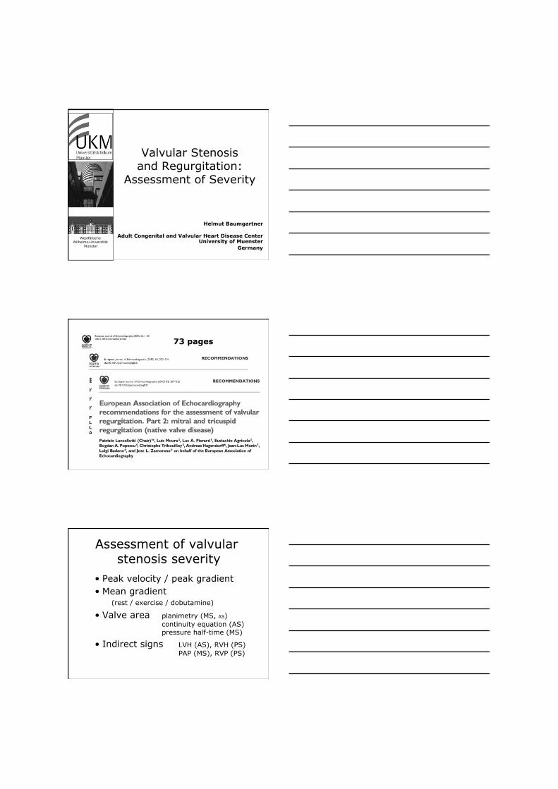
Assessment of valvular stenosis severity
• Peak velocity / peak gradient • Mean gradient
(rest / exercise / dobutamine)
• Valve area planimetry (MS, AS) continuity equation (AS) pressure half-time (MS)
• Indirect signs LVH (AS), RVH (PS) PAP (MS), RVP (PS)
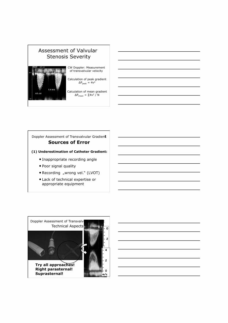
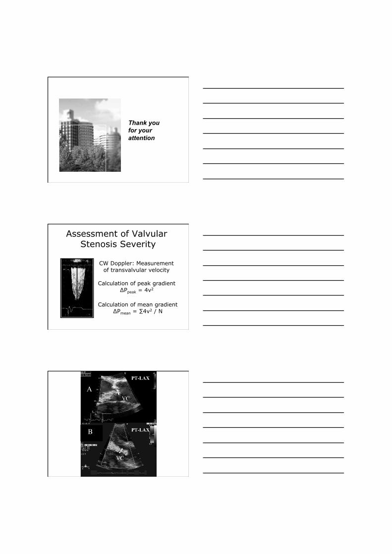
Assessment of Valvular Stenosis Severity
CW Doppler: Measurement of transvalvular velocity
Calculation of peak gradient ∆Ppeak = 4v2
Calculation of mean gradient ∆Pmean = ∑4v2 / N
• Inappropriate recording angle
• Poor signal quality
• Recording „wrong vel.“ (LVOT)
• Lack of technical expertise or appropriate equipment
(1) Underestimation of Catheter Gradient:
Doppler Assessment of Transvalvular Gradient Sources of Error
Try all approaches! Right parasternal! Suprasternal!
- 4 - - - 2 - - - 0 m/s
- 0 - - - 2 - -
Doppler Assessment of Transvalvular Gradient
Technical Aspects
• Failure to account for an increased subvalvular velocity
Doppler Assessment of Transvalvular Gradient
Sources of Error
(2) Overestimation of Catheter Gradient:
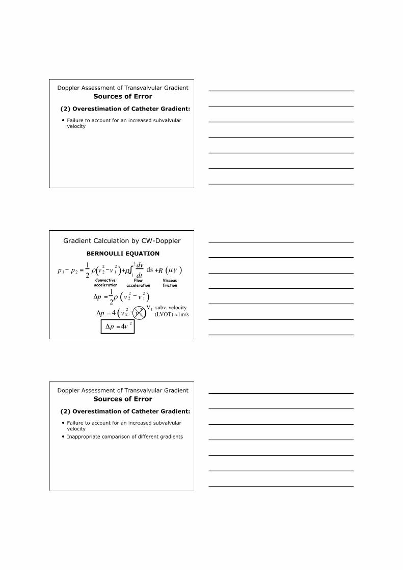
Gradient Calculation by CW-Doppler
BERNOULLI EQUATION
Δ p = 4 v 2 2 - v 1 2 ( ) Δ p = 4 v 2
1 ( p 1 - p 2 = 1 2
ρ v 2 2 - v 1
2 ) + ρ dv dt
+ R µ , v ( ) 2
∫ ds Convective acceleration
Flow acceleration
Viscous friction
Δ p = 1 2 ρ v 2
2 - v 1
2 ( ) V1: subv. velocity (LVOT) ≈1m/s
• Failure to account for an increased subvalvular velocity
• Inappropriate comparison of different gradients
Doppler Assessment of Transvalvular Gradient
Sources of Error
(2) Overestimation of Catheter Gradient:
• Failure to account for an increased subvalvular velocity
• Inappropriate comparison of different gradients
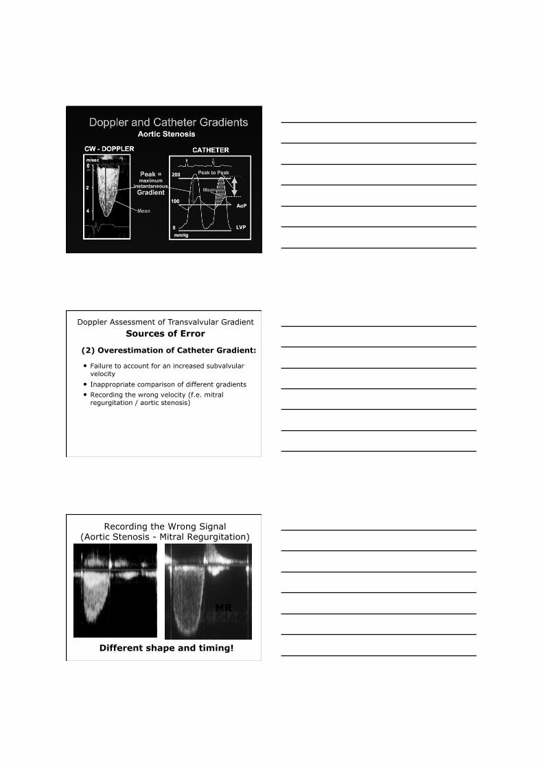
• Recording the wrong velocity (f.e. mitral regurgitation / aortic stenosis)
Doppler Assessment of Transvalvular Gradient
Sources of Error
(2) Overestimation of Catheter Gradient:
Recording the Wrong Signal (Aortic Stenosis - Mitral Regurgitation)
Different shape and timing!
AS MR
• Failure to account for an increased subvalvular velocity
• Inappropriate comparison of different gradients
• Recording the wrong velocity (f.e. mitral regurgitation / aortic stenosis)
• Nonrepresentative selection of velocity recording (arrhythmias - tendency to select highest velocities)
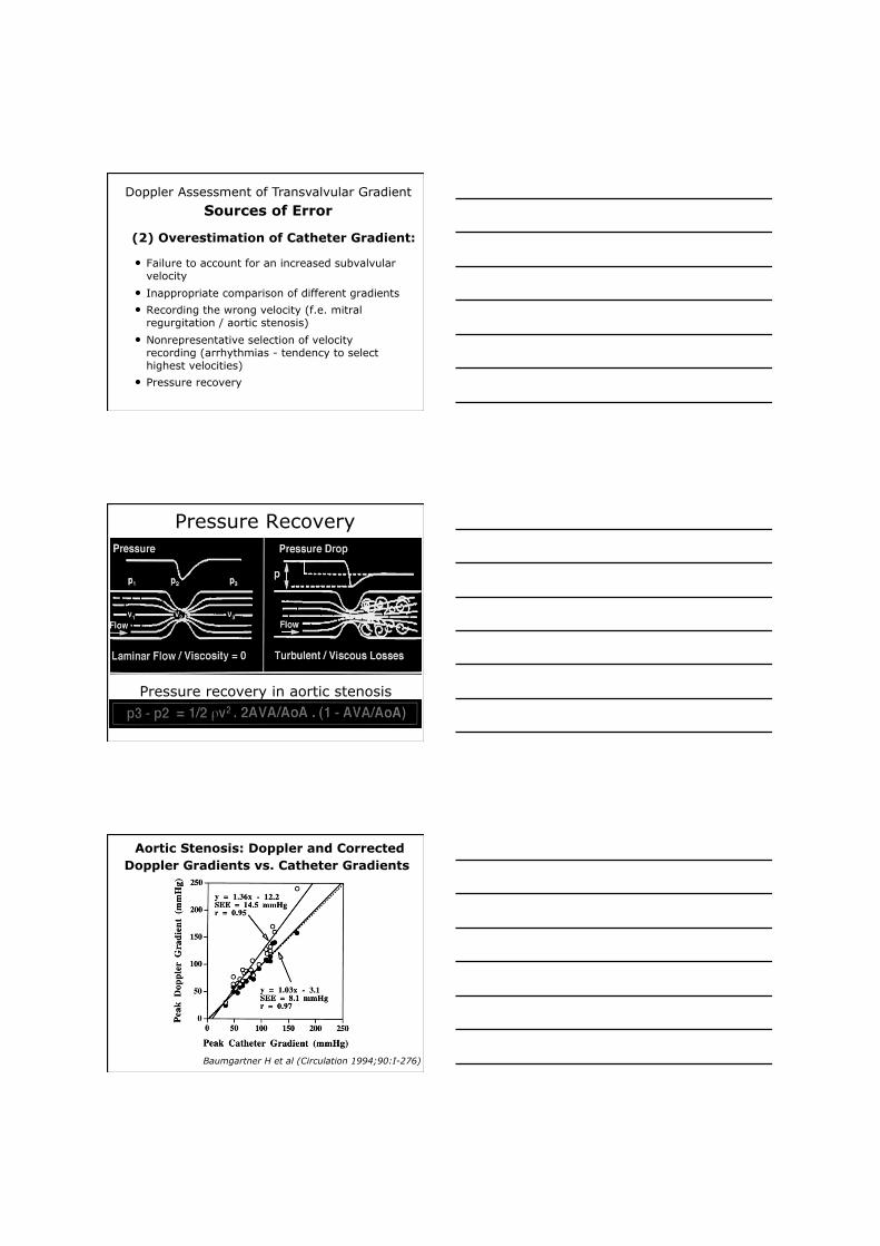
• Pressure recovery
Doppler Assessment of Transvalvular Gradient
Sources of Error
(2) Overestimation of Catheter Gradient:
Pressure Recovery
Pressure recovery in aortic stenosis
Aortic Stenosis: Doppler and Corrected Doppler Gradients vs. Catheter Gradients
Baumgartner H et al (Circulation 1994;90:I-276)
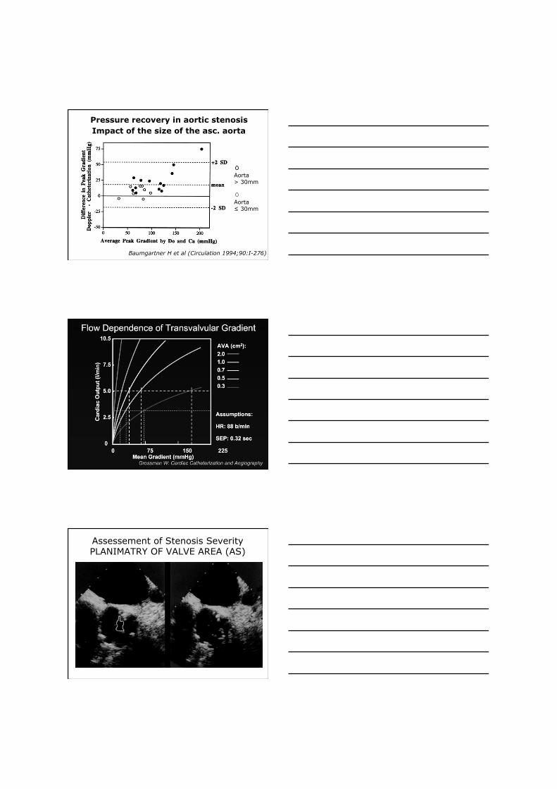
Pressure recovery in aortic stenosis Impact of the size of the asc. aorta
Baumgartner H et al (Circulation 1994;90:I-276)
Aorta > 30mm
Aorta ≤ 30mm
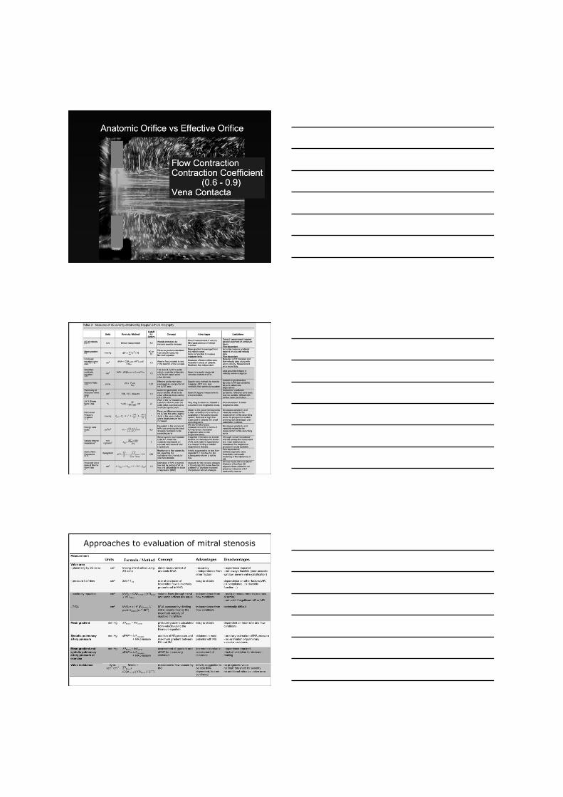
Assessement of Stenosis Severity PLANIMATRY OF VALVE AREA (AS)
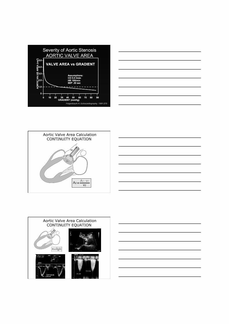
Aortic Valve Area Calculation CONTINUITY EQUATION
Aortic Valve Area Calculation CONTINUITY EQUATION
A1
V1
V2
Approaches to evaluation of mitral stenosis
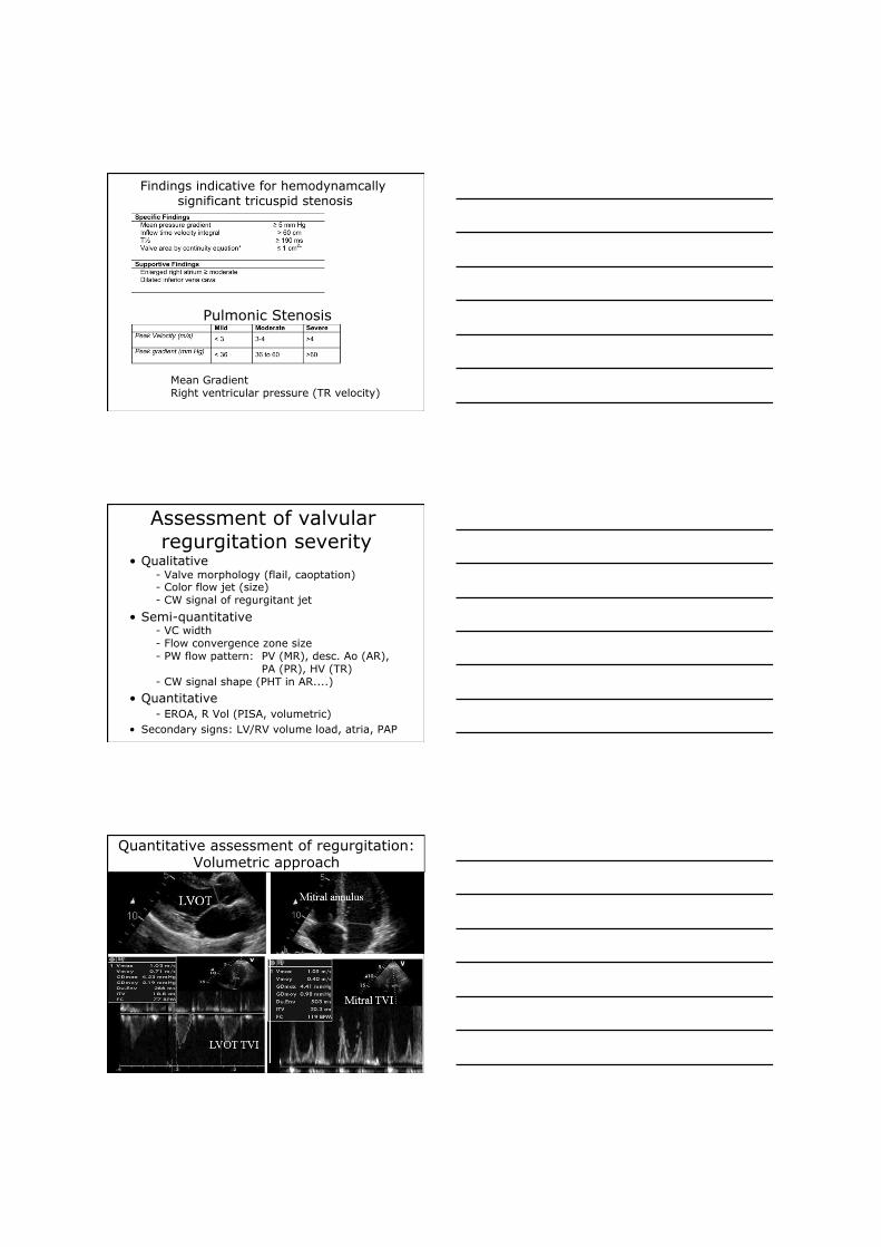
Findings indicative for hemodynamcally significant tricuspid stenosis
Pulmonic Stenosis
Mean Gradient Right ventricular pressure (TR velocity)
Assessment of valvular regurgitation severity
• Qualitative - Valve morphology (flail, caoptation) - Color flow jet (size) - CW signal of regurgitant jet
• Semi-quantitative - VC width - Flow convergence zone size - PW flow pattern: PV (MR), desc. Ao (AR), PA (PR), HV (TR) - CW signal shape (PHT in AR....)
• Quantitative - EROA, R Vol (PISA, volumetric)
• Secondary signs: LV/RV volume load, atria, PAP
Quantitative assessment of regurgitation: Volumetric approach
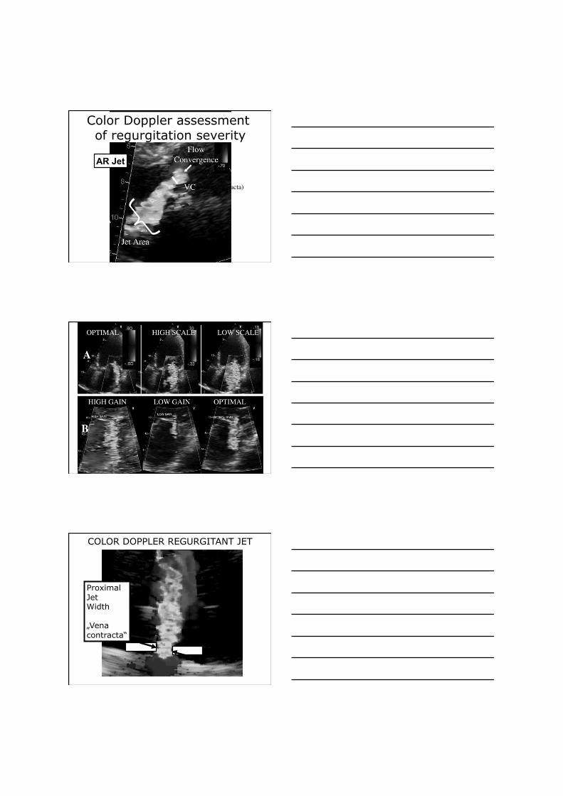
Color Doppler assessment of regurgitation severity
Ao LV
LA
(Vena Contracta)
AR Jet
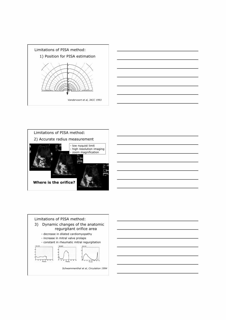
COLOR DOPPLER REGURGITANT JET
Proximal Jet Width
„Vena contracta“
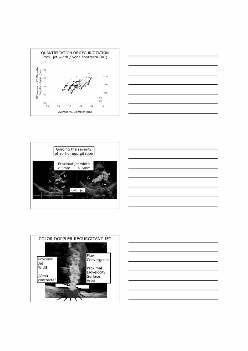
QUANTIFICATION OF REGURGITATION Prox. jet width / vena contracta (VC)
Diffe
rence
in V
C D
iam
eter
D
opple
r -
Lase
r (c
m)
Average VC Diameter (cm)
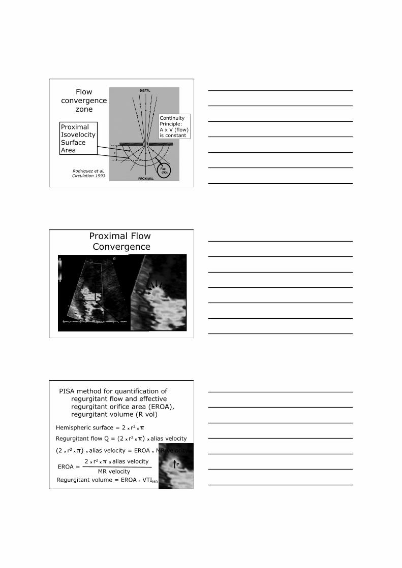
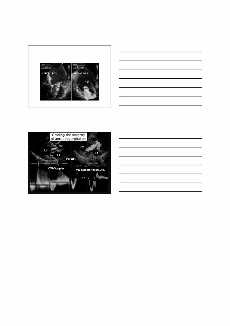
Grading the severity of aortic regurgitation
Proximal jet width < 3mm > 6mm
color jet
Flow Convergence
Proximal Isovelocity Surface Area
COLOR DOPPLER REGURGITANT JET
Proximal Jet Width
„Vena contracta“
Rodriguez et al, Circulation 1993
Flow convergence
zone
Proximal Isovelocity Surface Area
Continuity Principle: A x V (flow) is constant
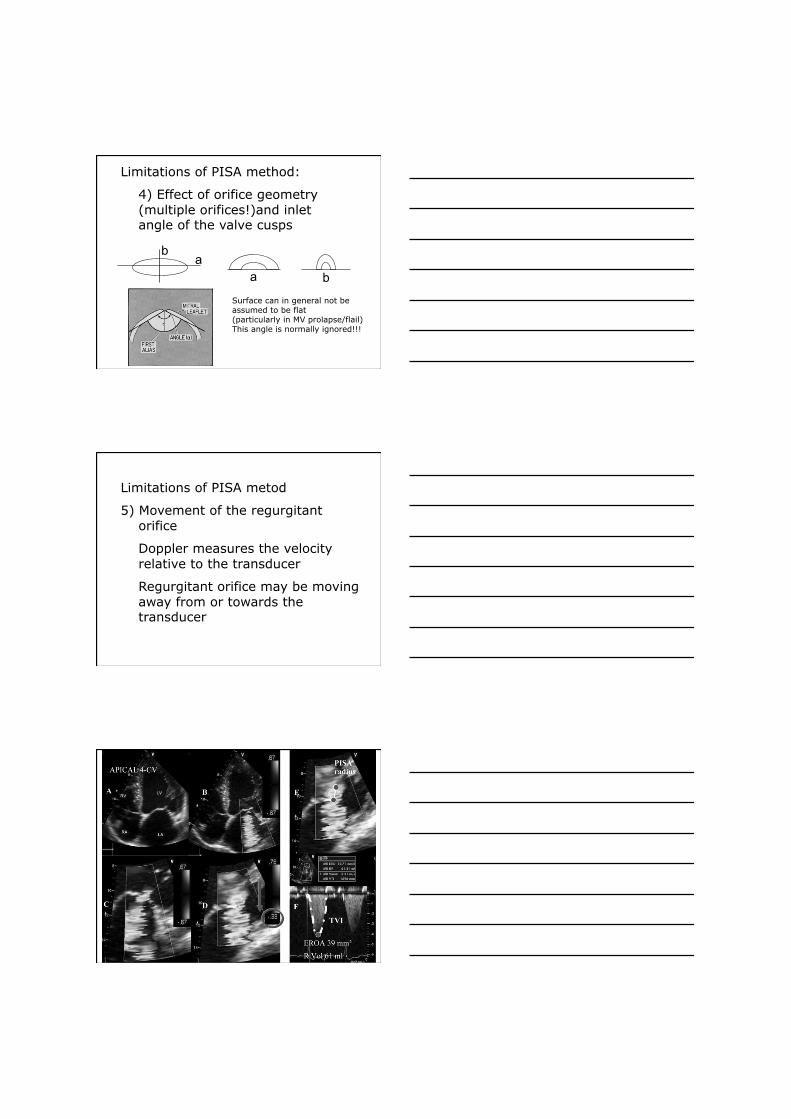
Proximal Flow Convergence
Hemispheric surface = 2 x r2 x π Regurgitant flow Q = (2 x r2 x π) x alias velocity
(2 x r2 x π) x alias velocity = EROA x MR velocity
PISA method for quantification of regurgitant flow and effective regurgitant orifice area (EROA), regurgitant volume (R vol)
MR velocity
2 x r2 x π x alias velocity EROA = r
Regurgitant volume = EROA x VTIMR
Limitations of PISA method:
1) Position for PISA estimation
Vandervoort et al, JACC 1993
Limitations of PISA method:
2) Accurate radius measurement
- low nyquist limit - high resolution imaging - zoom magnification
Where is the orifice?
Limitations of PISA method: 3) Dynamic changes of the anatomic
regurgitant orifice area - decrease in dilated cardiomyopathy
- increase in mitral valve prolaps - constant in rheumatic mitral regurgitation
Schwammenthal et al, Circulation 1994
Limitations of PISA method:
4) Effect of orifice geometry (multiple orifices!)and inlet angle of the valve cusps
a b
a b
Surface can in general not be assumed to be flat (particularly in MV prolapse/flail) This angle is normally ignored!!!
Limitations of PISA metod
5) Movement of the regurgitant orifice
Doppler measures the velocity relative to the transducer
Regurgitant orifice may be moving away from or towards the transducer

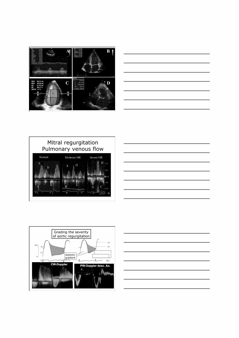
Mitral regurgitation Pulmonary venous flow
Grading the severity of aortic regurgitation
diastolic gradient
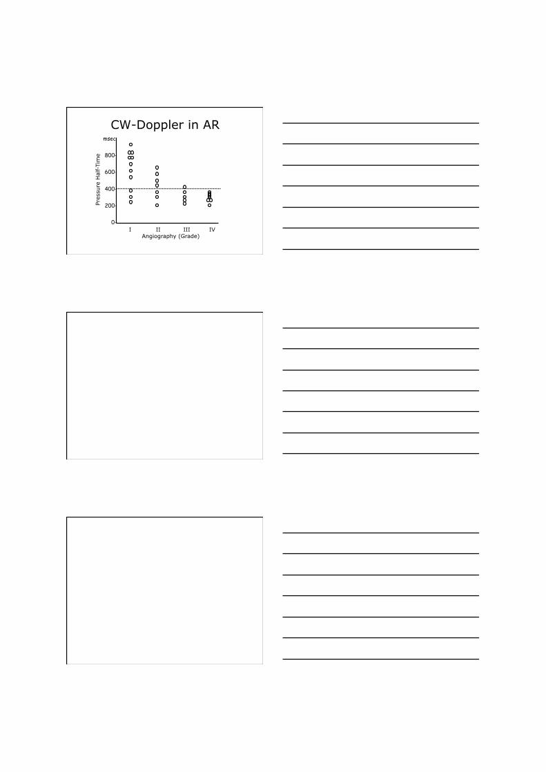
CW-Doppler in AR msec
800-
600-
400-
200-
0-
Pre
ssure
Hal
f-Tim
e
I II III IV Angiography (Grade)
Quantification of Valvular Regurgitation
Grading the severity of aortic regurgitation
EAE recommendations 2010
Grading the severity of mitral regurgitation
EAE recommendations 2010
Thank you for your attention
Assessment of Valvular Stenosis Severity
m/sec
2
4
0 CW Doppler: Measurement
of transvalvular velocity
Calculation of peak gradient ∆Ppeak = 4v2
Calculation of mean gradient ∆Pmean = ∑4v2 / N
Grading the severity of aortic regurgitation
Recommended