X-Ray Safety Training
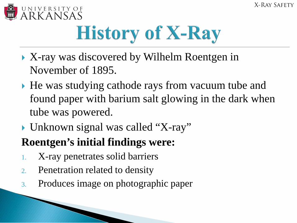
X-ray was discovered by Wilhelm Roentgen in November of 1895.
He was studying cathode rays from vacuum tube and found paper with barium salt glowing in the dark when tube was powered.
Unknown signal was called “X-ray”Roentgen’s initial findings were:1. X-ray penetrates solid barriers 2. Penetration related to density3. Produces image on photographic paper
X-Ray Safety
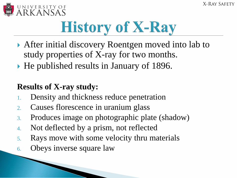
After initial discovery Roentgen moved into lab to study properties of X-ray for two months.
He published results in January of 1896.
Results of X-ray study:1. Density and thickness reduce penetration2. Causes florescence in uranium glass3. Produces image on photographic plate (shadow)4. Not deflected by a prism, not reflected5. Rays move with some velocity thru materials6. Obeys inverse square law
X-Ray Safety
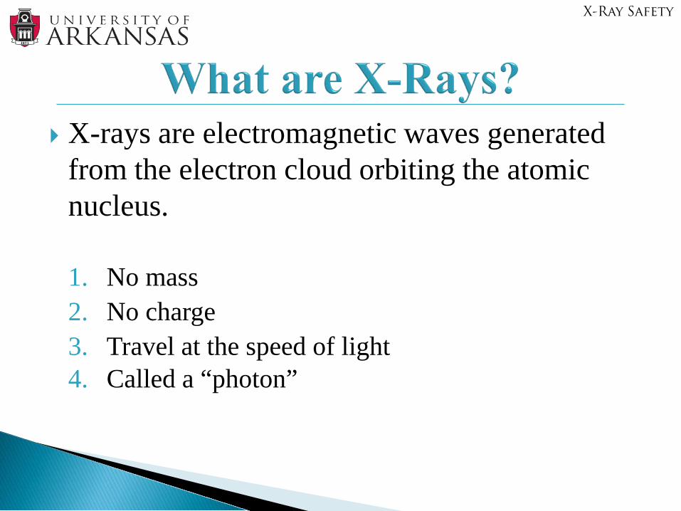
X-rays are electromagnetic waves generated from the electron cloud orbiting the atomic nucleus.
1. No mass2. No charge3. Travel at the speed of light 4. Called a “photon”
X-Ray Safety
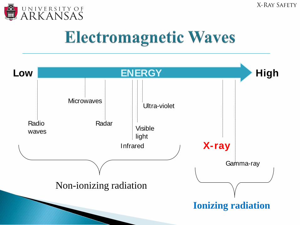
Low HighENERGY
Radio waves
Microwaves
Radar
Infrared
Visible light
Ultra-violet
X-ray
Gamma-ray
Non-ionizing radiation
Ionizing radiation
X-Ray Safety
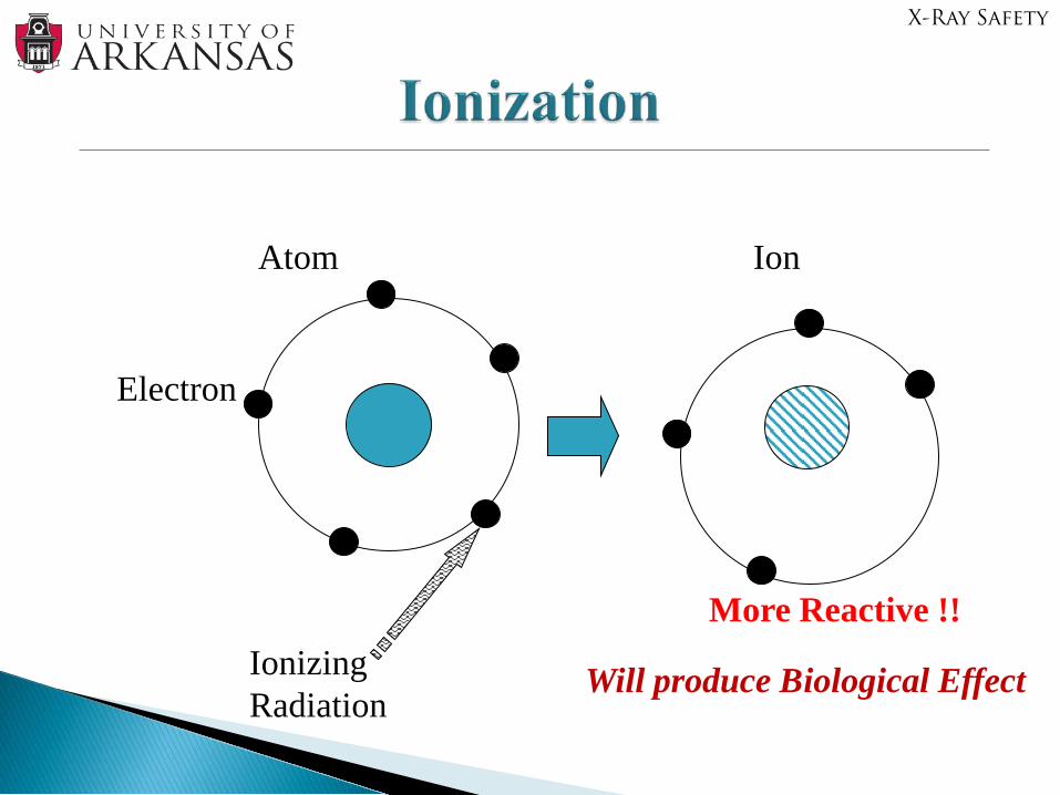
Atom
Electron
IonizingRadiation
Ion
More Reactive !!
Will produce Biological Effect
X-Ray Safety
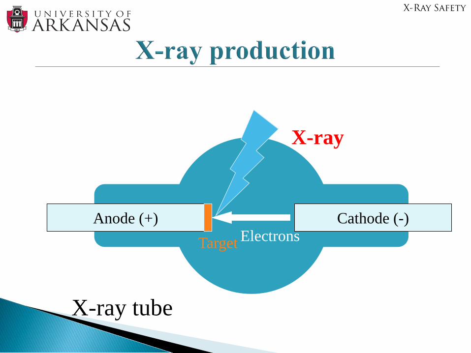
Anode (+) Cathode (-)ElectronsTarget
X-ray
X-ray tube
X-Ray Safety
Most X-ray devices emit electrons from a cathode, accelerated by a voltage
Electrons strike the anode (often Tungsten) Electrons slow down in the anode, and as a result of
interaction between the electrons and the atoms of the anode, x-rays are produced.
The energy of the x-ray shows different distribution depending on the anode material.
During this process, the device emits two different types of radiation.
X-Ray Safety
X-Ray Production – cont.
Nucleus
Electrons
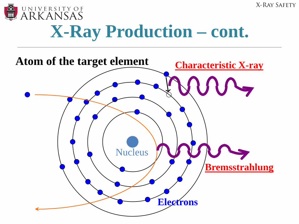
Atom of the target element Characteristic X-ray
Bremsstrahlung
X-Ray Safety
When the shell of the target (anode) atom has a vacancy, an electron from the outer orbit will fill the spot.
The energy difference is released as a form of X-ray.
The energy shows a peak as energy differences between shells are characteristic of each atom.
X-Ray Safety
X-Ray Safety
Bremsstrahlung X-Ray• Bremsstrahlung means “ breaking radiation” in
German• Bremsstrahlung occurs when high energy electrons
decelerate when interacting with the electric fields surrounding atomic nuclei. Excess energy is released in the form of an x-ray (photon).
• The energy of the resultant photon is related to the energy of the incident electron as well as the electric field strength.
• These forces are greater in nuclei with higher atomic numbers.
• This shows a continuous spectrum as each electron emits a different fraction of its energy.
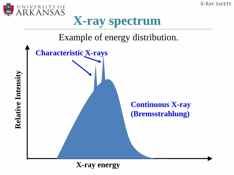
X-ray spectrum Example of energy distribution.
X-ray energy
Rel
ativ
e In
tens
ity
Characteristic X-rays
Continuous X-ray (Bremsstrahlung)
X-Ray Safety
Highly penetrating Electrically neutral Wide range of energies Travel in straight line at speed of light Cause ionization Cause fluorescence in crystals Cannot be focus by lens Produce photograph images Produce scattering
X-Ray Safety
Intensity (quantity)- measured as exposure rate in Roentgens
(mR/hr)
Control exposure (mR) by:- time of exposure - seconds- tube current (mA)- tube potential ( kVp)- Filtration (shielding)- distance (inverse square law
X-Ray Safety
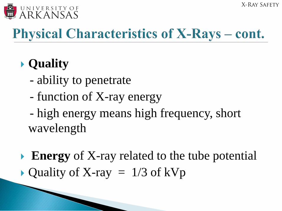
Quality - ability to penetrate- function of X-ray energy- high energy means high frequency, short wavelength
Energy of X-ray related to the tube potential Quality of X-ray = 1/3 of kVp
X-Ray Safety
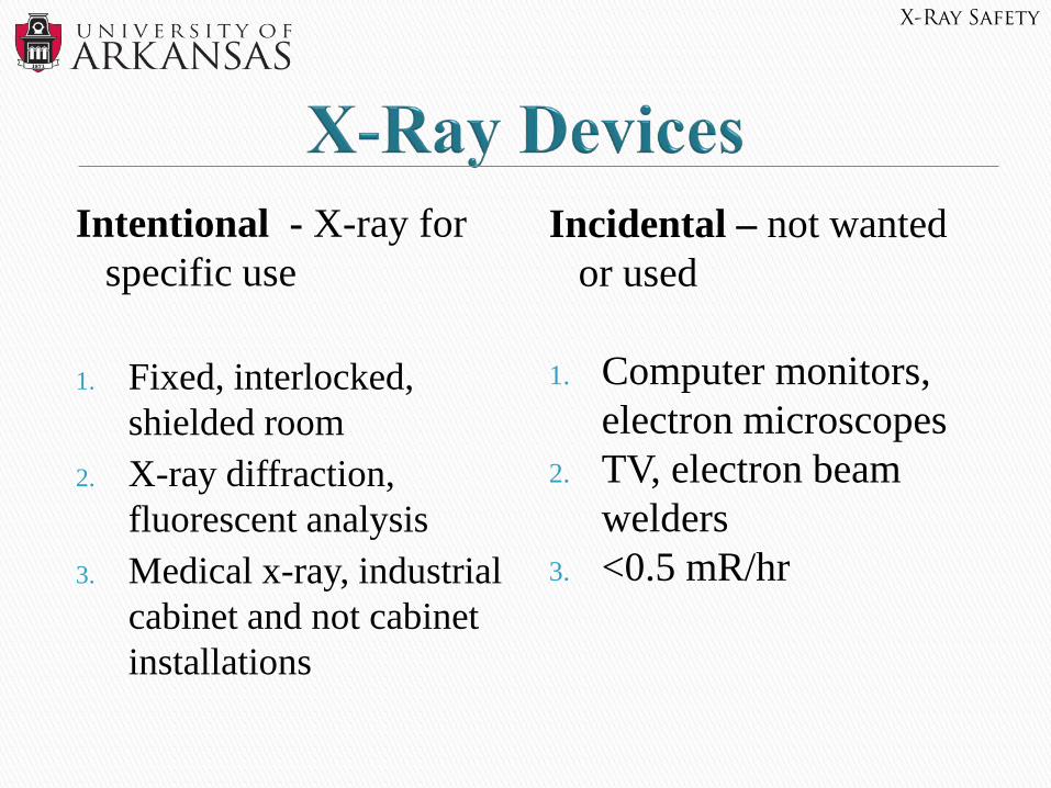
Intentional - X-ray for specific use
1. Fixed, interlocked, shielded room
2. X-ray diffraction, fluorescent analysis
3. Medical x-ray, industrial cabinet and not cabinet installations
Incidental – not wanted or used
1. Computer monitors, electron microscopes
2. TV, electron beam welders
3. <0.5 mR/hr
X-Ray Safety
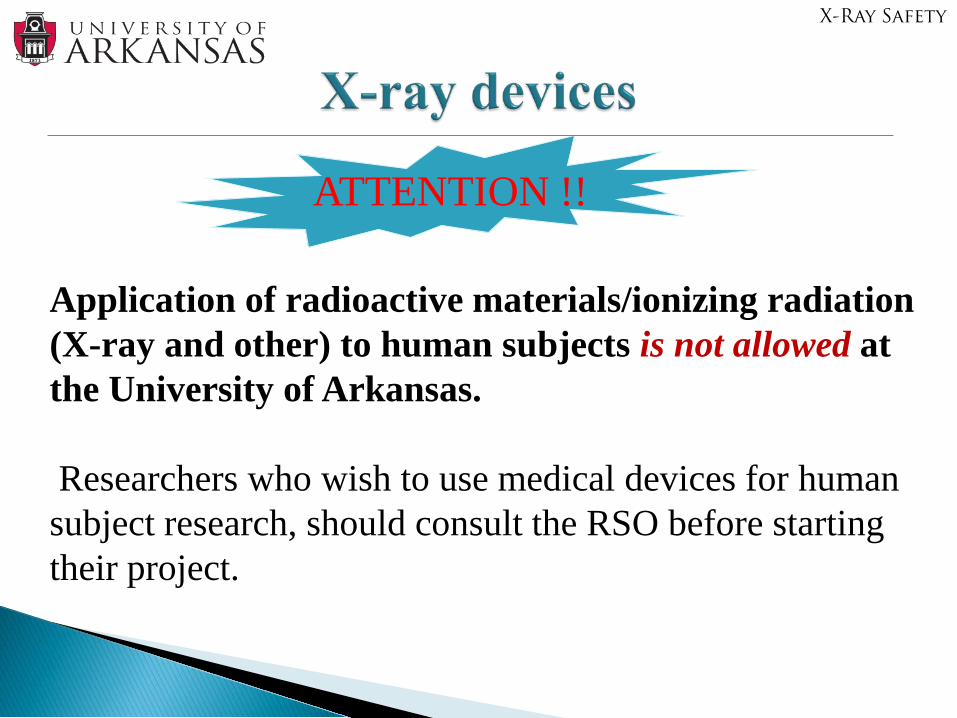
Application of radioactive materials/ionizing radiation (X-ray and other) to human subjects is not allowed at the University of Arkansas.
Researchers who wish to use medical devices for human subject research, should consult the RSO before starting their project.
ATTENTION !!
X-Ray Safety
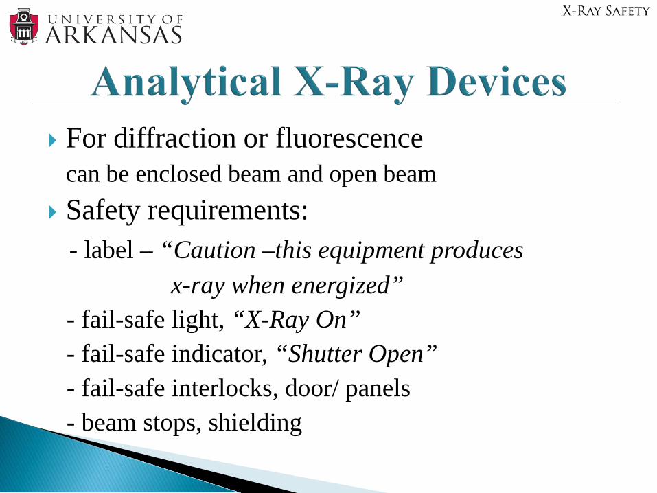
For diffraction or fluorescence can be enclosed beam and open beam
Safety requirements:- label – “Caution –this equipment produces
x-ray when energized”- fail-safe light, “X-Ray On”- fail-safe indicator, “Shutter Open”- fail-safe interlocks, door/ panels- beam stops, shielding
X-Ray Safety
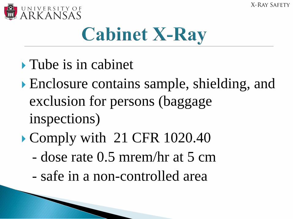
Tube is in cabinet Enclosure contains sample, shielding, and
exclusion for persons (baggage inspections)
Comply with 21 CFR 1020.40- dose rate 0.5 mrem/hr at 5 cm- safe in a non-controlled area
X-Ray Safety
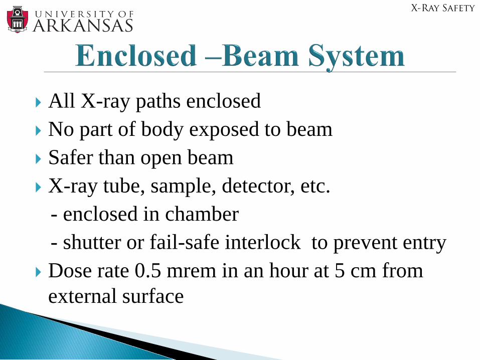
All X-ray paths enclosed No part of body exposed to beam Safer than open beam X-ray tube, sample, detector, etc.
- enclosed in chamber- shutter or fail-safe interlock to prevent entry
Dose rate 0.5 mrem in an hour at 5 cm from external surface
X-Ray Safety
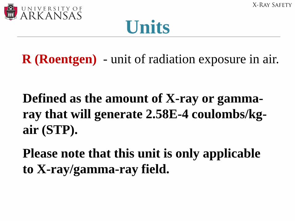
UnitsR (Roentgen) - unit of radiation exposure in air.
Defined as the amount of X-ray or gamma-ray that will generate 2.58E-4 coulombs/kg-air (STP).
Please note that this unit is only applicable to X-ray/gamma-ray field.
X-Ray Safety
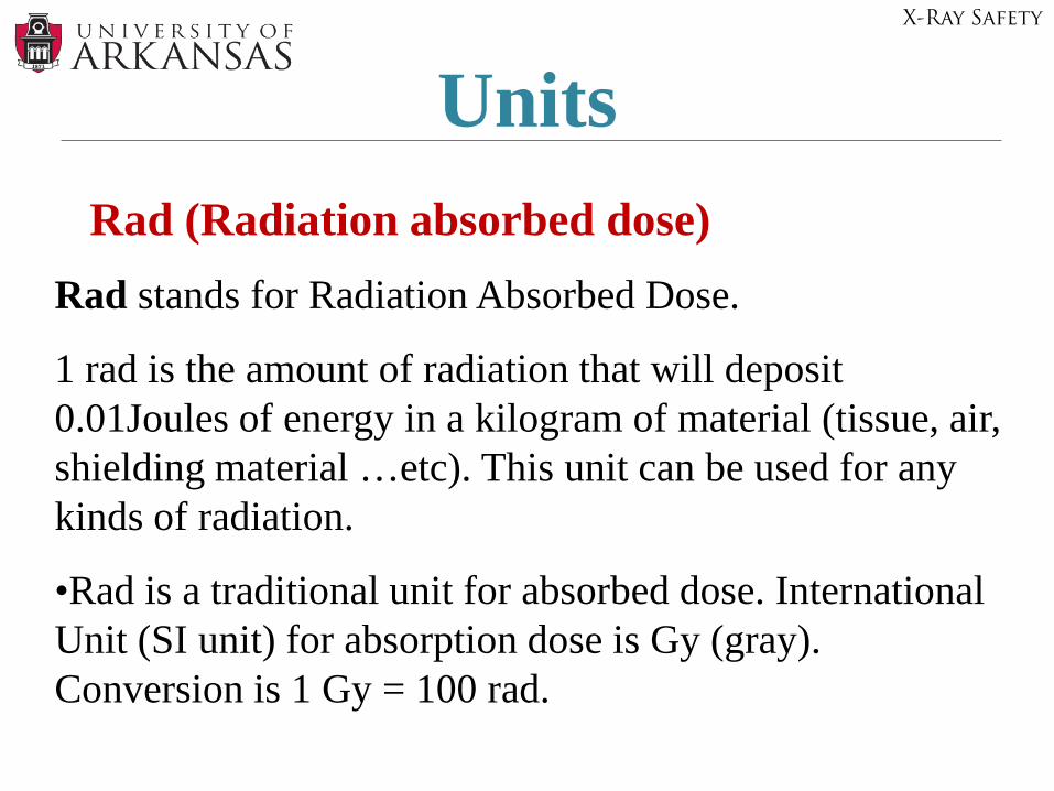
UnitsRad (Radiation absorbed dose)
Rad stands for Radiation Absorbed Dose.
1 rad is the amount of radiation that will deposit 0.01Joules of energy in a kilogram of material (tissue, air, shielding material …etc). This unit can be used for any kinds of radiation.
•Rad is a traditional unit for absorbed dose. International Unit (SI unit) for absorption dose is Gy (gray). Conversion is 1 Gy = 100 rad.
X-Ray Safety
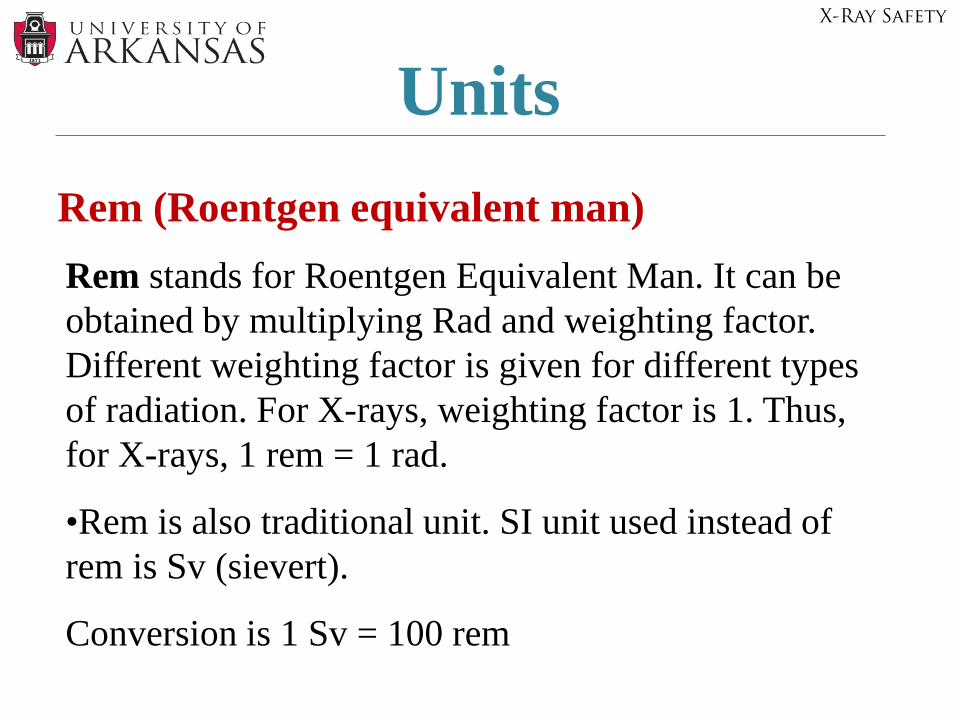
UnitsRem (Roentgen equivalent man)Rem stands for Roentgen Equivalent Man. It can be obtained by multiplying Rad and weighting factor. Different weighting factor is given for different types of radiation. For X-rays, weighting factor is 1. Thus, for X-rays, 1 rem = 1 rad.
•Rem is also traditional unit. SI unit used instead of rem is Sv (sievert).
Conversion is 1 Sv = 100 rem
X-Ray Safety
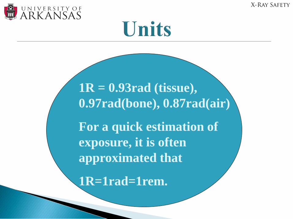
1R = 0.93rad (tissue), 0.97rad(bone), 0.87rad(air)
For a quick estimation of exposure, it is often approximated that
1R=1rad=1rem.
X-Ray Safety
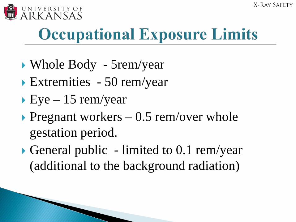
Whole Body - 5rem/year Extremities - 50 rem/year Eye – 15 rem/year Pregnant workers – 0.5 rem/over whole
gestation period. General public - limited to 0.1 rem/year
(additional to the background radiation)
X-Ray Safety
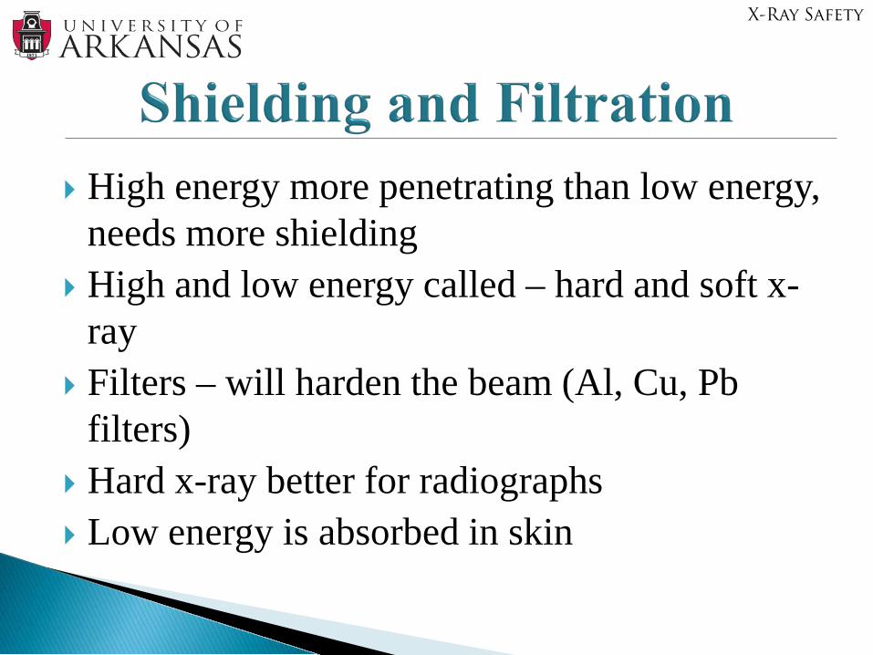
High energy more penetrating than low energy, needs more shielding
High and low energy called – hard and soft x-ray
Filters – will harden the beam (Al, Cu, Pbfilters)
Hard x-ray better for radiographs Low energy is absorbed in skin
X-Ray Safety
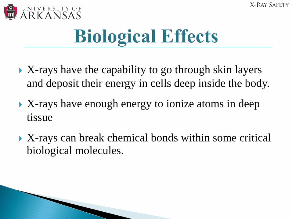
X-rays have the capability to go through skin layers and deposit their energy in cells deep inside the body.
X-rays have enough energy to ionize atoms in deep tissue
X-rays can break chemical bonds within some critical biological molecules.
X-Ray Safety

In some cases, those damaged cells are able to repair themselves. However, high dose or high dose rate exposure may create non curable damage.
When cells are not recovered, this damage can cause cell injury or even cell death. The effects may passed to daughter cells (with damaged characteristics). The division of this damaged cell may be the first step in tumor/cancer development.
If enough cells in a particular body organ are damaged, the function of the organ may be impaired.
X-Ray Safety
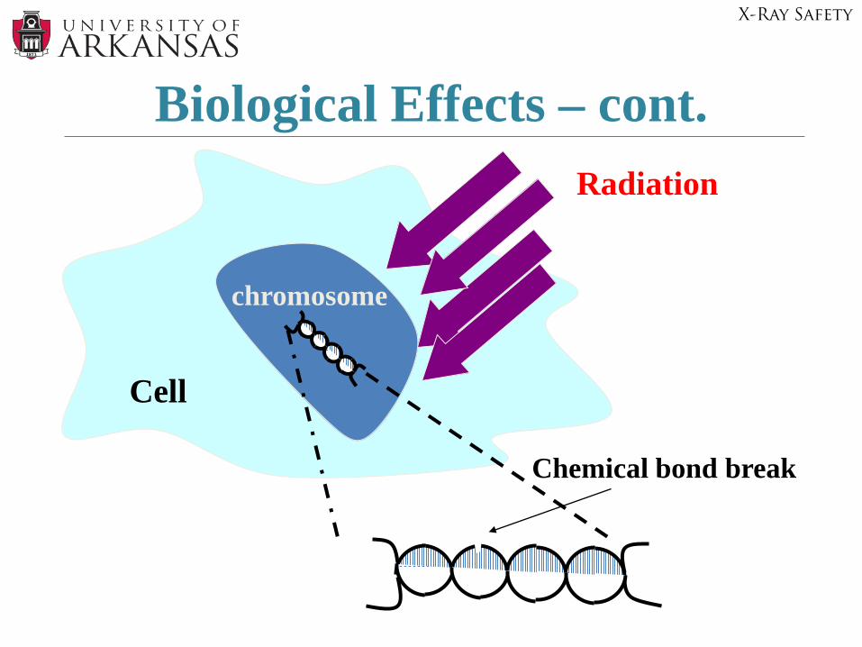
Biological Effects – cont.
chromosome
Cell
Radiation
Chemical bond break
X-Ray Safety
• Thermal Burns – nerve endings on skin surface gives early warning of thermal burns (Expl: immediate withdrawal of the hand from burning flame)
• X-Ray Burns – x-ray penetrate to deeper, basal skin cells, killing germinal cells that are intended to replace surface cells (no immediate pain)- Thus, skin may not heal- may require grafts or amputation
Direct Beam X-Ray Exposure vs. Thermal Burns
X-Ray Safety

• 500 rem (5000,000 mrem all at one time)– No immediate pain; warmth and itching may be
felt.– Reddening after a day, fading in few days.– Dry scaling or peeling will follow.– Avoid future injuries.– Recovery should be complete.
Acute X-Ray Effects
X-Ray Safety
• 1000 rem (In a single dose)– Serious tissue damage.– 2nd degree burns.– Reddening, inflammation.– Blisters in1-3 weeks, raw open wounds.– Hand exposure, fingers stiffness.– Need immediate medical attention.
Acute X-Ray Effects – cont.
X-Ray Safety
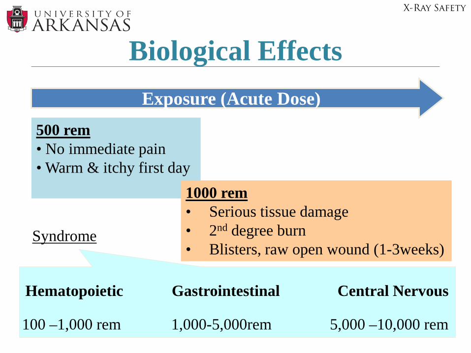
Biological EffectsExposure (Acute Dose)
500 rem• No immediate pain• Warm & itchy first day
1000 rem• Serious tissue damage• 2nd degree burn• Blisters, raw open wound (1-3weeks)
Hematopoietic Gastrointestinal Central Nervous
Syndrome
100 –1,000 rem 1,000-5,000rem 5,000 –10,000 rem
X-Ray Safety
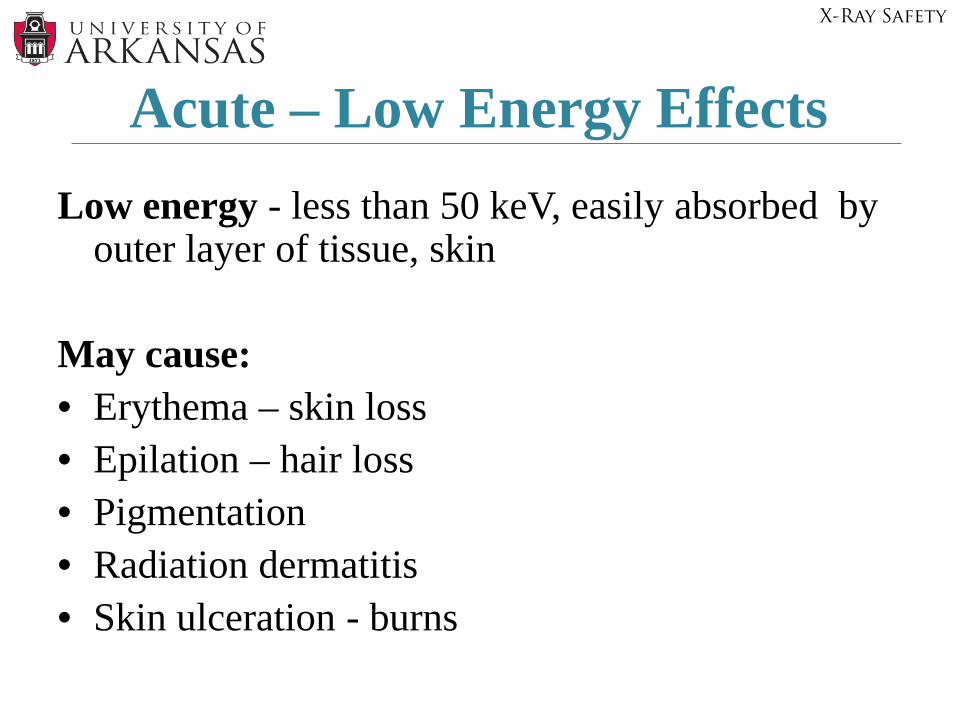
Low energy - less than 50 keV, easily absorbed by outer layer of tissue, skin
May cause:• Erythema – skin loss• Epilation – hair loss• Pigmentation • Radiation dermatitis• Skin ulceration - burns
Acute – Low Energy Effects
X-Ray Safety
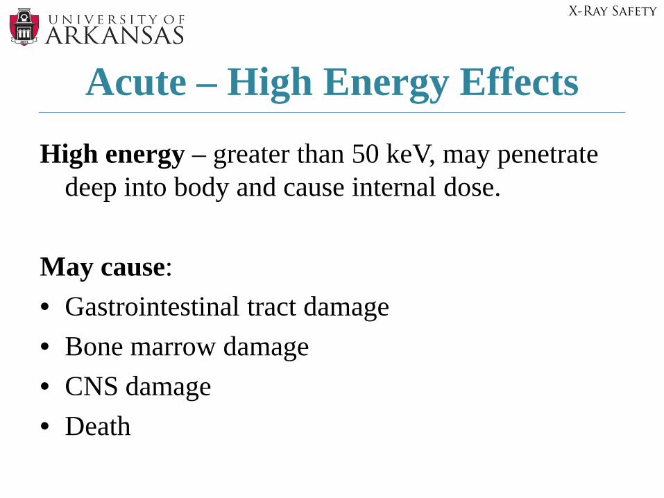
High energy – greater than 50 keV, may penetrate deep into body and cause internal dose.
May cause:• Gastrointestinal tract damage • Bone marrow damage• CNS damage• Death
Acute – High Energy Effects
X-Ray Safety
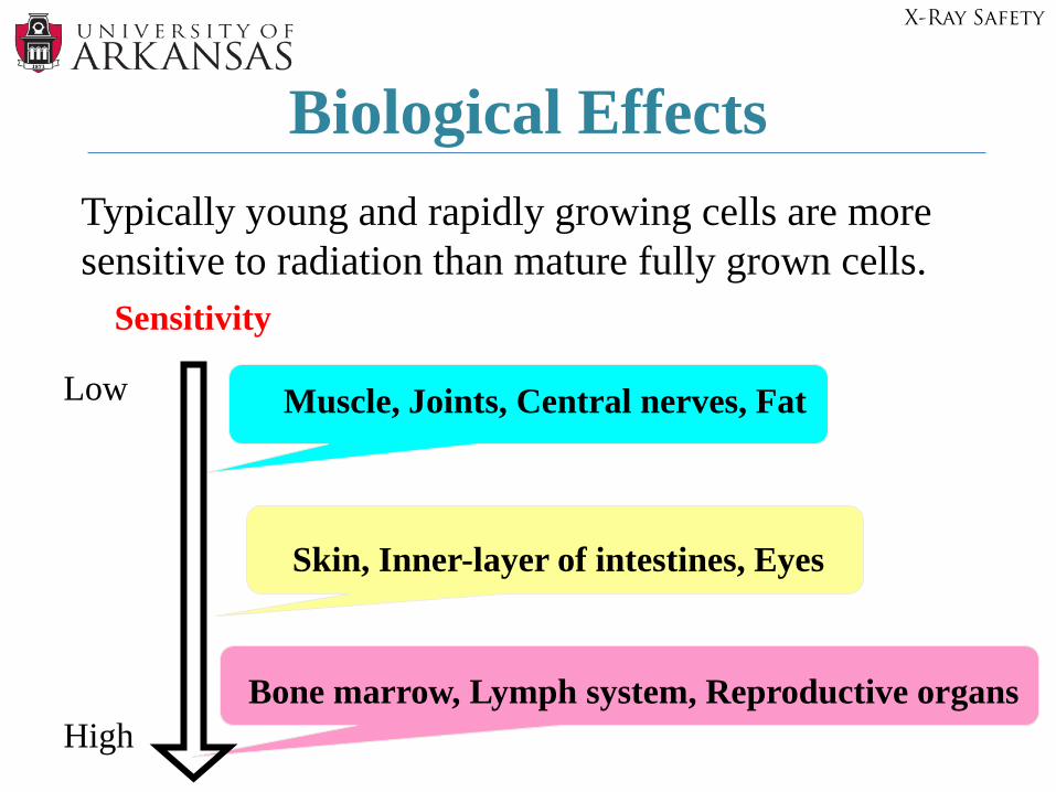
Biological Effects
Sensitivity
Low
High
Muscle, Joints, Central nerves, Fat
Skin, Inner-layer of intestines, Eyes
Bone marrow, Lymph system, Reproductive organs
Typically young and rapidly growing cells are more sensitive to radiation than mature fully grown cells.
X-Ray Safety
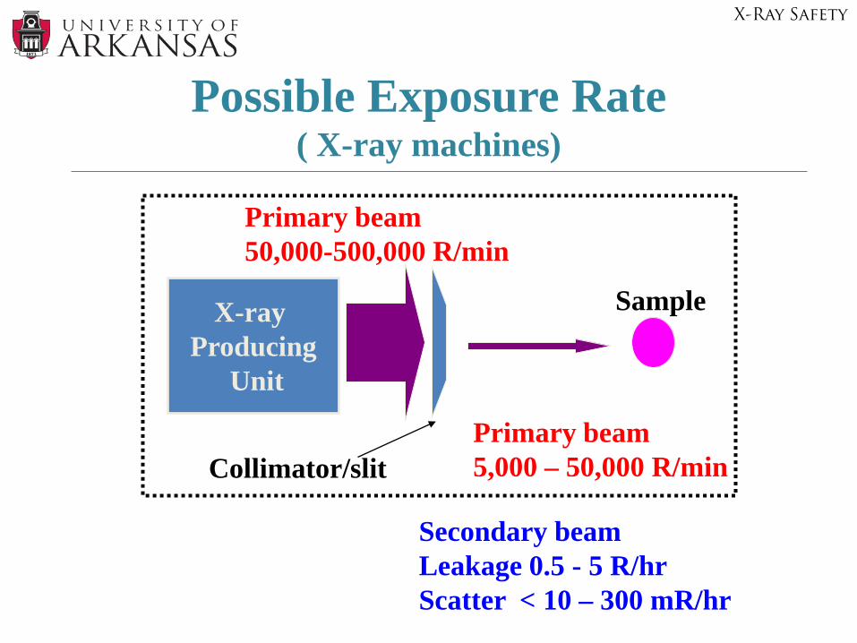
Possible Exposure Rate ( X-ray machines)
Primary beam50,000-500,000 R/min
Primary beam5,000 – 50,000 R/min
Secondary beamLeakage 0.5 - 5 R/hrScatter < 10 – 300 mR/hr
X-ray Producing
Unit
Collimator/slit
Sample
X-Ray Safety
Primary beam - very intensive exposure in beam- could be >100,000 R/min- small beam diameter, <1 cm
Scattered x-ray radiation – “sky shine”- low intensity- large area of exposure- from housing leakage- from any target material- from inadequate shielding
X-Ray Safety
Operator Error- adjustment or alignment of samples/cameras while beam is ON- not using safety features (bypassing interlocks, shielding of unused port, etc.- unauthorized user (untrained user, unsupervised operation)
X-ray leakage Not using protective equipment
- lead aprons, etc.
X-Ray Safety
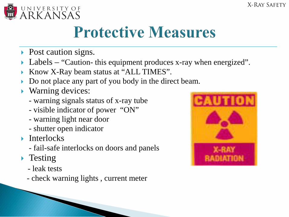
Post caution signs. Labels – “Caution- this equipment produces x-ray when energized”. Know X-Ray beam status at “ALL TIMES”. Do not place any part of you body in the direct beam. Warning devices:
- warning signals status of x-ray tube- visible indicator of power “ON”- warning light near door- shutter open indicator
Interlocks - fail-safe interlocks on doors and panels
Testing - leak tests- check warning lights , current meter
X-Ray Safety
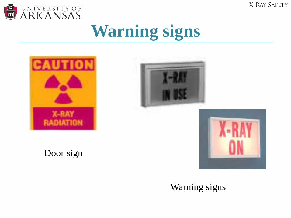
Warning signs
Door sign
Warning signs
X-Ray Safety
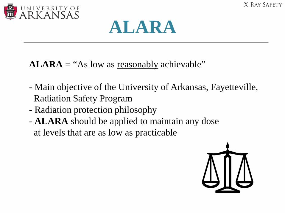
ALARA
ALARA = “As low as reasonably achievable”
- Main objective of the University of Arkansas, Fayetteville, Radiation Safety Program
- Radiation protection philosophy - ALARA should be applied to maintain any dose at levels that are as low as practicable
X-Ray Safety
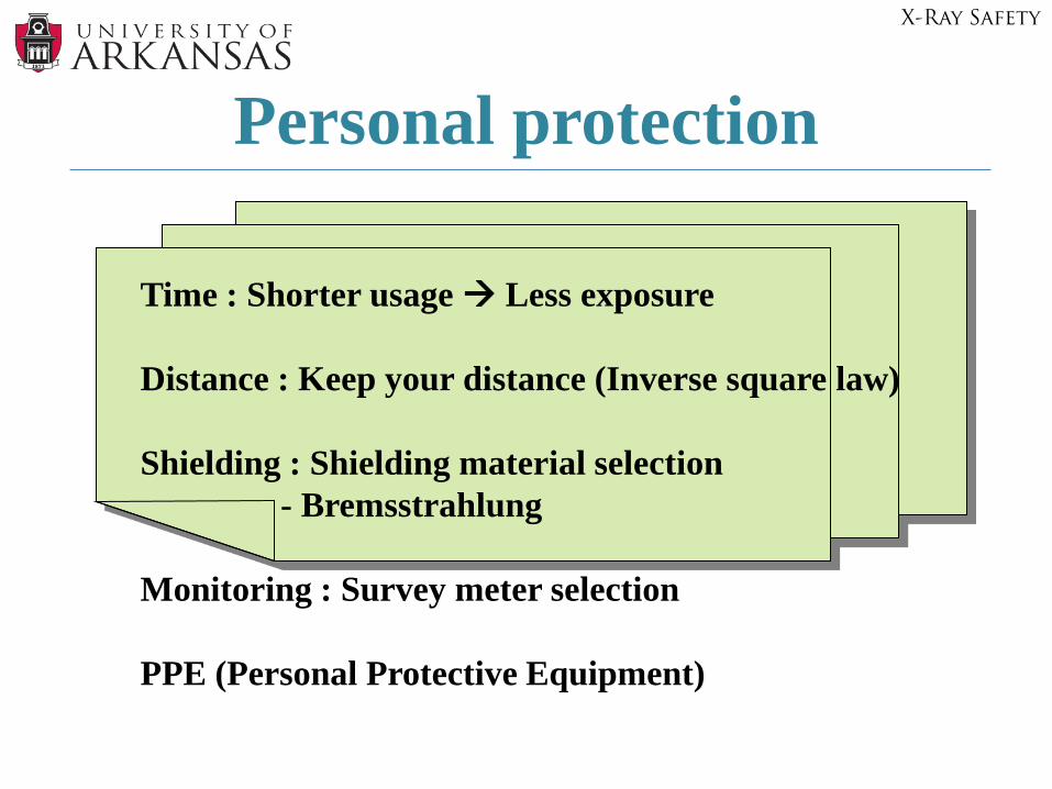
Personal protection
Time : Shorter usage Less exposure
Distance : Keep your distance (Inverse square law)
Shielding : Shielding material selection- Bremsstrahlung
Monitoring : Survey meter selection
PPE (Personal Protective Equipment)
X-Ray Safety
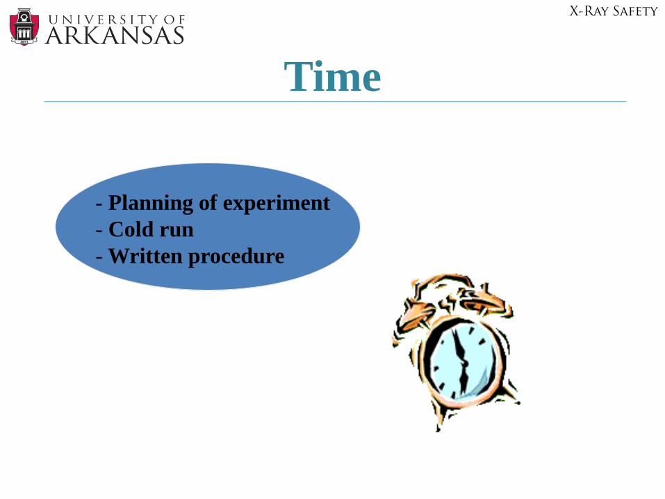
Time
- Planning of experiment- Cold run- Written procedure
X-Ray Safety
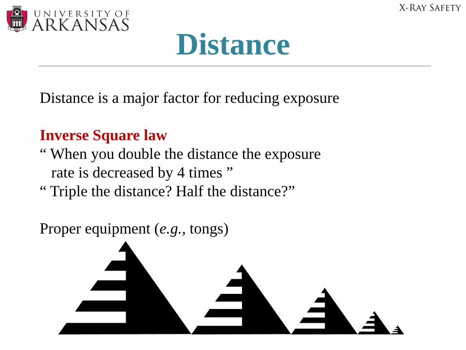
DistanceDistance is a major factor for reducing exposure
Inverse Square law“ When you double the distance the exposure
rate is decreased by 4 times ”“ Triple the distance? Half the distance?”
Proper equipment (e.g., tongs)
X-Ray Safety
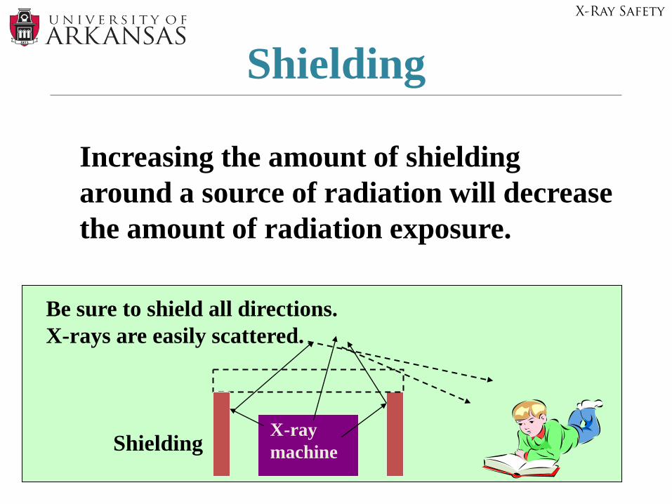
Shielding
Increasing the amount of shielding around a source of radiation will decrease the amount of radiation exposure.
X-ray machine
Be sure to shield all directions.X-rays are easily scattered.
Shielding
X-Ray Safety
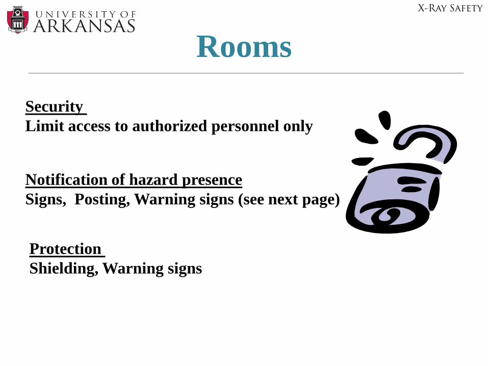
Rooms
Security Limit access to authorized personnel only
Notification of hazard presenceSigns, Posting, Warning signs (see next page)
Protection Shielding, Warning signs
X-Ray Safety
Frequency of Surveys- quarterly
- upon installation- upon changes/re-location- maintenance requirements- observed unsafe conditions
Survey Meters- Thin window Geiger-Muller (GM) counter may be used to check for leakage (to find where leakage occurs), indicate x-ray production (to verify if beam is “on” or “off”) , monitor routine operation. GM meters count individual photons (x-ray, gamma) in counts per minute. - Ion chamber can be used to determine dose rate at the x-ray field (how much). The ion chamber response in R/min (hour) and can be used to measure radiation dose rate.
X-Ray Safety
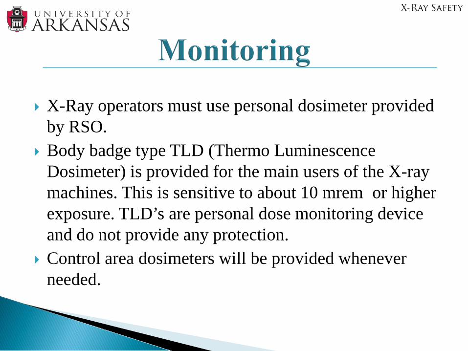
X-Ray operators must use personal dosimeter provided by RSO.
Body badge type TLD (Thermo Luminescence Dosimeter) is provided for the main users of the X-ray machines. This is sensitive to about 10 mrem or higher exposure. TLD’s are personal dose monitoring device and do not provide any protection.
Control area dosimeters will be provided whenever needed.
X-Ray Safety
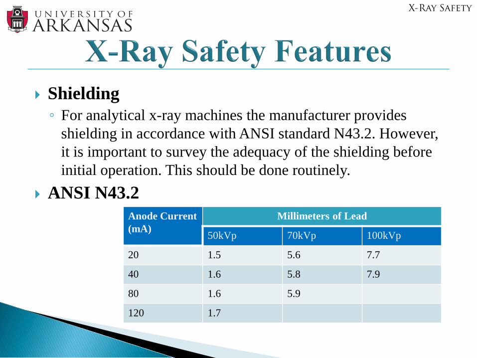
Shielding◦ For analytical x-ray machines the manufacturer provides
shielding in accordance with ANSI standard N43.2. However, it is important to survey the adequacy of the shielding before initial operation. This should be done routinely.
ANSI N43.2Anode Current (mA)
Millimeters of Lead
50kVp 70kVp 100kVp
20 1.5 5.6 7.7
40 1.6 5.8 7.9
80 1.6 5.9
120 1.7
X-Ray Safety
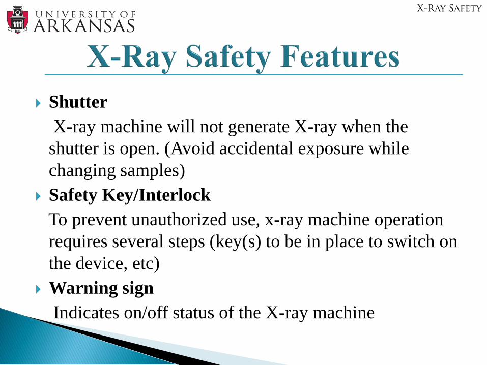
ShutterX-ray machine will not generate X-ray when the shutter is open. (Avoid accidental exposure while changing samples)
Safety Key/InterlockTo prevent unauthorized use, x-ray machine operation requires several steps (key(s) to be in place to switch on the device, etc)
Warning signIndicates on/off status of the X-ray machine
X-Ray Safety
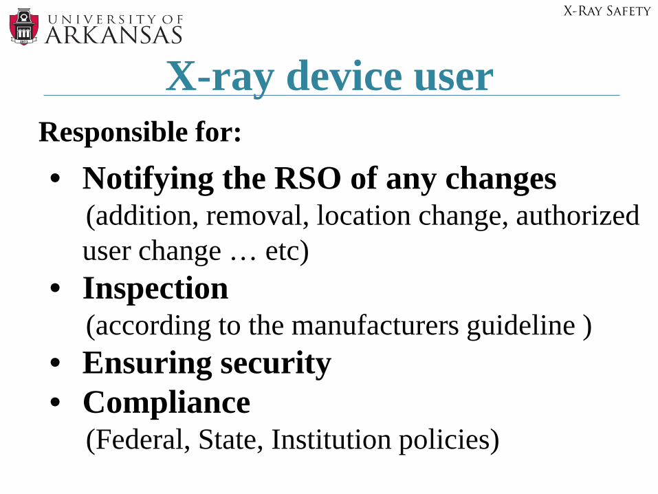
X-ray device user Responsible for: • Notifying the RSO of any changes
(addition, removal, location change, authorized user change … etc)
• Inspection (according to the manufacturers guideline )
• Ensuring security• Compliance
(Federal, State, Institution policies)
X-Ray Safety
Office of Environmental Health & Safety575-5448 (M-F, 7:30am – 4:00pm)
University of Arkansas Police Department (UAPD) 575-2222 (After hours & Holidays)
Radiation Safety Officer575-3379
Assistant Radiation Safety Officer575-8473
X-Ray Safety
Recommended