Embed Size (px)
Citation preview
2 Pulmonary Edema Pattern (Symmetric Bilateral Alveolar Pattern)
CLINICAL IMAGAGINGAN ATLAS OF DIFFERENTIAL DAIGNOSIS
EISENBERG
DR. Muhammad Bin Zulfiqar PGR-FCPS III SIMS/SHL
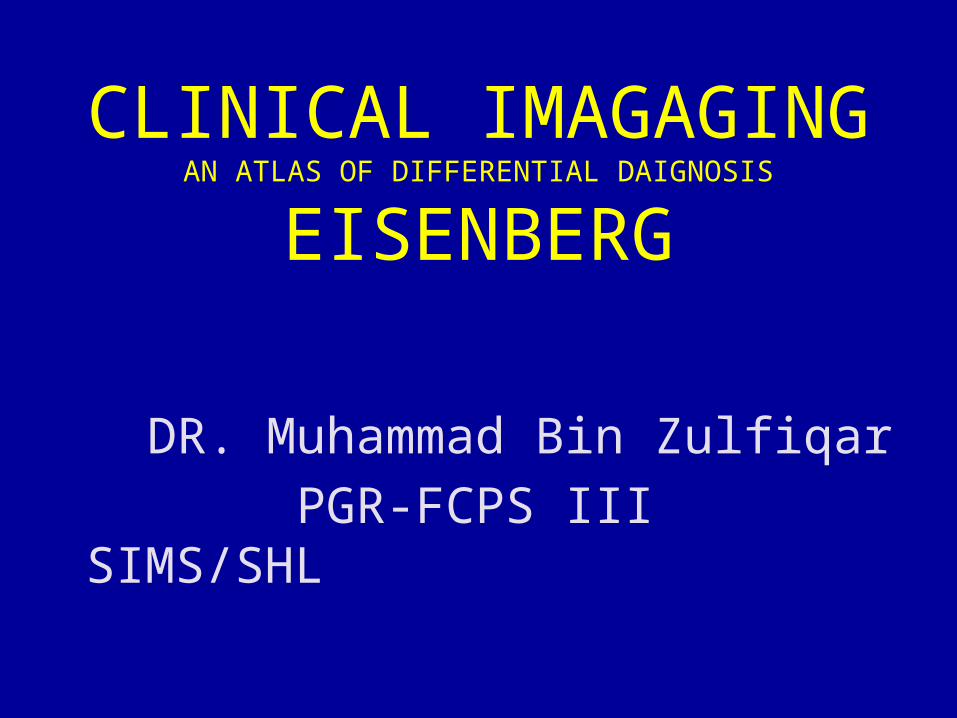
• Fig C 2-1 Congestive heart failure. Diffuse bilateral symmetric infiltration of the central portion of the lungs along with relative sparing of the periphery produces the butterfly, or bat's wing, pattern. The margins of the edematous lung are sharply defined. The consolidation is fairly homogeneous and is associated with a well-defined air bronchogram on both sides.7
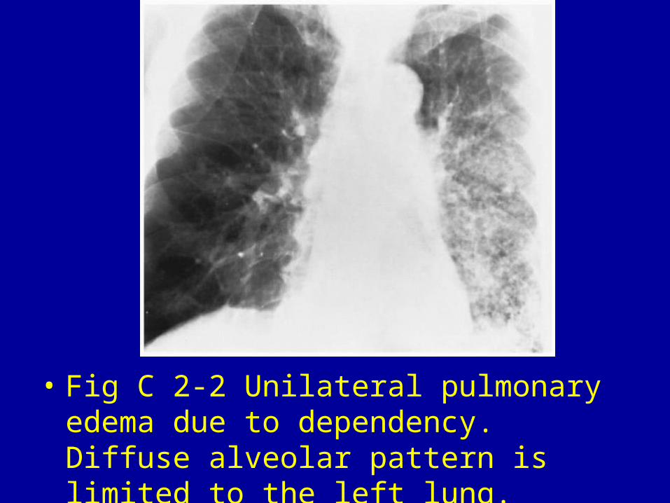
• Fig C 2-2 Unilateral pulmonary edema due to dependency. Diffuse alveolar pattern is limited to the left lung.
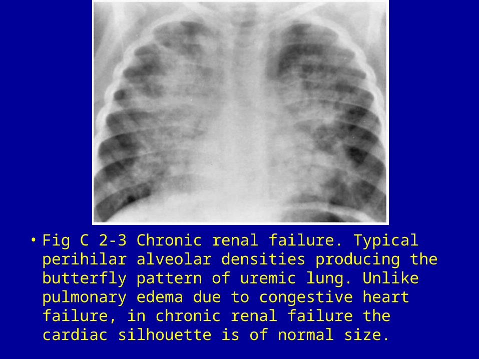
• Fig C 2-3 Chronic renal failure. Typical perihilar alveolar densities producing the butterfly pattern of uremic lung. Unlike pulmonary edema due to congestive heart failure, in chronic renal failure the cardiac silhouette is of normal size.
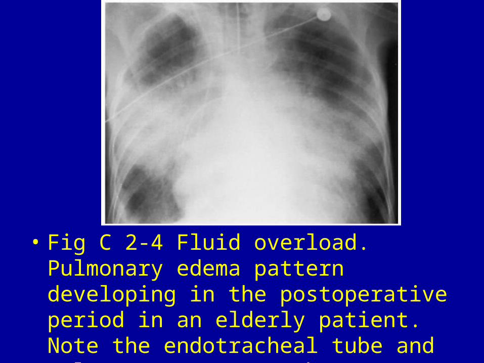
• Fig C 2-4 Fluid overload. Pulmonary edema pattern developing in the postoperative period in an elderly patient. Note the endotracheal tube and pulmonary artery catheter
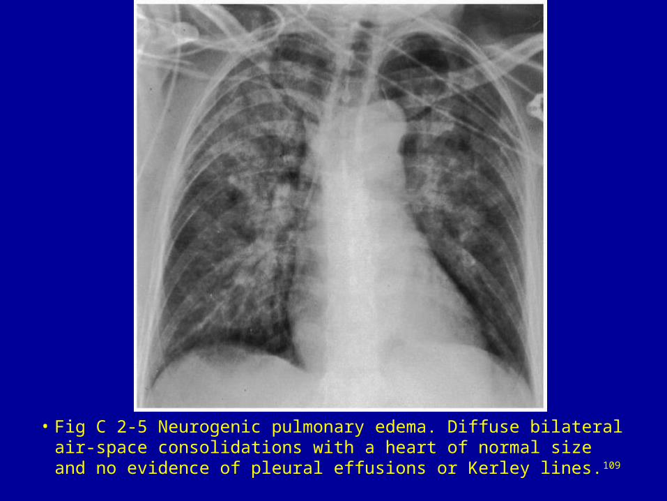
• Fig C 2-5 Neurogenic pulmonary edema. Diffuse bilateral air-space consolidations with a heart of normal size and no evidence of pleural effusions or Kerley lines.109
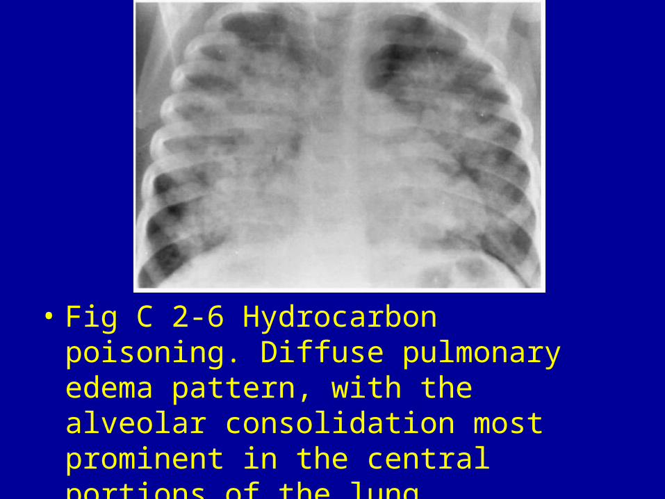
• Fig C 2-6 Hydrocarbon poisoning. Diffuse pulmonary edema pattern, with the alveolar consolidation most prominent in the central portions of the lung.
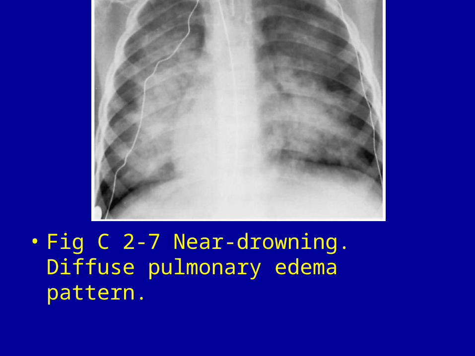
• Fig C 2-7 Near-drowning. Diffuse pulmonary edema pattern.
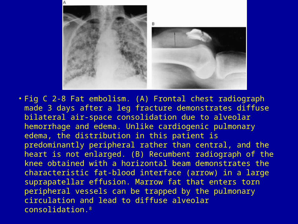
• Fig C 2-8 Fat embolism. (A) Frontal chest radiograph made 3 days after a leg fracture demonstrates diffuse bilateral air-space consolidation due to alveolar hemorrhage and edema. Unlike cardiogenic pulmonary edema, the distribution in this patient is predominantly peripheral rather than central, and the heart is not enlarged. (B) Recumbent radiograph of the knee obtained with a horizontal beam demonstrates the characteristic fat-blood interface (arrow) in a large suprapatellar effusion. Marrow fat that enters torn peripheral vessels can be trapped by the pulmonary circulation and lead to diffuse alveolar consolidation.8
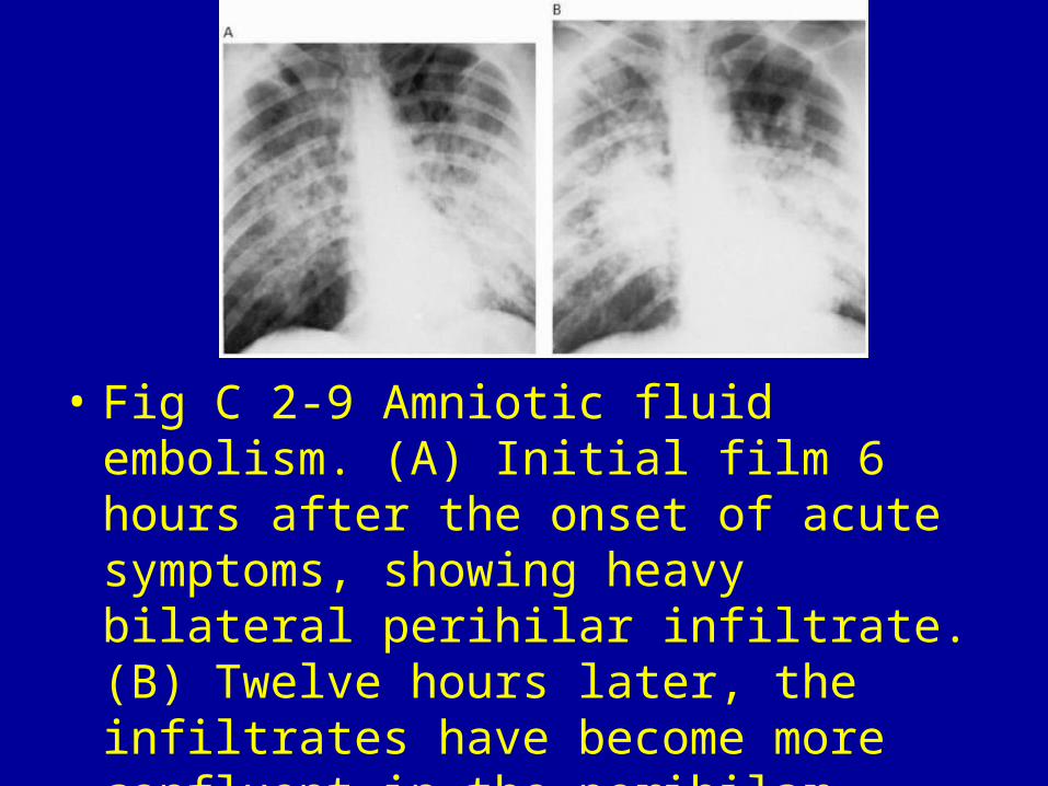
• Fig C 2-9 Amniotic fluid embolism. (A) Initial film 6 hours after the onset of acute symptoms, showing heavy bilateral perihilar infiltrate. (B) Twelve hours later, the infiltrates have become more confluent in the perihilar zones.9
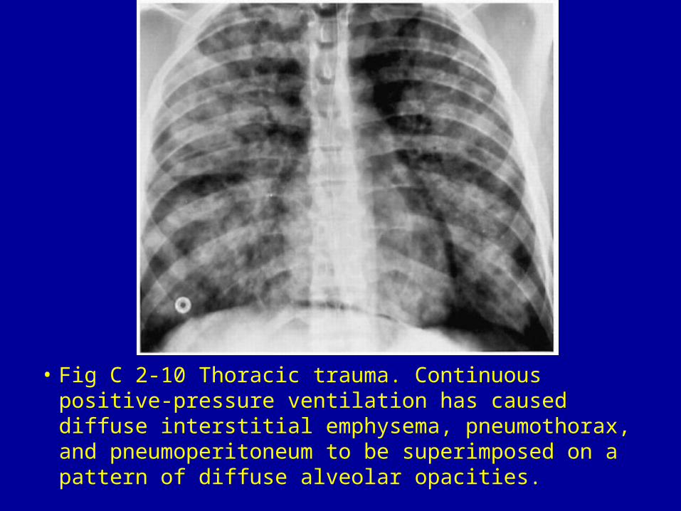
• Fig C 2-10 Thoracic trauma. Continuous positive-pressure ventilation has caused diffuse interstitial emphysema, pneumothorax, and pneumoperitoneum to be superimposed on a pattern of diffuse alveolar opacities.
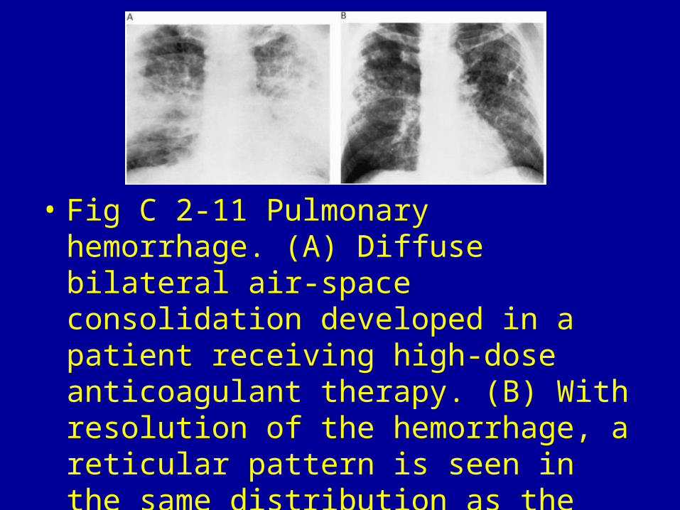
• Fig C 2-11 Pulmonary hemorrhage. (A) Diffuse bilateral air-space consolidation developed in a patient receiving high-dose anticoagulant therapy. (B) With resolution of the hemorrhage, a reticular pattern is seen in the same distribution as the alveolar infiltrate.
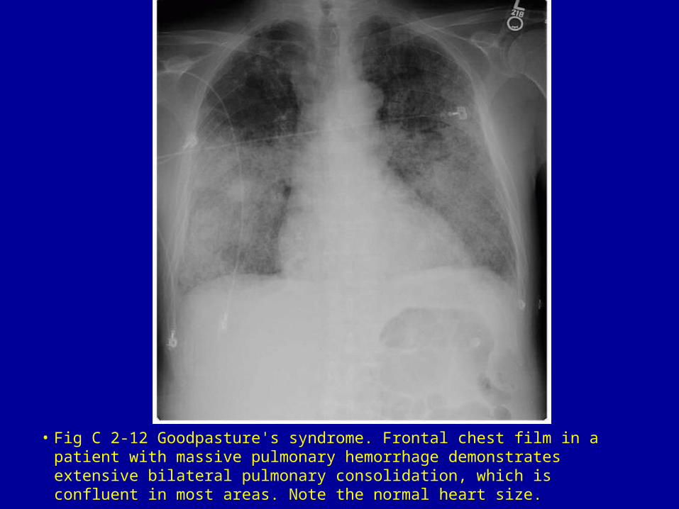
• Fig C 2-12 Goodpasture's syndrome. Frontal chest film in a patient with massive pulmonary hemorrhage demonstrates extensive bilateral pulmonary consolidation, which is confluent in most areas. Note the normal heart size.

• Fig C 2-13 Heroin abuse. (A) Initial radiograph obtained shortly after presentation to the emergency department reveals bilateral areas of increased opacity, a finding consistent with acute lung injury. (B) Follow-up study obtained two days later shows complete clearing of the areas of increased opacity. Such rapid clearing is common in heroin-induced lung injury.10
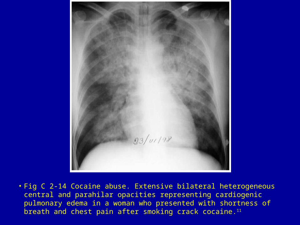
• Fig C 2-14 Cocaine abuse. Extensive bilateral heterogeneous central and parahilar opacities representing cardiogenic pulmonary edema in a woman who presented with shortness of breath and chest pain after smoking crack cocaine.11
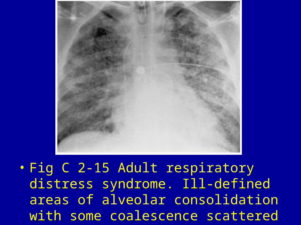
• Fig C 2-15 Adult respiratory distress syndrome. Ill-defined areas of alveolar consolidation with some coalescence scattered throughout both lungs.
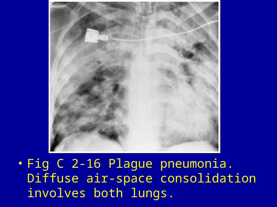
• Fig C 2-16 Plague pneumonia. Diffuse air-space consolidation involves both lungs.
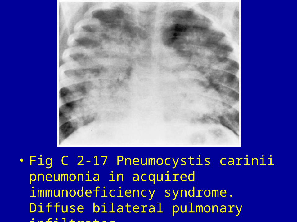
• Fig C 2-17 Pneumocystis carinii pneumonia in acquired immunodeficiency syndrome. Diffuse bilateral pulmonary infiltrates.
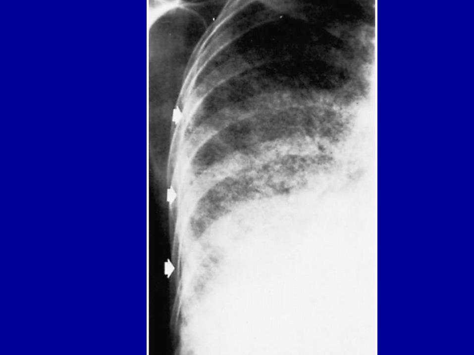


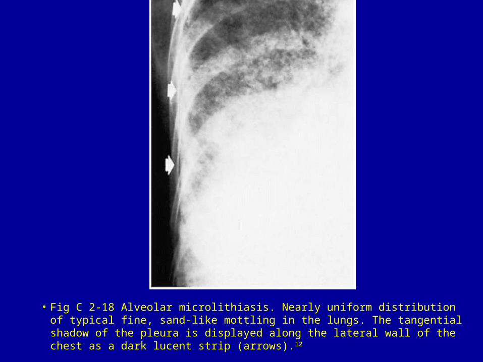
• Fig C 2-18 Alveolar microlithiasis. Nearly uniform distribution of typical fine, sand-like mottling in the lungs. The tangential shadow of the pleura is displayed along the lateral wall of the chest as a dark lucent strip (arrows).12
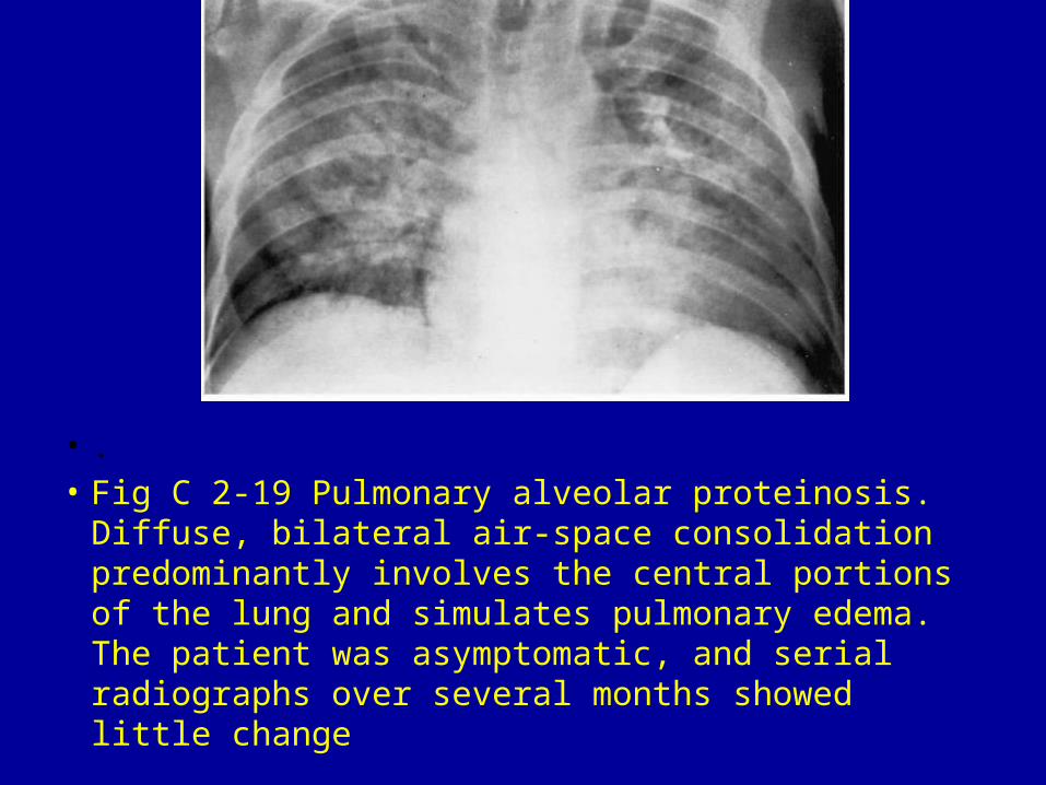
• .• Fig C 2-19 Pulmonary alveolar proteinosis. Diffuse,
bilateral air-space consolidation predominantly involves the central portions of the lung and simulates pulmonary edema. The patient was asymptomatic, and serial radiographs over several months showed little change
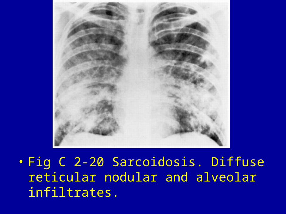
• Fig C 2-20 Sarcoidosis. Diffuse reticular nodular and alveolar infiltrates.