Embed Size (px)
Citation preview
24 Anterior Mediastinal Lesions on Computed Tomography

CLINICAL IMAGAGINGAN ATLAS OF DIFFERENTIAL DAIGNOSIS
EISENBERG
DR. Muhammad Bin Zulfiqar PGR-FCPS III SIMS/SHL
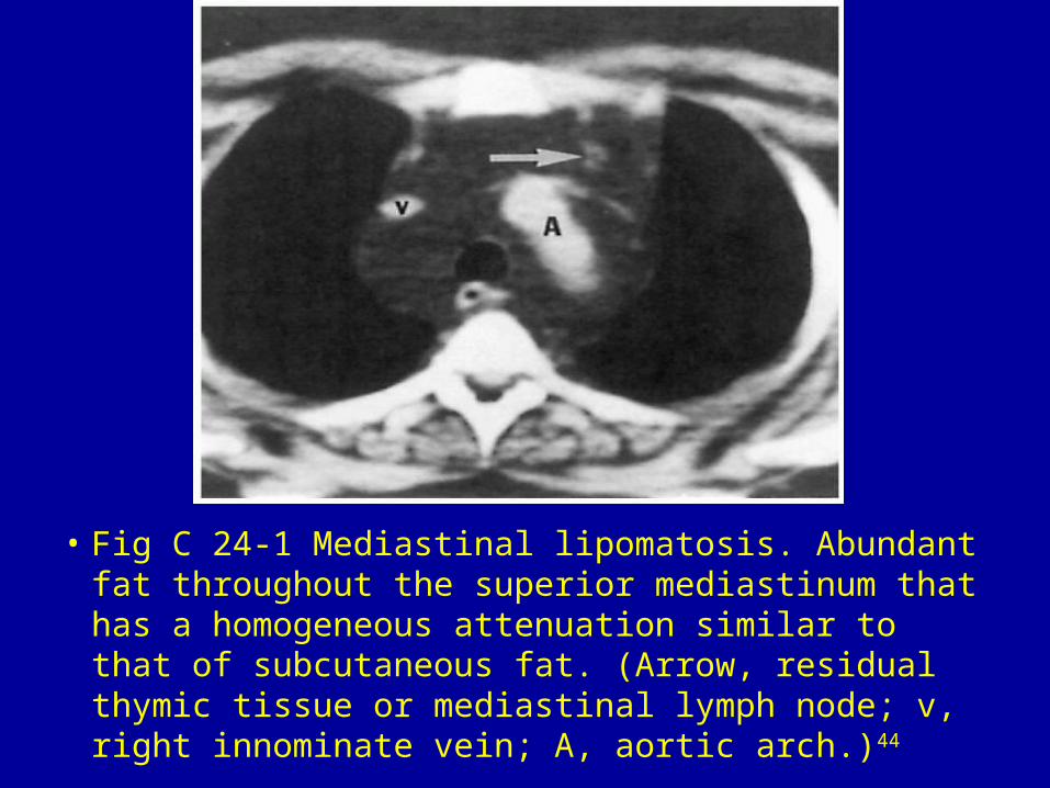
• Fig C 24-1 Mediastinal lipomatosis. Abundant fat throughout the superior mediastinum that has a homogeneous attenuation similar to that of subcutaneous fat. (Arrow, residual thymic tissue or mediastinal lymph node; v, right innominate vein; A, aortic arch.)44

• Fig C 24-2 Liposarcoma. Large, relatively inhomogeneous mass in the right side of the mediastinum. Note that the mass has a slightly higher attenuation than does the subcutaneous fat. The mass extended into the right side of the neck to involve the recurrent laryngeal nerve, paralyzing the right vocal cord.44

• Fig C 24-3 Morgagni hernia. The right inferior mediastinal mass contains large bowel and omental fat. Focal eventration of the diaphragm can be differentiated from a Morgagni's hernia by the intact diaphragm in the former entity.44

• Fig C 24-4 Thymic cyst. Incidentally noted well-circumscribed mass of fluid attenuation (arrow).45

• Fig C 24-5 Thymoma. Slightly lobulated mass (arrows) anterior to the main pulmonary artery (MPA) in a patient with myasthenia gravis.46

• Fig C 24-6 Thymoma. Enormous soft-tissue mass in the anterior mediastinum with posterior displacement of other mediastinal structures. No difference in density can be seen between the mass and the heart behind it.

• Fig C 24-7 Thymic carcinoma. Sagittal reformatted image shows that the anterior mediastinal mass is closely attached to the pericardium with loss of the fat plane (arrow) between the two entities, findings that suggest pericardial involvement. There is also a pericardial effusion.45

• Fig C 24-8 Thymic carcinoid. Lobulated, heretogeneously enhancing mass. Loss of the fat plane between the mass and the pericardium suggests invasiveness.45

• Fig C 24-9 Thymic hyperplasia. Bilobed, homogeneous soft-tissue lesion (arrows) in a patient with Graves' disease.45

• Fig C 24-10 Retrosternal goiter. Soft-tissue mass (arrow) extending into the anterior and middle mediastinum.47

• Fig C 24-11 Ectopic parathyroid adenoma. Small soft-tissue mass (arrow) in the anterior mediastinum. (A, aorta; a, major branches of the aorta; and v, brachiocephalic veins.)48

• Fig C 24-12 Mixed germ cell tumor. Contrast scan shows a huge tumor that is primarily solid, thought there is a relatively large cystic component (arrow).49

• Fig C 24-13 Lymphoma. Large mass that fills the anterior medaistinum. Note that the lung interfaces with the hilar vessels (arrow) and aorta (arrowhead) are well preserved. Thus, on plain radiographs these middle mediastinal structures were clearly seen through the mass (hilum overlay sign), indicating that the lesion was either in the anterior or posterior portion of the mediastinum.47

Fig C 24-14 Fibrosing mediastinitis. Soft-tissue attenuation mass in the anterior mediastinum. A, aorta; S, superior vena cava.)50