Embed Size (px)
Citation preview
80 Perihepatic Space* on Computed Tomography
CLINICAL IMAGAGINGAN ATLAS OF DIFFERENTIAL DAIGNOSIS
EISENBERG
DR. Muhammad Bin Zulfiqar PGR-FCPS III SIMS/SHL
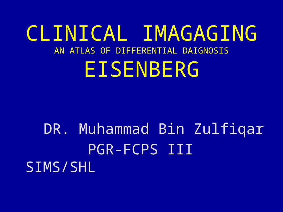
• Fig GI 80-1 Pneumoperitoneum from perforated diverticulitis. (A) Contrast scan (lung window) shows free intraperitoneal gas (arrowheads) in the perihepatic space. Free intraperitoneal gas is more easily identified by using lung window settings. (B) Scan of the pelvis shows the thickened wall of the sigmoid colon (arrow) and mild pericolonic inflammatory fat stranding, findings that represent diverticulitis.
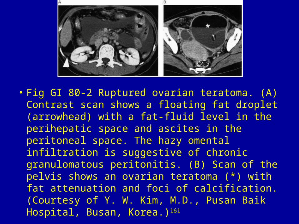
• Fig GI 80-2 Ruptured ovarian teratoma. (A) Contrast scan shows a floating fat droplet (arrowhead) with a fat-fluid level in the perihepatic space and ascites in the peritoneal space. The hazy omental infiltration is suggestive of chronic granulomatous peritonitis. (B) Scan of the pelvis shows an ovarian teratoma (*) with fat attenuation and foci of calcification. (Courtesy of Y. W. Kim, M.D., Pusan Baik Hospital, Busan, Korea.)161
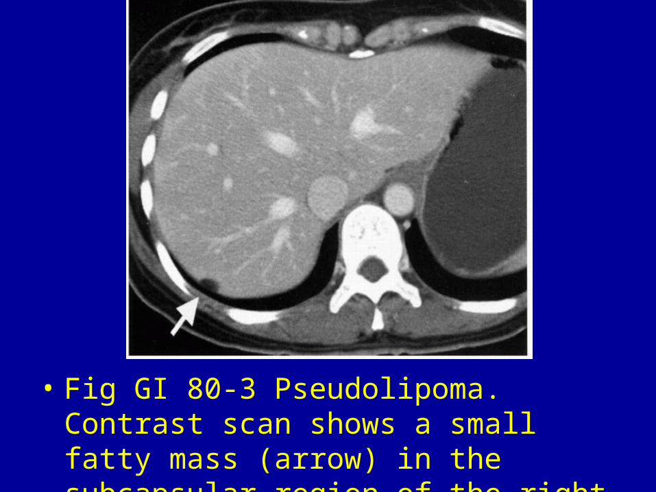
• Fig GI 80-3 Pseudolipoma. Contrast scan shows a small fatty mass (arrow) in the subcapsular region of the right hepatic lobe.161
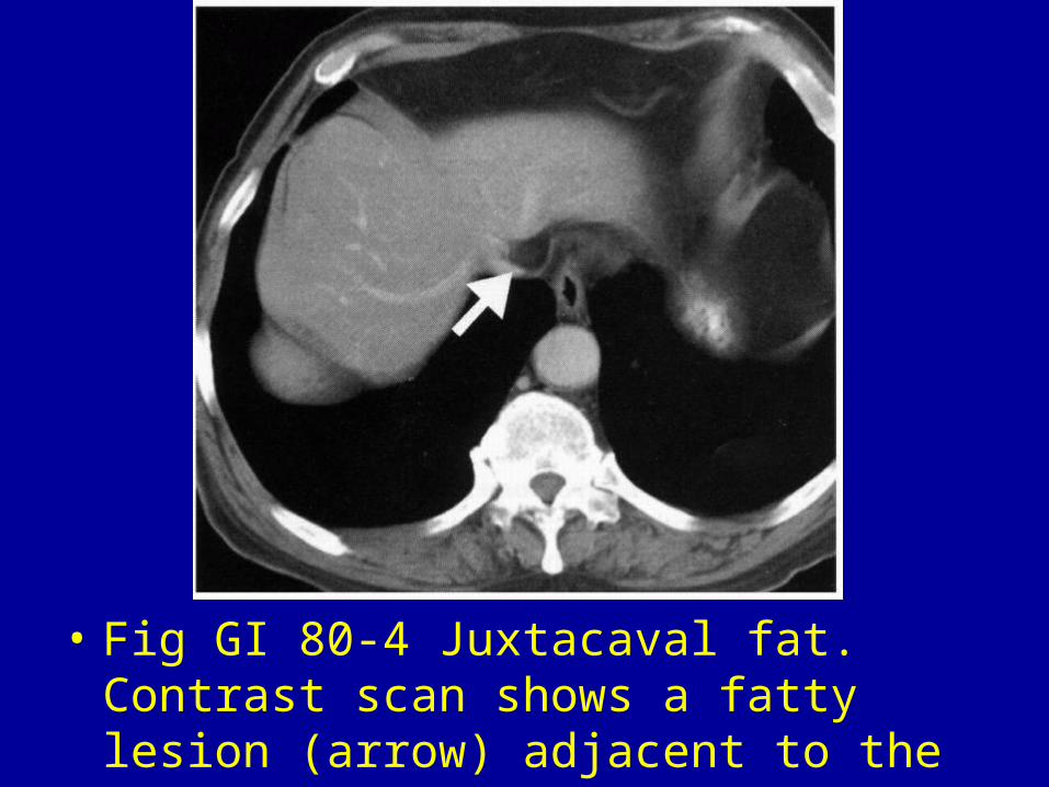
• Fig GI 80-4 Juxtacaval fat. Contrast scan shows a fatty lesion (arrow) adjacent to the intrahepatic vena cava.161
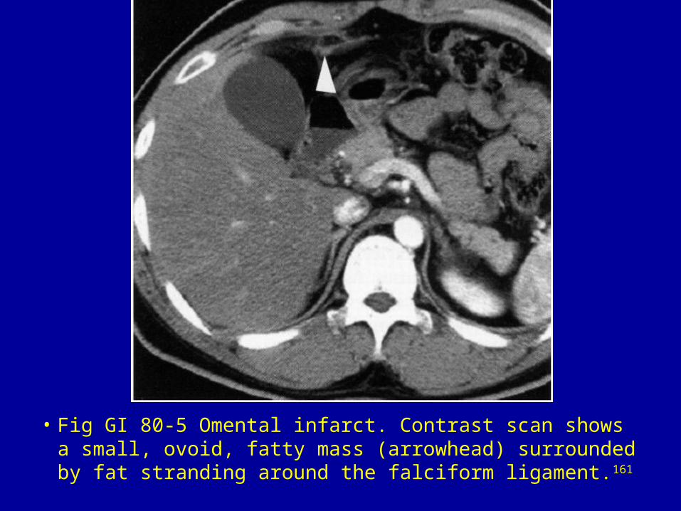
• Fig GI 80-5 Omental infarct. Contrast scan shows a small, ovoid, fatty mass (arrowhead) surrounded by fat stranding around the falciform ligament.161
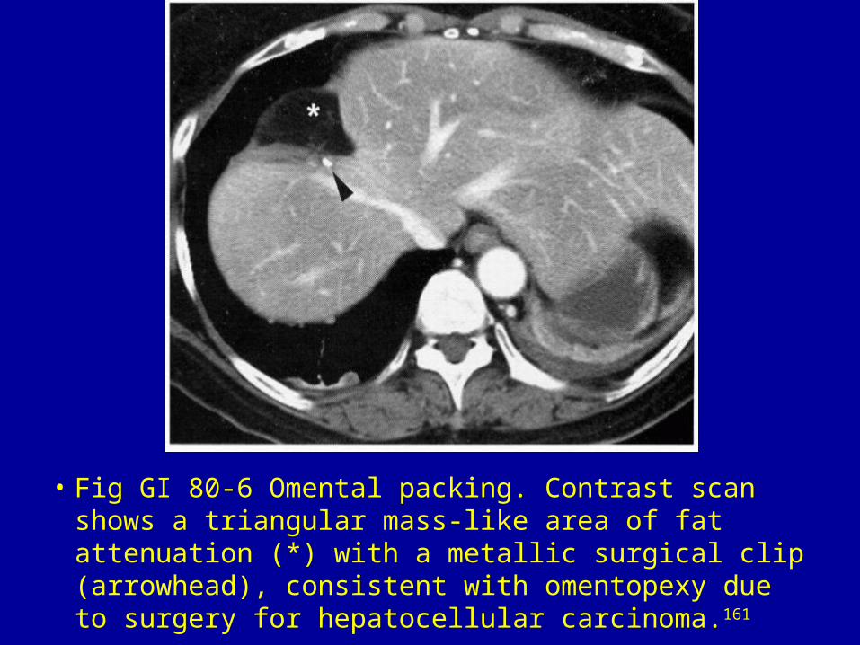
• Fig GI 80-6 Omental packing. Contrast scan shows a triangular mass-like area of fat attenuation (*) with a metallic surgical clip (arrowhead), consistent with omentopexy due to surgery for hepatocellular carcinoma.161
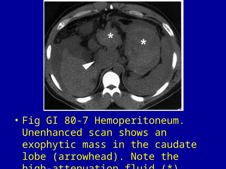
• Fig GI 80-7 Hemoperitoneum. Unenhanced scan shows an exophytic mass in the caudate lobe (arrowhead). Note the high-attenuation fluid (*) around the liver and spleen.161
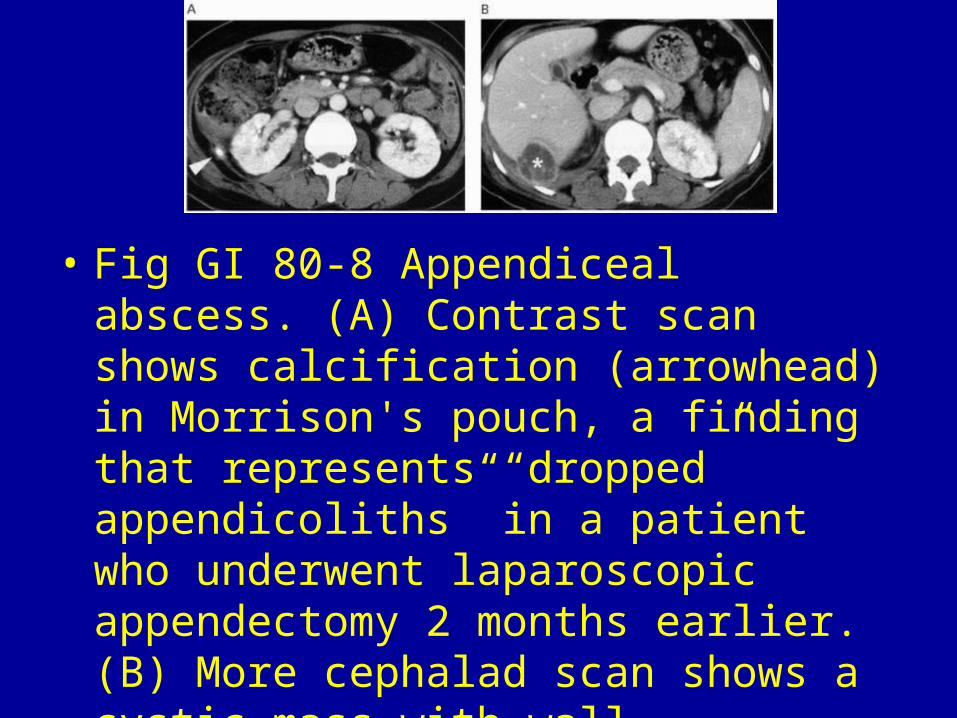
• Fig GI 80-8 Appendiceal abscess. (A) Contrast scan shows calcification (arrowhead) in Morrison's pouch, a finding that represents “dropped” appendicoliths” in a patient who underwent laparoscopic appendectomy 2 months earlier. (B) More cephalad scan shows a cystic mass with wall enhancement (*). This appearance is consistent with an abscess in the posterior right subhepatic space.161
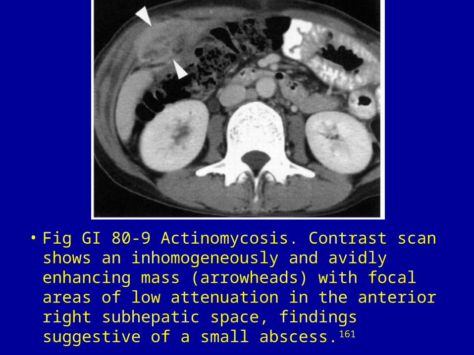
• Fig GI 80-9 Actinomycosis. Contrast scan shows an inhomogeneously and avidly enhancing mass (arrowheads) with focal areas of low attenuation in the anterior right subhepatic space, findings suggestive of a small abscess.161
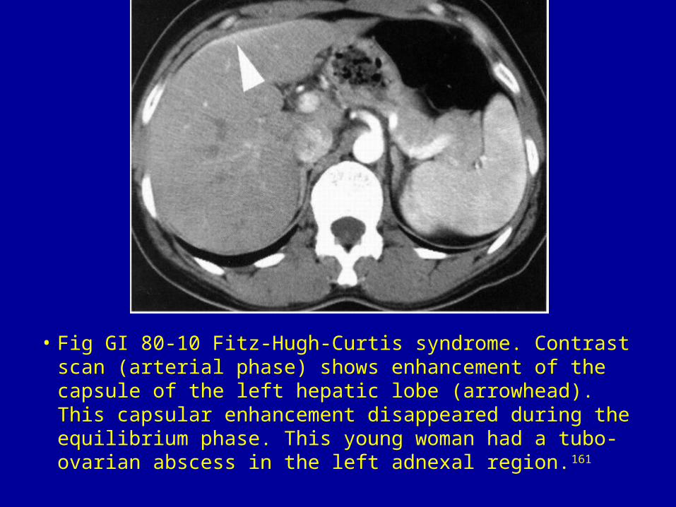
• Fig GI 80-10 Fitz-Hugh-Curtis syndrome. Contrast scan (arterial phase) shows enhancement of the capsule of the left hepatic lobe (arrowhead). This capsular enhancement disappeared during the equilibrium phase. This young woman had a tubo-ovarian abscess in the left adnexal region.161
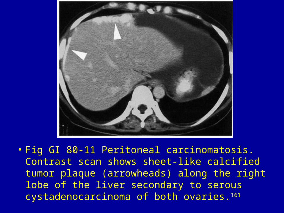
• Fig GI 80-11 Peritoneal carcinomatosis. Contrast scan shows sheet-like calcified tumor plaque (arrowheads) along the right lobe of the liver secondary to serous cystadenocarcinoma of both ovaries.161
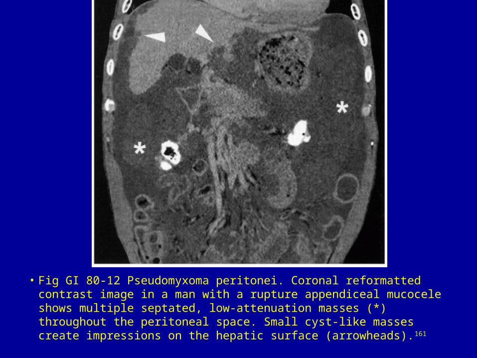
• Fig GI 80-12 Pseudomyxoma peritonei. Coronal reformatted contrast image in a man with a rupture appendiceal mucocele shows multiple septated, low-attenuation masses (*) throughout the peritoneal space. Small cyst-like masses create impressions on the hepatic surface (arrowheads).161
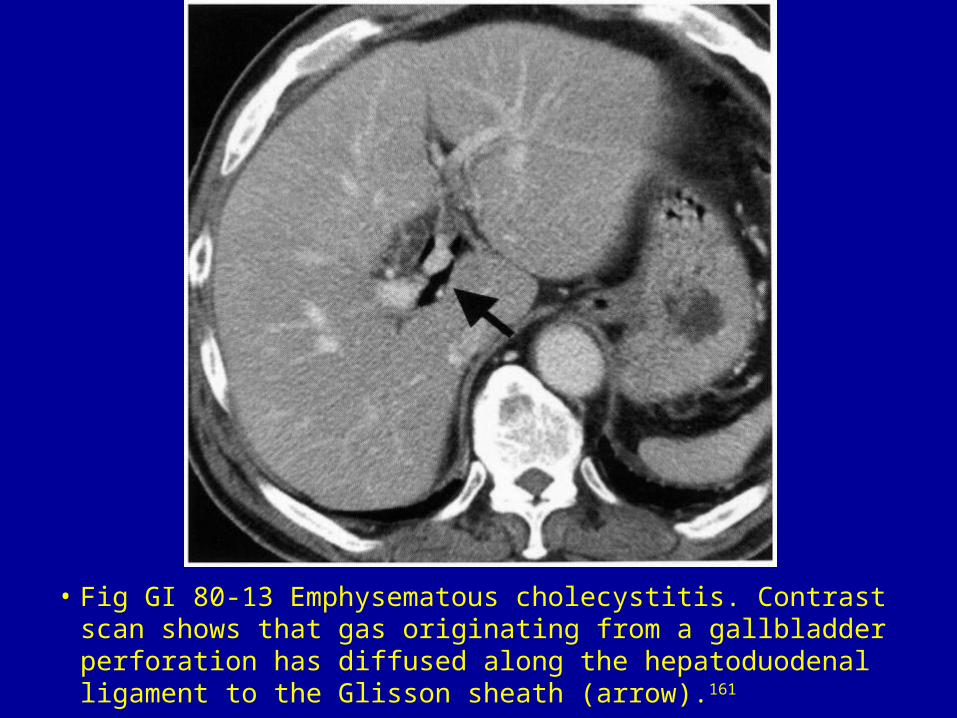
• Fig GI 80-13 Emphysematous cholecystitis. Contrast scan shows that gas originating from a gallbladder perforation has diffused along the hepatoduodenal ligament to the Glisson sheath (arrow).161
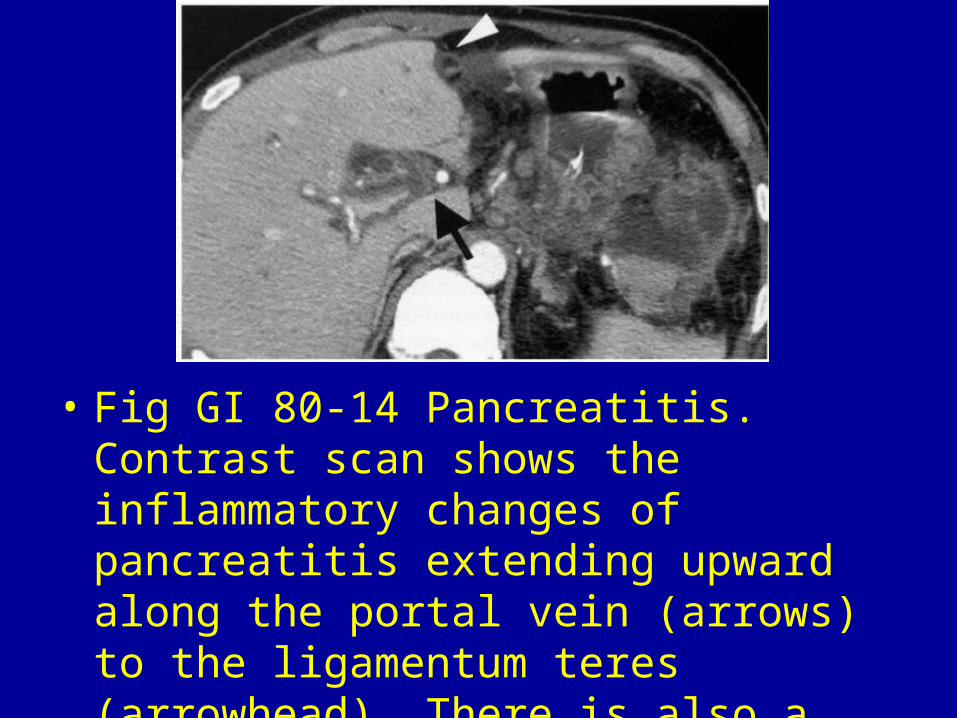
• Fig GI 80-14 Pancreatitis. Contrast scan shows the inflammatory changes of pancreatitis extending upward along the portal vein (arrows) to the ligamentum teres (arrowhead). There is also a peripancreatic fluid collection.161