Embed Size (px)
DESCRIPTION
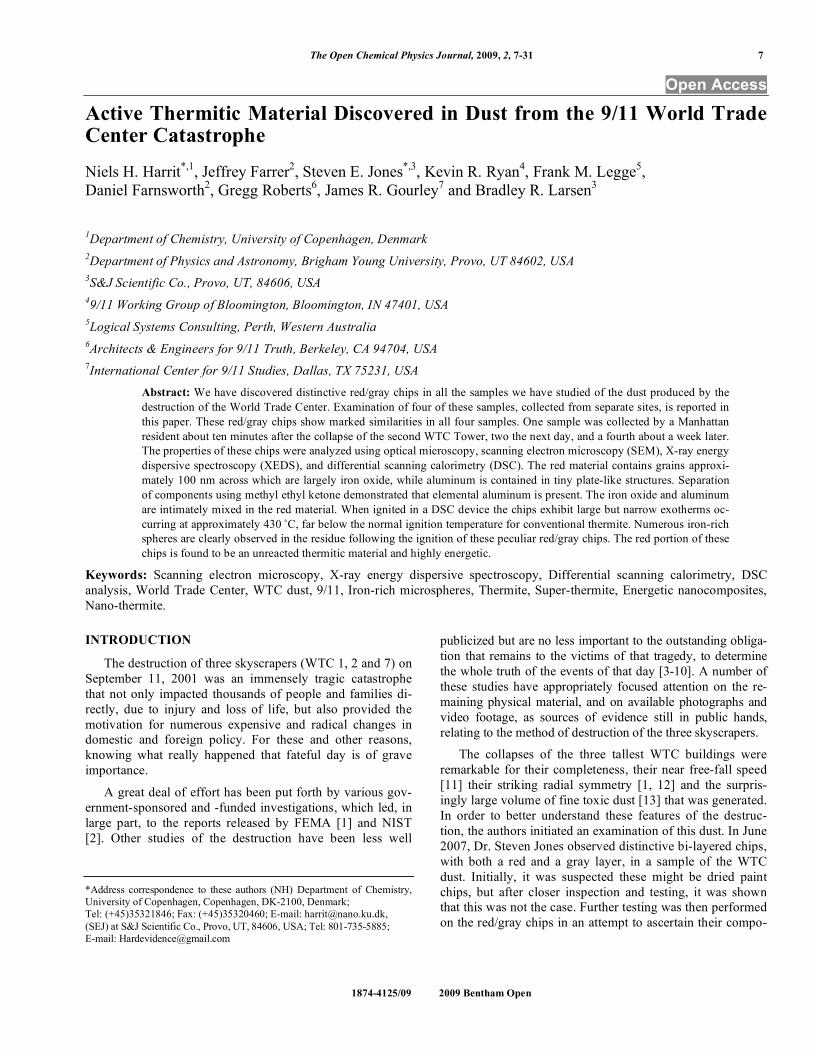
Abstract: We have discovered distinctive red/gray chips in all the samples we have studied of the dust produced by the destruction of the World Trade Center. Examination of four of these samples, collected from separate sites, is reported in this paper. These red/gray chips show marked similarities in all four samples. One sample was collected by a Manhattan resident about ten minutes after the collapse of the second WTC Tower, two the next day, and a fourth about a week later. The properties of these chips were analyzed using optical microscopy, scanning electron microscopy (SEM), X-ray energy dispersive spectroscopy (XEDS), and differential scanning calorimetry (DSC). The red material contains grains approximately 100 nm across which are largely iron oxide, while aluminum is contained in tiny plate-like structures. Separation of components using methyl ethyl ketone demonstrated that elemental aluminum is present. The iron oxide and aluminum are intimately mixed in the red material. When ignited in a DSC device the chips exhibit large but narrow exotherms occurring at approximately 430 °C, far below the normal ignition temperature for conventional thermite. Numerous iron-rich spheres are clearly observed in the residue following the ignition of these peculiar red/gray chips. The red portion of these chips is found to be an unreacted thermitic material and highly energetic.
Citation preview
The Open Chemical Physics Journal, 2009, 2, 7-31 7
1874-4125/09 2009 Bentham Open
Open Access
Active Thermitic Material Discovered in Dust from the 9/11 World Trade Center Catastrophe
Niels H. Harrit*,1
, Jeffrey Farrer2, Steven E. Jones
*,3, Kevin R. Ryan
4, Frank M. Legge
5,
Daniel Farnsworth2, Gregg Roberts
6, James R. Gourley
7 and Bradley R. Larsen
3
1Department of Chemistry, University of Copenhagen, Denmark
2Department of Physics and Astronomy, Brigham Young University, Provo, UT 84602, USA
3S&J Scientific Co., Provo, UT, 84606, USA
49/11 Working Group of Bloomington, Bloomington, IN 47401, USA
5Logical Systems Consulting, Perth, Western Australia
6Architects & Engineers for 9/11 Truth, Berkeley, CA 94704, USA
7International Center for 9/11 Studies, Dallas, TX 75231, USA
Abstract: We have discovered distinctive red/gray chips in all the samples we have studied of the dust produced by the
destruction of the World Trade Center. Examination of four of these samples, collected from separate sites, is reported in
this paper. These red/gray chips show marked similarities in all four samples. One sample was collected by a Manhattan
resident about ten minutes after the collapse of the second WTC Tower, two the next day, and a fourth about a week later.
The properties of these chips were analyzed using optical microscopy, scanning electron microscopy (SEM), X-ray energy
dispersive spectroscopy (XEDS), and differential scanning calorimetry (DSC). The red material contains grains approxi-
mately 100 nm across which are largely iron oxide, while aluminum is contained in tiny plate-like structures. Separation
of components using methyl ethyl ketone demonstrated that elemental aluminum is present. The iron oxide and aluminum
are intimately mixed in the red material. When ignited in a DSC device the chips exhibit large but narrow exotherms oc-
curring at approximately 430 ˚C, far below the normal ignition temperature for conventional thermite. Numerous iron-rich
spheres are clearly observed in the residue following the ignition of these peculiar red/gray chips. The red portion of these
chips is found to be an unreacted thermitic material and highly energetic.
Keywords: Scanning electron microscopy, X-ray energy dispersive spectroscopy, Differential scanning calorimetry, DSC
analysis, World Trade Center, WTC dust, 9/11, Iron-rich microspheres, Thermite, Super-thermite, Energetic nanocomposites,
Nano-thermite.
INTRODUCTION
The destruction of three skyscrapers (WTC 1, 2 and 7) on
September 11, 2001 was an immensely tragic catastrophe
that not only impacted thousands of people and families di-
rectly, due to injury and loss of life, but also provided the
motivation for numerous expensive and radical changes in
domestic and foreign policy. For these and other reasons,
knowing what really happened that fateful day is of grave
importance.
A great deal of effort has been put forth by various gov-
ernment-sponsored and -funded investigations, which led, in
large part, to the reports released by FEMA [1] and NIST
[2]. Other studies of the destruction have been less well
*Address correspondence to these authors (NH) Department of Chemistry,
University of Copenhagen, Copenhagen, DK-2100, Denmark;
Tel: (+45)35321846; Fax: (+45)35320460; E-mail: [email protected],
(SEJ) at S&J Scientific Co., Provo, UT, 84606, USA; Tel: 801-735-5885;
E-mail: [email protected]
publicized but are no less important to the outstanding obliga-
tion that remains to the victims of that tragedy, to determine
the whole truth of the events of that day [3-10]. A number of
these studies have appropriately focused attention on the re-
maining physical material, and on available photographs and
video footage, as sources of evidence still in public hands,
relating to the method of destruction of the three skyscrapers.
The collapses of the three tallest WTC buildings were
remarkable for their completeness, their near free-fall speed
[11] their striking radial symmetry [1, 12] and the surpris-
ingly large volume of fine toxic dust [13] that was generated.
In order to better understand these features of the destruc-
tion, the authors initiated an examination of this dust. In June
2007, Dr. Steven Jones observed distinctive bi-layered chips,
with both a red and a gray layer, in a sample of the WTC
dust. Initially, it was suspected these might be dried paint
chips, but after closer inspection and testing, it was shown
that this was not the case. Further testing was then performed
on the red/gray chips in an attempt to ascertain their compo-
8 The Open Chemical Physics Journal, 2009, Volume 2 Harrit et al.
sition and properties. The authors also obtained and exam-
ined additional samples of WTC dust which had been col-
lected by independent observers on, or very soon after, 9/11.
All of the samples examined contained these very small,
peculiar red/gray chips. Previous studies discussing observa-
tions of the WTC dust include reports by the RJ Lee Com-
pany [14], the U.S. Geological Survey (USGS) [15], McGee
et al. [13] and Lioy et al. [16] Some of these studies con-
firmed the finding of iron-rich microspheres, which are also
peculiar [5, 8, 11, 13-15] but the red/gray chips analyzed in
this study have apparently not been discussed in previously
published reports. It is worth emphasizing that one sample
was collected about ten minutes after the collapse of the sec-
ond Tower, so it cannot possibly have been contaminated by
clean-up operations [17].
MATERIALS AND METHODS
1. Provenance of the Samples Analyzed for this Report
In a paper presented first online in autumn 2006 regard-
ing anomalies observed in the World Trade Center destruc-
tion [6], a general request was issued for samples of the
WTC dust. The expectation at that time was that a careful
examination of the dust might yield evidence to support the
hypothesis that explosive materials other than jet fuel caused
the extraordinarily rapid and essentially total destruction of
the WTC buildings.
It was learned that a number of people had saved samples
of the copious, dense dust, which spread and settled across
Manhattan. Several of these people sent portions of their
samples to members of this research group. This paper dis-
cusses four separate dust samples collected on or shortly
after 9/11/2001. Each sample was found to contain red/gray
chips. All four samples were originally collected by private
citizens who lived in New York City at the time of the trag-
edy. These citizens came forward and provided samples for
analysis in the public interest, allowing study of the 9/11
dust for whatever facts about the day might be learned from
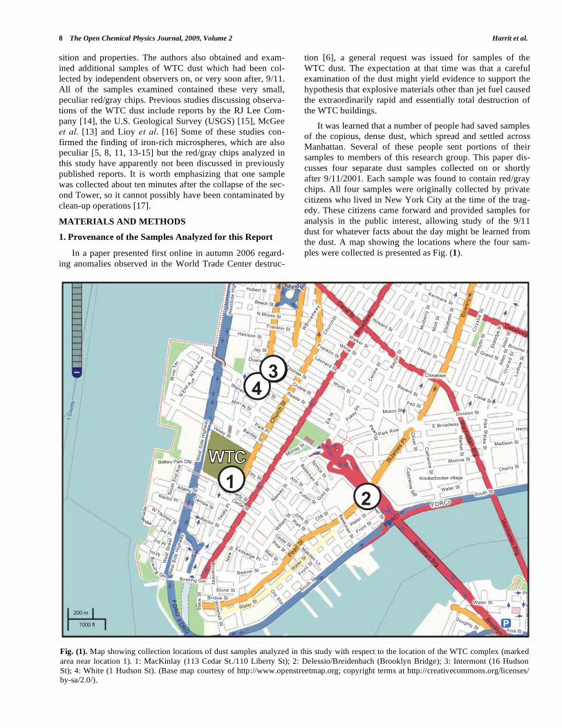
the dust. A map showing the locations where the four sam-
ples were collected is presented as Fig. (1).
Fig. (1). Map showing collection locations of dust samples analyzed in this study with respect to the location of the WTC complex (marked
area near location 1). 1: MacKinlay (113 Cedar St./110 Liberty St); 2: Delessio/Breidenbach (Brooklyn Bridge); 3: Intermont (16 Hudson
St); 4: White (1 Hudson St). (Base map courtesy of http://www.openstreetmap.org; copyright terms at http://creativecommons.org/licenses/ by-sa/2.0/).
�����
�������
�
�
��
�� �
� �
��
���
�
� �
��
��
������
����
����������
����������
� !�������
"��#$���
��%������
&��
����
���
�'$
&"
����
(��
)$�
�#$�
�&��������&$������
����������*���������
&���$���
)��������
)$��������
��������
����
)$�
�#$�
��
)�������
"���
�(����(������
+���������
"�(
���
!��
%���
���
�!
����
��
���,
�%��
����
�
)��
����
���
��
����
����
�
��#�
��#�
����
��
�����
��
-��
���
���
����
���
�
*���
�(��
�
"�������.�������
���������
)�������
����#��
���������
)$�����(�
"��
����
��
"��������
)��
����
��
/������!��#����
/������( -�� ����
� )��$�������
�
��"����(��
� ��������
�����
!���������
)$�������!���#����
!�������
�!
��$������"�'
/�����
�/�����
�
����$���
�)�
�)�
&�������
+��#���%�#���� ����'�
)�
�$ ���#��
�
/�
����
���
�/0
����
�1
���
���
/������
"��������"�'
!��$������"
�'
&�������
2��� ��
��'$�����
"�������
/$
3
/
&����
/����/0"��������
3�������
!����� ��
����� ������
!��������
�4��#����
"���������
.�����
�
��������
)���������$����/�������&
������
���
�����
����
)�������/������&������
�5����'��/0��(
���
"�� �����
��������
"��������
���
#���
�
"�(���' .�
&����$�����
�
"��
��
(�
�
6���/0����/0
����/0
.�
"�
(�� �
'
&��
���
����
�
��#������
)���������
��#����/�
����
��/�
)�#������
��������
&���� ��
-������4
����$���
������
�&
������
�
!������*�
"����
�����
�������&������
�
� �����
/����
��
&��
�����
���
�'$(
��
����
�
/���
&�����������
������ ��
&��
���
����
��'
$(��
���
�$��
����
�
"�������/����)���
��
��)
����
�
����
�
Active Thermitic Material Found in WTC Dust The Open Chemical Physics Journal, 2009, Volume 2 9
The earliest-collected sample came from Mr. Frank De-
lessio who, according to his videotaped testimony [17], was
on the Manhattan side of the Brooklyn Bridge about the time
the second tower, the North Tower, fell to the ground. He
saw the tower fall and was enveloped by the resulting thick
dust which settled throughout the area. He swept a handful
of the dust from a rail on the pedestrian walkway near the
end of the bridge, about ten minutes after the fall of the
North Tower. He then went to visit his friend, Mr. Tom
Breidenbach, carrying the dust in his hand, and the two of
them discussed the dust and decided to save it in a plastic
bag. On 11/15/2007, Breidenbach sent a portion of this dust
to Dr. Jones for analysis. Breidenbach has also recorded his
testimony about the collection of this dust sample on video-
tape [17]. Thus, the Delessio/Breidenbach sample was col-
lected about ten minutes after the second tower collapsed. It
was, therefore, definitely not contaminated by the steel-
cutting or clean-up operations at Ground Zero, which began
later. Furthermore, it is not mixed with dust from WTC 7,
which fell hours later.
On the morning of 9/12/2001, Mr. Stephen White of New
York City entered a room in his apartment on the 8th floor of
1 Hudson Street, about five blocks from the WTC. He found
a layer of dust about an inch thick on a stack of folded laun-
dry near a window which was open about 4 inches (10 cm).
Evidently the open window had allowed a significant amount
of dust from the WTC destruction the day before to enter the
room and cover the laundry. He saved some of the dust and,
on 2/02/2008, sent a sample directly to Dr. Jones for analy-
sis.
Another sample was collected from the apartment build-
ing at 16 Hudson Street by Mr. Jody Intermont at about 2 pm
on 9/12/2001. Two small samples of this dust were simulta-
neously sent to Dr. Jones and to Kevin Ryan on 2/02/2008
for analysis. Intermont sent a signed affidavit with each
sample verifying that he had personally collected the (now-
split) sample; he wrote:
“This dust, which came from the ‘collapsed’
World Trade Center Towers, was collected from
my loft at the corner of Reade Street and Hud-
son Street on September 12, 2001. I give per-
mission to use my name in connection to this
evidence”. [Signed 31 January 2008 in the pres-
ence of a witness who also signed his name].
On the morning of 9/11/2001, Ms. Janette MacKinlay
was in her fourth-floor apartment at 113 Cedar St./110 Lib-
erty St. in New York City, across the street from the WTC
plaza. As the South Tower collapsed, the flowing cloud of
dust and debris caused windows of her apartment to break
inward and dust filled her apartment. She escaped by quickly
wrapping a wet towel around her head and exiting the build-
ing. The building was closed for entry for about a week. As
soon as Ms. MacKinlay was allowed to re-enter her apart-
ment, she did so and began cleaning up. There was a thick
layer of dust on the floor. She collected some of it into a
large sealable plastic bag for possible later use in an art
piece. Ms. MacKinlay responded to the request in the 2006
paper by Dr. Jones by sending him a dust sample. In No-
vember 2006, Dr. Jones traveled to California to visit Ms.
MacKinlay at her new location, and in the company of sev-
eral witnesses collected a second sample of the WTC dust
directly from her large plastic bag where the dust was stored.
She has also sent samples directly to Dr. Jeffrey Farrer and
Kevin Ryan. Results from their studies form part of this re-
port.
Another dust sample was collected by an individual from
a window sill of a building on Potter Street in NYC. He has
not given permission for his name to be disclosed, therefore
his material is not included in this study. That sample, how-
ever, contained red/gray chips of the same general composi-
tion as the samples described here.
2. Chip Size, Isolation, and Examination
For clarification, the dust samples collected and sent to
the authors by Ms. Janette MacKinlay will be sample 1; the
sample collected by Mr. Frank Delassio, or the Delas-
sio/Breidenbach sample, will be sample 2; the sample col-
lected by Mr. Jody Intermont will be sample 3; and the sam-
ple collected by Mr. Stephen White will be sample 4. The
red/gray chips are attracted by a magnet, which facilitates
collection and separation of the chips from the bulk of the
dust. A small permanent magnet in its own plastic bag was
used to attract and collect the chips from dust samples. The
chips are typically small but readily discernible by eye due to
their distinctive color. They are of variable size with major
dimensions of roughly 0.2 to 3 mm. Thicknesses vary from
roughly 10 to 100 microns for each layer (red and gray).
Samples of WTC dust from these and other collectors have
been sent directly from collectors to various scientists (in-
cluding some not on this research team) who have also found
such red/gray chips in the dust from the World Trade Center
destruction.
An FEI XL30-SFEG scanning electron microscope
(SEM) was used to perform secondary-electron (SE) imag-
ing and backscattered electron (BSE) imaging. The SE imag-
ing was used to look at the surface topography and porosity
of the red/gray chips, while the BSE imaging was used to
distinguish variations in average atomic number, Z. The mi-
croscope was also equipped with an EDAX X-ray energy
dispersive spectrometry (XEDS) system. The XEDS system
uses a silicon detector (SiLi) with resolution better than 135
eV. The spectrum resolution was set to 10 eV per channel.
Operating conditions for the acquired XEDS spectra were 20
keV beam energy (unless otherwise specified) and 40-120
second acquisition time (livetime). XEDS maps were ac-
quired using the same system at a beam energy of 10 keV.
For general surface analysis in the SEM, dust samples
were mounted to carbon conductive tabs. The samples were
left unwashed and uncoated unless otherwise specified. In
order to more closely observe the characteristics of the red
and gray layers, and to eliminate the possibility of surface
contamination from other dust particles, several red/gray
chips from each of the four WTC dust samples were frac-
tured. The clean, cross-section surfaces were then studied by
BSE imaging and XEDS.
10 The Open Chemical Physics Journal, 2009, Volume 2 Harrit et al.
Some samples were also tested in a differential scanning
calorimeter (Netzsch DSC 404C) to measure heat flow into
or out of the red/gray chips. The DSC tests were conducted
with a linear heating rate of 10 ˚C per minute up to a tem-
perature of 700 ˚C. During heating, the samples were con-
tained in alumina pans and air was allowed to flow at 55
milliliters per minute during the heating. The plots were gen-
erated by acquiring data points at a rate of 20 points per ˚C
or 200 points per minute. The equipment was calibrated to
display the data in watts per gram. The plots were set to dis-
play positive heat flow out of the sample such that exother-
mic behavior of the sample would yield a peak and endo-
thermic behavior a trough.
The dust samples were also examined by visible-light
microscopy (VLM) through a Nikon Epiphot 200 stereomi-
croscope, an Olympus BX60 stereomicroscope and a Nikon
Labophot microscope and camera.
RESULTS
1. Characterization of the Red/Gray Chips
Red/gray chips were found in all of the dust samples col-
lected. An analysis of the chips was performed to assess the
similarity of the chips and to determine the chemistry and
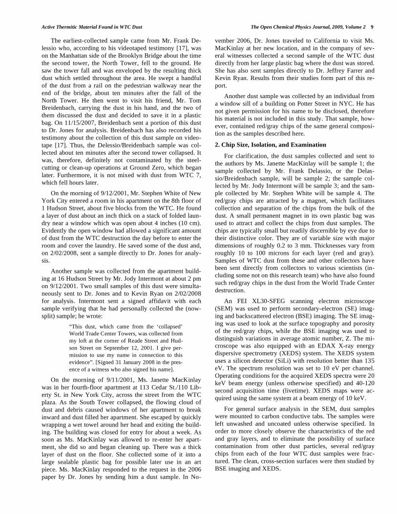
materials that make up the chips. Fig. (2) displays photomi-
crographs of red/gray chips from each of the four WTC dust
samples. Note the scale marker in each image as they were
acquired at different magnifications. At approximately
2.5 mm in length, the chip in Fig. (2a) was one of the larger
chips collected. The mass of this chip was approximately 0.7
mg. All of the chips used in the study had a gray layer and a
red layer and were attracted by a magnet. The inset image in
Fig. (2d) shows the chip in cross section, which reveals the
gray layer. The gray layer is also partially visible in Fig.
(2b). Similarities between the samples are already evident
from these photographs.
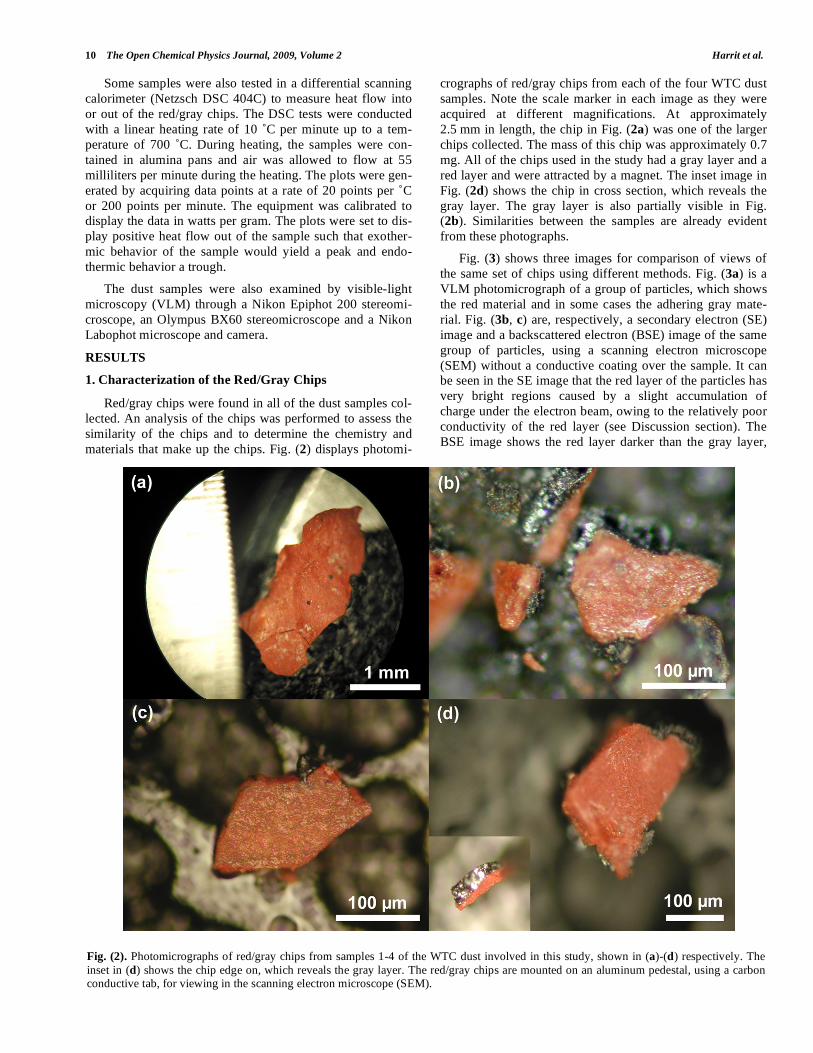
Fig. (3) shows three images for comparison of views of
the same set of chips using different methods. Fig. (3a) is a
VLM photomicrograph of a group of particles, which shows
the red material and in some cases the adhering gray mate-
rial. Fig. (3b, c) are, respectively, a secondary electron (SE)
image and a backscattered electron (BSE) image of the same
group of particles, using a scanning electron microscope
(SEM) without a conductive coating over the sample. It can
be seen in the SE image that the red layer of the particles has
very bright regions caused by a slight accumulation of
charge under the electron beam, owing to the relatively poor
conductivity of the red layer (see Discussion section). The
BSE image shows the red layer darker than the gray layer,
Fig. (2). Photomicrographs of red/gray chips from samples 1-4 of the WTC dust involved in this study, shown in (a)-(d) respectively. The
inset in (d) shows the chip edge on, which reveals the gray layer. The red/gray chips are mounted on an aluminum pedestal, using a carbon conductive tab, for viewing in the scanning electron microscope (SEM).
Active Thermitic Material Found in WTC Dust The Open Chemical Physics Journal, 2009, Volume 2 11
indicating that the red layer is composed of material that has
a relatively lower average atomic number than the gray
layer.
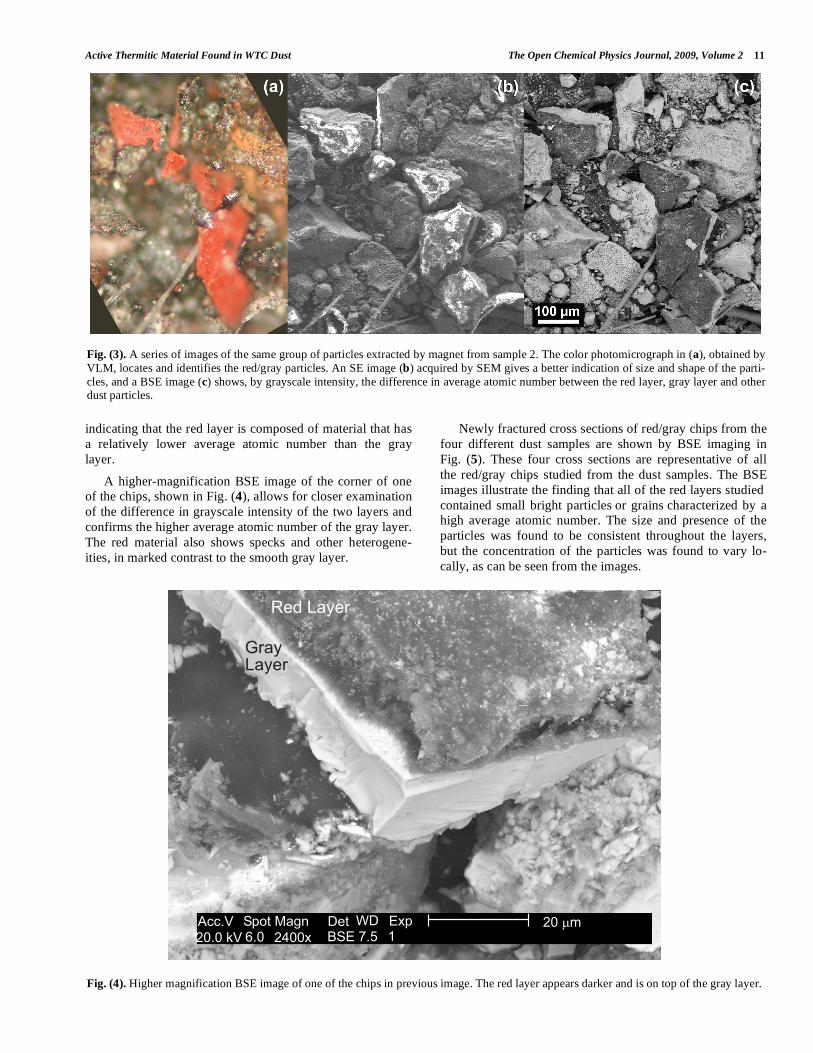
A higher-magnification BSE image of the corner of one
of the chips, shown in Fig. (4), allows for closer examination
of the difference in grayscale intensity of the two layers and
confirms the higher average atomic number of the gray layer.
The red material also shows specks and other heterogene-
ities, in marked contrast to the smooth gray layer.
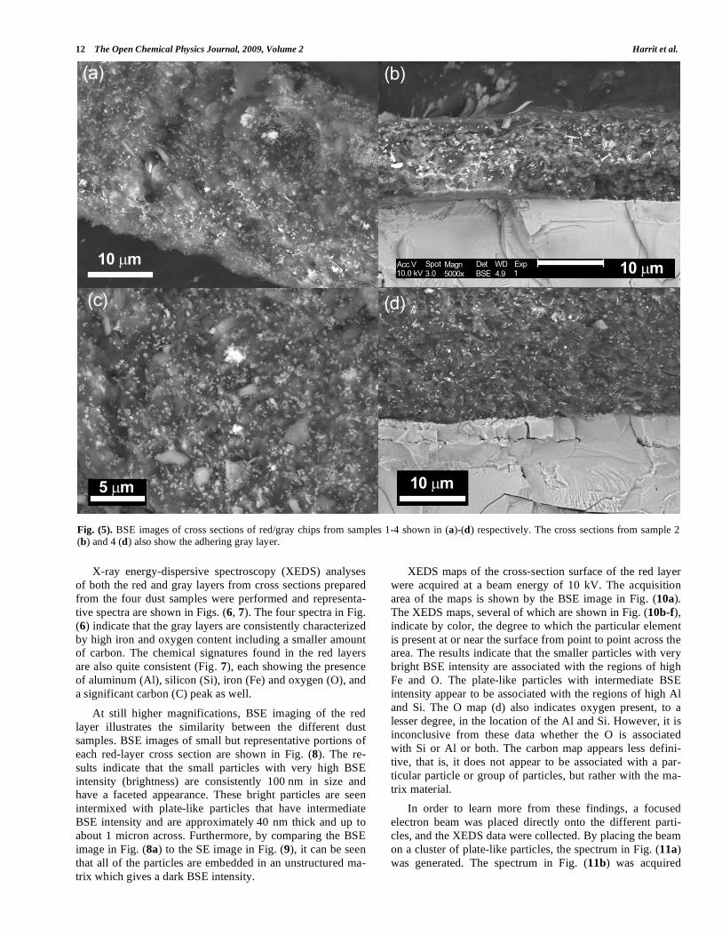
Newly fractured cross sections of red/gray chips from the
four different dust samples are shown by BSE imaging in
Fig. (5). These four cross sections are representative of all
the red/gray chips studied from the dust samples. The BSE
images illustrate the finding that all of the red layers studied
contained small bright particles or grains characterized by a
high average atomic number. The size and presence of the
particles was found to be consistent throughout the layers,
but the concentration of the particles was found to vary lo-
cally, as can be seen from the images.
Fig. (3). A series of images of the same group of particles extracted by magnet from sample 2. The color photomicrograph in (a), obtained by
VLM, locates and identifies the red/gray particles. An SE image (b) acquired by SEM gives a better indication of size and shape of the parti-
cles, and a BSE image (c) shows, by grayscale intensity, the difference in average atomic number between the red layer, gray layer and other dust particles.
Fig. (4). Higher magnification BSE image of one of the chips in previous image. The red layer appears darker and is on top of the gray layer.
��������
������
��� � ��������� ��� �� ��������� ��� �� �!� "
� ��#
12 The Open Chemical Physics Journal, 2009, Volume 2 Harrit et al.
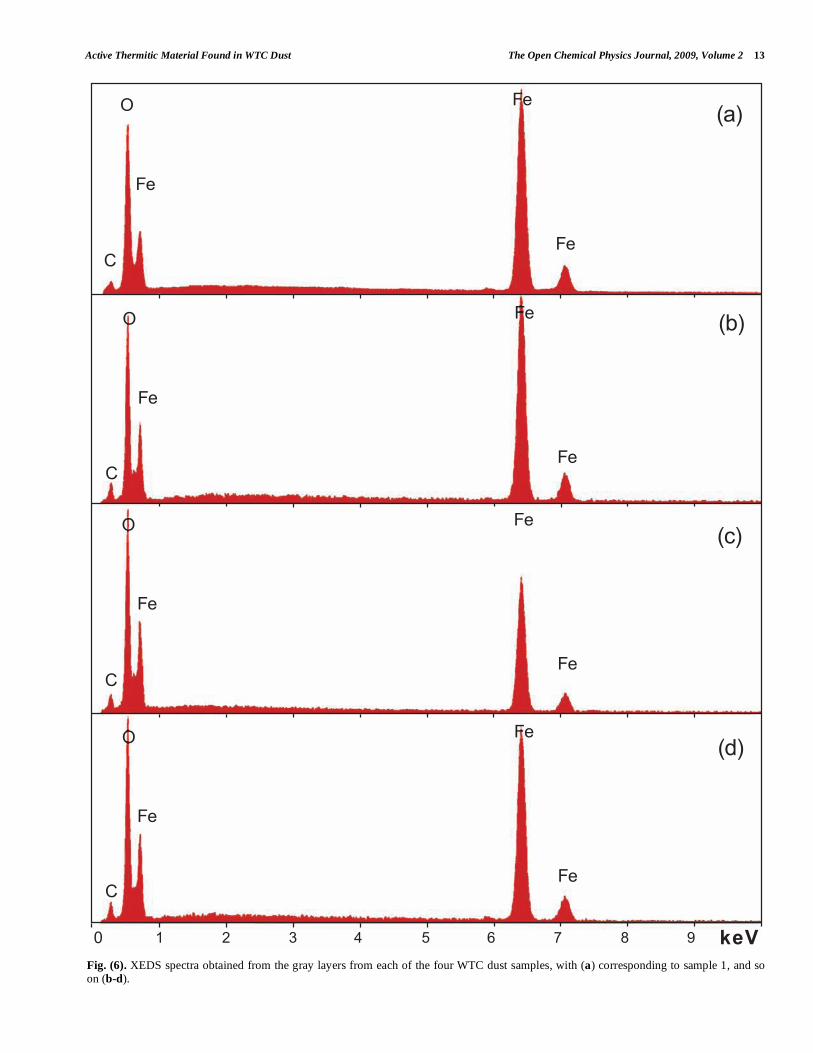
X-ray energy-dispersive spectroscopy (XEDS) analyses
of both the red and gray layers from cross sections prepared
from the four dust samples were performed and representa-
tive spectra are shown in Figs. (6, 7). The four spectra in Fig.
(6) indicate that the gray layers are consistently characterized
by high iron and oxygen content including a smaller amount
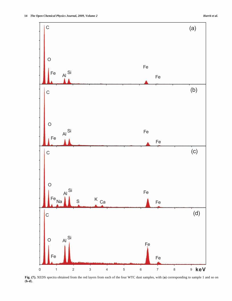
of carbon. The chemical signatures found in the red layers
are also quite consistent (Fig. 7), each showing the presence
of aluminum (Al), silicon (Si), iron (Fe) and oxygen (O), and
a significant carbon (C) peak as well.
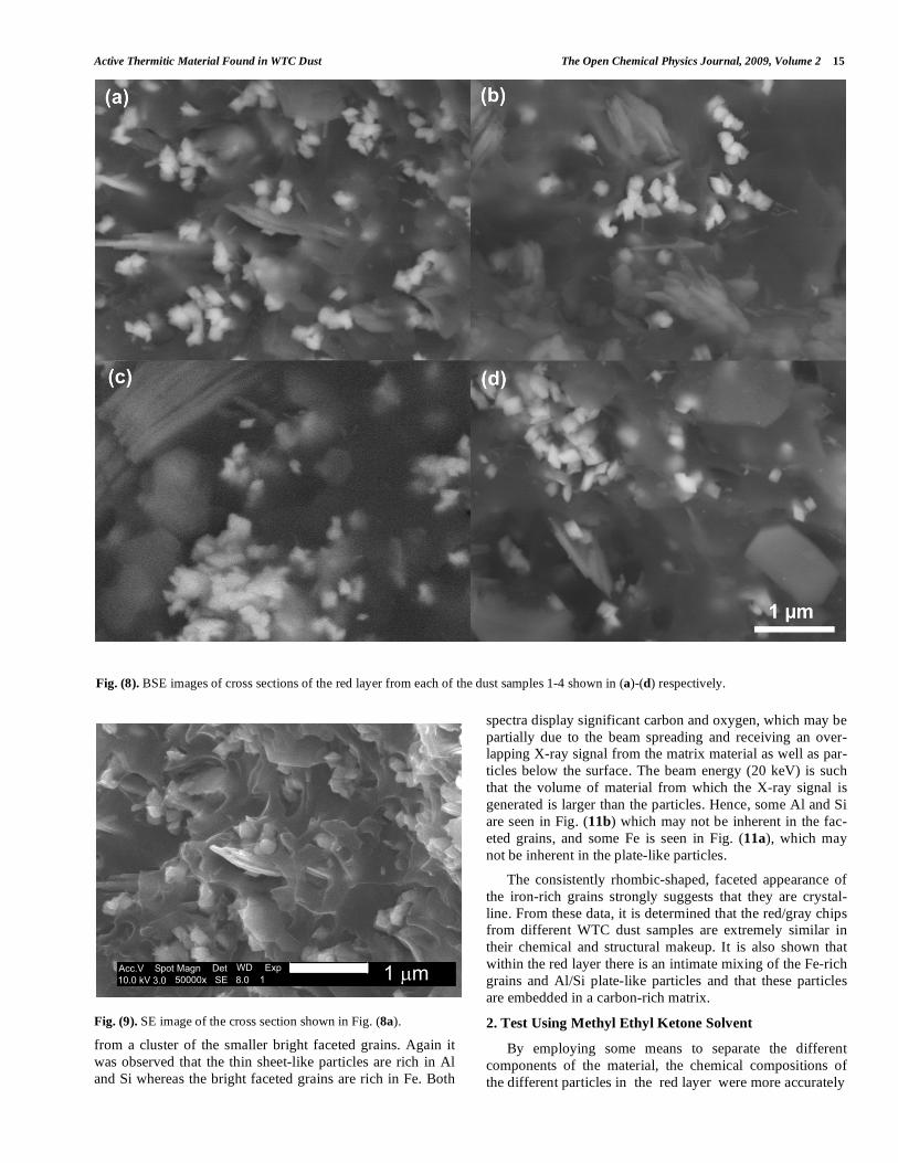
At still higher magnifications, BSE imaging of the red
layer illustrates the similarity between the different dust
samples. BSE images of small but representative portions of
each red-layer cross section are shown in Fig. (8). The re-
sults indicate that the small particles with very high BSE
intensity (brightness) are consistently 100 nm in size and
have a faceted appearance. These bright particles are seen
intermixed with plate-like particles that have intermediate
BSE intensity and are approximately 40 nm thick and up to
about 1 micron across. Furthermore, by comparing the BSE
image in Fig. (8a) to the SE image in Fig. (9), it can be seen
that all of the particles are embedded in an unstructured ma-
trix which gives a dark BSE intensity.
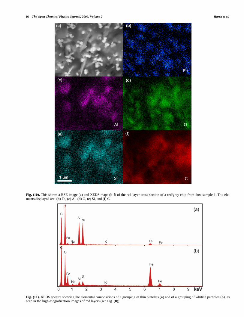
XEDS maps of the cross-section surface of the red layer
were acquired at a beam energy of 10 kV. The acquisition
area of the maps is shown by the BSE image in Fig. (10a).
The XEDS maps, several of which are shown in Fig. (10b-f),
indicate by color, the degree to which the particular element
is present at or near the surface from point to point across the
area. The results indicate that the smaller particles with very
bright BSE intensity are associated with the regions of high
Fe and O. The plate-like particles with intermediate BSE
intensity appear to be associated with the regions of high Al
and Si. The O map (d) also indicates oxygen present, to a
lesser degree, in the location of the Al and Si. However, it is
inconclusive from these data whether the O is associated
with Si or Al or both. The carbon map appears less defini-
tive, that is, it does not appear to be associated with a par-
ticular particle or group of particles, but rather with the ma-
trix material.
In order to learn more from these findings, a focused
electron beam was placed directly onto the different parti-
cles, and the XEDS data were collected. By placing the beam
on a cluster of plate-like particles, the spectrum in Fig. (11a)
was generated. The spectrum in Fig. (11b) was acquired
Fig. (5). BSE images of cross sections of red/gray chips from samples 1-4 shown in (a)-(d) respectively. The cross sections from sample 2 (b) and 4 (d) also show the adhering gray layer.
7�8 7%8
7�87#8
��� � ���
� ����## 3�� ���3 6 �
�4�� !�'�9���5 "�� : ;
&�� �54�
��� ���
������
���� �����
����������� � ��
���� ��������� ��� ��
����� ����
�����
Active Thermitic Material Found in WTC Dust The Open Chemical Physics Journal, 2009, Volume 2 13
Fig. (6). XEDS spectra obtained from the gray layers from each of the four WTC dust samples, with (a) corresponding to sample 1, and so on (b-d).
� � � 6 : 9 < = > ; ���
)
-
�
�
�
7�8
�
7#8�
�
-
)
�
7%8�
�
-
)
�
7�8�
�
-
)
14 The Open Chemical Physics Journal, 2009, Volume 2 Harrit et al.
Fig. (7). XEDS spectra obtained from the red layers from each of the four WTC dust samples, with (a) corresponding to sample 1 and so on (b-d).
)
-
� ����
�
�
)
-
���
�� �
�
)
-
���
�� �
��� � + )�
)
-
�
����
�
�
7�8
7#8
7%8
7�8
� � � 6 : 9 < = > ; ���
Active Thermitic Material Found in WTC Dust The Open Chemical Physics Journal, 2009, Volume 2 15
Fig. (9). SE image of the cross section shown in Fig. (8a).
from a cluster of the smaller bright faceted grains. Again it
was observed that the thin sheet-like particles are rich in Al
and Si whereas the bright faceted grains are rich in Fe. Both
spectra display significant carbon and oxygen, which may be
partially due to the beam spreading and receiving an over-
lapping X-ray signal from the matrix material as well as par-
ticles below the surface. The beam energy (20 keV) is such
that the volume of material from which the X-ray signal is
generated is larger than the particles. Hence, some Al and Si
are seen in Fig. (11b) which may not be inherent in the fac-
eted grains, and some Fe is seen in Fig. (11a), which may
not be inherent in the plate-like particles.
The consistently rhombic-shaped, faceted appearance of
the iron-rich grains strongly suggests that they are crystal-
line. From these data, it is determined that the red/gray chips
from different WTC dust samples are extremely similar in
their chemical and structural makeup. It is also shown that
within the red layer there is an intimate mixing of the Fe-rich
grains and Al/Si plate-like particles and that these particles
are embedded in a carbon-rich matrix.
2. Test Using Methyl Ethyl Ketone Solvent
By employing some means to separate the different
components of the material, the chemical compositions of
the different particles in the red layer were more accurately
Fig. (8). BSE images of cross sections of the red layer from each of the dust samples 1-4 shown in (a)-(d) respectively.
�## 3 �4���!�'�������> �9����5�� ���3 6 � ������ & �54
16 The Open Chemical Physics Journal, 2009, Volume 2 Harrit et al.
Fig. (10). This shows a BSE image (a) and XEDS maps (b-f) of the red-layer cross section of a red/gray chip from dust sample 1. The ele-
ments displayed are: (b) Fe, (c) Al, (d) O, (e) Si, and (f) C.
Fig. (11). XEDS spectra showing the elemental compositions of a grouping of thin platelets (a) and of a grouping of whitish particles (b), as seen in the high-magnification images of red layers (see Fig. (8)).
� � � 6 : 9 < = > ; ���
�
�
+
����
��
�
-)
7%8
��+
����
���
-
)7�8
Active Thermitic Material Found in WTC Dust The Open Chemical Physics Journal, 2009, Volume 2 17
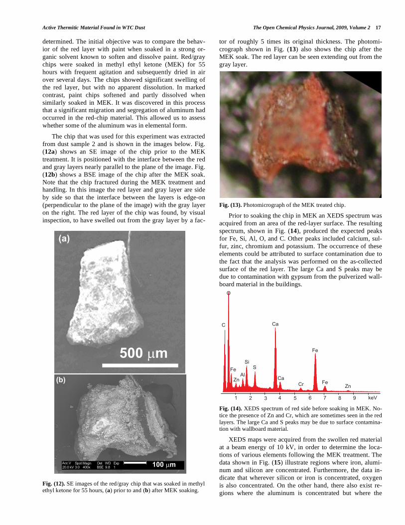
determined. The initial objective was to compare the behav-
ior of the red layer with paint when soaked in a strong or-
ganic solvent known to soften and dissolve paint. Red/gray
chips were soaked in methyl ethyl ketone (MEK) for 55
hours with frequent agitation and subsequently dried in air
over several days. The chips showed significant swelling of
the red layer, but with no apparent dissolution. In marked
contrast, paint chips softened and partly dissolved when
similarly soaked in MEK. It was discovered in this process
that a significant migration and segregation of aluminum had
occurred in the red-chip material. This allowed us to assess
whether some of the aluminum was in elemental form.
The chip that was used for this experiment was extracted
from dust sample 2 and is shown in the images below. Fig.
(12a) shows an SE image of the chip prior to the MEK
treatment. It is positioned with the interface between the red
and gray layers nearly parallel to the plane of the image. Fig.
(12b) shows a BSE image of the chip after the MEK soak.
Note that the chip fractured during the MEK treatment and
handling. In this image the red layer and gray layer are side
by side so that the interface between the layers is edge-on
(perpendicular to the plane of the image) with the gray layer
on the right. The red layer of the chip was found, by visual
inspection, to have swelled out from the gray layer by a fac-
tor of roughly 5 times its original thickness. The photomi-
crograph shown in Fig. (13) also shows the chip after the
MEK soak. The red layer can be seen extending out from the
gray layer.
Fig. (13). Photomicrograph of the MEK treated chip.
Prior to soaking the chip in MEK an XEDS spectrum was
acquired from an area of the red-layer surface. The resulting
spectrum, shown in Fig. (14), produced the expected peaks
for Fe, Si, Al, O, and C. Other peaks included calcium, sul-
fur, zinc, chromium and potassium. The occurrence of these
elements could be attributed to surface contamination due to
the fact that the analysis was performed on the as-collected
surface of the red layer. The large Ca and S peaks may be
due to contamination with gypsum from the pulverized wall-
board material in the buildings.
Fig. (14). XEDS spectrum of red side before soaking in MEK. No-
tice the presence of Zn and Cr, which are sometimes seen in the red
layers. The large Ca and S peaks may be due to surface contamina-tion with wallboard material.
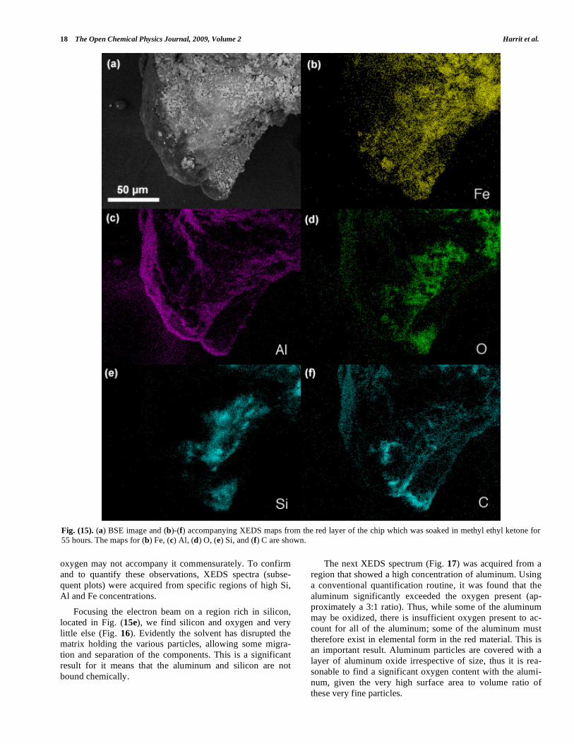
XEDS maps were acquired from the swollen red material
at a beam energy of 10 kV, in order to determine the loca-
tions of various elements following the MEK treatment. The
data shown in Fig. (15) illustrate regions where iron, alumi-
num and silicon are concentrated. Furthermore, the data in-
dicate that wherever silicon or iron is concentrated, oxygen
is also concentrated. On the other hand, there also exist re-
gions where the aluminum is concentrated but where the
Fig. (12). SE images of the red/gray chip that was soaked in methyl ethyl ketone for 55 hours, (a) prior to and (b) after MEK soaking.
�����������
�� �������� ����
�� ��
����� �
��� ������
���
��
�����
�
� ��
��
����
��
����
��
� � � � � � � � ���
��
18 The Open Chemical Physics Journal, 2009, Volume 2 Harrit et al.
oxygen may not accompany it commensurately. To confirm
and to quantify these observations, XEDS spectra (subse-
quent plots) were acquired from specific regions of high Si,
Al and Fe concentrations.
Focusing the electron beam on a region rich in silicon,
located in Fig. (15e), we find silicon and oxygen and very
little else (Fig. 16). Evidently the solvent has disrupted the
matrix holding the various particles, allowing some migra-
tion and separation of the components. This is a significant
result for it means that the aluminum and silicon are not
bound chemically.
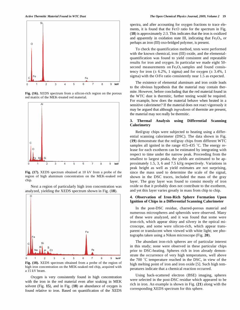
The next XEDS spectrum (Fig. 17) was acquired from a
region that showed a high concentration of aluminum. Using
a conventional quantification routine, it was found that the
aluminum significantly exceeded the oxygen present (ap-
proximately a 3:1 ratio). Thus, while some of the aluminum
may be oxidized, there is insufficient oxygen present to ac-
count for all of the aluminum; some of the aluminum must
therefore exist in elemental form in the red material. This is
an important result. Aluminum particles are covered with a
layer of aluminum oxide irrespective of size, thus it is rea-
sonable to find a significant oxygen content with the alumi-
num, given the very high surface area to volume ratio of
these very fine particles.
Fig. (15). (a) BSE image and (b)-(f) accompanying XEDS maps from the red layer of the chip which was soaked in methyl ethyl ketone for
55 hours. The maps for (b) Fe, (c) Al, (d) O, (e) Si, and (f) C are shown.
Active Thermitic Material Found in WTC Dust The Open Chemical Physics Journal, 2009, Volume 2 19
Fig. (16). XEDS spectrum from a silicon-rich region on the porous red matrix of the MEK-treated red material.
Fig. (17). XEDS spectrum obtained at 10 kV from a probe of the
region of high aluminum concentration on the MEK-soaked red chip.
Next a region of particularly high iron concentration was
analyzed, yielding the XEDS spectrum shown in Fig. (18).
Fig. (18). XEDS spectrum obtained from a probe of the region of
high iron concentration on the MEK-soaked red chip, acquired with a 15 kV beam.
Oxygen is very consistently found in high concentration
with the iron in the red material even after soaking in MEK
solvent (Fig. 15), and in Fig. (18) an abundance of oxygen is
found relative to iron. Based on quantification of the XEDS
spectra, and after accounting for oxygen fractions to trace ele-
ments, it is found that the Fe:O ratio for the spectrum in Fig.
(18) is approximately 2:3. This indicates that the iron is oxidized
and apparently in oxidation state III, indicating that Fe2O3, or
perhaps an iron (III) oxo-bridged polymer, is present.
To check the quantification method, tests were performed
with the known chemical, iron (III) oxide, and the elemental-
quantification was found to yield consistent and repeatable
results for iron and oxygen. In particular we made eight 50-
second measurements on Fe2O3 samples and found consis-
tency for iron (± 6.2%, 1 sigma) and for oxygen (± 3.4%, 1
sigma) with the O/Fe ratio consistently near 1.5 as expected.
The existence of elemental aluminum and iron oxide leads
to the obvious hypothesis that the material may contain ther-
mite. However, before concluding that the red material found in
the WTC dust is thermitic, further testing would be required.
For example, how does the material behave when heated in a
sensitive calorimeter? If the material does not react vigorously it
may be argued that although ingredients of thermite are present,
the material may not really be thermitic.
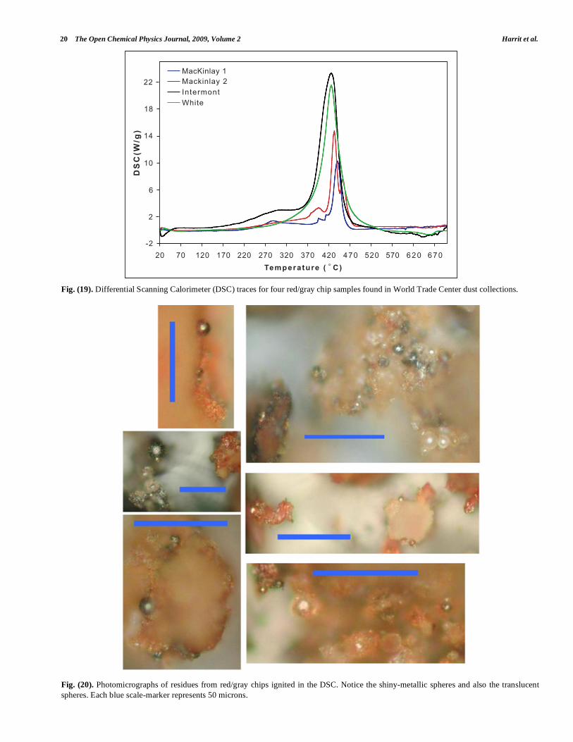
3. Thermal Analysis using Differential Scanning Calorimetry
Red/gray chips were subjected to heating using a differ-
ential scanning calorimeter (DSC). The data shown in Fig.
(19) demonstrate that the red/gray chips from different WTC
samples all ignited in the range 415-435 ˚C. The energy re-
lease for each exotherm can be estimated by integrating with
respect to time under the narrow peak. Proceeding from the
smallest to largest peaks, the yields are estimated to be ap-
proximately 1.5, 3, 6 and 7.5 kJ/g respectively. Variations in
peak height as well as yield estimates are not surprising,
since the mass used to determine the scale of the signal,
shown in the DSC traces, included the mass of the gray
layer. The gray layer was found to consist mostly of iron
oxide so that it probably does not contribute to the exotherm,
and yet this layer varies greatly in mass from chip to chip.
4. Observation of Iron-Rich Sphere Formation Upon Ignition of Chips in a Differential Scanning Calorimeter
In the post-DSC residue, charred-porous material and
numerous microspheres and spheroids were observed. Many
of these were analyzed, and it was found that some were
iron-rich, which appear shiny and silvery in the optical mi-
croscope, and some were silicon-rich, which appear trans-
parent or translucent when viewed with white light; see pho-
tographs taken using a Nikon microscope (Fig. 20).
The abundant iron-rich spheres are of particular interest
in this study; none were observed in these particular chips
prior to DSC-heating. Spheres rich in iron already demon-
strate the occurrence of very high temperatures, well above
the 700 ˚C temperature reached in the DSC, in view of the
high melting point of iron and iron oxide [5]. Such high tem-
peratures indicate that a chemical reaction occurred.
Using back-scattered electron (BSE) imaging, spheres
were selected in the post-DSC residue which appeared to be
rich in iron. An example is shown in Fig. (21) along with the
corresponding XEDS spectrum for this sphere.
-
)
�
�0
��
�
�
� � � 6 : 9 < = > ; ���
���
/ )0
� � � � � � � � ���
� �
�
�
��
� � � � � � � � ���
� �� ��
�
�
��
20 The Open Chemical Physics Journal, 2009, Volume 2 Harrit et al.
Fig. (19). Differential Scanning Calorimeter (DSC) traces for four red/gray chip samples found in World Trade Center dust collections.
Fig. (20). Photomicrographs of residues from red/gray chips ignited in the DSC. Notice the shiny-metallic spheres and also the translucent
spheres. Each blue scale-marker represents 50 microns.
��
��
��
��
�
�
��
�� � ��� �� ��� �� �� � ����� ��� �� � � � � �
���������� � � �
��
���
��
� ����� ���� ����� �����������������
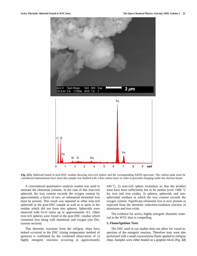
Active Thermitic Material Found in WTC Dust The Open Chemical Physics Journal, 2009, Volume 2 21
A conventional quantitative analysis routine was used to
estimate the elemental contents. In the case of this iron-rich
spheroid, the iron content exceeds the oxygen content by
approximately a factor of two, so substantial elemental iron
must be present. This result was repeated in other iron-rich
spheroids in the post-DSC sample as well as in spots in the
residue which did not form into spheres. Spheroids were
observed with Fe:O ratios up to approximately 4:1. Other
iron-rich spheres were found in the post-DSC residue which
contained iron along with aluminum and oxygen (see Dis-
cussion section).
That thermitic reactions from the red/gray chips have
indeed occurred in the DSC (rising temperature method of
ignition) is confirmed by the combined observation of 1)
highly energetic reactions occurring at approximately
430 ˚C, 2) iron-rich sphere formation so that the product
must have been sufficiently hot to be molten (over 1400 ˚C
for iron and iron oxide), 3) spheres, spheroids and non-
spheroidal residues in which the iron content exceeds the
oxygen content. Significant elemental iron is now present as
expected from the thermitic reduction-oxidation reaction of
aluminum and iron oxide.
The evidence for active, highly energetic thermitic mate-
rial in the WTC dust is compelling.
5. Flame/Ignition Tests
The DSC used in our studies does not allow for visual in-
spection of the energetic reaction. Therefore tests were also
performed with a small oxyacetylene flame applied to red/gray
chips. Samples were either heated on a graphite block (Fig. 22)
Fig. (21). Spheroid found in post-DSC residue showing iron-rich sphere and the corresponding XEDS spectrum. The carbon peak must be considered indeterminate here since this sample was flashed with a thin carbon layer in order to preclude charging under the electron beam.
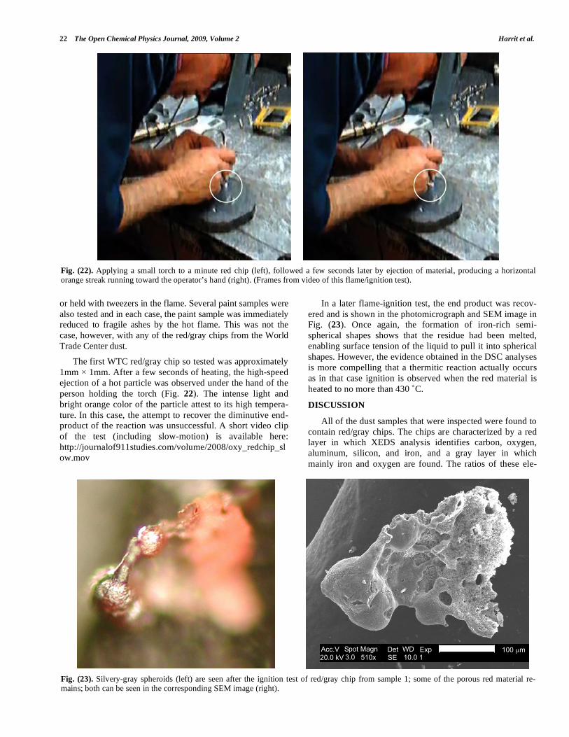
22 The Open Chemical Physics Journal, 2009, Volume 2 Harrit et al.
or held with tweezers in the flame. Several paint samples were
also tested and in each case, the paint sample was immediately
reduced to fragile ashes by the hot flame. This was not the
case, however, with any of the red/gray chips from the World
Trade Center dust.
The first WTC red/gray chip so tested was approximately
1mm 1mm. After a few seconds of heating, the high-speed
ejection of a hot particle was observed under the hand of the
person holding the torch (Fig. 22). The intense light and
bright orange color of the particle attest to its high tempera-
ture. In this case, the attempt to recover the diminutive end-
product of the reaction was unsuccessful. A short video clip
of the test (including slow-motion) is available here:
http://journalof911studies.com/volume/2008/oxy_redchip_sl
ow.mov
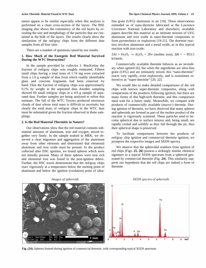
In a later flame-ignition test, the end product was recov-
ered and is shown in the photomicrograph and SEM image in
Fig. (23). Once again, the formation of iron-rich semi-
spherical shapes shows that the residue had been melted,
enabling surface tension of the liquid to pull it into spherical
shapes. However, the evidence obtained in the DSC analyses
is more compelling that a thermitic reaction actually occurs
as in that case ignition is observed when the red material is
heated to no more than 430 ˚C.
DISCUSSION
All of the dust samples that were inspected were found to
contain red/gray chips. The chips are characterized by a red
layer in which XEDS analysis identifies carbon, oxygen,
aluminum, silicon, and iron, and a gray layer in which
mainly iron and oxygen are found. The ratios of these ele-
Fig. (22). Applying a small torch to a minute red chip (left), followed a few seconds later by ejection of material, producing a horizontal orange streak running toward the operator’s hand (right). (Frames from video of this flame/ignition test).
Fig. (23). Silvery-gray spheroids (left) are seen after the ignition test of red/gray chip from sample 1; some of the porous red material re-mains; both can be seen in the corresponding SEM image (right).
����� ����� � �� �� ��������� ��� ���� �� �����
�����
Active Thermitic Material Found in WTC Dust The Open Chemical Physics Journal, 2009, Volume 2 23
ments appear to be similar especially when this analysis is
performed on a clean cross-section of the layers. The BSE
imaging also shows the consistency of the red layers by re-
vealing the size and morphology of the particles that are con-
tained in the bulk of the layers. The results clearly show the
similarities of the red/gray chips from the different dust
samples from all four sites.
There are a number of questions raised by our results.
1. How Much of the Energetic Red Material Survived During the WTC Destruction?
In the sample provided by collector J. MacKinlay the
fraction of red/gray chips was roughly estimated. Fifteen
small chips having a total mass of 1.74 mg were extracted
from a 1.6 g sample of dust from which readily identifiable
glass and concrete fragments had been removed by
hand. Thus the fraction of red/gray chips was approximately
0.1% by weight in the separated dust Another sampling
showed 69 small red/gray chips in a 4.9 g sample of sepa-
rated dust. Further samples are being analyzed to refine this
estimate. The fall of the WTC Towers produced enormous
clouds of dust whose total mass is difficult to ascertain; but
clearly the total mass of red/gray chips in the WTC dust
must be substantial given the fraction observed in these sam-
plings.
2. Is the Red Material Thermitic in Nature?
Our observations show that the red material contains sub-
stantial amounts of aluminum, iron and oxygen, mixed to-
gether very finely. In the sample soaked in MEK, we ob-
served a clear migration and aggregation of the aluminum
away from other elements and determined that elemental
aluminum and iron oxide must be present. In the product
collected after DSC ignition, we found spheres which were
not initially present. Many of these spheres were iron rich
and elemental iron was found in the post-ignition debris.
Further, the DSC traces demonstrate that the red/gray chips
react vigorously at a temperature below the melting point of
aluminum and below the ignition (oxidation) point of ultra-
fine grain (UFG) aluminum in air [18]. These observations
reminded us of nano-thermite fabricated at the Lawrence
Livermore National Laboratory and elsewhere; available
papers describe this material as an intimate mixture of UFG
aluminum and iron oxide in nano-thermite composites to
form pyrotechnics or explosives [19-21]. The thermite reac-
tion involves aluminum and a metal oxide, as in this typical
reaction with iron oxide:
2Al + Fe2O3 Al2O3 + 2Fe (molten iron), H = 853.5
kJ/mole.
Commercially available thermite behaves as an incendi-
ary when ignited [6], but when the ingredients are ultra-fine
grain (UFG) and are intimately mixed, this “nano-thermite”
reacts very rapidly, even explosively, and is sometimes re-
ferred to as “super-thermite” [20, 22].
We would like to make detailed comparisons of the red
chips with known super-thermite composites, along with
comparisons of the products following ignition, but there are
many forms of this high-tech thermite, and this comparison
must wait for a future study. Meanwhile, we compare with
products of commercially available (macro-) thermite. Dur-
ing ignition of thermite, we have observed that many spheres
and spheroids are formed as part of the molten product of the
reaction is vigorously scattered. These particles tend to be-
come spherical due to surface tension and, being small, are
rapidly cooled and solidify as they fall through the air, thus
their spherical shape is preserved.
To facilitate comparisons between the products of
red/gray chip ignition and commercial thermite ignition, we
juxtapose the respective images and XEDS spectra.
We observe that the spheroidal residues from ignition of
red chips (Figs. 25, 26) possess a strikingly similar chemical
signature to a typical XEDS spectrum from a spheroid gen-
erated by commercial thermite (Fig. 24). This similarity sup-
ports our hypothesis that the red chips are indeed a form of
thermite.
Images of spheroids XEDS spectra of spheroids
Fig. (24). Spheres formed during ignition of commercial thermite, with corresponding typical XEDS spectrum.
����������� ��
�� ���������
��� �� ���� ������ ���
24 The Open Chemical Physics Journal, 2009, Volume 2 Harrit et al.
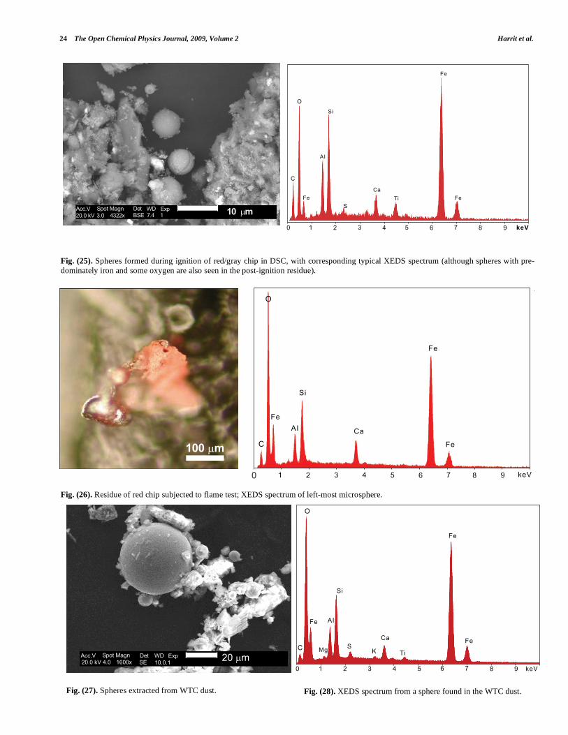
Fig. (25). Spheres formed during ignition of red/gray chip in DSC, with corresponding typical XEDS spectrum (although spheres with pre-
dominately iron and some oxygen are also seen in the post-ignition residue).
Fig. (26). Residue of red chip subjected to flame test; XEDS spectrum of left-most microsphere.
Fig. (27). Spheres extracted from WTC dust.
Fig. (28). XEDS spectrum from a sphere found in the WTC dust.
�
-
)
�
�0
��
)�
�
�
� � 6 : 9 < = > ; ��3
� ���
�
�
��
��
�
�
�
� ��
��
� � � � � � � � � ���
������������ ���
������� ����! "�#�����
$�� %$ #!� ������
����� ����� � �� �� ��� ������������� ����� �� ������
�
� ��
�� �
�
�
�
��
��
��
� � � � � � � � � � ���
Active Thermitic Material Found in WTC Dust The Open Chemical Physics Journal, 2009, Volume 2 25
In addition to the red/gray chips, many small spheres
have been found by our group in the WTC dust. These con-
tain the same elements as the residue of thermite, as noted in
a previous paper [5]. We show spheres found in the WTC
dust (Fig. 27) and a representative XEDS spectrum from
such a sphere (Fig. 28); we invite the reader to compare
these results with those found for ignition of commercial
thermite and for ignition of red/gray chips (above).
3. Could the Red Material Be Unreacted “Super-Thermite”?
We have noted that ordinary thermite acts as an incendi-
ary when ignited. However, when the ingredients are ultra-
fine-grain and are intimately mixed, the mixture reacts very
rapidly, even explosively [20]. Thus, there is a highly ener-
getic form of thermite known as an energetic nanocomposite
or “super-thermite,” composed of aluminum and iron oxide
with at least one component being approximately 100 nm or
less, often along with silicon and carbon [19-28].
“Reaction rates between nanosize aluminum
and metal oxides can be significantly greater
than those observed with traditional micron-size
thermite powders. Reactions occurring between
metal and metal oxide powders are accompa-
nied by the generation of high temperatures
(>3000 K). Super-thermites, formed by mixing
of aluminum and metal oxide nanopowders re-
sult in energy release rate by two orders of
magnitude higher than similar mixtures consist-
ing of micron size reactants” [22].
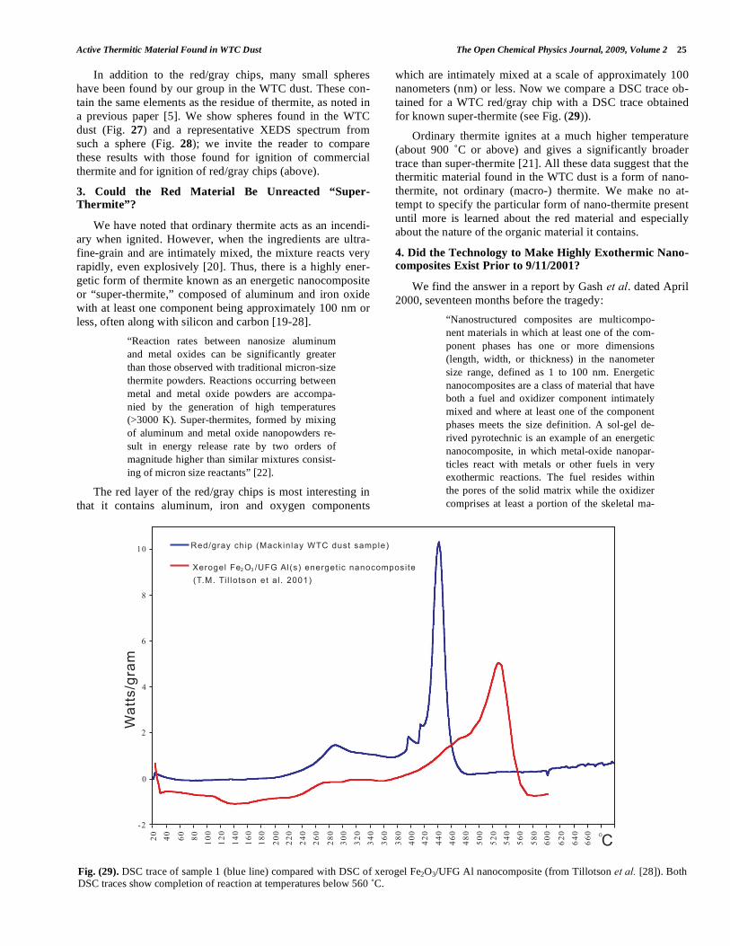
The red layer of the red/gray chips is most interesting in
that it contains aluminum, iron and oxygen components
which are intimately mixed at a scale of approximately 100
nanometers (nm) or less. Now we compare a DSC trace ob-
tained for a WTC red/gray chip with a DSC trace obtained
for known super-thermite (see Fig. (29)).
Ordinary thermite ignites at a much higher temperature
(about 900 ˚C or above) and gives a significantly broader
trace than super-thermite [21]. All these data suggest that the
thermitic material found in the WTC dust is a form of nano-
thermite, not ordinary (macro-) thermite. We make no at-
tempt to specify the particular form of nano-thermite present
until more is learned about the red material and especially
about the nature of the organic material it contains.
4. Did the Technology to Make Highly Exothermic Nano-composites Exist Prior to 9/11/2001?
We find the answer in a report by Gash et al. dated April
2000, seventeen months before the tragedy:
“Nanostructured composites are multicompo-
nent materials in which at least one of the com-
ponent phases has one or more dimensions
(length, width, or thickness) in the nanometer
size range, defined as 1 to 100 nm. Energetic
nanocomposites are a class of material that have
both a fuel and oxidizer component intimately
mixed and where at least one of the component
phases meets the size definition. A sol-gel de-
rived pyrotechnic is an example of an energetic
nanocomposite, in which metal-oxide nanopar-
ticles react with metals or other fuels in very
exothermic reactions. The fuel resides within
the pores of the solid matrix while the oxidizer
comprises at least a portion of the skeletal ma-
Fig. (29). DSC trace of sample 1 (blue line) compared with DSC of xerogel Fe2O3/UFG Al nanocomposite (from Tillotson et al. [28]). Both DSC traces show completion of reaction at temperatures below 560 ˚C.
��
�
�
�
�
�
��
�� �� �� �� ���
���
���
���
���
���
���
���
���
���
��
��
��
��
��
���
���
���
���
���
��
��
��
��
��
���
���
���
���
��
������� ������ ���������������������
������������� �!�"������������ ���� ��������# $
��%�%��� � ����������� % �#&&'�
�������
26 The Open Chemical Physics Journal, 2009, Volume 2 Harrit et al.
trix.” “As an example, energetic nanocompo-
sites of FexOy and metallic aluminum are easily
synthesized. The compositions are stable, safe
and can be readily ignited” [19].
We gather that the technology to make materials re-
markably fitting the characterization of the red chips was
available by April 2000. In the same report, the scientists
noted that “polymers” can be added to the nanocomposite:
“This sol-gel method allows for the addition of
insoluble materials (e.g., metals or polymers) to
the viscous sol, just before gelation, to produce
a uniformly distributed and energetic nanocom-
posite upon gelation. Al metal (as a fine pow-
der, ~6μm diameter) was added to some FexOy
gel syntheses just before gelation to produce
FexOy /Al(s) pyrotechnic nanocomposites….
These nanocomposites were subsequently proc-
essed to make both a xerogel and aerogel of the
material…. The pyrotechnic nanocomposite can
be ignited using a propane torch” [19].
Indeed, the red chips can be ignited using a torch and
they have properties of a pyrotechnic nanocomposite. All the
required ingredients are present – aluminum, iron, oxygen,
silicon, and carbon – and they are incorporated in such a way
that the chip forms (and sometimes ejects) very hot material
when ignited. The Gash report describes FTIR spectra which
characterize this energetic material. We have performed
these same tests and will report the results elsewhere. We
note that polymers in the matrix may be responsible for ab-
sorption of MEK and the subsequent swelling which we ob-
served [29].
A report on an April 2001 conference discloses who was
known to be working on such explosives at that time:
The 221st National Meeting of the American
Chemical Society held during April 2001 in San
Diego featured a symposium on Defense Appli-
cations of Nanomaterials. One of the 4 sessions
was titled nanoenergetics…. This session pro-
vided a good representation of the breadth of
work ongoing in this field, which is roughly 10
years old.… At this point in time, all of the
military services and some DOE and academic
laboratories have active R&D programs aimed
at exploiting the unique properties of nano-
materials that have potential to be used in
energetic formulations for advanced explo-
sives…. nanoenergetics hold promise as use-
ful ingredients for the thermobaric (TBX)
and TBX-like weapons, particularly due to
their high degree of tailorability with regards to
energy release and impulse management [20].
The feature of “impulse management” may be signifi-
cant. It is possible that formulations may be chosen to have
just sufficient percussive effect to achieve the desired frag-
mentation while minimizing the noise level.
5. Can Super-Thermite be Handled Safely?
The April 2000 report by Gash et al. states:
“The nature of the wet nanocomposites also af-
fords an additional degree of safety. In our
hands, the wet pyrotechnic nanocomposites
cannot be ignited until the drying process is
complete. This property should allow the pro-
duction of a large quantity of the pyrotechnics
that can be stored safely for some time and
dried shortly before its use” [19].
Safe handling of the malleable sol-gel material allows
easy coating of surfaces (such as steel), which the same
group, in a subsequent report, says they have achieved.
“The sol-gel process is very amenable to dip-,
spin-, and spray-coating technologies to coat
surfaces. We have utilized this property to dip-
coat various substrates to make sol-gel
Fe2O3/Al/Viton coatings. The energetic coating
dries to give a nice adherent film.” “We have
prepared fine powders, pressed pellets, cast
monoliths, and thin films of the hybrid inor-
ganic/organic energetic nanocomposite” [25].
Thus, the energetic nano-composite can be sprayed or
even “painted” onto surfaces, effectively forming an ener-
getic or even explosive paint. The red chips we found in the
WTC dust conform to their description of “thin films” of
“hybrid inorganic/organic energetic nanocomposite”. Indeed,
the descriptive terms “energetic coating” and “nice adherent
film” fit very well with our observations of the red-chips
which survived the WTC destruction. We cannot determine
at this time, however, whether the thinness of the chips re-
sulted from the application method or the manner of reac-
tion. While the application of a thin film might have suited
specific desired outcomes, it is also possible that the quench-
ing effect of the steel the material was in contact with may
have prevented a thin film of a larger mass from reacting.
The fact that most of the chips have a distinctive gray layer
suggests that the unreacted material was in close contact
with something else, either its target, a container, or an adhe-
sive.
Clapsaddle et al. further noted in their report:
“These results indicate that under ambient con-
ditions the hybrid inorganic/organic energetic
composite is very stable to impact, is spark in-
sensitive, and only very slightly friction sensi-
tive. As noted in the Experimental section of
this report, in our hands wet hybrid nanocompo-
sites are safe to handle and difficult to thermal
[sic] ignite. However, once dry the material
burns very vigorously and rapidly with the evo-
lution of significant amounts of gaseous spe-
cies” [24].
The organic component contributes to the rapid gas evo-
lution and explosive nature of these energetic super-
thermites when dry [24].
“Super-thermite electric matches” have been developed
at Los Alamos National Laboratory for which “applications
include triggering explosives for ... demolition” [30]. It is
indeed possible that such matches, which are designed to be
ignited by a simple electric pulse, could contain material
Active Thermitic Material Found in WTC Dust The Open Chemical Physics Journal, 2009, Volume 2 27
similar to the red material we have found in the WTC dust.
With regard to the safety of super-thermite matches, the Los
Alamos announcement notes:
“Unfortunately, conventional electric matches
use lead containing compounds that are ex-
tremely sensitive to impact, friction, static, and
heat stimuli, thereby making them dangerous to
handle. In addition, these compounds produce
toxic smoke. The Super-Thermite electric
matches produce no toxic lead smoke and are
safer to use because they resist friction, im-
pact, heat, and static discharge through the
composition, thereby minimizing accidental ig-
nition. They can be designed to create various
thermal-initiating outputs—simple sparks, hot
slag, droplets, or flames—depending on the
needs of different applications” [30].
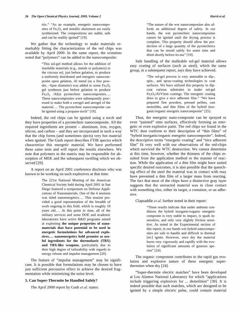
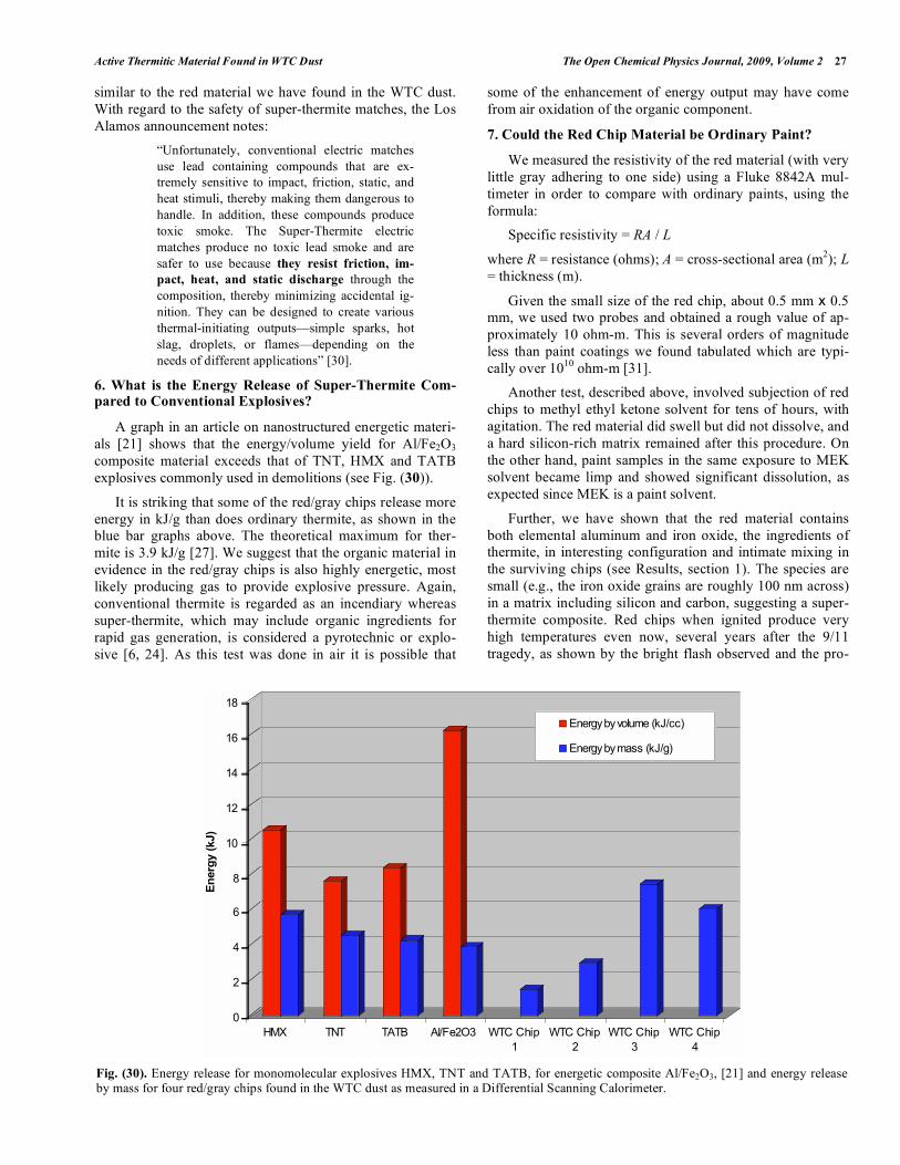
6. What is the Energy Release of Super-Thermite Com-pared to Conventional Explosives?
A graph in an article on nanostructured energetic materi-
als [21] shows that the energy/volume yield for Al/Fe2O3
composite material exceeds that of TNT, HMX and TATB
explosives commonly used in demolitions (see Fig. (30)).
It is striking that some of the red/gray chips release more
energy in kJ/g than does ordinary thermite, as shown in the
blue bar graphs above. The theoretical maximum for ther-
mite is 3.9 kJ/g [27]. We suggest that the organic material in
evidence in the red/gray chips is also highly energetic, most
likely producing gas to provide explosive pressure. Again,
conventional thermite is regarded as an incendiary whereas
super-thermite, which may include organic ingredients for
rapid gas generation, is considered a pyrotechnic or explo-
sive [6, 24]. As this test was done in air it is possible that
some of the enhancement of energy output may have come
from air oxidation of the organic component.
7. Could the Red Chip Material be Ordinary Paint?
We measured the resistivity of the red material (with very
little gray adhering to one side) using a Fluke 8842A mul-
timeter in order to compare with ordinary paints, using the
formula:
Specific resistivity = RA / L
where R = resistance (ohms); A = cross-sectional area (m2); L
= thickness (m).
Given the small size of the red chip, about 0.5 mm x 0.5
mm, we used two probes and obtained a rough value of ap-
proximately 10 ohm-m. This is several orders of magnitude
less than paint coatings we found tabulated which are typi-
cally over 1010
ohm-m [31].
Another test, described above, involved subjection of red
chips to methyl ethyl ketone solvent for tens of hours, with
agitation. The red material did swell but did not dissolve, and
a hard silicon-rich matrix remained after this procedure. On
the other hand, paint samples in the same exposure to MEK
solvent became limp and showed significant dissolution, as
expected since MEK is a paint solvent.
Further, we have shown that the red material contains
both elemental aluminum and iron oxide, the ingredients of
thermite, in interesting configuration and intimate mixing in
the surviving chips (see Results, section 1). The species are
small (e.g., the iron oxide grains are roughly 100 nm across)
in a matrix including silicon and carbon, suggesting a super-
thermite composite. Red chips when ignited produce very
high temperatures even now, several years after the 9/11
tragedy, as shown by the bright flash observed and the pro-
0
2
4
6
8
10
12
14
16
18
En
erg
y (
kJ)
HMX TNT TATB Al/Fe2O3 WTC Chip
1
WTC Chip
2
WTC Chip
3
WTC Chip
4
Energy by volume (kJ/cc)
Energy by mass (kJ/g)
Fig. (30). Energy release for monomolecular explosives HMX, TNT and TATB, for energetic composite Al/Fe2O3, [21] and energy release by mass for four red/gray chips found in the WTC dust as measured in a Differential Scanning Calorimeter.
28 The Open Chemical Physics Journal, 2009, Volume 2 Harrit et al.
duction of molten iron-rich spheres (see photomicrographs in
Fig. (20) above). Correspondingly, the DSC tests demon-
strate the release of high enthalpy, actually exceeding that of
pure thermite. Furthermore, the energy is released over a
short period of time, shown by the narrowness of the peak in
Fig. (29). The post-DSC-test residue contains microspheres
in which the iron exceeds the oxygen content, implying that
at least some of the iron oxide has been reduced in the reac-
tion. If a paint were devised that incorporated these very
energetic materials, it would be highly dangerous when dry
and most unlikely to receive regulatory approval for building
use. To merit consideration, any assertion that a prosaic sub-
stance such as paint could match the characteristics we have
described would have to be accompanied by empirical dem-
onstration using a sample of the proposed material, including
SEM/XEDS and DSC analyses.
8. What Future Studies are Contemplated?
We observe that the total energy released from some of
the red chips exceeds the theoretical limit for thermite alone
(3.9 kJ/g). One possibility is that the organic material in the
red layer is itself energetic. Determination of the chemical
compound(s) involved in the organic component of the red
material would promote understanding. Further studies of the
red material (separated from the gray material) compared to
known super-thermite variants using DSC, TGA, FTIR (etc.)
analyses would certainly be in order. In particular, NMR and
GC-mass spectroscopy and related studies are urged to iden-
tify the organic material.
We have observed that some chips have additional ele-
ments such as potassium, lead, barium and copper. Are these
significant, and why do such elements appear in some red
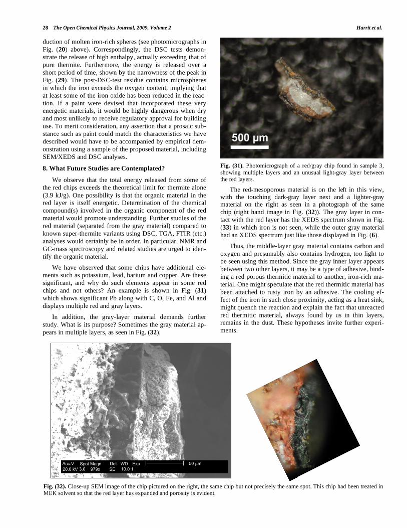
chips and not others? An example is shown in Fig. (31)
which shows significant Pb along with C, O, Fe, and Al and
displays multiple red and gray layers.
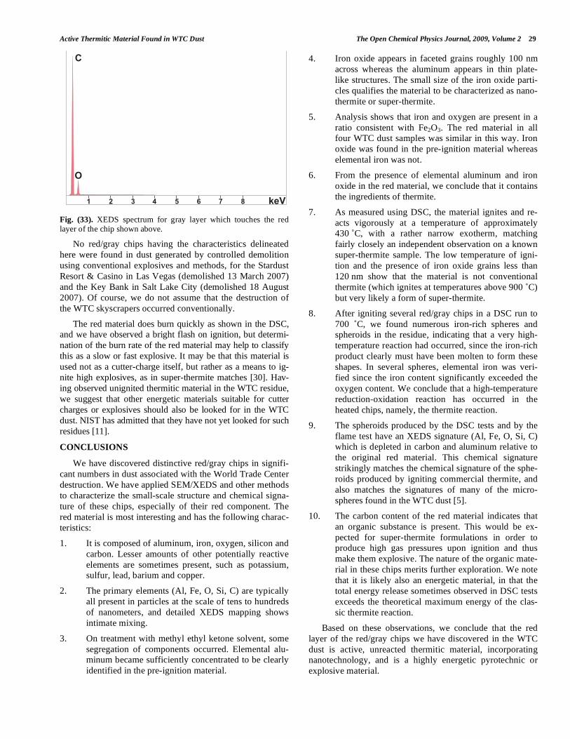
In addition, the gray-layer material demands further
study. What is its purpose? Sometimes the gray material ap-
pears in multiple layers, as seen in Fig. (32).
Fig. (31). Photomicrograph of a red/gray chip found in sample 3,
showing multiple layers and an unusual light-gray layer between the red layers.
The red-mesoporous material is on the left in this view, with the touching dark-gray layer next and a lighter-gray material on the right as seen in a photograph of the same
chip (right hand image in Fig. (32)). The gray layer in con-
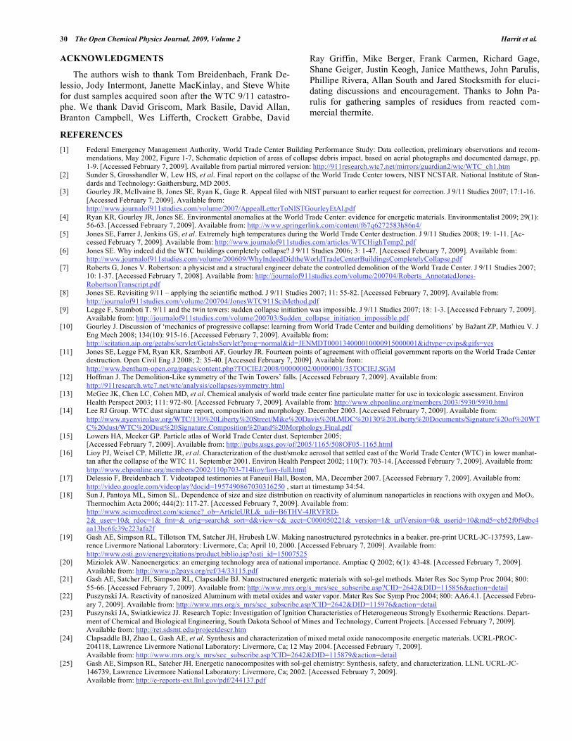
tact with the red layer has the XEDS spectrum shown in Fig.
(33) in which iron is not seen, while the outer gray material
had an XEDS spectrum just like those displayed in Fig. (6).
Thus, the middle-layer gray material contains carbon and
oxygen and presumably also contains hydrogen, too light to
be seen using this method. Since the gray inner layer appears
between two other layers, it may be a type of adhesive, bind-
ing a red porous thermitic material to another, iron-rich ma-
terial. One might speculate that the red thermitic material has
been attached to rusty iron by an adhesive. The cooling ef-
fect of the iron in such close proximity, acting as a heat sink,
might quench the reaction and explain the fact that unreacted
red thermitic material, always found by us in thin layers,
remains in the dust. These hypotheses invite further experi-
ments.
Fig. (32). Close-up SEM image of the chip pictured on the right, the same chip but not precisely the same spot. This chip had been treated in MEK solvent so that the red layer has expanded and porosity is evident.
����� ����� � �� �� ��������� ��� ���� �� �����
����
Active Thermitic Material Found in WTC Dust The Open Chemical Physics Journal, 2009, Volume 2 29
Fig. (33). XEDS spectrum for gray layer which touches the red layer of the chip shown above.
No red/gray chips having the characteristics delineated
here were found in dust generated by controlled demolition
using conventional explosives and methods, for the Stardust
Resort & Casino in Las Vegas (demolished 13 March 2007)
and the Key Bank in Salt Lake City (demolished 18 August
2007). Of course, we do not assume that the destruction of
the WTC skyscrapers occurred conventionally.
The red material does burn quickly as shown in the DSC,
and we have observed a bright flash on ignition, but determi-
nation of the burn rate of the red material may help to classify
this as a slow or fast explosive. It may be that this material is
used not as a cutter-charge itself, but rather as a means to ig-
nite high explosives, as in super-thermite matches [30]. Hav-
ing observed unignited thermitic material in the WTC residue,
we suggest that other energetic materials suitable for cutter
charges or explosives should also be looked for in the WTC
dust. NIST has admitted that they have not yet looked for such
residues [11].
CONCLUSIONS
We have discovered distinctive red/gray chips in signifi-
cant numbers in dust associated with the World Trade Center
destruction. We have applied SEM/XEDS and other methods
to characterize the small-scale structure and chemical signa-
ture of these chips, especially of their red component. The
red material is most interesting and has the following charac-
teristics:
1. It is composed of aluminum, iron, oxygen, silicon and
carbon. Lesser amounts of other potentially reactive
elements are sometimes present, such as potassium,
sulfur, lead, barium and copper.
2. The primary elements (Al, Fe, O, Si, C) are typically
all present in particles at the scale of tens to hundreds
of nanometers, and detailed XEDS mapping shows
intimate mixing.
3. On treatment with methyl ethyl ketone solvent, some
segregation of components occurred. Elemental alu-
minum became sufficiently concentrated to be clearly
identified in the pre-ignition material.
4. Iron oxide appears in faceted grains roughly 100 nm
across whereas the aluminum appears in thin plate-
like structures. The small size of the iron oxide parti-
cles qualifies the material to be characterized as nano-
thermite or super-thermite.
5. Analysis shows that iron and oxygen are present in a
ratio consistent with Fe2O3. The red material in all
four WTC dust samples was similar in this way. Iron
oxide was found in the pre-ignition material whereas
elemental iron was not.
6. From the presence of elemental aluminum and iron
oxide in the red material, we conclude that it contains
the ingredients of thermite.
7. As measured using DSC, the material ignites and re-
acts vigorously at a temperature of approximately
430 ˚C, with a rather narrow exotherm, matching
fairly closely an independent observation on a known
super-thermite sample. The low temperature of igni-
tion and the presence of iron oxide grains less than
120 nm show that the material is not conventional
thermite (which ignites at temperatures above 900 ˚C)
but very likely a form of super-thermite.
8. After igniting several red/gray chips in a DSC run to
700 ˚C, we found numerous iron-rich spheres and
spheroids in the residue, indicating that a very high-
temperature reaction had occurred, since the iron-rich
product clearly must have been molten to form these
shapes. In several spheres, elemental iron was veri-
fied since the iron content significantly exceeded the
oxygen content. We conclude that a high-temperature
reduction-oxidation reaction has occurred in the
heated chips, namely, the thermite reaction.
9. The spheroids produced by the DSC tests and by the
flame test have an XEDS signature (Al, Fe, O, Si, C)
which is depleted in carbon and aluminum relative to
the original red material. This chemical signature
strikingly matches the chemical signature of the sphe-
roids produced by igniting commercial thermite, and
also matches the signatures of many of the micro-
spheres found in the WTC dust [5].
10. The carbon content of the red material indicates that
an organic substance is present. This would be ex-
pected for super-thermite formulations in order to
produce high gas pressures upon ignition and thus
make them explosive. The nature of the organic mate-
rial in these chips merits further exploration. We note
that it is likely also an energetic material, in that the
total energy release sometimes observed in DSC tests
exceeds the theoretical maximum energy of the clas-
sic thermite reaction.
Based on these observations, we conclude that the red
layer of the red/gray chips we have discovered in the WTC
dust is active, unreacted thermitic material, incorporating
nanotechnology, and is a highly energetic pyrotechnic or
explosive material.
���������
�
30 The Open Chemical Physics Journal, 2009, Volume 2 Harrit et al.
ACKNOWLEDGMENTS
The authors wish to thank Tom Breidenbach, Frank De-
lessio, Jody Intermont, Janette MacKinlay, and Steve White
for dust samples acquired soon after the WTC 9/11 catastro-
phe. We thank David Griscom, Mark Basile, David Allan,
Branton Campbell, Wes Lifferth, Crockett Grabbe, David
Ray Griffin, Mike Berger, Frank Carmen, Richard Gage,
Shane Geiger, Justin Keogh, Janice Matthews, John Parulis,
Phillipe Rivera, Allan South and Jared Stocksmith for eluci-
dating discussions and encouragement. Thanks to John Pa-
rulis for gathering samples of residues from reacted com-
mercial thermite.
REFERENCES
[1] Federal Emergency Management Authority, World Trade Center Building Performance Study: Data collection, preliminary observations and recom-mendations, May 2002, Figure 1-7, Schematic depiction of areas of collapse debris impact, based on aerial photographs and documented damage, pp.
1-9. [Accessed February 7, 2009]. Available from partial mirrored version: http://911research.wtc7.net/mirrors/guardian2/wtc/WTC_ch1.htm [2] Sunder S, Grosshandler W, Lew HS, et al. Final report on the collapse of the World Trade Center towers, NIST NCSTAR. National Institute of Stan-
dards and Technology: Gaithersburg, MD 2005. [3] Gourley JR, McIlvaine B, Jones SE, Ryan K, Gage R. Appeal filed with NIST pursuant to earlier request for correction. J 9/11 Studies 2007; 17:1-16.
[Accessed February 7, 2009]. Available from: http://www.journalof911studies.com/volume/2007/AppealLetterToNISTGourleyEtAl.pdf
[4] Ryan KR, Gourley JR, Jones SE. Environmental anomalies at the World Trade Center: evidence for energetic materials. Environmentalist 2009; 29(1): 56-63. [Accessed February 7, 2009]. Available from: http://www.springerlink.com/content/f67q6272583h86n4/
[5] Jones SE, Farrer J, Jenkins GS, et al. Extremely high temperatures during the World Trade Center destruction. J 9/11 Studies 2008; 19: 1-11. [Ac-cessed February 7, 2009]. Available from: http://www.journalof911studies.com/articles/WTCHighTemp2.pdf
[6] Jones SE. Why indeed did the WTC buildings completely collapse? J 9/11 Studies 2006; 3: 1-47. [Accessed February 7, 2009]. Available from: http://www.journalof911studies.com/volume/200609/WhyIndeedDidtheWorldTradeCenterBuildingsCompletelyCollapse.pdf
[7] Roberts G, Jones V. Robertson: a physicist and a structural engineer debate the controlled demolition of the World Trade Center. J 9/11 Studies 2007; 10: 1-37. [Accessed February 7, 2008]. Available from: http://journalof911studies.com/volume/200704/Roberts_AnnotatedJones-
RobertsonTranscript.pdf [8] Jones SE. Revisiting 9/11 – applying the scientific method. J 9/11 Studies 2007; 11: 55-82. [Accessed February 7, 2009]. Available from:
http://journalof911studies.com/volume/200704/JonesWTC911SciMethod.pdf [9] Legge F, Szamboti T. 9/11 and the twin towers: sudden collapse initiation was impossible. J 9/11 Studies 2007; 18: 1-3. [Accessed February 7, 2009].
Available from: http://journalof911studies.com/volume/200703/Sudden_collapse_initiation_impossible.pdf [10] Gourley J. Discussion of ‘mechanics of progressive collapse: learning from World Trade Center and building demolitions’ by Ba ant ZP, Mathieu V. J
Eng Mech 2008; 134(10): 915-16. [Accessed February 7, 2009]. Available from: http://scitation.aip.org/getabs/servlet/GetabsServlet?prog=normal&id=JENMDT000134000010000915000001&idtype=cvips&gifs=yes
[11] Jones SE, Legge FM, Ryan KR, Szamboti AF, Gourley JR. Fourteen points of agreement with official government reports on the World Trade Center destruction. Open Civil Eng J 2008; 2: 35-40. [Accessed February 7, 2009]. Available from:
http://www.bentham-open.org/pages/content.php?TOCIEJ/2008/00000002/00000001/35TOCIEJ.SGM [12] Hoffman J. The Demolition-Like symmetry of the Twin Towers’ falls. [Accessed February 7, 2009]. Available from:
http://911research.wtc7.net/wtc/analysis/collapses/symmetry.html [13] McGee JK, Chen LC, Cohen MD, et al. Chemical analysis of world trade center fine particulate matter for use in toxicologic assessment. Environ
Health Perspect 2003; 111: 972-80. [Accessed February 7, 2009]. Available from: http://www.ehponline.org/members/2003/5930/5930.html [14] Lee RJ Group. WTC dust signature report, composition and morphology. December 2003. [Accessed February 7, 2009]. Available from:
http://www.nyenvirolaw.org/WTC/130%20Liberty%20Street/Mike%20Davis%20LMDC%20130%20Liberty%20Documents/Signature%20of%20WTC%20dust/WTC%20Dust%20Signature.Composition%20and%20Morphology.Final.pdf
[15] Lowers HA, Meeker GP. Particle atlas of World Trade Center dust. September 2005; [Accessed February 7, 2009]. Available from: http://pubs.usgs.gov/of/2005/1165/508OF05-1165.html
[16] Lioy PJ, Weisel CP, Millette JR, et al. Characterization of the dust/smoke aerosol that settled east of the World Trade Center (WTC) in lower manhat-tan after the collapse of the WTC 11. September 2001. Environ Health Perspect 2002; 110(7): 703-14. [Accessed February 7, 2009]. Available from:
http://www.ehponline.org/members/2002/110p703-714lioy/lioy-full.html [17] Delessio F, Breidenbach T. Videotaped testimonies at Faneuil Hall, Boston, MA, December 2007. [Accessed February 7, 2009]. Available from:
http://video.google.com/videoplay?docid=1957490867030316250 , start at timestamp 34:54. [18] Sun J, Pantoya ML, Simon SL. Dependence of size and size distribution on reactivity of aluminum nanoparticles in reactions with oxygen and MoO3.
Thermochim Acta 2006; 444(2): 117-27. [Accessed February 7, 2009]. Available from: http://www.sciencedirect.com/science?_ob=ArticleURL&_udi=B6THV-4JRVFRD-
2&_user=10&_rdoc=1&_fmt=&_orig=search&_sort=d&view=c&_acct=C000050221&_version=1&_urlVersion=0&_userid=10&md5=cb52f0f9dbc4aa13bc6fc39e223afa2f
[19] Gash AE, Simpson RL, Tillotson TM, Satcher JH, Hrubesh LW. Making nanostructured pyrotechnics in a beaker. pre-print UCRL-JC-137593, Law-rence Livermore National Laboratory: Livermore, Ca; April 10, 2000. [Accessed February 7, 2009]. Available from:
http://www.osti.gov/energycitations/product.biblio.jsp?osti_id=15007525 [20] Miziolek AW. Nanoenergetics: an emerging technology area of national importance. Amptiac Q 2002; 6(1): 43-48. [Accessed February 7, 2009].
Available from: http://www.p2pays.org/ref/34/33115.pdf [21] Gash AE, Satcher JH, Simpson RL, Clapsaddle BJ. Nanostructured energetic materials with sol-gel methods. Mater Res Soc Symp Proc 2004; 800:
55-66. [Accessed February 7, 2009]. Available from: http://www.mrs.org/s_mrs/sec_subscribe.asp?CID=2642&DID=115856&action=detail [22] Puszynski JA. Reactivity of nanosized Aluminum with metal oxides and water vapor. Mater Res Soc Symp Proc 2004; 800: AA6.4.1. [Accessed Febru-
ary 7, 2009]. Available from: http://www.mrs.org/s_mrs/sec_subscribe.asp?CID=2642&DID=115976&action=detail [23] Puszynski JA, Swiatkiewicz JJ. Research Topic: Investigation of Ignition Characteristics of Heterogeneous Strongly Exothermic Reactions. Depart-
ment of Chemical and Biological Engineering, South Dakota School of Mines and Technology, Current Projects. [Accessed February 7, 2009]. Available from: http://ret.sdsmt.edu/projectdescr.htm
[24] Clapsaddle BJ, Zhao L, Gash AE, et al. Synthesis and characterization of mixed metal oxide nanocomposite energetic materials. UCRL-PROC-204118, Lawrence Livermore National Laboratory: Livermore, Ca; 12 May 2004. [Accessed February 7, 2009].
Available from: http://www.mrs.org/s_mrs/sec_subscribe.asp?CID=2642&DID=115879&action=detail [25] Gash AE, Simpson RL, Satcher JH. Energetic nanocomposites with sol-gel chemistry: Synthesis, safety, and characterization. LLNL UCRL-JC-
146739, Lawrence Livermore National Laboratory: Livermore, Ca; 2002. [Accessed February 7, 2009]. Available from: http://e-reports-ext.llnl.gov/pdf/244137.pdf
Active Thermitic Material Found in WTC Dust The Open Chemical Physics Journal, 2009, Volume 2 31
[26] Zhao L, Clapsaddle BJ, Satcher JH, Jr, Schaefer DW, Shea KJ. Integrated chemical systems: the simultaneous formation of hybrid nanocomposites of
iron oxide and organo silsesquioxanes. Chem Mater 2005; 17(6): 1358-66. [Accessed February 7, 2009]. Available from: http://pubs.acs.org/cgi-bin/abstract.cgi/cmatex/2005/17/i06/abs/cm048231i.html
[27] Clapsaddle BJ, Zhao L, Prentice D, et al. Formulation and performance of novel energetic nanocomposites and gas generators prepared by sol–gel methods. LLNL UCRL-PROC–210871, Lawrence Livermore National Laboratory: Livermore, Ca; March 2005;
[Accessed February 7, 2009]. Available from: http://e-reports-ext.llnl.gov/pdf/318263.pdf [28] Tillotson TM, Gash AE, Simpson RL, Hrubesh LW, Satcher JH, Jr, Poco JF. Nanostructured energetic materials using sol-gel methodologies. J Non-
Cryst Sol 2001; 285: 338-345. [Accessed February 7, 2009]. Available from: http://www.sciencedirect.com/science?_ob=ArticleURL&_udi=B6TXM-435KKJV-
2G&_user=10&_rdoc=1&_fmt=&_orig=search&_sort=d&view=c&_acct=C000050221&_version=1&_urlVersion=0&_userid=10&md5=96168ef14a007c2cc1dee1667b0d1b2f
[29] Bandyopadhyay A, de Sarkar M, Bhowmick AK. Polymer-filler interactions in sol-gel derived polymer/silica hybrid nanocomposites. J Polym Sci Part B. Polym Phys 2005; 43(17): 2399-412. [Accessed August 4, 2008]. Available from: http://www3.interscience.wiley.com/journal/110572549/abstract
[30] R&D Awards. super-thermite electric matches. [Accessed February 7, 2009]. Available from: http://awards.lanl.gov/PDFfiles/Super-Thermite_Electric_Matches_2003.pdf
[31] Abu Ayana YM, El-Sawy SM, Salah SH. Zinc-ferrite pigment for corrosion protection. Anti-Corros Methods Mater 1997; 44(6): 381-8. Available from: http://www.emeraldinsight.com/Insight/ViewContentServlet?Filename=Published/EmeraldFullTextArticle/Articles/1280440604.html
Received: August 12, 2008 Revised: February 10, 2009 Accepted: February 13, 2009
© Harrit et al.; Licensee Bentham Open.
This is an open access article licensed under the terms of the Creative Commons Attribution Non-Commercial License (http: //creativecommons.org/licenses/by- nc/3.0/) which permits unrestricted, non-commercial use, distribution and reproduction in any medium, provided the work is properly cited.