Embed Size (px)
Citation preview
Dr Khin Soe
Department of Oral Medicine
A well circumscribed lesion derived from
odontogenic epithelium that usually occurs
around the crowns of unerupted anterior
teeth of young patients and consists of
epithelium in swirls and ductal patterns
interspersed with spherical calcifications
The adenomatoid odontogenic tumour
usually presents during the second and third
decades of life.
The majority of tumours arise in the anterior
part of the maxilla, especially in the canine
areas, and
there are usually few symptoms apart from a
slowly enlarging swelling.
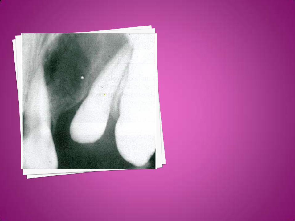
On radiographs it usually appears as a well-
defined radiolucency but in some cases
calcification within the tumour may produce
faint radiopacities.
The lesion is often associated with an
unerupted tooth and may simulate a
dentigerous cyst.
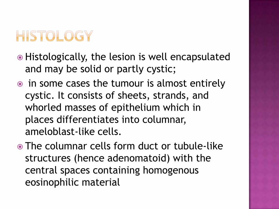
Histologically, the lesion is well encapsulated
and may be solid or partly cystic;
in some cases the tumour is almost entirely
cystic. It consists of sheets, strands, and
whorled masses of epithelium which in
places differentiates into columnar,
ameloblast-like cells.
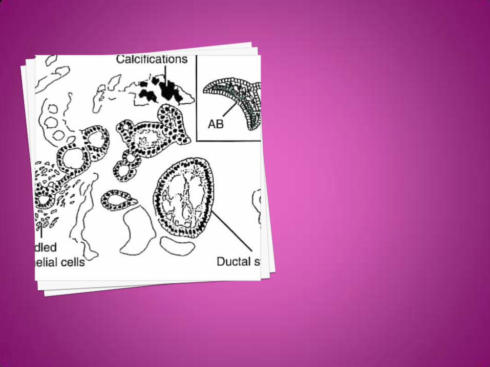
The columnar cells form duct or tubule-like
structures (hence adenomatoid) with the
central spaces containing homogenous
eosinophilic material
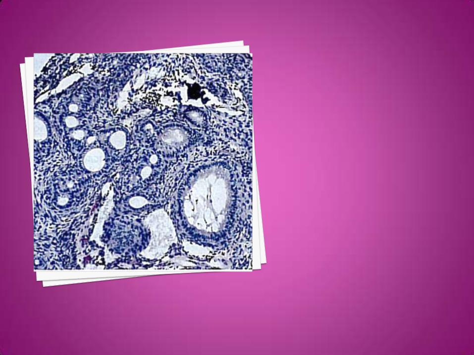
They are thought to represent abortive
attempts at enamel organ formation. There
is very little supporting stroma.
Small foci of calcification are scattered
throughout the tumour and occasionally
tubular dentine and enamel matrix may be
seen.
The nature of the lesion is uncertain and it
may be hamartomatous rather than truly
neoplastic.
It must be differentiated from
ameloblastoma.

The adenomatoid odontogenic tumour is
readily enucleated and does not recur:
it does not require radical excision.
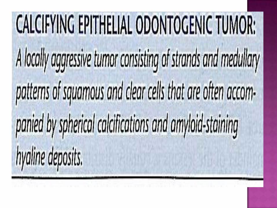
The calcifying epithelial odontogenic tumour
is a rare, benign epithelial neoplasm.
It occurs over a wide age range and is about
twice as common in the mandible as in the
maxilla.
Most of the tumours arise in the molar or
premolar area and about half are associated
with the crown of an unerupted tooth.
Although most tumours arise within bone,
extraosseous lesions have been reported.
Radiographs of intraosseous tumours show an
irregular radiolucent area which may or may
not be clearly demarcated from the
surrounding normal bone.
The radiolucency contains varying amounts
of radiopaque bodies due to calcification
within the tumour.


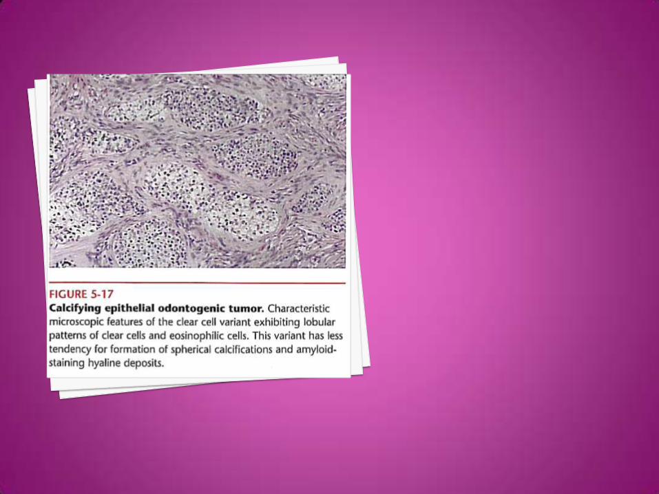
Histologically, the tumour consists of sheets
and strands of polyhedral epithelial cells
with abundant eosinophilic cytoplasm lying in
a fibrous stroma.
The epithelial cells often show prominent
intercellular bridges and marked nuclear
pleomorphism but the latter is not indicative
of malignancy.
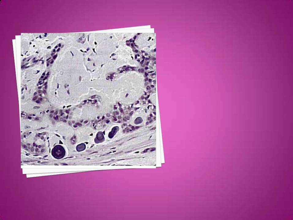
A characteristic feature is the presence
within the sheets of epithelial cells of
homogeneous, amyloid-like material which
may become calcified.
The calcifications are concentric laminated
structures that may fuse into complex
masses.
The nature of the amyloid-like material is
uncertain but is probably derived from
products synthesized by the epithelial cells.
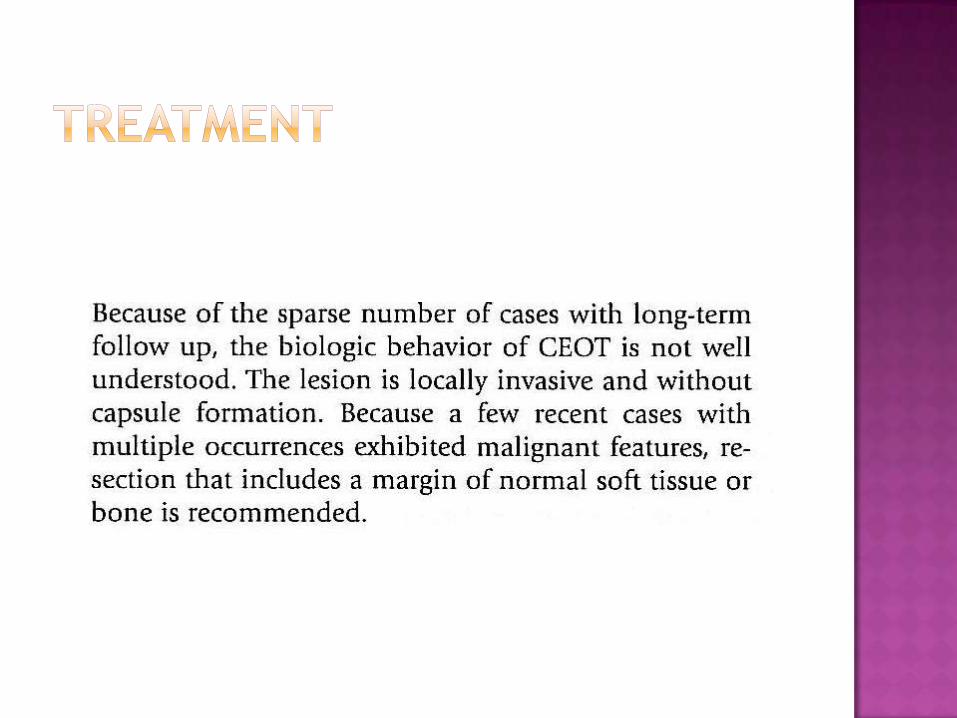
Although the tumour is generally regarded to
be locally invasive it appears to be less
aggressive than the ameloblastoma.
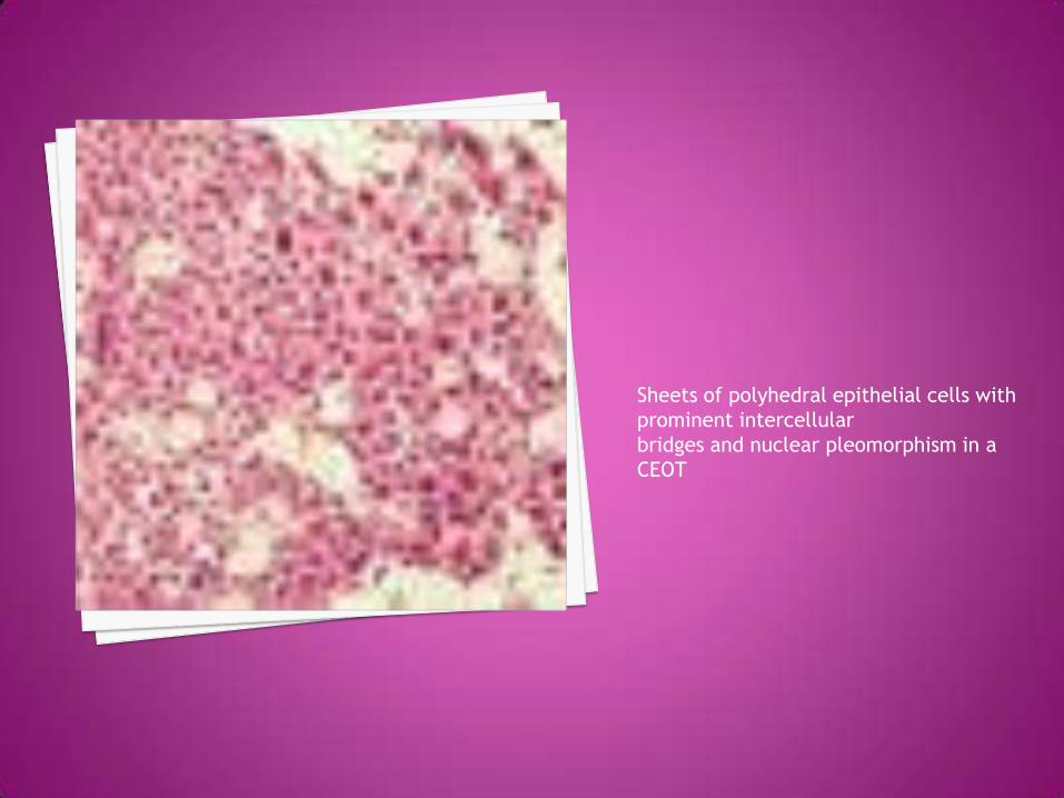
Sheets of polyhedral epithelial cells with
prominent intercellular
bridges and nuclear pleomorphism in a
CEOT
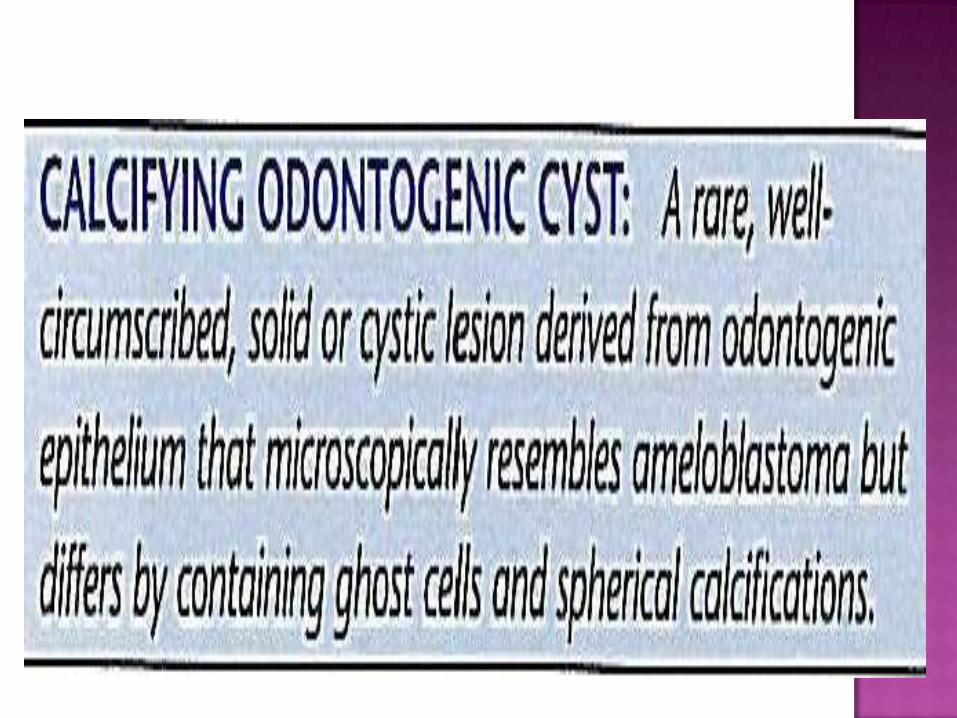
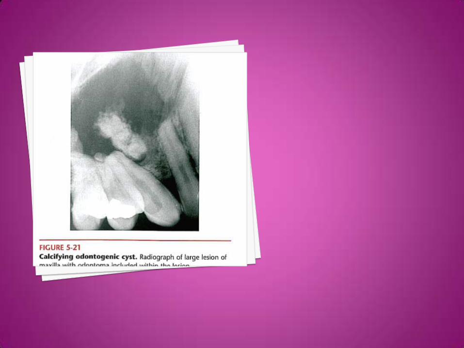
The calcifying cystic odontogenic tumour is a
grossly cystic odontogenic tumour and may
be a hamartoma rather than a true benign
neoplasm.
The dentinogenic ghost cell tumour is
histologically very similar except that it is a
solid lesion.
It was originally considered to represent the
solid variant of the calcifying cystic
odontogenic tumour.
However, as more cases are reported there is
increasing evidence that the dentinogenic
ghost cell tumour is a distinct pathological
entity and is a true benign neoplasm.
Both present mainly as central lesions within
the jaws but peripheral, gingival lesions also
occur.
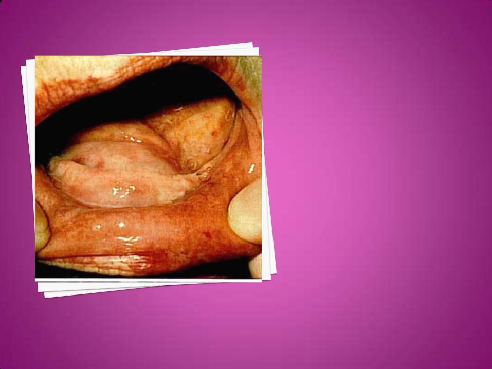
The calcifying cystic odontogenic tumouroccurs over a wide age range but is usually seen below 40 years of age.
About 75 per cent are intraosseous and either jaw may be involved.
The majority, including those located in the gingival or alveolar soft tissues, arise anteriorly to the first permanent molar tooth.
The lesion usually presents as a slowly enlarging but otherwise symptomless swelling.
Radiographically, the lesion appears as a
well-defined unilocular or multilocular
radiolucent area containing varying amounts
of radiopaque, calcified material.
It may be associated with the crown of an
unerupted tooth.

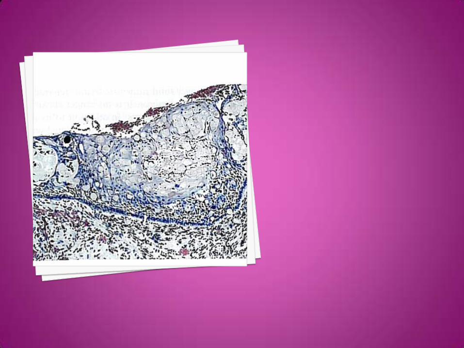
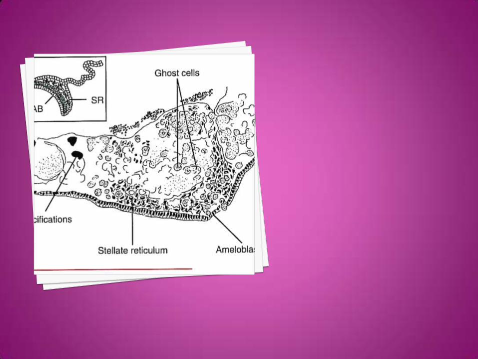
Histologically, the cyst is lined by epithelium
which shows a well-defined basal layer of
columnar, ameloblast-like cells and overlying
layers of more loosely arranged cells that
may resemble stellate reticulum.
A characteristic feature is the presence
within the lining of masses of swollen and
keratinized epithelial cells which are usually
referred to as 'ghost' cells since the original
cell outlines can still be discerned.
The 'ghost' epithelial cells may calcify.
Breakdown of the epithelium may release
keratinous debris into the supporting
connective tissue resulting in a prominent
foreign-body, giant-cell reaction.
Irregular masses of dentine-like matrix
material (dentinoid) are frequently found in
the supporting fibrous tissue in direct
contact with the basal layer of the
epithelium.
Less commonly, more extensive formation of
dental hard tissues is seen, including enamel,
producing a structure similar to a complex or
compound odontome as an integral part of
the lesion.
Calcifying cystic odontogenic tumour
associated with odontomes tend to occur in a
younger age group and most have presented
in the anterior maxilla.
The dentinogenic ghost cell tumour is a
predominantly solid lesion which comprises
the same epithelial, keratinized ghost cells
and dentinoid components as the calcifying
cystic odontogenic tumour, but as a
disorganized mass.
It tends to occur in an older age group than
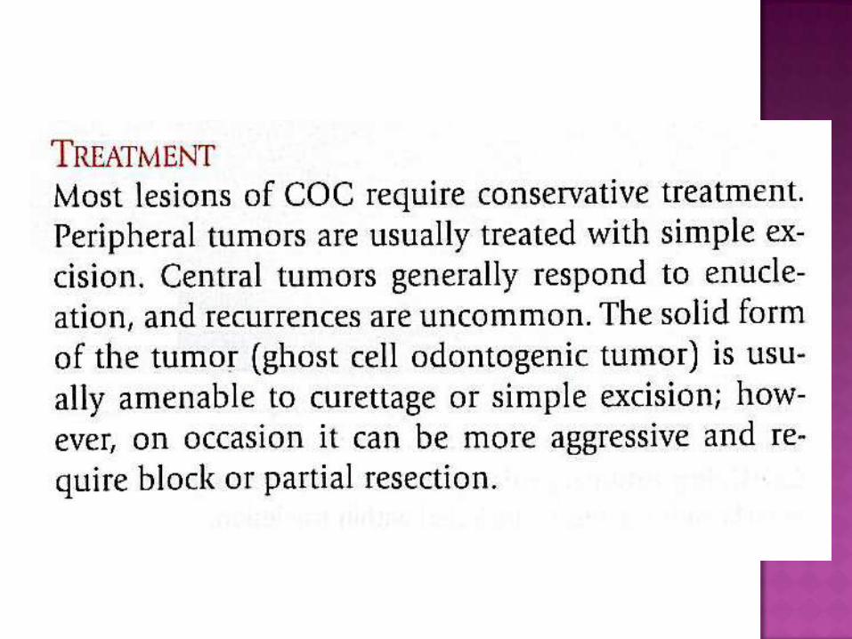
the calcifying cystic odontogenic tumour.
Like the calcifying cystic odontogenic tumour
some respond well to conservative
treatment.
However, others pursue a more aggressive
course and, like the ameloblastoma, are
locally invasive neoplasms.


Odontomas are mixed odontogenic tumors in
which both the epithelial and mesenchymal
components have undergone functional
differentiation to the point that both enamel
and dentin are formed.
The most common of the odontogenic
tumors, odontomas are believed to be
hamartomatous rather than neoplastic in
nature.
The compound odontoma is a lesion in which
all the dental tissues are represented in an
orderly fashion so that there is at least
superficial anatomic resemblance to teeth.
In a complex odontoma, on the other hand,
although all the dental tissues are
represented, they are formed in such a
rudimentary fashion that there is little or no
morphologic similarity to normal tooth
formation.
compound odontomas have a propensity for
occurrence in the canine and incisor region,
being found more often in the maxilla than in
the mandible,
whereas complex odontomas show a
predilection for occurrence in the posterior
jaws.
Compound odontomas have been reported by
Slootweg as having a mean age of occurrence
of 14.8 years compared to 20.3 years of age
for complex odontomas, possibly because the
odontogenic tissue in the anterior jaws
where the compound odontoma
predominantly occurs has finished. its
differentiation earlier than tissues in the
posterior part of the jaw."
Although odontomas are usually
asymptomatic, they may be the cause of
noneruption or impaction of teeth and
retained primary teeth.
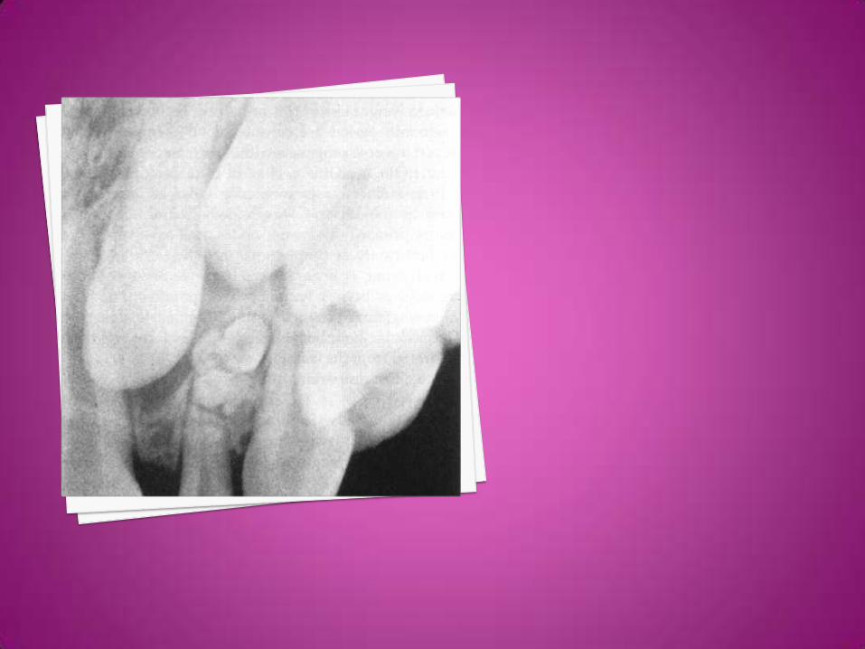
Odontomas are most commonly found on
routine radiographic examination, presenting
as an irregular radiopaque mass or as small,
toothlike structures.
The recommended treatment for an
odontoma is conservative surgical excision,
with care taken to remove the surrounding
soft tissue.
No propensity for recurrence has been noted.
Invaginated odontomes (dens invaginatus)
arise as a result of invagination of a portion
of the enamel organ into the dental papilla
at an early stage in odontogenesis, before
the formation of calcified dental tissues.
The majority of invaginations originate in the
coronal part of the tooth but radicular
invaginations also occur.
Although coronal invaginations may involve any type of tooth, including supernumerary teeth, the permanent maxillary lateral incisors are the teeth most frequently affected.
The anomaly is often bilateral. The condition is uncommon in mandibular teeth and cases reported involving the primary dentition are exceedingly rare.
The prevalence of dens invaginatus varies in different series from less than 1 to about 10 per cent, based on studies of extracted maxillary permanent lateral incisors, or on radiographic surveys.
The degree of invagination varies but three
main types are identified:
type 1, where the invagination is confined to
the crown of the involved tooth;
type 2, where the invagination extends into
the root; and
type 3, where the invagination extends
through the root apex.
In the permanent maxillary lateral incisor the invagination arises from the cingulum pit or, in the case of peg-shaped lateral incisors, from the incisal tip.
Where the invagination is of a minor degree the tooth may be of normal appearance, but with the more extensive forms the crown, and particularly the root, may be considerably dilated.
The terms 'dilated' or 'gestant odontome' are sometimes applied to describe such anomalies
Radiographs reveal an invagination lined by
enamel which is continuous with the normal
enamel covering of the tooth.
The appearances may resemble a tooth
within a tooth, hence the term 'dens-in-
dente'.
Key points - Invaginated odontome
· mainly permanent maxillary lateral incisors
· enamel-lined invagination on radiograph
· extent of invagination varies
· enamel and dentine in the base of the
invagination often defective in quantity
and/or quality
· pulpitis and sequelae common
· abnormalities of crown/root morphology
Evaginated odontomes (dens evaginatus) are uncommon and are characterized by extra cusp-like tubercles which usually arise from the occlusal surfaces of premolars or the palatal surfaces of the maxillary central or lateral incisors.
The anomaly presents as an enamel-covered, teat-like tubercle projecting from the occlusal surface of an otherwise normal premolar.
The evagination is easily fractured resulting in exposure of the pulp and its sequelae.
Evaginated odontomes involving the occlusal
surfaces of premolars occur predominantly in
people of Mongoloid stock. Those involving
the anterior teeth, predominantly the
permanent maxillary lateral
incisors, originate from the palatal cingulum.
They are usually referred to as talon cusps
because of their resemblance to an eagle's
talon.
The enamel pearl presents as a small droplet
of enamel on the root of a tooth and is found
most frequently near or in the furcation of
the roots of maxillary permanent molar
teeth.
Most arise close to the amelocemental
junction but they are occasionally found near
the root apex.
The lesion is symptomless and is discovered
as an incidental finding on radiographs or
when the tooth is extracted.
Microscopically, some consist entirely of
enamel but others contain a core of dentine
and even a small amount of pulp tissue .
The anomaly is thought to arise as a result of
a growth disturbance of Hertwig's sheath
resulting in budding of the sheath followed
by differentiation of ameloblasts and
amelogenesis.
The complex odontome is A Developmental
tumour- Like Mass Consisting Of Disorderly
arranged dental tissues.
The complex odontome occurs predominantly
in the second and third decades of life and
the majority arise in the molar region of the
mandible.
They are often associated with the crowns of
unerupted teeth and occasionally may take
the place of a tooth.
For these reasons they may be discovered, when small, as incidental findings when investigating a patient with a tooth missing from the dental arch. As the lesion enlarges it usually presents as a painless, slow-growing expansion of the jaw, but may become infected and present with pain, particularly if it communicates with the mouth.
Multiple odontomes are rare. In some cases complex odontomes develop in association with calcifying odontogenic cysts .
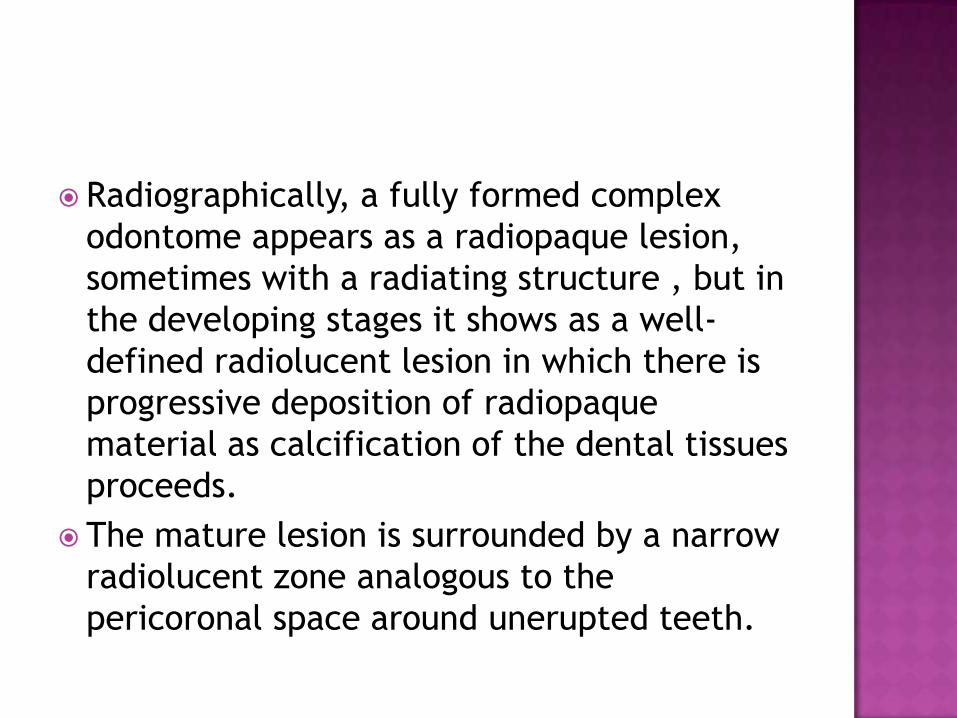
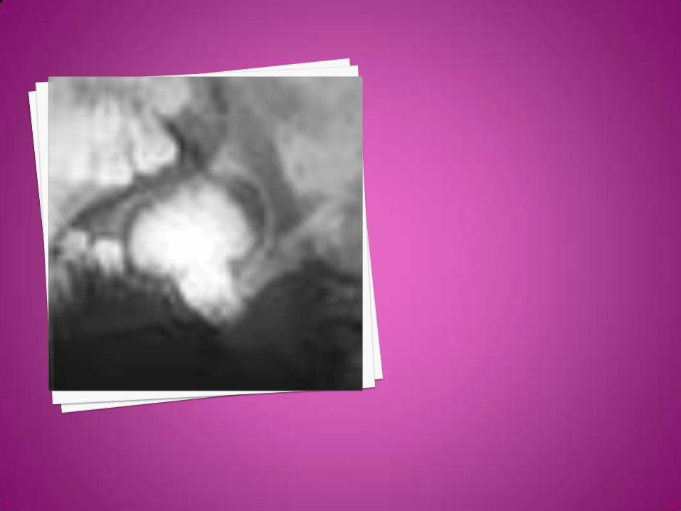
Radiographically, a fully formed complex
odontome appears as a radiopaque lesion,
sometimes with a radiating structure , but in
the developing stages it shows as a well-
defined radiolucent lesion in which there is
progressive deposition of radiopaque
material as calcification of the dental tissues
proceeds.
The mature lesion is surrounded by a narrow
radiolucent zone analogous to the
pericoronal space around unerupted teeth.
Histologically, the fully developed complex
odontome consists of a mass of disorderly
arranged, but well-formed enamel, dentine,
and cementum.
Key points - Complex odontome
· developmental lesion resulting in
disorganized mass of dental tissues
· 2nd/3rd decade; predominantly molar
region mandible
· may overlie/replace a tooth
· radiolucent/radiopaque depending on
maturity
· dentine forms bulk of lesion
Key points - Compound odontome
· developmental lesion resulting in the formation of a bag of discrete denticles
· 1st/2nd decade; predominantly anterior maxilla
· often overlies the crown of an unerupted tooth
· separate denticles identifiable on radiograph
· denticles comprise enamel, dentine, cementum, and pulp in their normal anatomical relationship