Embed Size (px)
Citation preview

Avian Encephalomyelitis (AE)

Plan of Talk
Introduction
Etiology
Transmission
Clinical signs
Post mortem lesions
Diagnosis
Treatment
Prevention and control

Plan of Talk
Introduction
Etiology
Transmission
Clinical signs
Post mortem lesions
Diagnosis
Treatment
Prevention and control

Introduction
Avian encephalomyelitis (AE) is a viral disease of youngchickens, turkeys, Japanese quail, pheasants and pigeons.
AE is characterized by neurologic signs that result frominfection of the CNS with an RNA virus in the familyPicornaviridae.

Cont. …
Turkeys
They are less susceptible to naturalinfection
They generally develop a milder clinicaldisease than chickens.

Cont. …
Infection occurs via vertical and horizontal transmission.
If a breeder flock becomes infected during egg production,the virus is vertically transmitted to the offspring and a majoroutbreak occurs.
The disease often appears in a series of flocks hatched fromthe infected breeder flock.

Cont. …
Field strains of the virus are enterotropic and multiply in theintestine.
Infected birds shed the virus in their feces for a few days to afew weeks, which serves to spread the infection to hatchmates.
AE virus is resistant to environmental conditions and mayremain infectious for long periods.

Plan of Talk
Introduction
Etiology
Transmission
Clinical signs
Post mortem lesions
Diagnosis
Treatment
Prevention and control

Etiology
AEV
Serologic uniformity, strains differ in virulence.

Strain Classification and Pathogenicity
Pathotype One
Represented by natural field strains,is enterotropic.
These strains infect chickens readilyvia the oral route, multiply in theintestine, and are shed in the feces.
Cont. …
Pathotype Two
Embryo-adapted strains constitutethe other pathotype.
They do not infect via the oral routeexcept with very high doses, andthey do not spread horizontally.
X
X
Cont. …
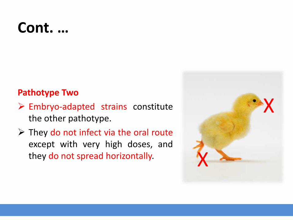
Adaptation may occur aftermultiple passages in antibody-freechicken embryos.

Plan of Talk
Introduction
Etiology
Transmission
Clinical signs
Post mortem lesions
Diagnosis
Treatment
Prevention and control
Transmission
Egg transmission is the major route of transmission.
Infected breeders will transmit the A.E. virus for severalweeks and cause a decrease in egg hatchability.
Infected chicks that hatch will show clinical signs of thedisease and spread the infection in the incubator to othernew hatched susceptible chicks.
Young chicks can also be infected on the farm.

Transmission
The horizontal transmission by direct contact may be morecommon, but vertical transmission is clinically moreimportant.
The incubation period varies from 5 to 14 days depending onthe route of infection.

Plan of Talk
Introduction
Etiology
Transmission
Clinical signs
Post mortem lesions
Diagnosis
Treatment
Prevention and control

Clinical Signs
Vertically infected chicks
Clinical signs appear during the first week after hatching,although signs may be present in a few birds at hatching.
Vertical infection followed by horizontal infection causes acharacteristic biphasic mortality pattern.

Cont. …
Horizontally infected chicks
Clinical signs appear at 2–4 week of age, incubation period of5-14 days.
Clinical disease progresses through the flock for the first fewweeks, and the episode is usually over by the time the flock is4 weeks old.
After 4 weeks of age, chickens are resistant to disease but notinfection.
Morbidity and mortality rates vary and depend on the level ofegg transmission and degree of immunity in the flock.
In severe outbreaks, both morbidity and mortality may exceed50%.

The main clinical signs are ataxia and leg weakness that variesfrom sitting on hocks to paresis that progresses to paralysis anddecumbency.

Fine tremors of the head and neck.
Cupping the bird in one's hands often results in a buzzingfeeling because of rapid and fine tremors.
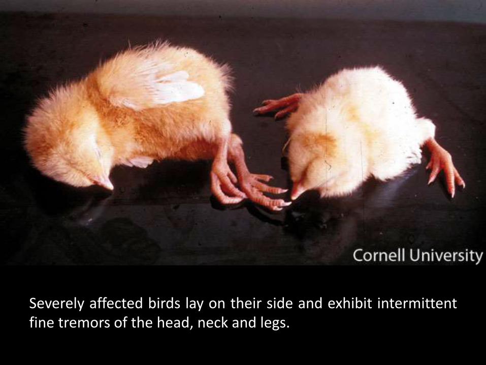
Severely affected birds lay on their side and exhibit intermittentfine tremors of the head, neck and legs.


Clinical Signs
In laying chickens, there is a sudden, 5%–10% drop in eggproduction, which usually lasts for less than 2 weeks, followedby a return to normal production.
There is no deterioration in egg shell quality.
Hatchability may drop as much as 5% during the decline inegg production due to late embryonic mortality.
Infected eggs are laid during the period of viremia, whichusually lasts 1–2 wk.

Plan of Talk
Introduction
Etiology
Transmission
Clinical signs
Post mortem lesions
Diagnosis
Treatment
Prevention and control

Post Mortem Lesions
No gross lesions are seen in the brain of infected birds.
Gray to white foci may be visible on cut surfaces of the muscleof the gizzard.
Weeks after infection, opacity of eye lenses (cataracts) mayoccur in a small percentage of chickens that survive theinfection.

Plan of Talk
Introduction
Etiology
Transmission
Clinical signs
Post mortem lesions
Diagnosis
Treatment
Prevention and control

Diagnosis
Diagnosis is based on:
1. History
2. Clinical signs
3. Characteristic histopathologic lesions in the brain and spinalcord.

Cont. …
The following methods may help in making a diagnosis for AvianEncephalomyelitis.
1. Virus Neutralization test.
2. Agar Gel test.
3. Elisa test.
4. Embryo Susceptibility test.
The above tests are only indicative of antibody present but notnecessarily disease.

Cont. …
The diagnosis is best confirmed by isolation and identification ofthe virus.
1. Tissues collected for virus isolation must include the brainand duodenum with the pancreas.
2. Demonstration of AE virus antigen in the brain, spinal cord,and other tissues by immunofluorescent andimmunohistochemical staining is a reliable method ofdiagnosis.

Plan of Talk
Introduction
Etiology
Transmission
Clinical signs
Post mortem lesions
Diagnosis
Treatment
Prevention and control

Treatment
Sick birds should be isolated and potentially destroyed as fewof them recover.
Good supportive care may be helpful in some cases.
Sanitize the premises.

Plan of Talk
Introduction
Etiology
Transmission
Clinical signs
Post mortem lesions
Diagnosis
Treatment
Prevention and control

Prevention and Control
Immunization of breeder pullets 10-15 weeks old with acommercial live vaccine is advised to prevent verticaltransmission of the virus to progeny and to provide them withmaternal immunity against the disease.
Vaccination of layers is advisable to prevent a temporary dropin egg production.