Embed Size (px)
Citation preview
Chapter 13: CENTRAL NERVOUS SYSTEMS
1• Brain and Spinal Cord are the
main organs.• Brain - single organ.• Spinal cord – 4 main regions
cerebrumdiencephalonbrain stemcerebellum
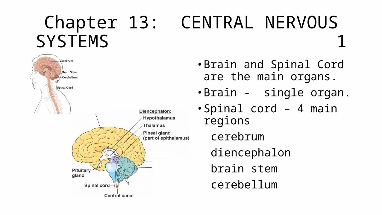
Cerebrum 2
• The iconic gray mantle of the human brain, which appears to make up most of the mass of the brain, is the cerebrum. • Wrinkled portion is the cerebral
cortex.• Deep within the cerebrum, the
white matter of the corpus callosum provides the major pathway for communication between the two hemispheres of the cerebral cortex.
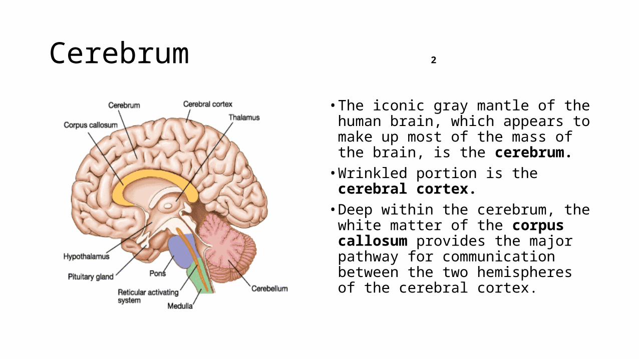
LIMBIC SYSTEM 3
• Many of the higher neurological functions, such as memory, emotion, and consciousness, are the result of cerebral function.• The limbic cortex is the region of
the cerebral cortex that is part of the limbic system, a collection of structures involved in emotion, memory, and behavior.
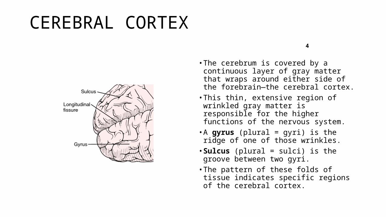
CEREBRAL CORTEX 4
• The cerebrum is covered by a continuous layer of gray matter that wraps around either side of the forebrain—the cerebral cortex.
• This thin, extensive region of wrinkled gray matter is responsible for the higher functions of the nervous system.
• A gyrus (plural = gyri) is the ridge of one of those wrinkles.
• Sulcus (plural = sulci) is the groove between two gyri.
• The pattern of these folds of tissue indicates specific regions of the cerebral cortex.
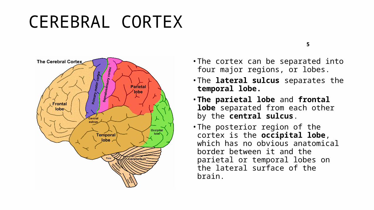
CEREBRAL CORTEX 5
• The cortex can be separated into four major regions, or lobes.• The lateral sulcus separates the
temporal lobe. • The parietal lobe and frontal lobe
separated from each other by the central sulcus. • The posterior region of the cortex is
the occipital lobe, which has no obvious anatomical border between it and the parietal or temporal lobes on the lateral surface of the brain.
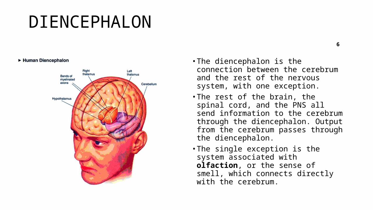
DIENCEPHALON 6
• The diencephalon is the connection between the cerebrum and the rest of the nervous system, with one exception. • The rest of the brain, the spinal cord,
and the PNS all send information to the cerebrum through the diencephalon. Output from the cerebrum passes through the diencephalon. • The single exception is the system
associated with olfaction, or the sense of smell, which connects directly with the cerebrum.
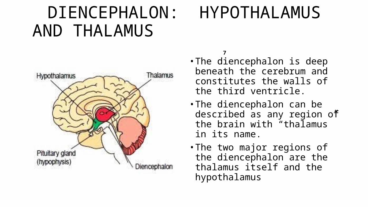
DIENCEPHALON: HYPOTHALAMUS AND THALAMUS 7
• The diencephalon is deep beneath the cerebrum and constitutes the walls of the third ventricle. • The diencephalon can be
described as any region of the brain with “thalamus” in its name. • The two major regions of the
diencephalon are the thalamus itself and the hypothalamus
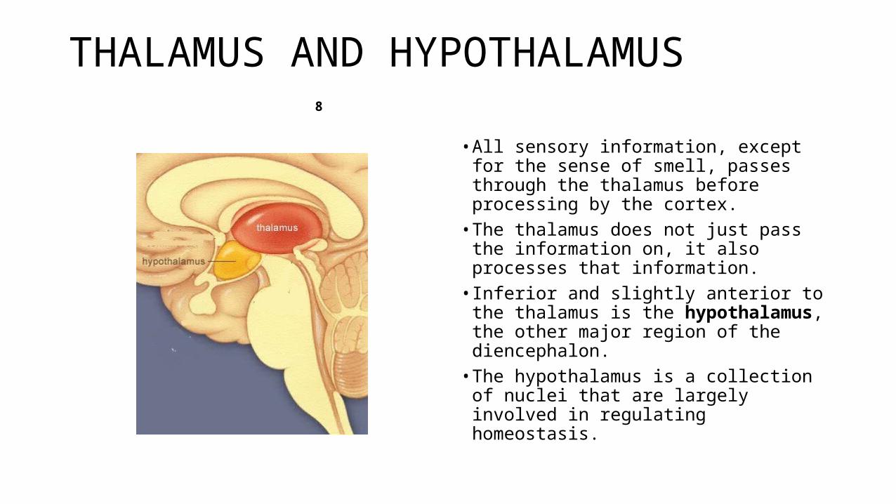
THALAMUS AND HYPOTHALAMUS 8
• All sensory information, except for the sense of smell, passes through the thalamus before processing by the cortex.
• The thalamus does not just pass the information on, it also processes that information.
• Inferior and slightly anterior to the thalamus is the hypothalamus, the other major region of the diencephalon.
• The hypothalamus is a collection of nuclei that are largely involved in regulating homeostasis.
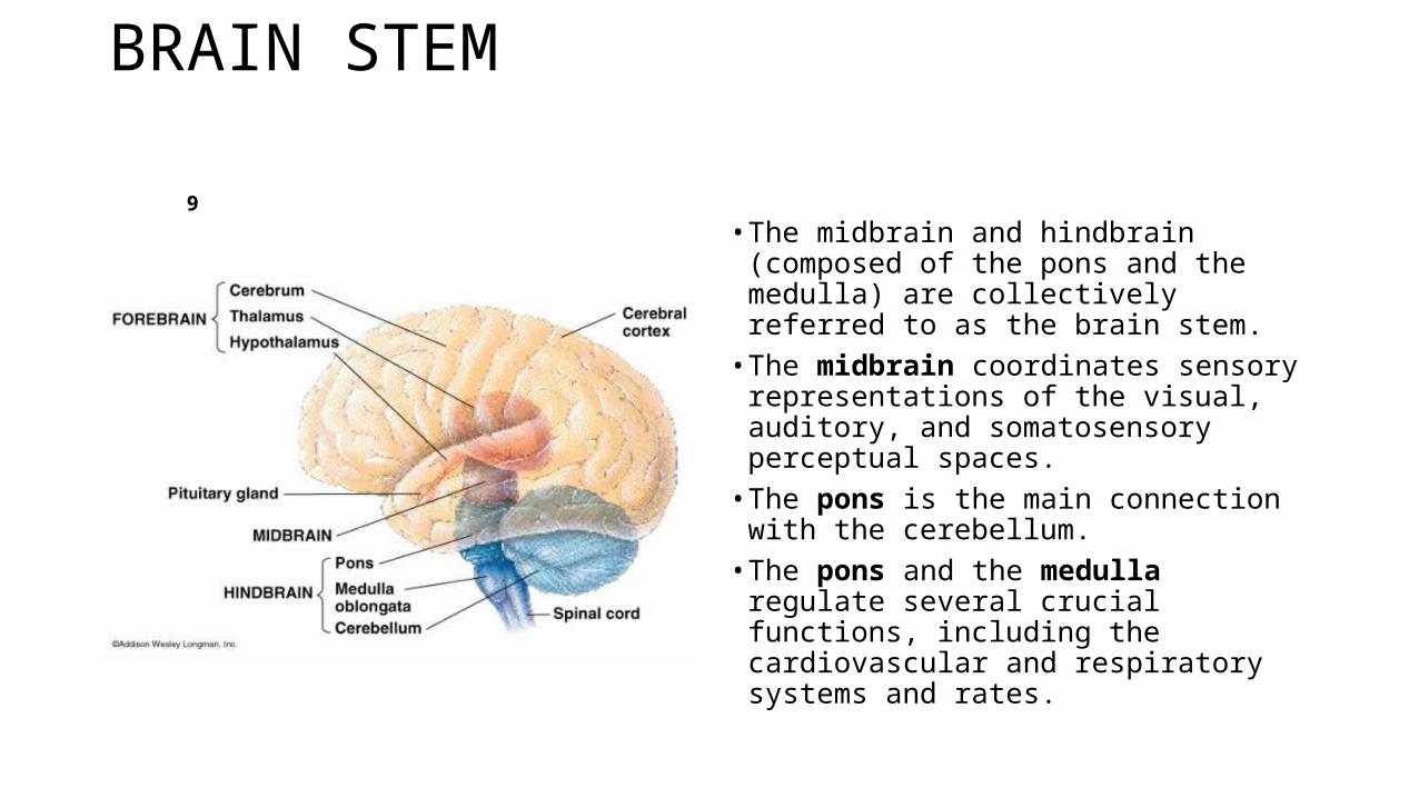
BRAIN STEM 9
• The midbrain and hindbrain (composed of the pons and the medulla) are collectively referred to as the brain stem.
• The midbrain coordinates sensory representations of the visual, auditory, and somatosensory perceptual spaces.
• The pons is the main connection with the cerebellum.
• The pons and the medulla regulate several crucial functions, including the cardiovascular and respiratory systems and rates.
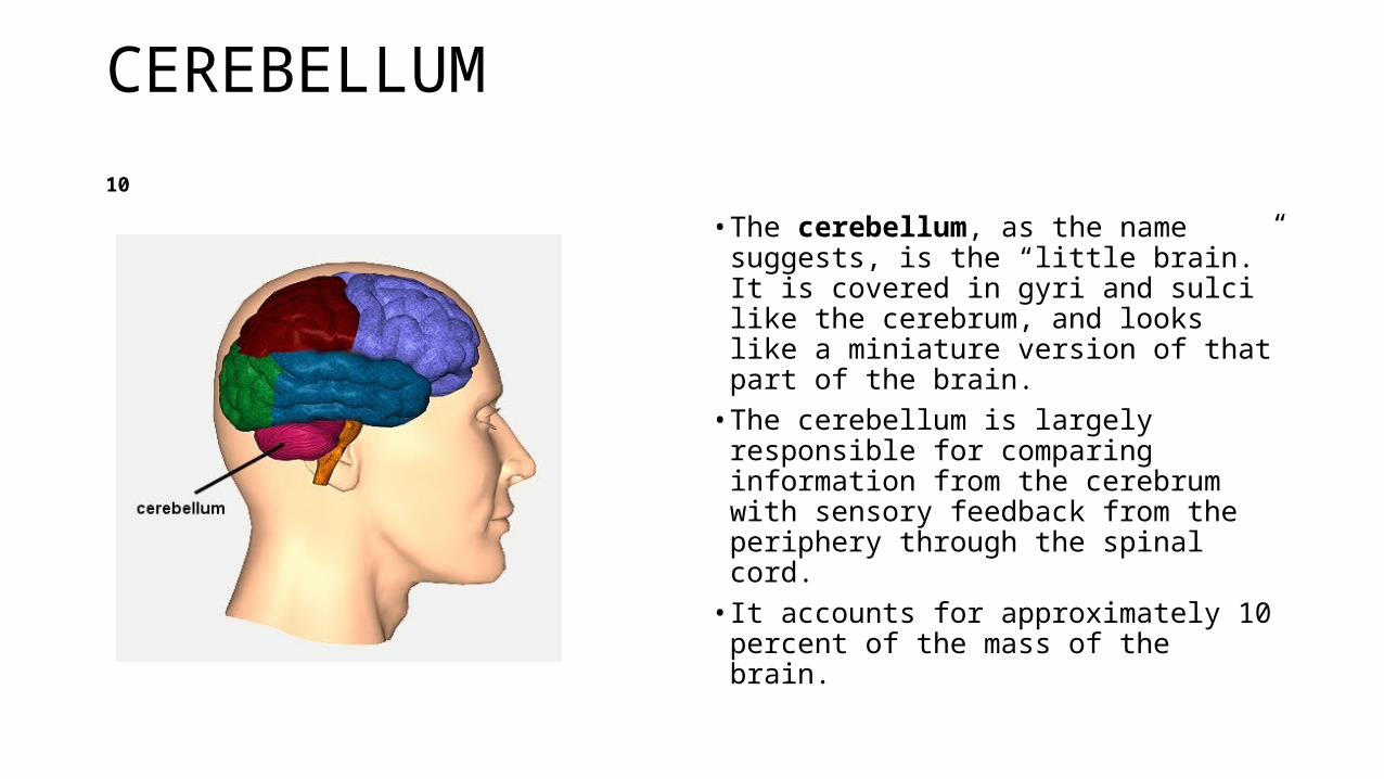
CEREBELLUM 10
• The cerebellum, as the name suggests, is the “little brain.” It is covered in gyri and sulci like the cerebrum, and looks like a miniature version of that part of the brain. • The cerebellum is largely responsible
for comparing information from the cerebrum with sensory feedback from the periphery through the spinal cord. • It accounts for approximately 10
percent of the mass of the brain.
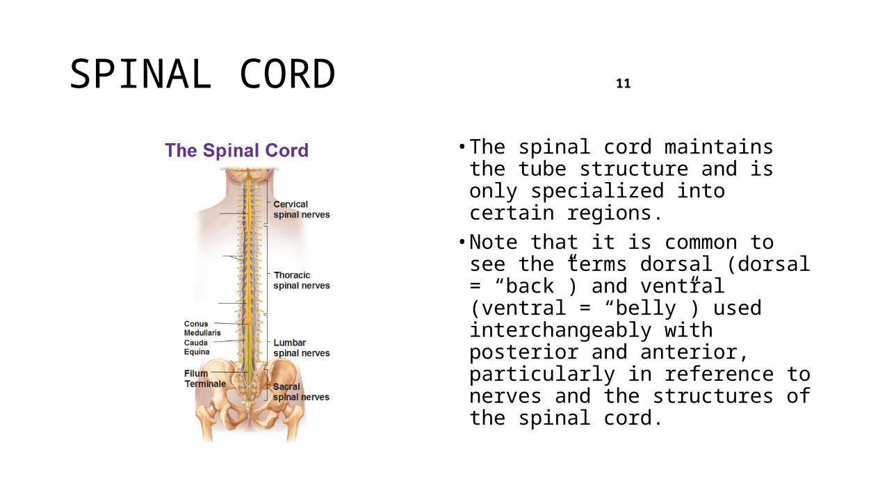
SPINAL CORD 11
• The spinal cord maintains the tube structure and is only specialized into certain regions.• Note that it is common to see the
terms dorsal (dorsal = “back”) and ventral (ventral = “belly”) used interchangeably with posterior and anterior, particularly in reference to nerves and the structures of the spinal cord.
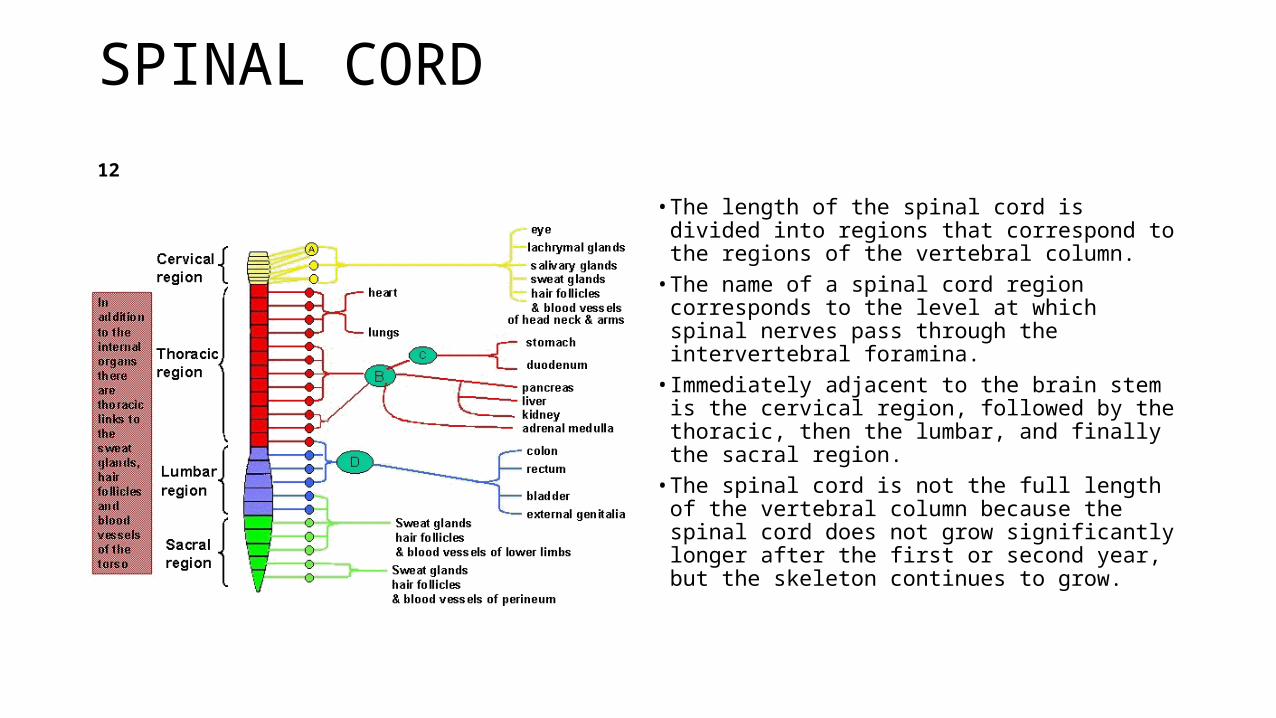
SPINAL CORD 12
• The length of the spinal cord is divided into regions that correspond to the regions of the vertebral column.
• The name of a spinal cord region corresponds to the level at which spinal nerves pass through the intervertebral foramina.
• Immediately adjacent to the brain stem is the cervical region, followed by the thoracic, then the lumbar, and finally the sacral region.
• The spinal cord is not the full length of the vertebral column because the spinal cord does not grow significantly longer after the first or second year, but the skeleton continues to grow.
ARTERIAL SUPPLY 13
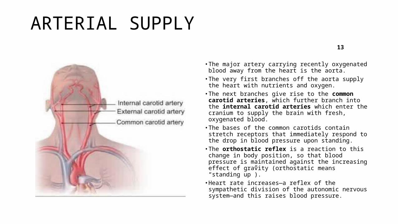
• The major artery carrying recently oxygenated blood away from the heart is the aorta.
• The very first branches off the aorta supply the heart with nutrients and oxygen.
• The next branches give rise to the common carotid arteries, which further branch into the internal carotid arteries which enter the cranium to supply the brain with fresh, oxygenated blood.
• The bases of the common carotids contain stretch receptors that immediately respond to the drop in blood pressure upon standing.
• The orthostatic reflex is a reaction to this change in body position, so that blood pressure is maintained against the increasing effect of gravity (orthostatic means “standing up”).
• Heart rate increases—a reflex of the sympathetic division of the autonomic nervous system—and this raises blood pressure.
PROTECTIVE COVERING OF BRAIN AND SPINAL CORD 14
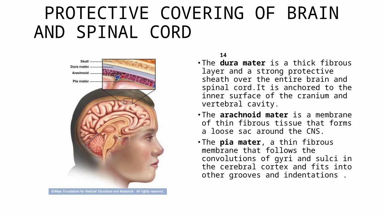
• The dura mater is a thick fibrous layer and a strong protective sheath over the entire brain and spinal cord.It is anchored to the inner surface of the cranium and vertebral cavity.
• The arachnoid mater is a membrane of thin fibrous tissue that forms a loose sac around the CNS.
• The pia mater, a thin fibrous membrane that follows the convolutions of gyri and sulci in the cerebral cortex and fits into other grooves and indentations .
DURA MATER 15
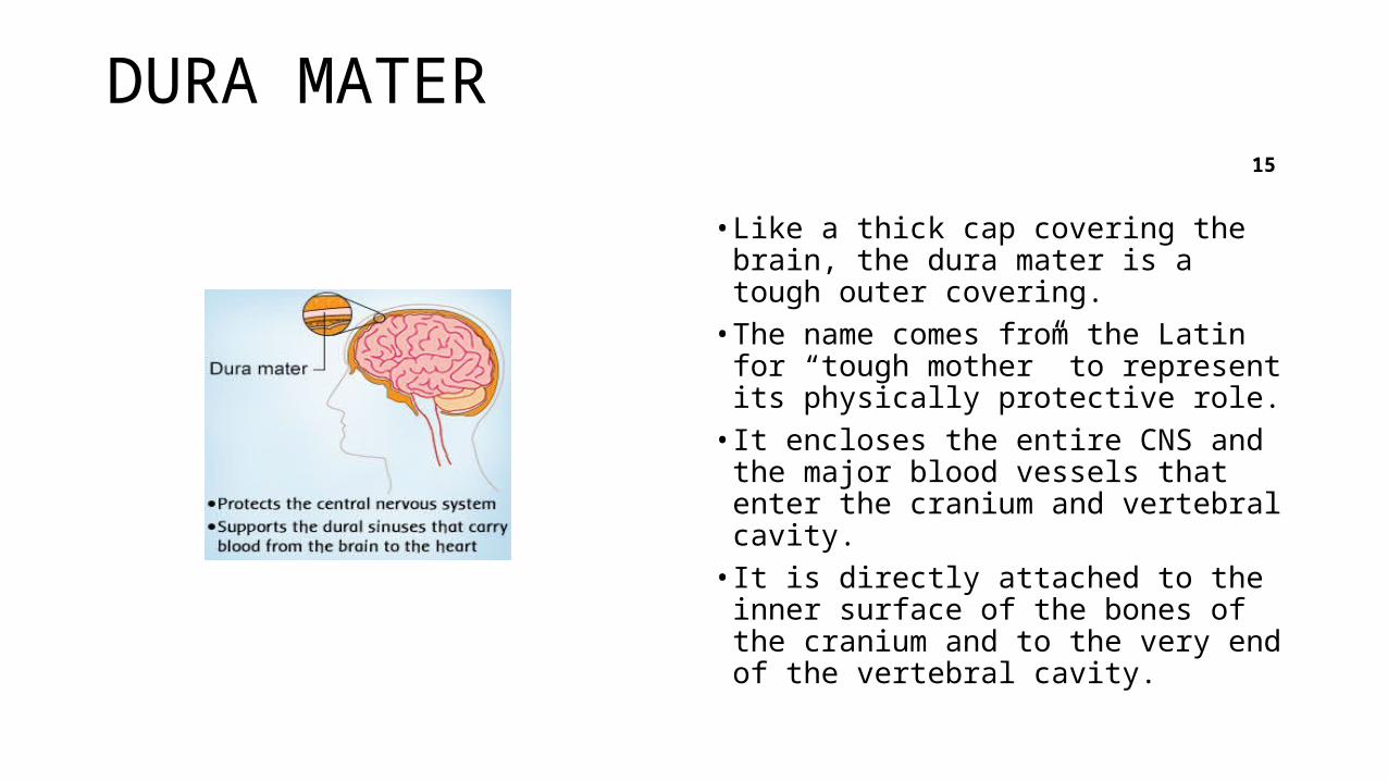
• Like a thick cap covering the brain, the dura mater is a tough outer covering. • The name comes from the Latin for
“tough mother” to represent its physically protective role. • It encloses the entire CNS and the
major blood vessels that enter the cranium and vertebral cavity. • It is directly attached to the inner
surface of the bones of the cranium and to the very end of the vertebral cavity.
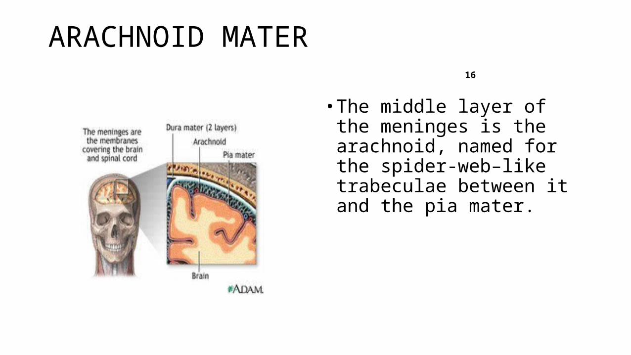
ARACHNOID MATER 16
• The middle layer of the meninges is the arachnoid, named for the spider-web–like trabeculae between it and the pia mater.
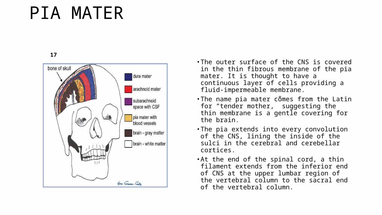
PIA MATER 17
• The outer surface of the CNS is covered in the thin fibrous membrane of the pia mater. It is thought to have a continuous layer of cells providing a fluid-impermeable membrane.
• The name pia mater comes from the Latin for “tender mother,” suggesting the thin membrane is a gentle covering for the brain.
• The pia extends into every convolution of the CNS, lining the inside of the sulci in the cerebral and cerebellar cortices.
• At the end of the spinal cord, a thin filament extends from the inferior end of CNS at the upper lumbar region of the vertebral column to the sacral end of the vertebral column.
LUMBAR PUNCTURE 18
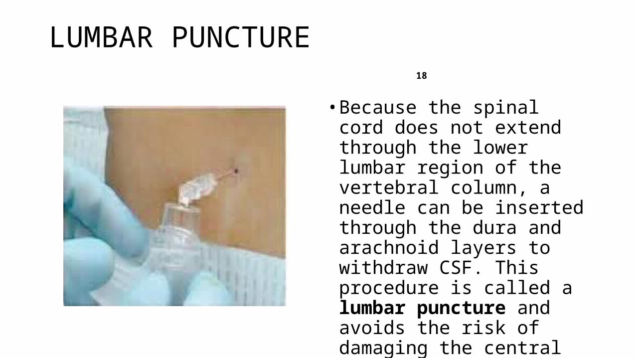
• Because the spinal cord does not extend through the lower lumbar region of the vertebral column, a needle can be inserted through the dura and arachnoid layers to withdraw CSF. This procedure is called a lumbar puncture and avoids the risk of damaging the central tissue of the spinal cord.
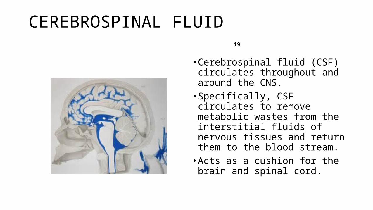
CEREBROSPINAL FLUID 19
• Cerebrospinal fluid (CSF) circulates throughout and around the CNS. • Specifically, CSF circulates to
remove metabolic wastes from the interstitial fluids of nervous tissues and return them to the blood stream.• Acts as a cushion for the brain
and spinal cord.
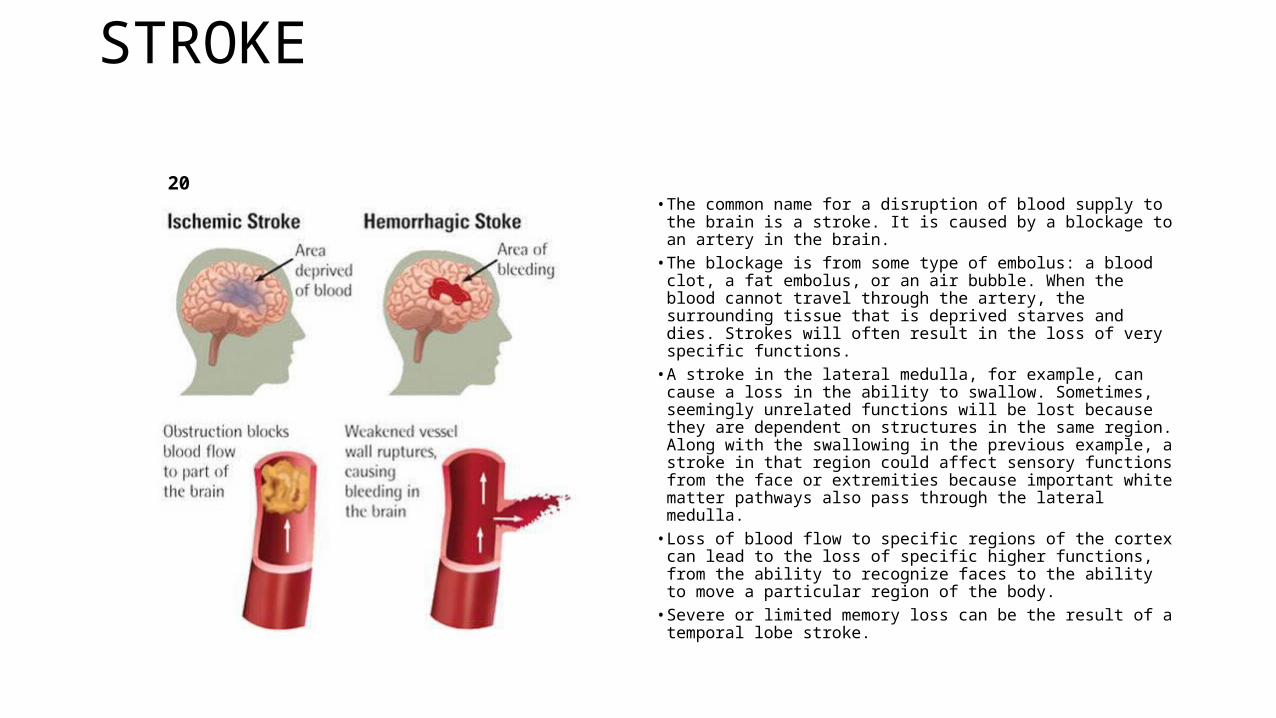
STROKE 20
• The common name for a disruption of blood supply to the brain is a stroke. It is caused by a blockage to an artery in the brain.
• The blockage is from some type of embolus: a blood clot, a fat embolus, or an air bubble. When the blood cannot travel through the artery, the surrounding tissue that is deprived starves and dies. Strokes will often result in the loss of very specific functions.
• A stroke in the lateral medulla, for example, can cause a loss in the ability to swallow. Sometimes, seemingly unrelated functions will be lost because they are dependent on structures in the same region. Along with the swallowing in the previous example, a stroke in that region could affect sensory functions from the face or extremities because important white matter pathways also pass through the lateral medulla.
• Loss of blood flow to specific regions of the cortex can lead to the loss of specific higher functions, from the ability to recognize faces to the ability to move a particular region of the body.
• Severe or limited memory loss can be the result of a temporal lobe stroke.
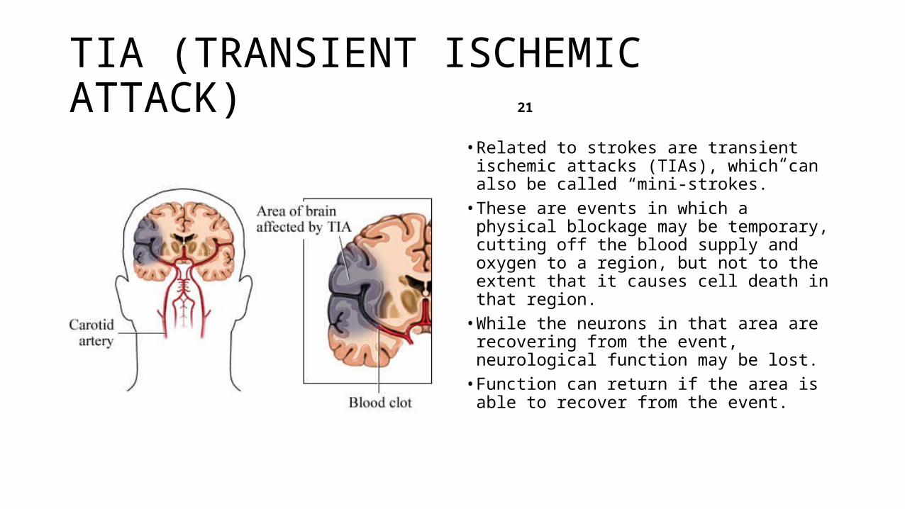
TIA (TRANSIENT ISCHEMIC ATTACK) 21
• Related to strokes are transient ischemic attacks (TIAs), which can also be called “mini-strokes.”
• These are events in which a physical blockage may be temporary, cutting off the blood supply and oxygen to a region, but not to the extent that it causes cell death in that region.
• While the neurons in that area are recovering from the event, neurological function may be lost.
• Function can return if the area is able to recover from the event.
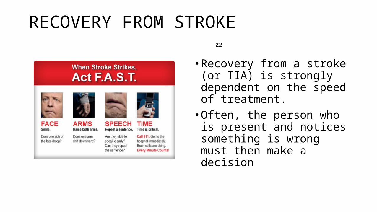
RECOVERY FROM STROKE 22
• Recovery from a stroke (or TIA) is strongly dependent on the speed of treatment. • Often, the person who is present
and notices something is wrong must then make a decision
STROKE RECOVERY 23
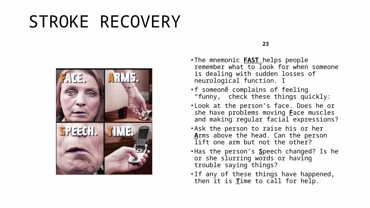
• The mnemonic FAST helps people remember what to look for when someone is dealing with sudden losses of neurological function. I
• f someone complains of feeling “funny,” check these things quickly:
• Look at the person’s face. Does he or she have problems moving Face muscles and making regular facial expressions?
• Ask the person to raise his or her Arms above the head. Can the person lift one arm but not the other?
• Has the person’s Speech changed? Is he or she slurring words or having trouble saying things?
• If any of these things have happened, then it is Time to call for help.
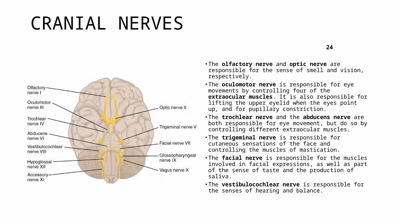
CRANIAL NERVES 24
• The olfactory nerve and optic nerve are responsible for the sense of smell and vision, respectively.
• The oculomotor nerve is responsible for eye movements by controlling four of the extraocular muscles. It is also responsible for lifting the upper eyelid when the eyes point up, and for pupillary constriction.
• The trochlear nerve and the abducens nerve are both responsible for eye movement, but do so by controlling different extraocular muscles.
• The trigeminal nerve is responsible for cutaneous sensations of the face and controlling the muscles of mastication.
• The facial nerve is responsible for the muscles involved in facial expressions, as well as part of the sense of taste and the production of saliva.
• The vestibulocochlear nerve is responsible for the senses of hearing and balance.
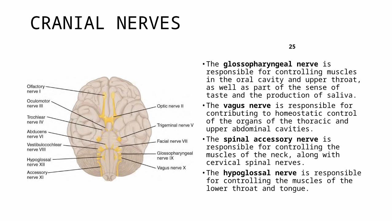
CRANIAL NERVES 25
• The glossopharyngeal nerve is responsible for controlling muscles in the oral cavity and upper throat, as well as part of the sense of taste and the production of saliva.
• The vagus nerve is responsible for contributing to homeostatic control of the organs of the thoracic and upper abdominal cavities.
• The spinal accessory nerve is responsible for controlling the muscles of the neck, along with cervical spinal nerves.
• The hypoglossal nerve is responsible for controlling the muscles of the lower throat and tongue.
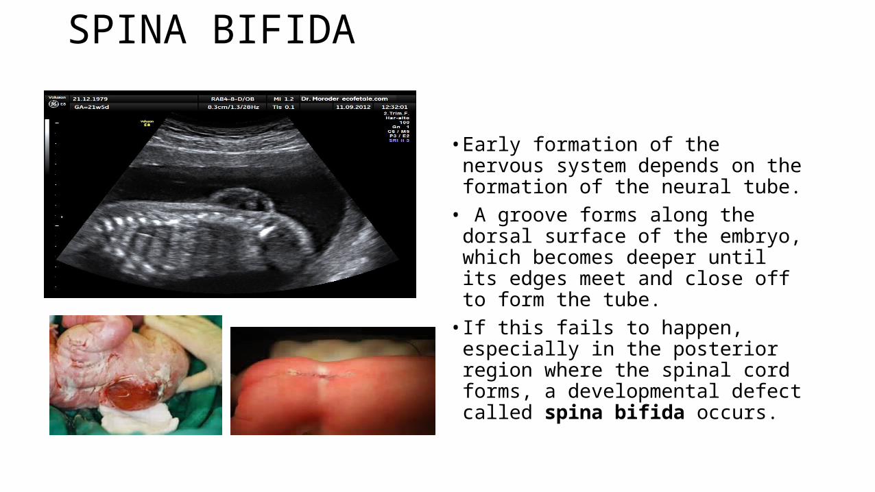
SPINA BIFIDA 26
• Early formation of the nervous system depends on the formation of the neural tube.• A groove forms along the dorsal
surface of the embryo, which becomes deeper until its edges meet and close off to form the tube. • If this fails to happen, especially in
the posterior region where the spinal cord forms, a developmental defect called spina bifida occurs.