Embed Size (px)
Citation preview
Basic Dysrhythmia Interpretation
Presented by :Rina Hadi Abdullah Lestari
CRN/ACLS Instructor/Nursing Director Secretary
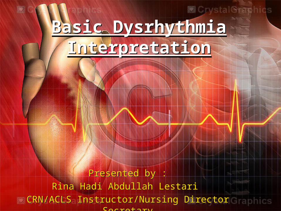
Heart

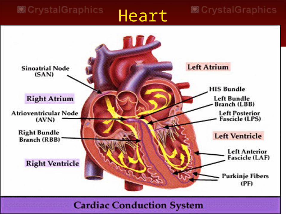
Recording of the ECG
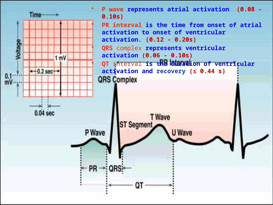
• P wave represents atrial activation (0.08 - 0.10s)• PR interval is the time from onset of atrial activation to
onset of ventricular activation. (0.12 - 0.20s)• QRS complex represents ventricular activation (0.06 -
0.10s)• QT interval is the duration of ventricular activation and
recovery (≤ 0.44 s)
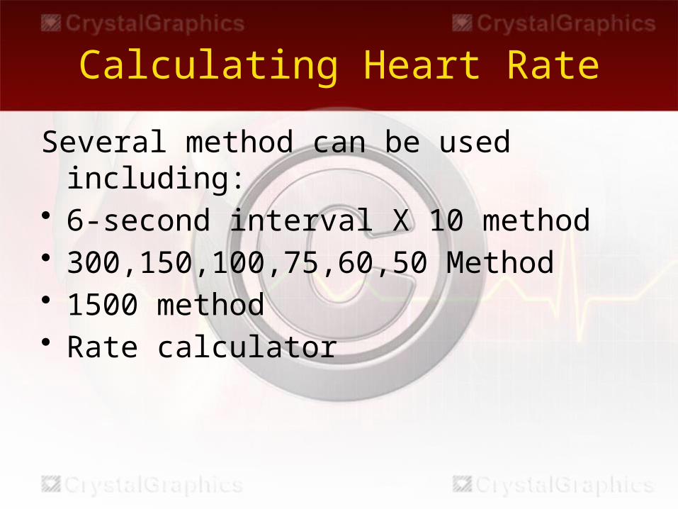
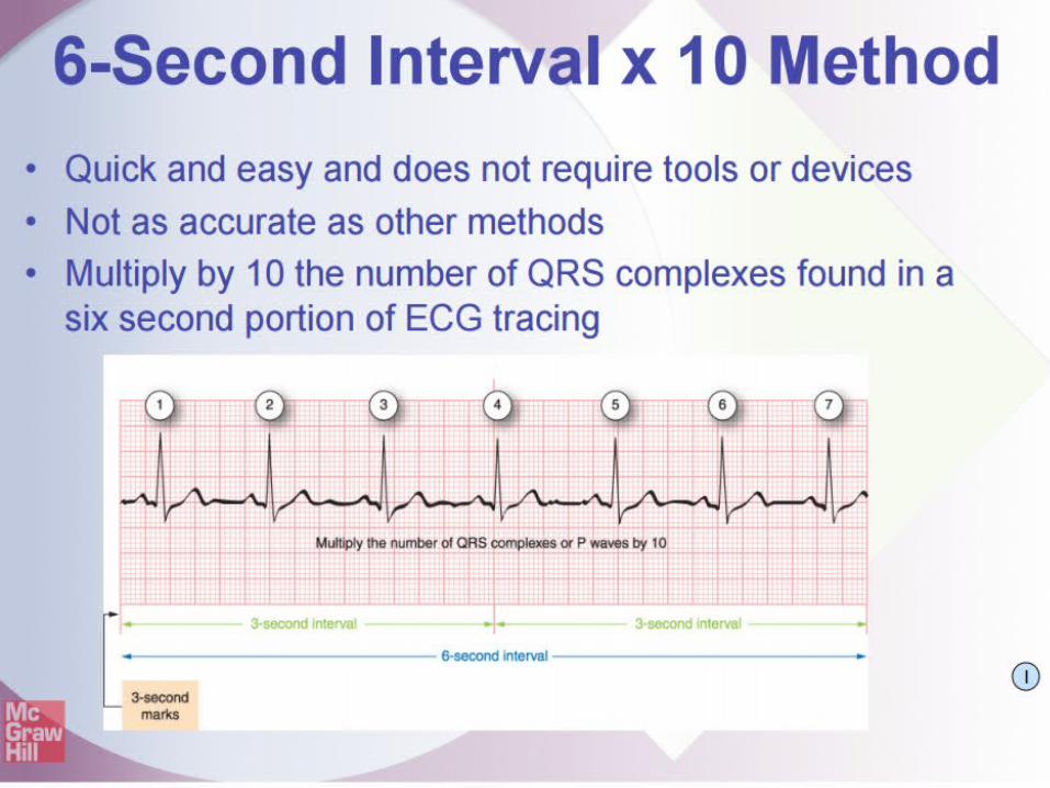
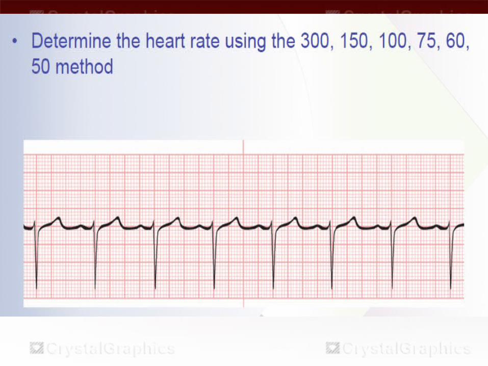
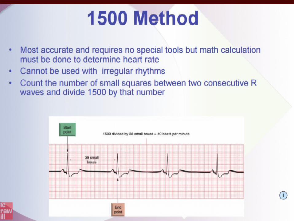
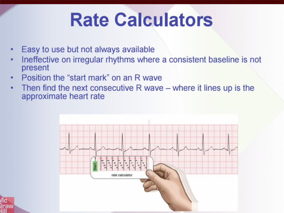
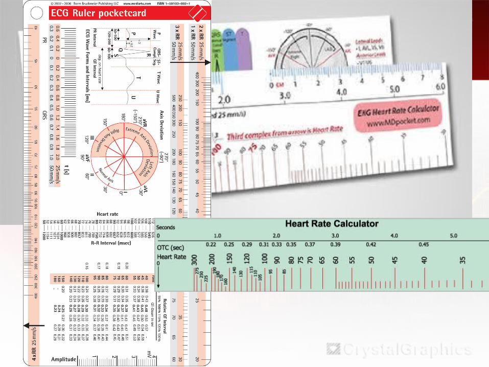
Calculating Heart Rate
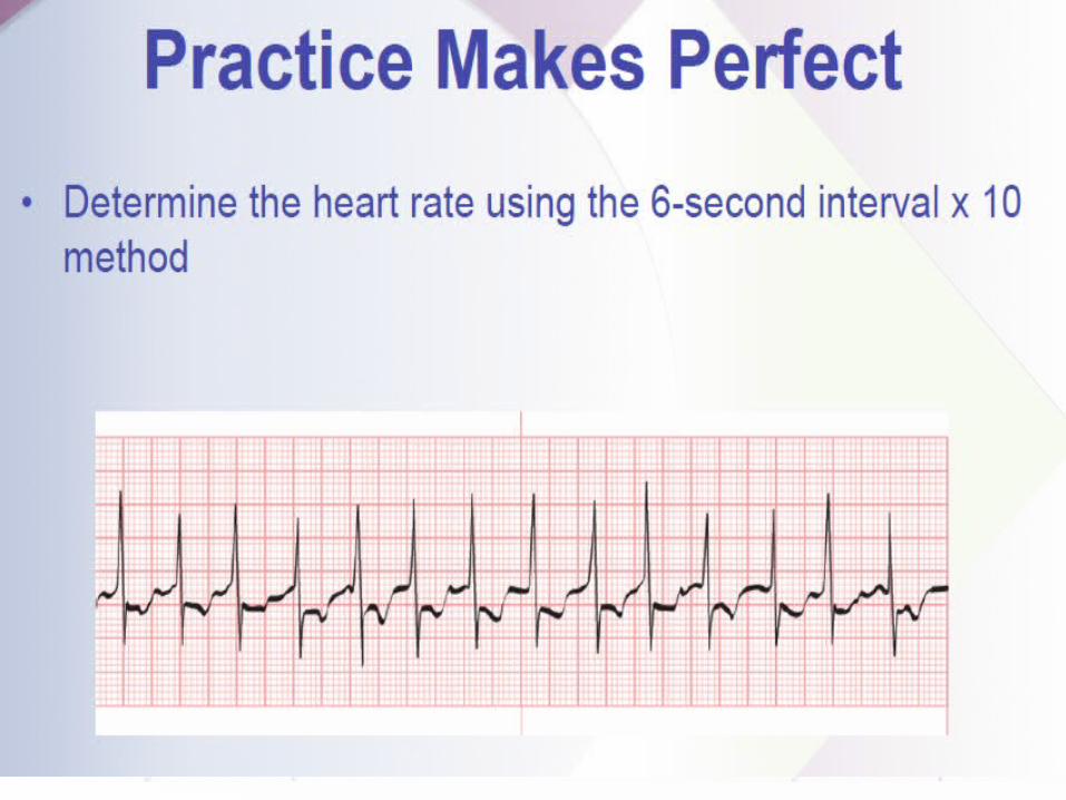
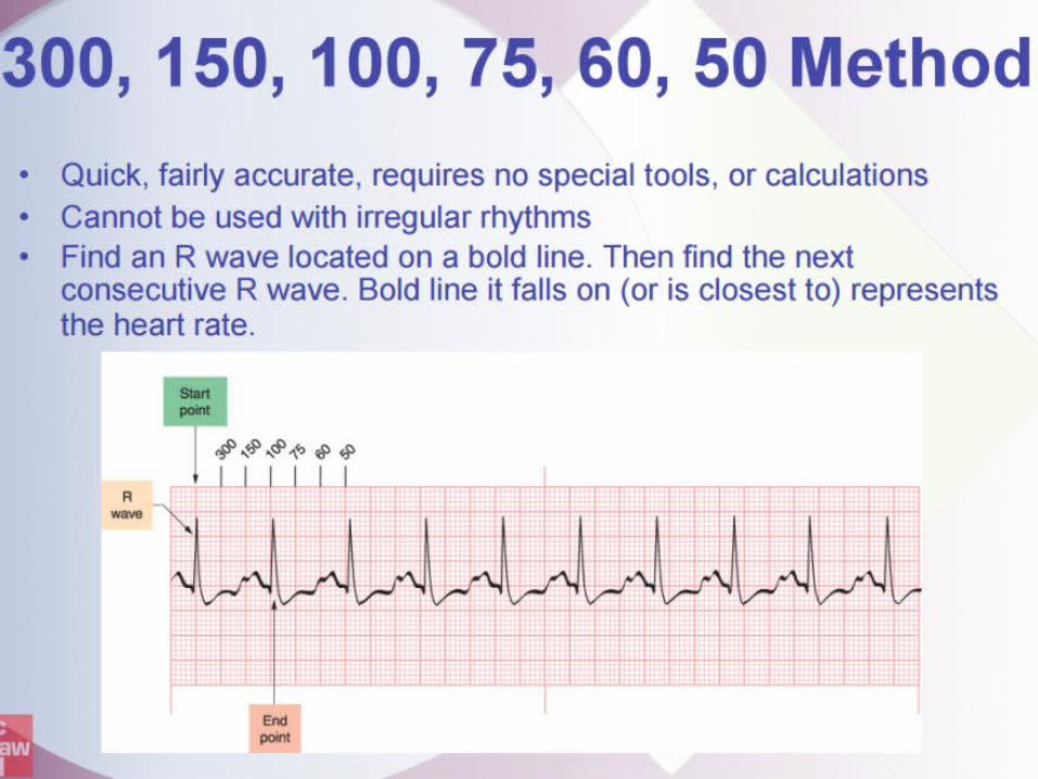
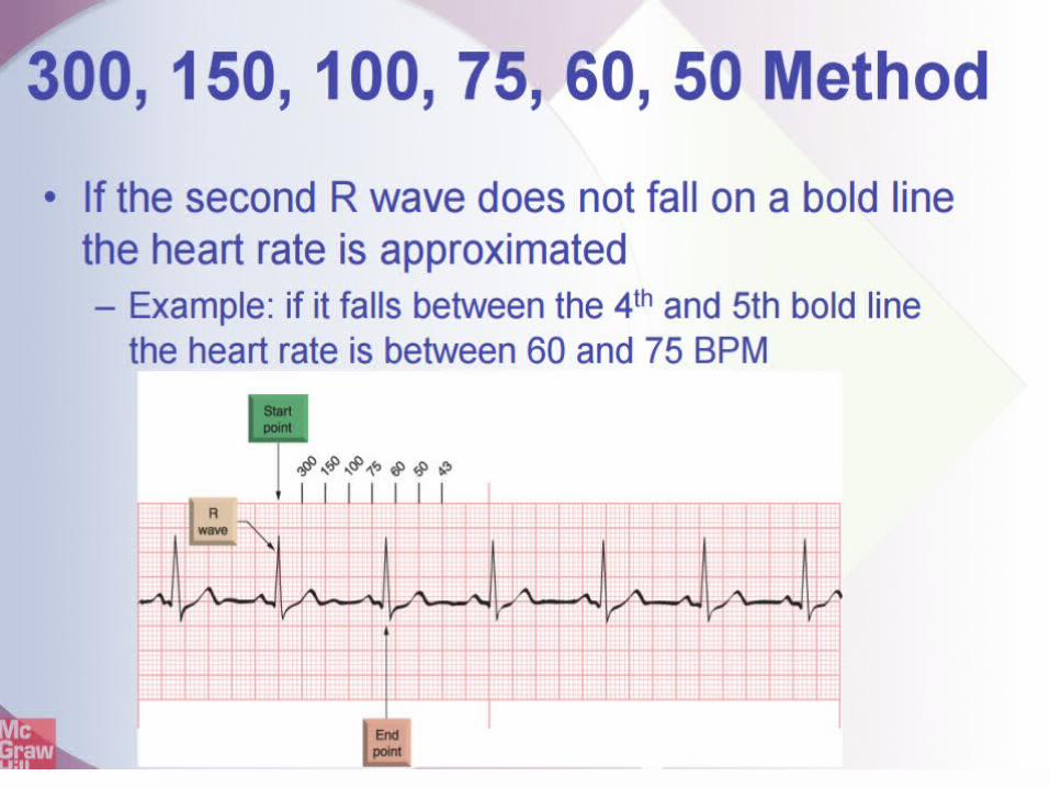
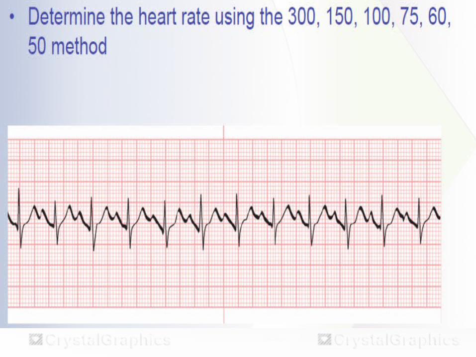
Several method can be used including:• 6-second interval X 10 method• 300,150,100,75,60,50 Method• 1500 method• Rate calculator
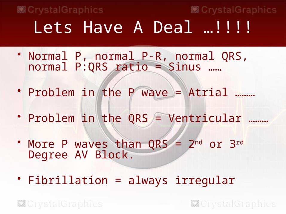
Lets Have A Deal …!!!!• Normal P, normal P-R, normal QRS, normal
P:QRS ratio = Sinus ……
• Problem in the P wave = Atrial ………
• Problem in the QRS = Ventricular ………
• More P waves than QRS = 2nd or 3rd Degree AV Block.
• Fibrillation = always irregular
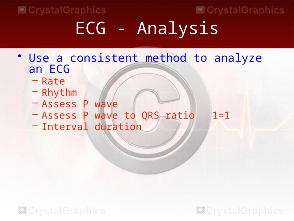
ECG - Analysis• Use a consistent method to analyze an ECG
– Rate– Rhythm– Assess P wave– Assess P wave to QRS ratio 1=1– Interval duration
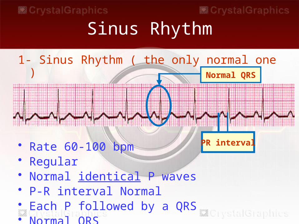
Sinus Rhythm
1- Sinus Rhythm ( the only normal one )
• Rate 60-100 bpm• Regular• Normal identical P waves• P-R interval Normal• Each P followed by a QRS• Normal QRS
Normal QRS
PR interval
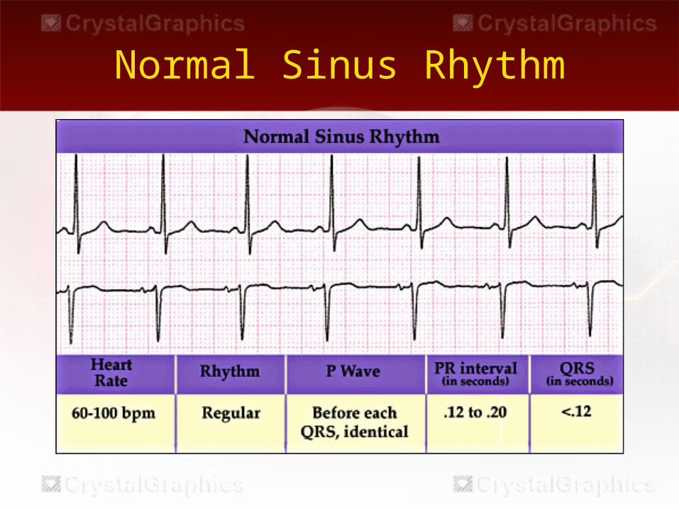
Normal Sinus Rhythm
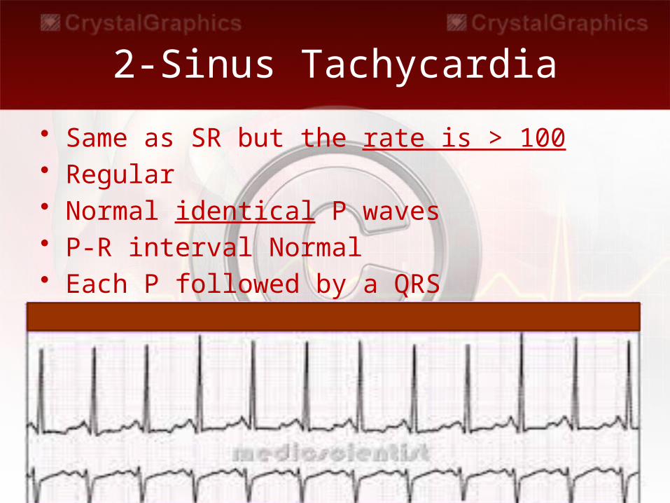
2-Sinus Tachycardia
• Same as SR but the rate is > 100• Regular• Normal identical P waves• P-R interval Normal• Each P followed by a QRS
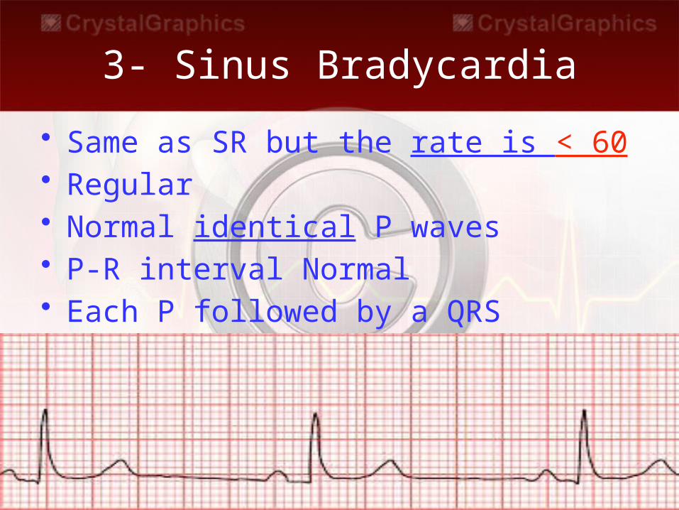
3- Sinus Bradycardia
• Same as SR but the rate is < 60• Regular• Normal identical P waves• P-R interval Normal• Each P followed by a QRS
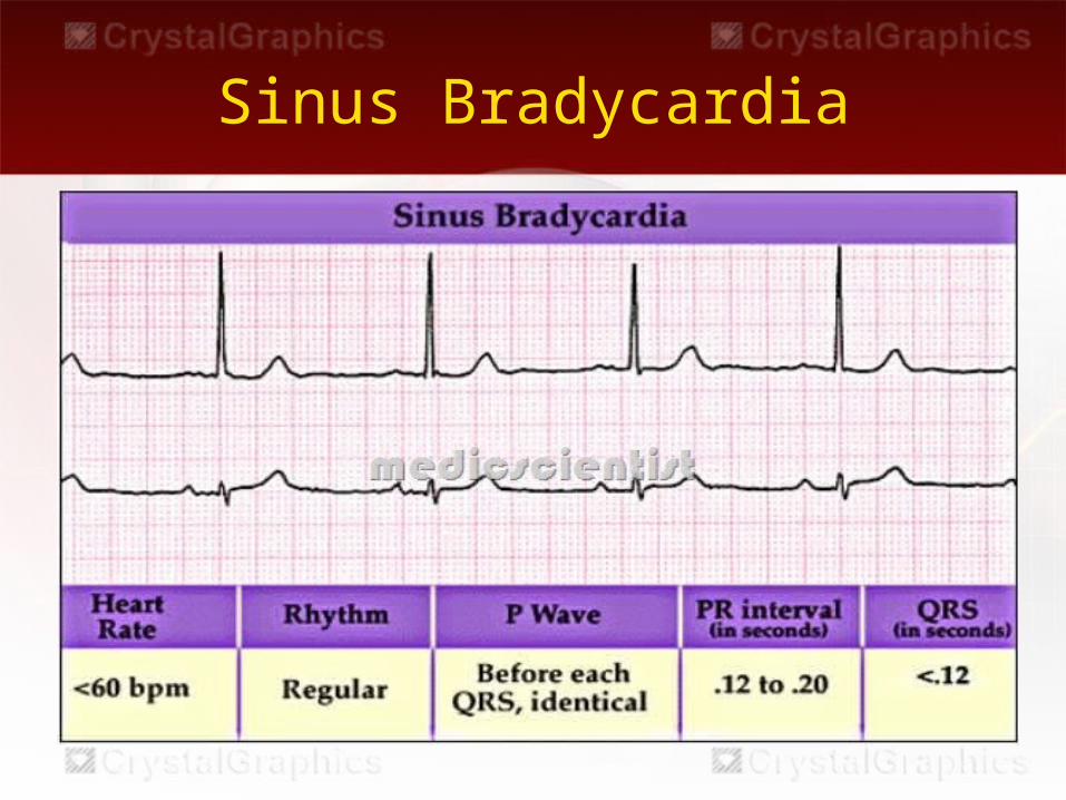
Sinus Bradycardia
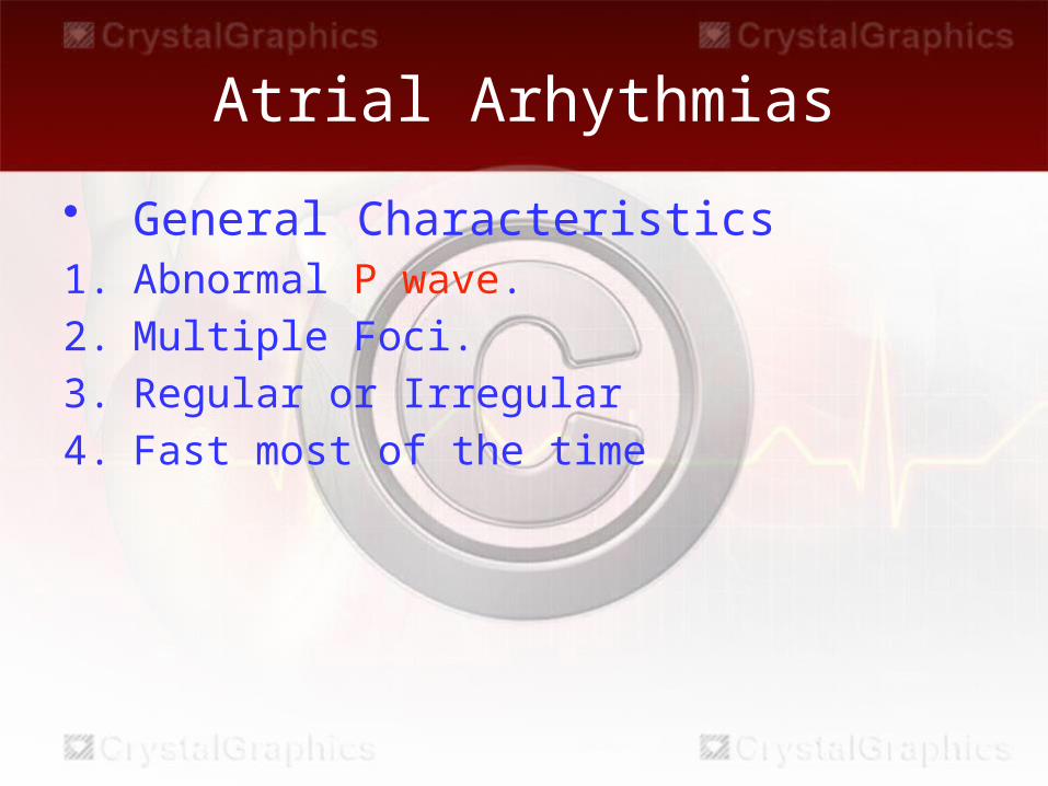
Atrial Arhythmias
• General Characteristics 1. Abnormal P wave. 2. Multiple Foci. 3. Regular or Irregular4. Fast most of the time
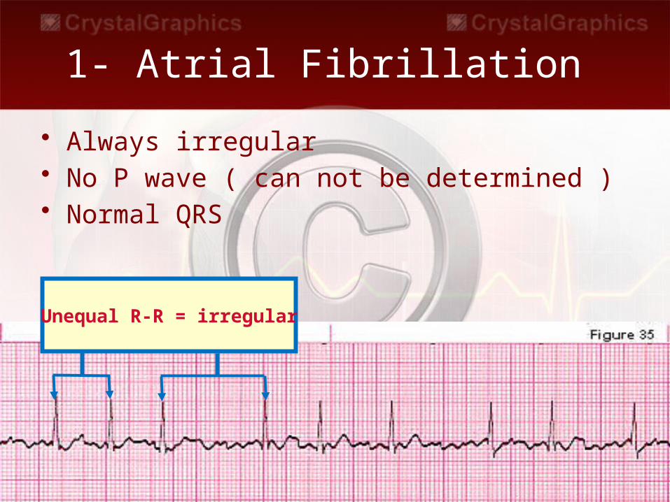
1- Atrial Fibrillation
• Always irregular• No P wave ( can not be determined ) • Normal QRS
Unequal R-R = irregular
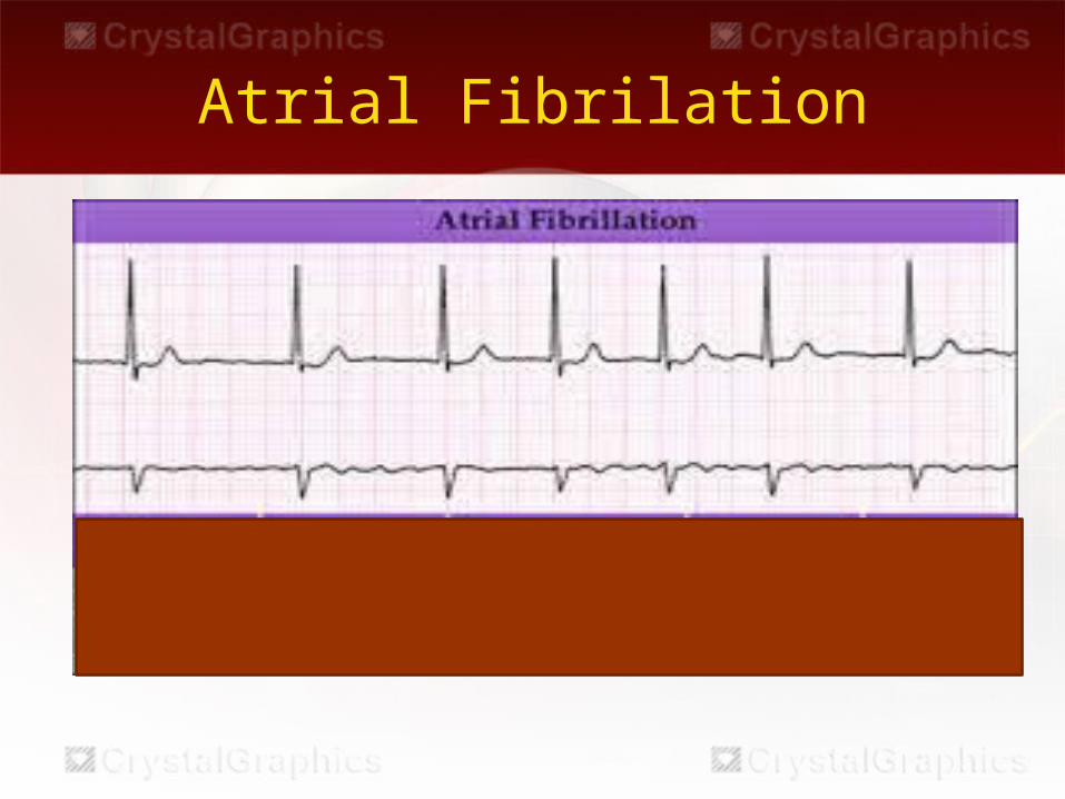
Atrial Fibrilation
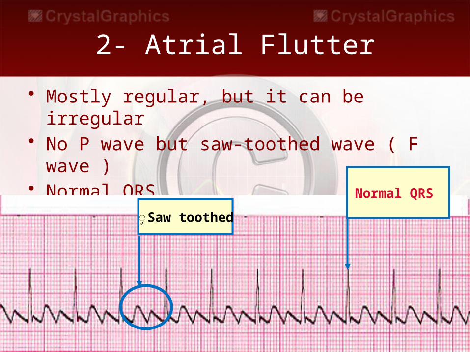
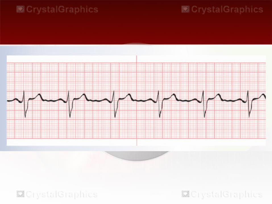
2- Atrial Flutter
• Mostly regular, but it can be irregular• No P wave but saw-toothed wave ( F wave )• Normal QRS
�Saw toothed
Normal QRS
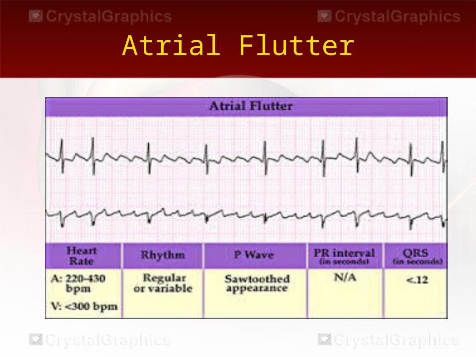
Atrial Flutter
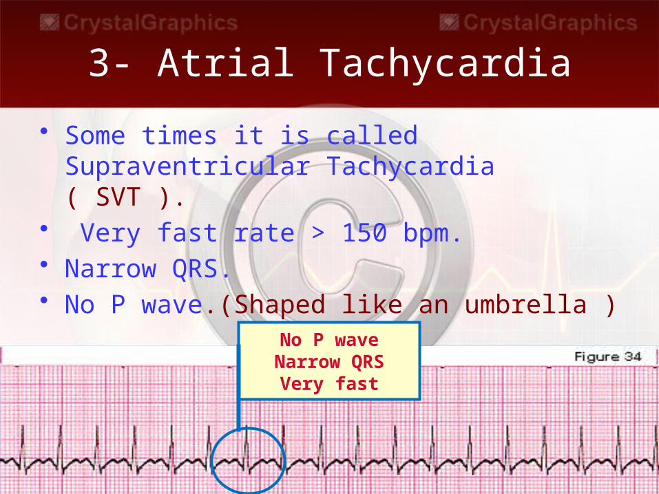
3- Atrial Tachycardia
• Some times it is called Supraventricular Tachycardia ( SVT ).
• Very fast rate > 150 bpm.• Narrow QRS.• No P wave.(Shaped like an umbrella )
No P waveNarrow QRS
Very fast
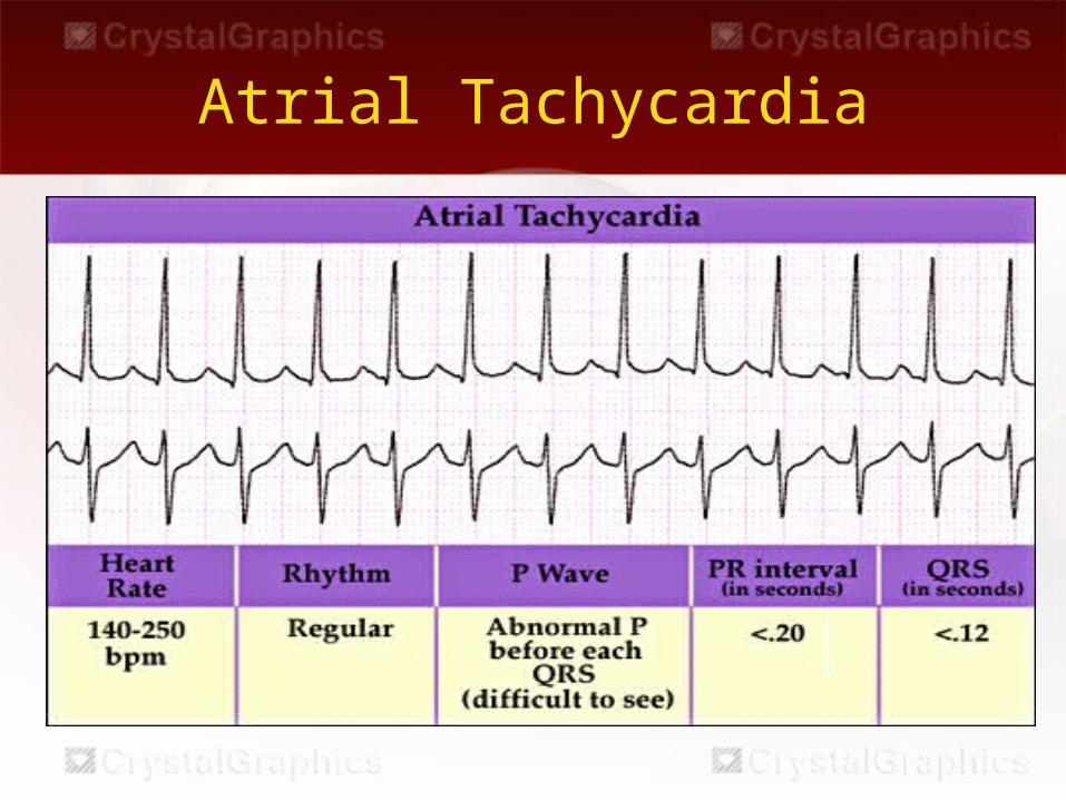
Atrial Tachycardia
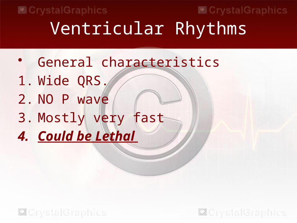
Ventricular Rhythms
• General characteristics1. Wide QRS.2. NO P wave3. Mostly very fast4. Could be Lethal
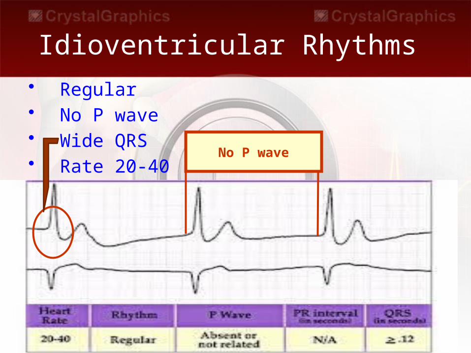
Idioventricular Rhythms • Regular • No P wave• Wide QRS• Rate 20-40
No P wave
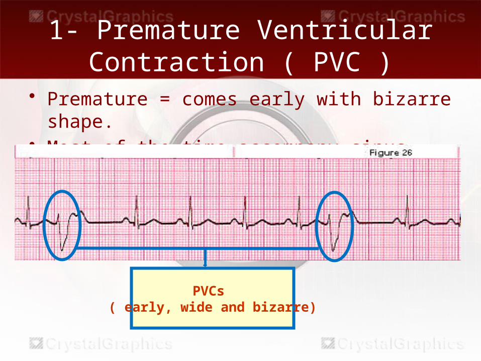
• Premature = comes early with bizarre shape.• Most of the time accompany sinus rhythm.
1- Premature Ventricular Contraction ( PVC )
PVCs ( early, wide and bizarre)
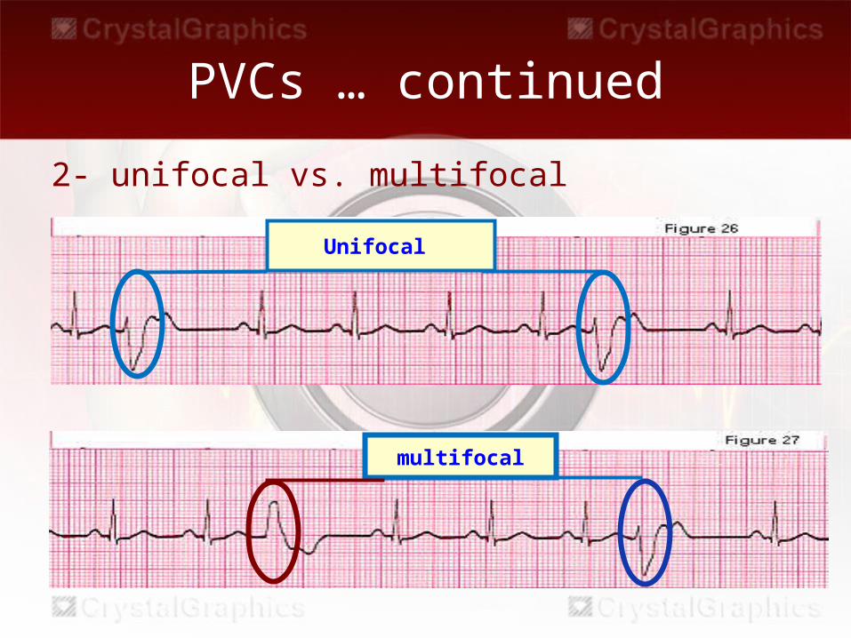
PVCs … continued
2- unifocal vs. multifocal
Unifocal
multifocal
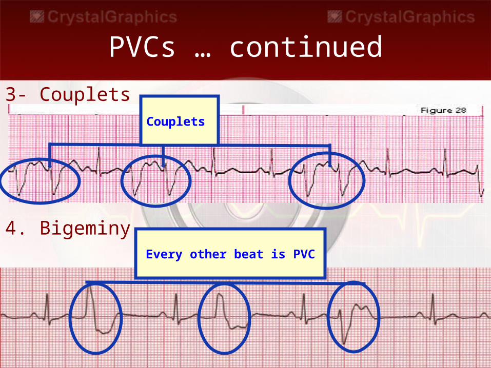
PVCs … continued3- Couplets
4. Bigeminy
Couplets
Every other beat is PVC
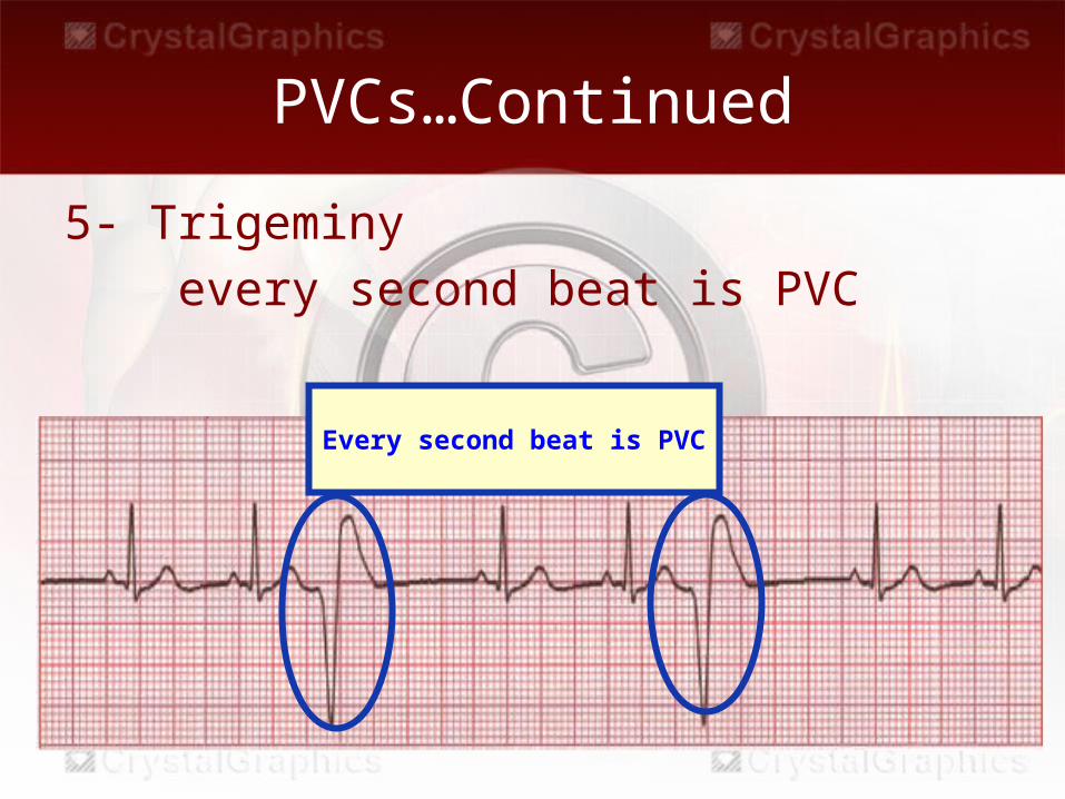
PVCs…Continued
5- Trigeminy every second beat is PVC
Every second beat is PVC
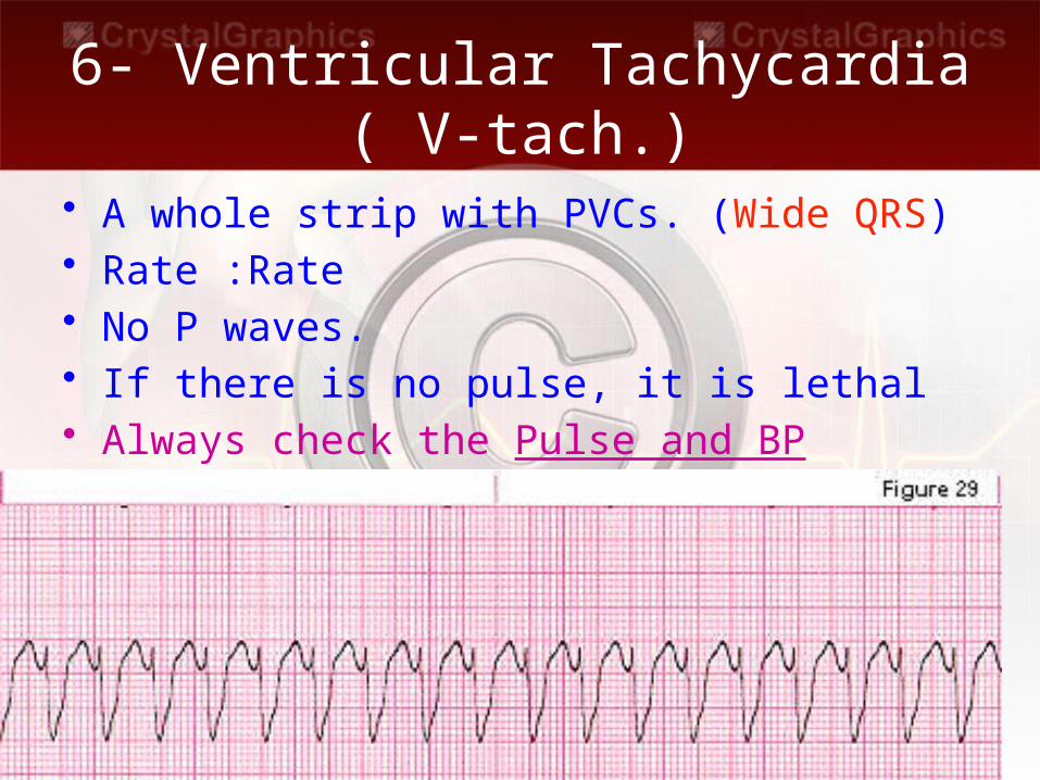
6- Ventricular Tachycardia( V-tach.)
• A whole strip with PVCs. (Wide QRS)• Rate :Rate• No P waves.• If there is no pulse, it is lethal• Always check the Pulse and BP
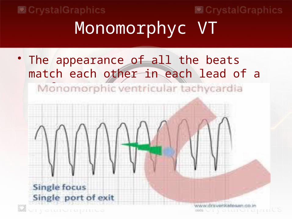
Monomorphyc VT
• The appearance of all the beats match each other in each lead of a surface ECG
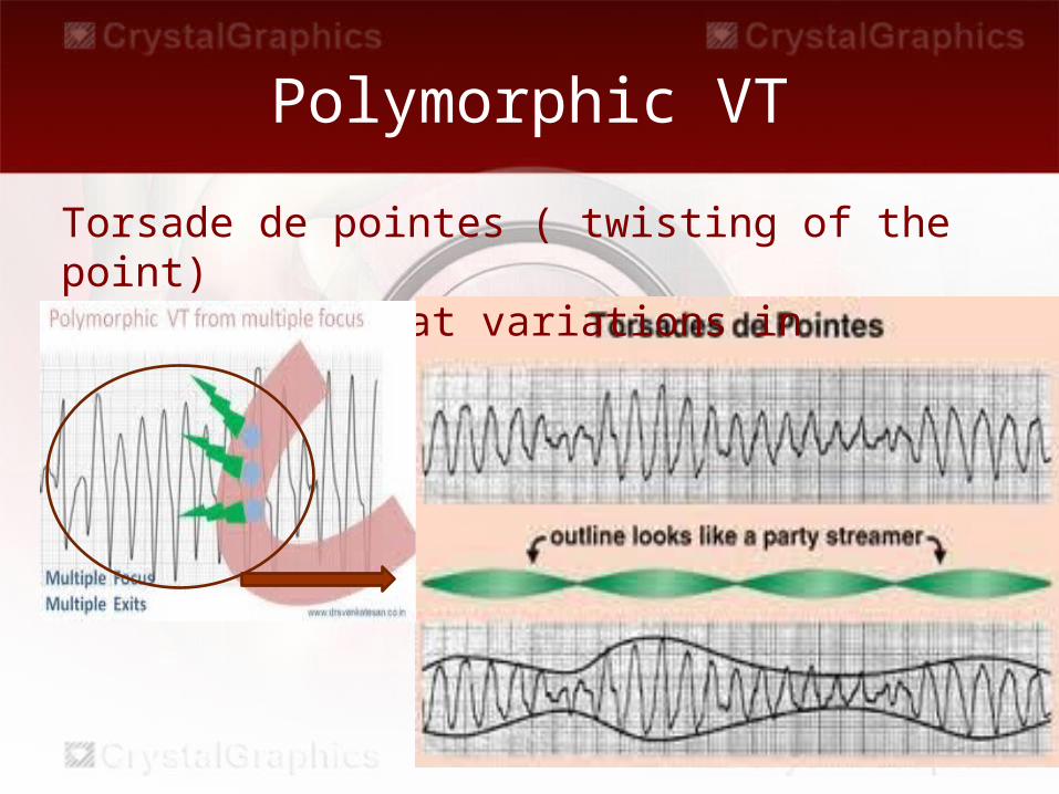
Polymorphic VT
Torsade de pointes ( twisting of the point)has beat-to-beat variations in morphology
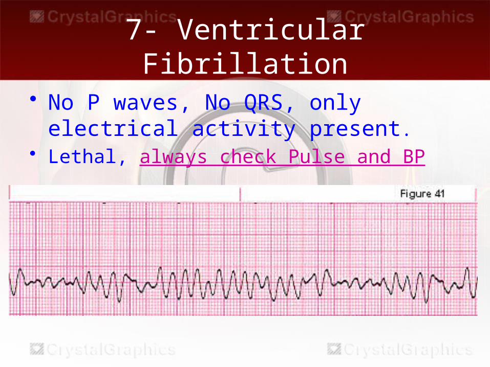
7- Ventricular Fibrillation
• No P waves, No QRS, only electrical activity present.
• Lethal, always check Pulse and BP
AV Blocks
• Prolonged P-R interval or more P waves than QRS
• Block means DELAY.
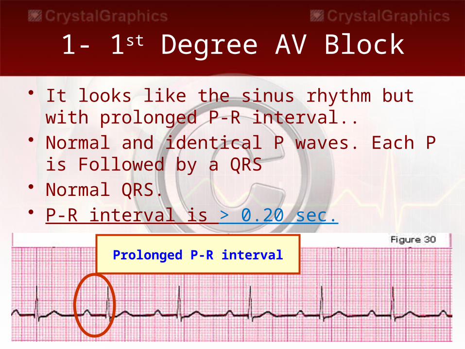
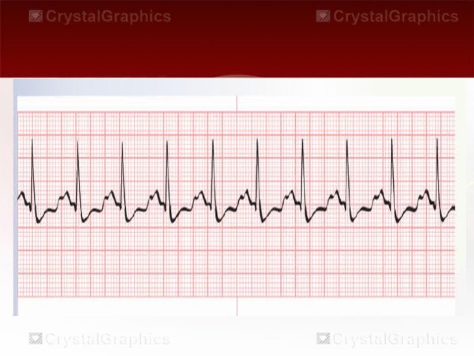
1- 1st Degree AV Block
• It looks like the sinus rhythm but with prolonged P-R interval..
• Normal and identical P waves. Each P is Followed by a QRS
• Normal QRS.• P-R interval is > 0.20 sec.
Prolonged P-R interval
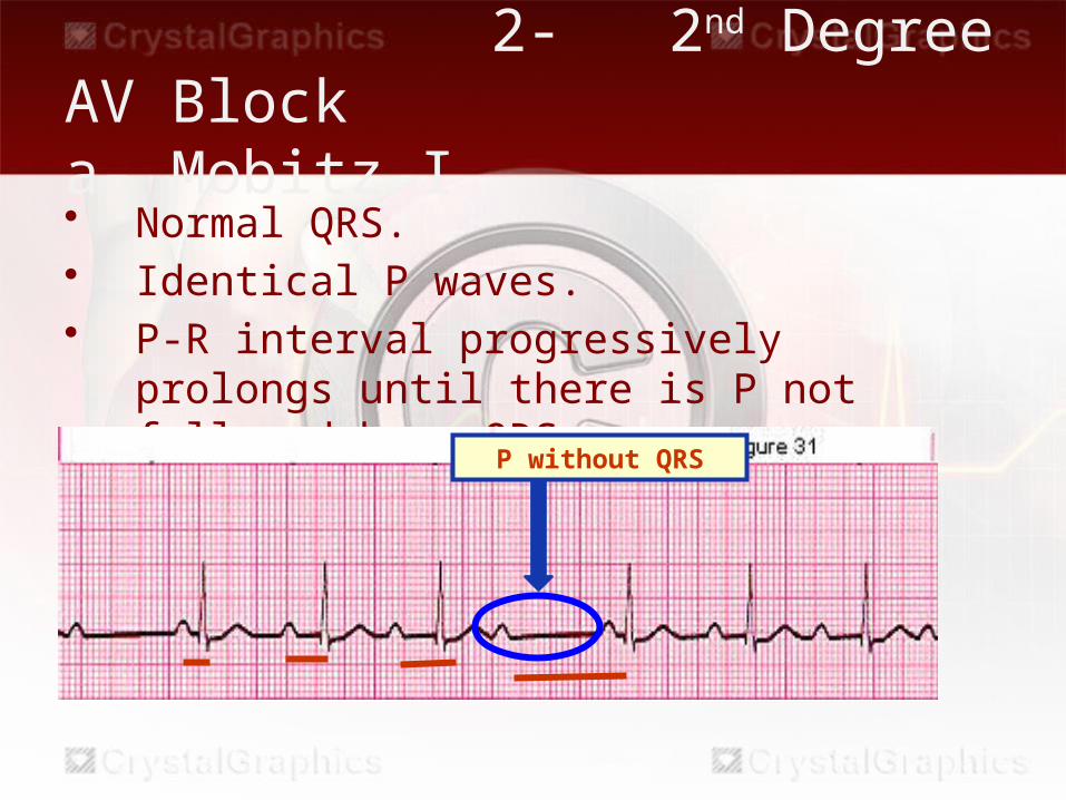
2- 2nd Degree AV Block a- Mobitz I• Normal QRS.• Identical P waves.• P-R interval progressively prolongs until there
is P not followed by a QRSP without QRS
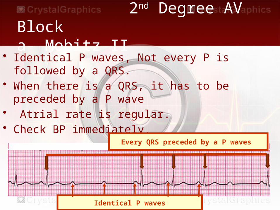
• Identical P waves, Not every P is followed by a QRS.• When there is a QRS, it has to be preceded by a P
wave • Atrial rate is regular.• Check BP immediately.
2nd Degree AV Block a- Mobitz II
Identical P waves
Every QRS preceded by a P waves
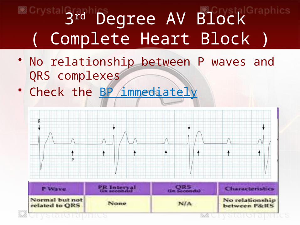
• No relationship between P waves and QRS complexes
• Check the BP immediately
3rd Degree AV Block( Complete Heart Block )
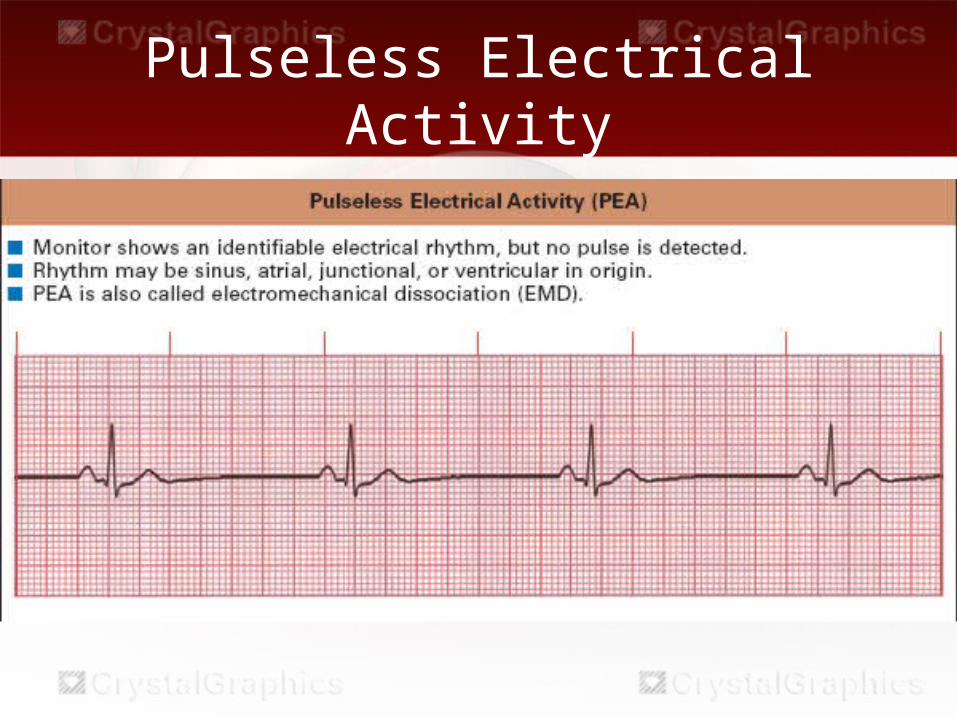
Pulseless Electrical Activity
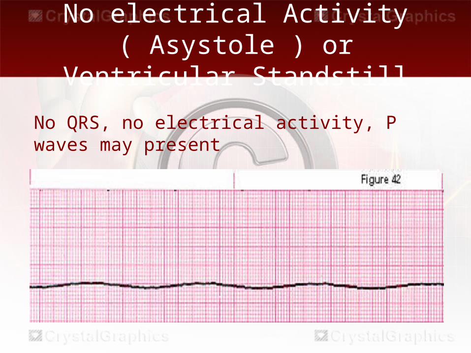
No electrical Activity( Asystole ) or Ventricular Standstill
No QRS, no electrical activity, P waves may present
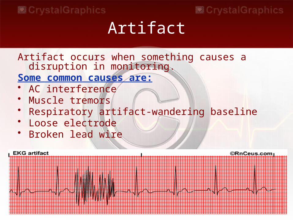
Artifact
Artifact occurs when something causes a disruption in monitoring.
Some common causes are:• AC interference • Muscle tremors • Respiratory artifact-wandering baseline • Loose electrode • Broken lead wire

Instant Feedback
1.Sinus tachycardia is a normal response to pain.
TrueFalse
2. Sinus bradycardia is always abnormal and must be treated.TrueFalse
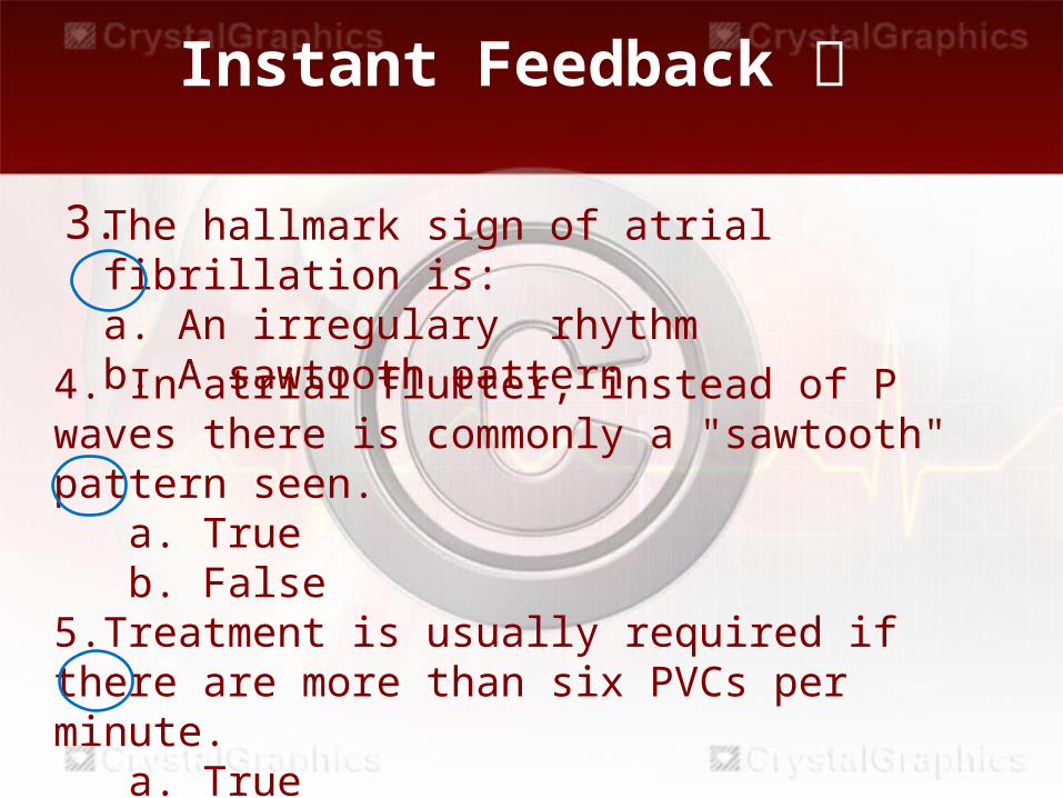
3.
Instant Feedback
The hallmark sign of atrial fibrillation is: a. An irregulary rhythmb. A sawtooth pattern
4. In atrial flutter, instead of P waves there is commonly a "sawtooth" pattern seen. a. True b. False5.Treatment is usually required if there are more than six PVCs per minute. a. True b. False
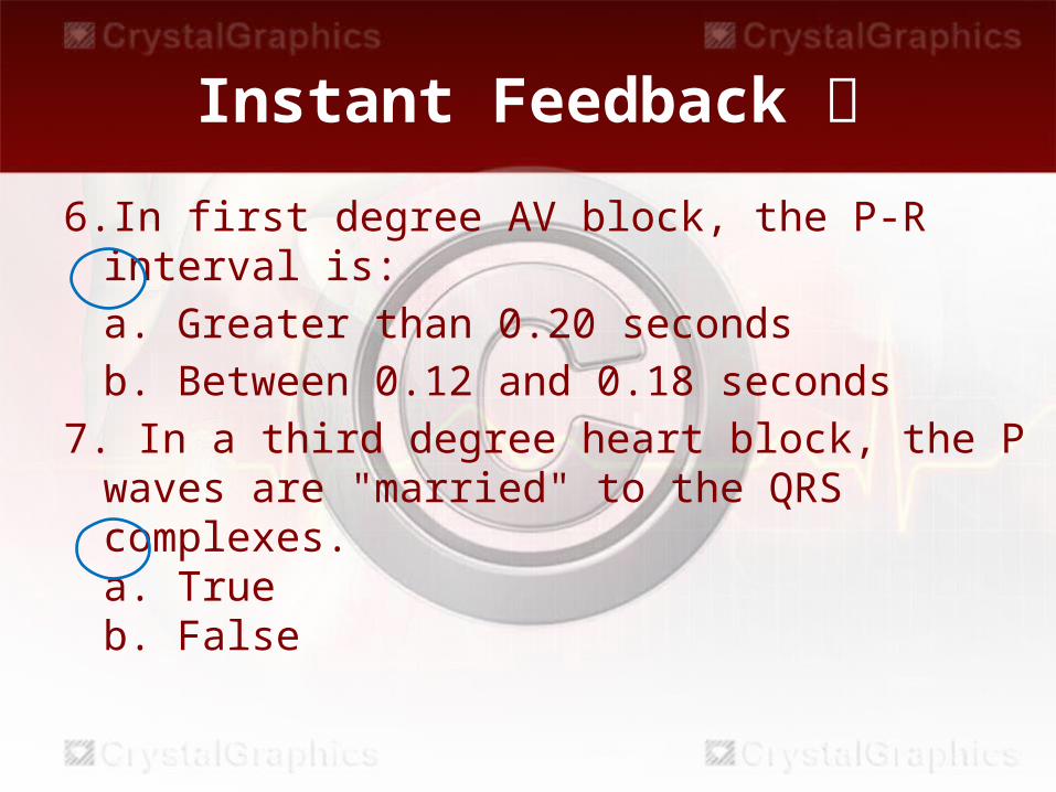
Instant Feedback 6.In first degree AV block, the P-R interval is:
a. Greater than 0.20 secondsb. Between 0.12 and 0.18 seconds
7. In a third degree heart block, the P waves are "married" to the QRS complexes.a. Trueb. False
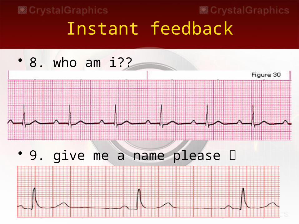
Instant feedback
• 8. who am i??
• 9. give me a name please
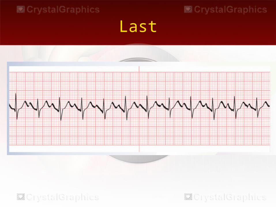
Last