Embed Size (px)
Citation preview
Symptoms, Sign and Identification of disease
Course- B.Sc. (Agri.)
Subject- Principles of Plant Pathology
Unit-2

Symptom and sign
Symptom - are the expression of the disease caused by the
manifestation of the physiological reaction of the plant due to
harmful activity of the pathogen
Sign-Physical evidence of the presence of disease agent (e.g.,
mold or fungel spores, bacterial ooze)
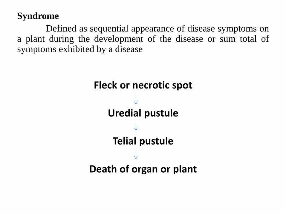
Syndrome
Defined as sequential appearance of disease symptoms ona plant during the development of the disease or sum total ofsymptoms exhibited by a disease
Fleck or necrotic spot
Uredial pustule
Telial pustule
Death of organ or plant
Local infection:
An infection affecting a limited part of a plant e.g. leaf spot.
Systemic infection:
infection that spread point of infection to different parts of the
plants e.g. wilts, virus infection, loose smut
Lesion
A localized necrotic or chlorotic areas of diseased tissue/ organ.
Local lesion:
A localized spot produced on a leaf upon mechanical inoculation
with a virus.
Types of symptoms
Morphological symptoms
Histological symptoms
Morphological symptoms
Morphological: (Externally detectable symptoms caused by any
pathogen e.g. blight, leaf spot
– Necrosis
– Hypoplasia
– Hyperplasia & Hypertrophy
Necrosis degeneration of protoplast followed by death of the
tissue or organ or plant
Plesionecrosis (Nearly dead): necrotic symptoms
expressed before the death of the protoplast are calledplesionecrosis.
E.g. yellowing, hydrosis, wilting
Holonecrosis: necrotic symptoms expressed after the death ofthe protoplast are called holonecrosis. In this the affected tissueturns brown in colour
E.g. Rots, spots, blights
Plesionecrotic symptoms
• Yellowing
• Wilting
• Hydrosis
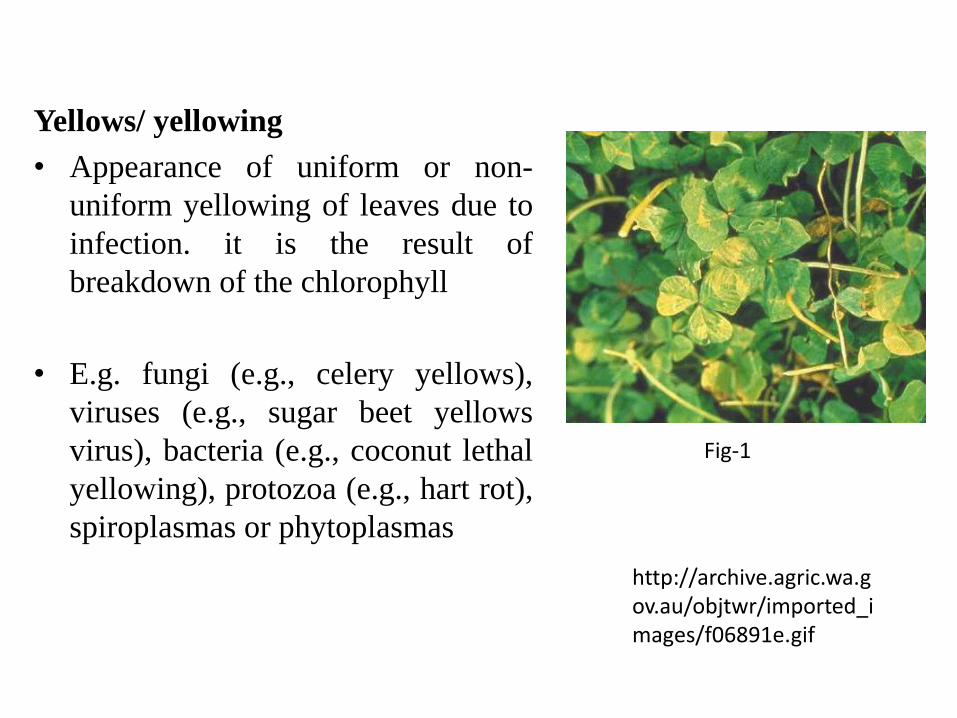
Yellows/ yellowing
• Appearance of uniform or non-
uniform yellowing of leaves due to
infection. it is the result of
breakdown of the chlorophyll
• E.g. fungi (e.g., celery yellows),
viruses (e.g., sugar beet yellows
virus), bacteria (e.g., coconut lethal
yellowing), protozoa (e.g., hart rot),
spiroplasmas or phytoplasmas
http://archive.agric.wa.gov.au/objtwr/imported_images/f06891e.gif
Fig-1
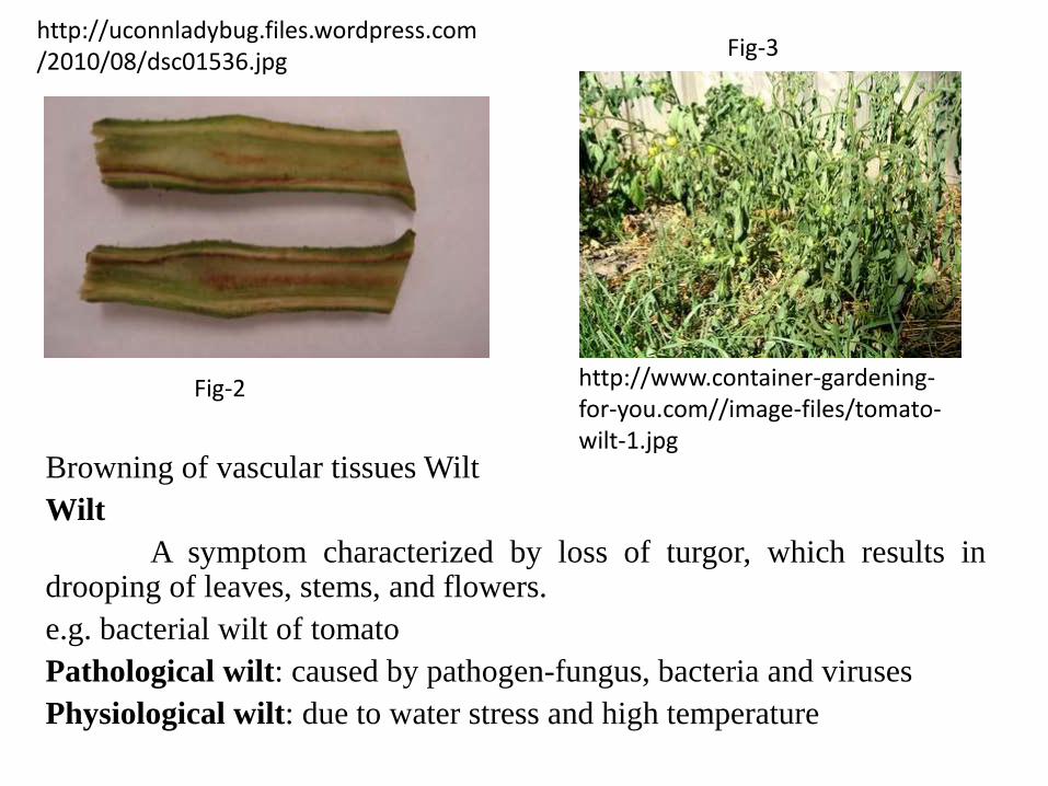
Browning of vascular tissues Wilt
Wilt
A symptom characterized by loss of turgor, which results indrooping of leaves, stems, and flowers.
e.g. bacterial wilt of tomato
Pathological wilt: caused by pathogen-fungus, bacteria and viruses
Physiological wilt: due to water stress and high temperature
http://www.container-gardening-for-you.com//image-files/tomato-wilt-1.jpg
http://uconnladybug.files.wordpress.com/2010/08/dsc01536.jpg
Fig-2
Fig-3
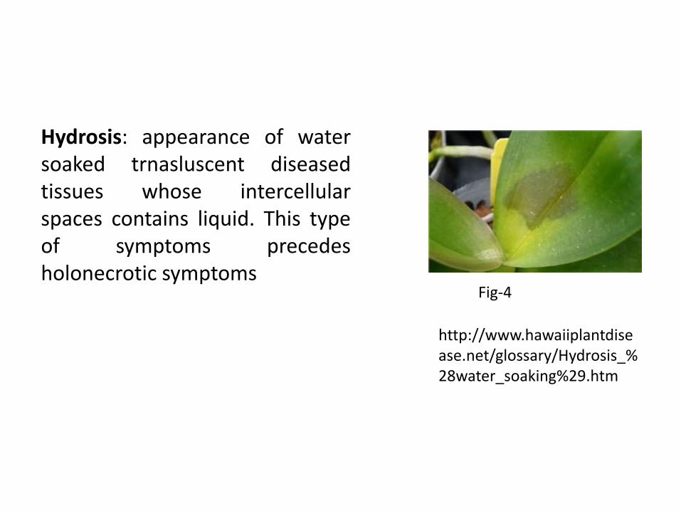
Hydrosis: appearance of watersoaked trnasluscent diseasedtissues whose intercellularspaces contains liquid. This typeof symptoms precedesholonecrotic symptoms
http://www.hawaiiplantdisease.net/glossary/Hydrosis_%28water_soaking%29.htm
Fig-4
Holonecrotic symptoms
May develop on any part of the plant and generally the infected
tissues turns brown.
Holonecrotic symptoms can be divided into three categories
– Necrosis of the green plant parts
– Necrosis of the storage organs
– Necrosis of woody tissues
Necrosis of the storage organs
– Rots
– Leak:
– Mummification:
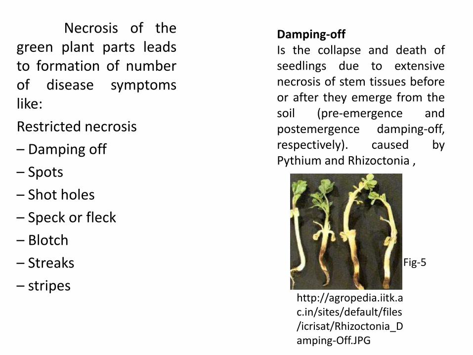
Necrosis of thegreen plant parts leadsto formation of numberof disease symptomslike:
Restricted necrosis
– Damping off
– Spots
– Shot holes
– Speck or fleck
– Blotch
– Streaks
– stripes
Damping-offIs the collapse and death ofseedlings due to extensivenecrosis of stem tissues beforeor after they emerge from thesoil (pre-emergence andpostemergence damping-off,respectively). caused byPythium and Rhizoctonia ,
http://agropedia.iitk.ac.in/sites/default/files/icrisat/Rhizoctonia_Damping-Off.JPG
Fig-5
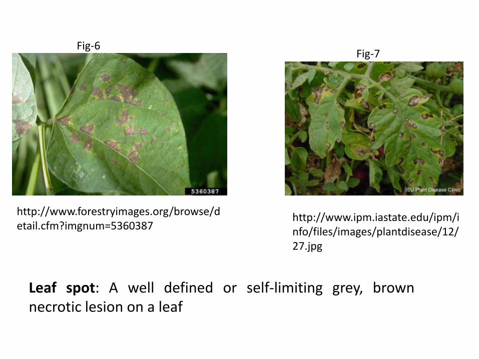
Leaf spot: A well defined or self-limiting grey, brownnecrotic lesion on a leaf
http://www.forestryimages.org/browse/detail.cfm?imgnum=5360387
http://www.ipm.iastate.edu/ipm/info/files/images/plantdisease/12/27.jpg
Fig-6Fig-7
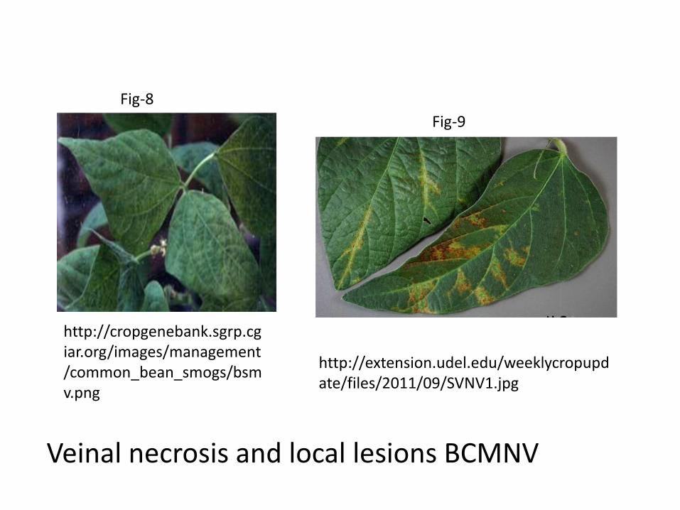
Veinal necrosis and local lesions BCMNV
http://cropgenebank.sgrp.cgiar.org/images/management/common_bean_smogs/bsmv.png
http://extension.udel.edu/weeklycropupdate/files/2011/09/SVNV1.jpg
Fig-8
Fig-9
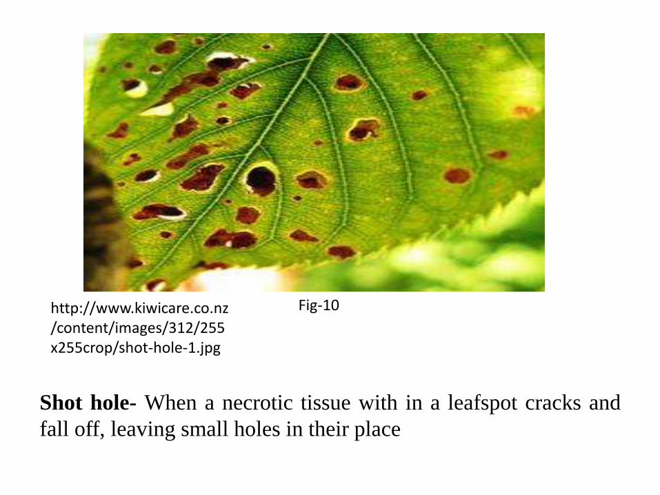
Shot hole- When a necrotic tissue with in a leafspot cracks and
fall off, leaving small holes in their place
http://www.kiwicare.co.nz/content/images/312/255x255crop/shot-hole-1.jpg
Fig-10
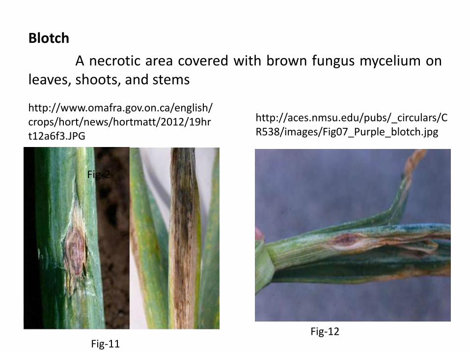
Blotch
A necrotic area covered with brown fungus mycelium onleaves, shoots, and stems
http://www.omafra.gov.on.ca/english/crops/hort/news/hortmatt/2012/19hrt12a6f3.JPG
http://aces.nmsu.edu/pubs/_circulars/CR538/images/Fig07_Purple_blotch.jpg
Fig-2
Fig-11Fig-12
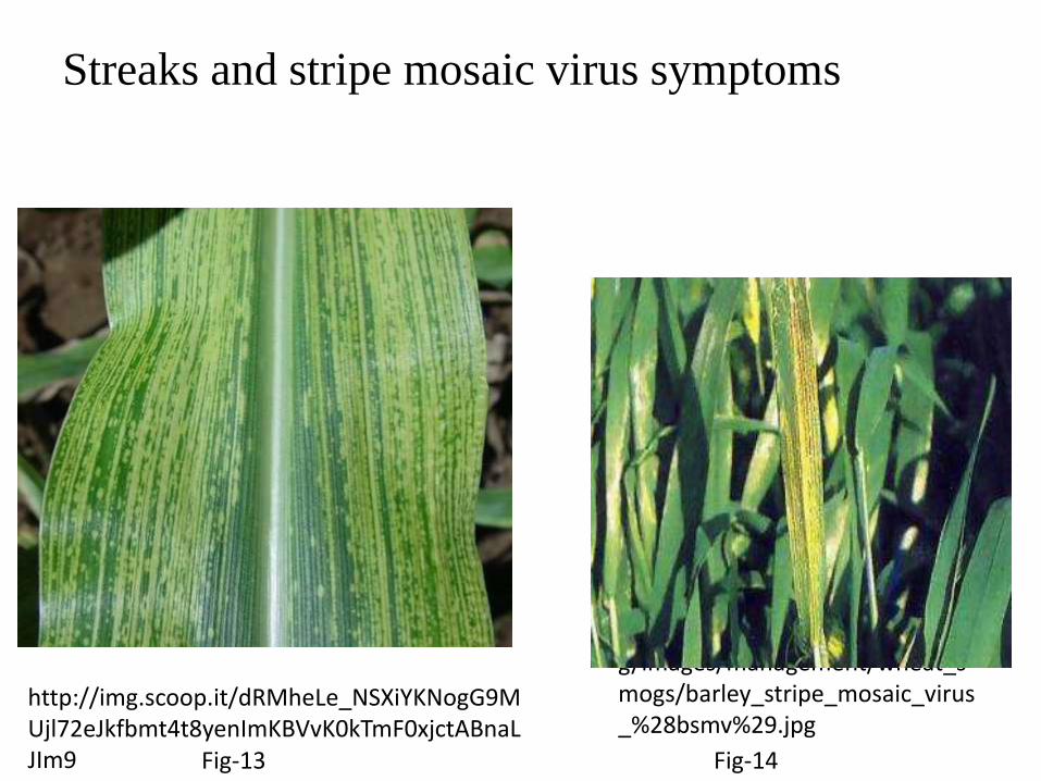
Streaks and stripe mosaic virus symptoms
http://img.scoop.it/dRMheLe_NSXiYKNogG9MUjl72eJkfbmt4t8yenImKBVvK0kTmF0xjctABnaLJIm9
http://cropgenebank.sgrp.cgiar.org/images/management/wheat_smogs/barley_stripe_mosaic_virus_%28bsmv%29.jpg
Fig-13 Fig-14
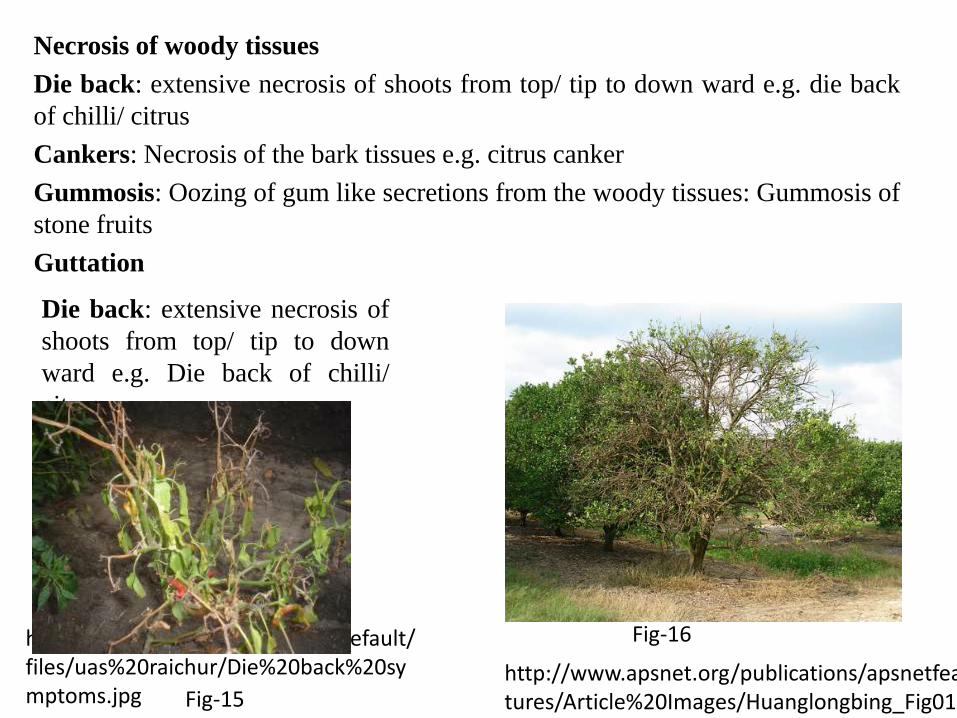
Necrosis of woody tissues
Die back: extensive necrosis of shoots from top/ tip to down ward e.g. die back
of chilli/ citrus
Cankers: Necrosis of the bark tissues e.g. citrus canker
Gummosis: Oozing of gum like secretions from the woody tissues: Gummosis of
stone fruits
Guttation
http://www.apsnet.org/publications/apsnetfeatures/Article%20Images/Huanglongbing_Fig01.jpg
Die back: extensive necrosis of
shoots from top/ tip to down
ward e.g. Die back of chilli/
citrus
http://agropedia.iitk.ac.in/sites/default/files/uas%20raichur/Die%20back%20symptoms.jpg Fig-15
Fig-16
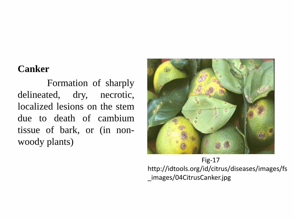
Canker
Formation of sharply
delineated, dry, necrotic,
localized lesions on the stem
due to death of cambium
tissue of bark, or (in non-
woody plants)
http://idtools.org/id/citrus/diseases/images/fs_images/04CitrusCanker.jpg
Fig-17
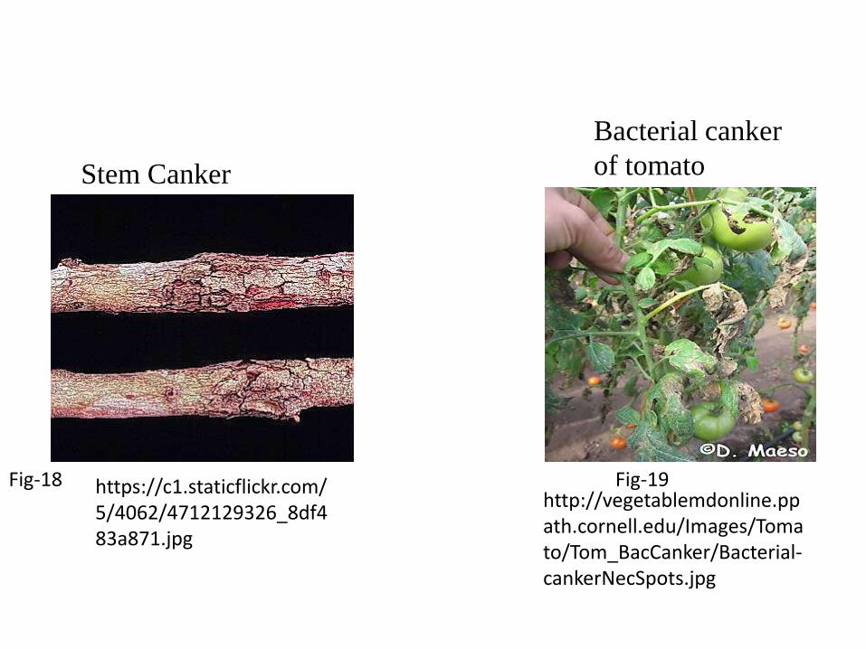
https://c1.staticflickr.com/5/4062/4712129326_8df483a871.jpg
Stem Canker
http://vegetablemdonline.ppath.cornell.edu/Images/Tomato/Tom_BacCanker/Bacterial-cankerNecSpots.jpg
Bacterial canker
of tomato
Fig-19Fig-18
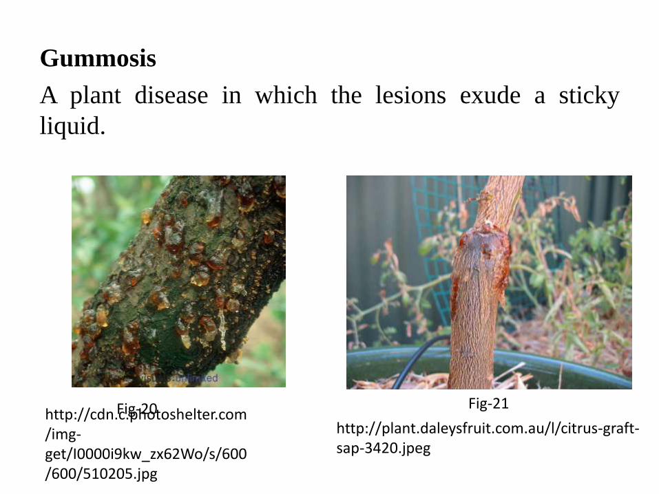
Gummosis
A plant disease in which the lesions exude a sticky
liquid.
http://cdn.c.photoshelter.com/img-get/I0000i9kw_zx62Wo/s/600/600/510205.jpg
http://plant.daleysfruit.com.au/l/citrus-graft-sap-3420.jpeg
Fig-20 Fig-21
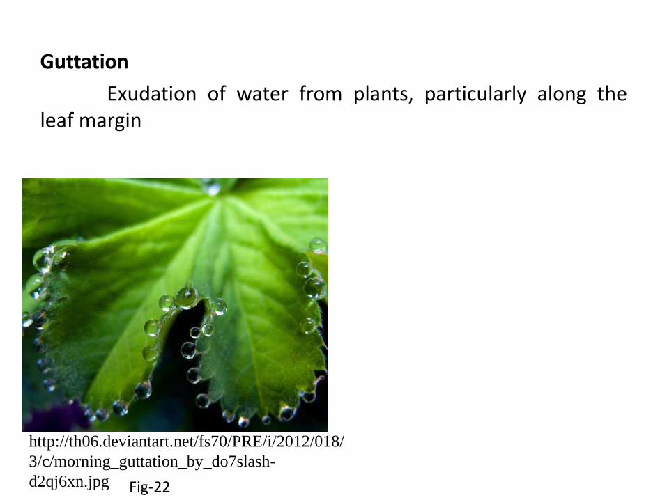
Guttation
Exudation of water from plants, particularly along theleaf margin
http://th06.deviantart.net/fs70/PRE/i/2012/018/
3/c/morning_guttation_by_do7slash-
d2qj6xn.jpg Fig-22
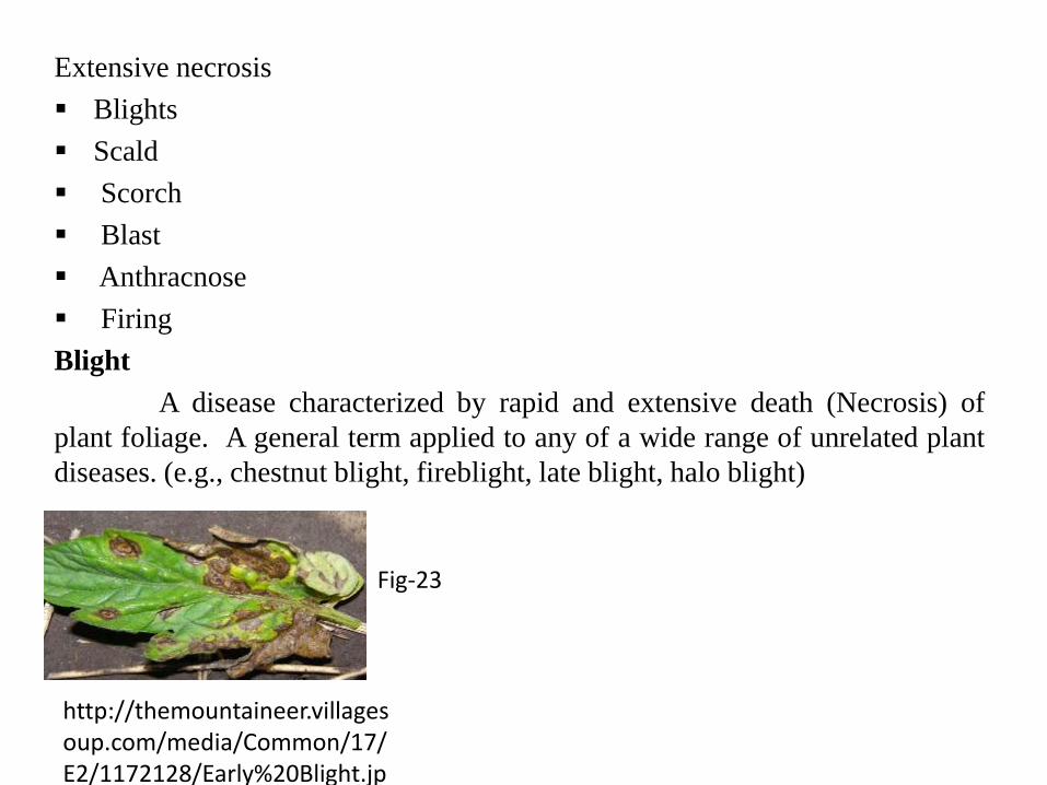
Extensive necrosis
Blights
Scald
Scorch
Blast
Anthracnose
Firing
Blight
A disease characterized by rapid and extensive death (Necrosis) of
plant foliage. A general term applied to any of a wide range of unrelated plant
diseases. (e.g., chestnut blight, fireblight, late blight, halo blight)
http://themountaineer.villagesoup.com/media/Common/17/E2/1172128/Early%20Blight.jpg
Fig-23
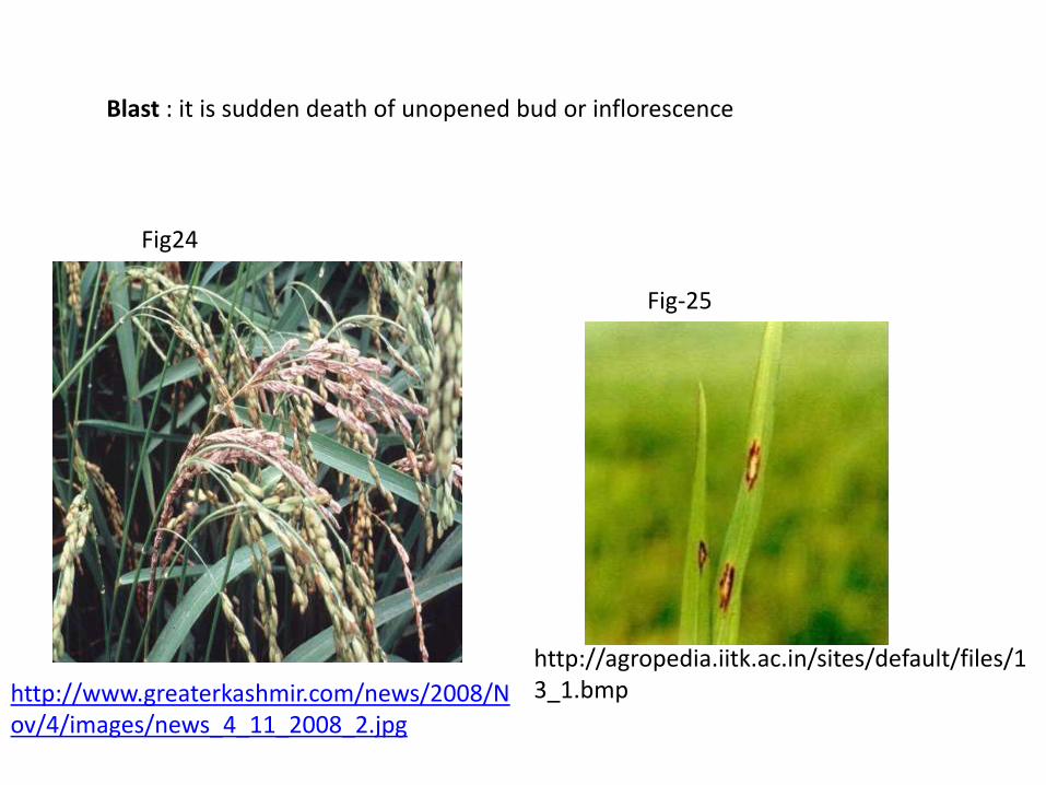
Blast : it is sudden death of unopened bud or inflorescence
http://agropedia.iitk.ac.in/sites/default/files/13_1.bmphttp://www.greaterkashmir.com/news/2008/N
ov/4/images/news_4_11_2008_2.jpg
Fig24
Fig-25
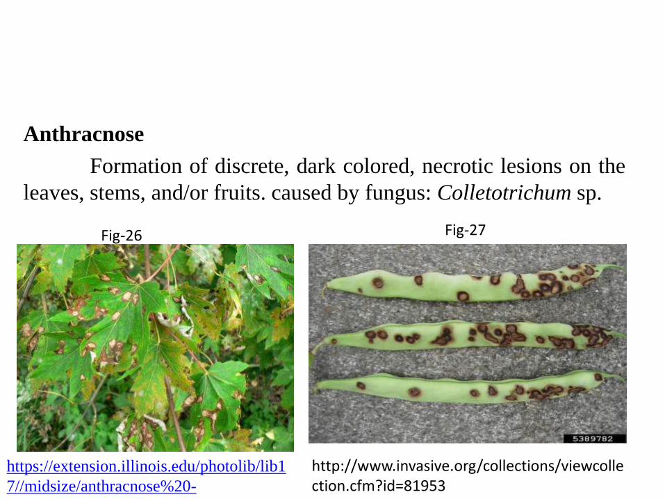
Anthracnose
Formation of discrete, dark colored, necrotic lesions on the
leaves, stems, and/or fruits. caused by fungus: Colletotrichum sp.
http://www.invasive.org/collections/viewcollection.cfm?id=81953
https://extension.illinois.edu/photolib/lib1
7//midsize/anthracnose%20-
%20silver%20maple.jpg
Fig-26 Fig-27

Necrosis of the storage organs
• Rots
• Leak
• Mummification
Rot
The softening, discoloration, and often decay or disintegrationof a succulent plant tissue as a result of fungal or bacterialinfection.
http://www7.inra.fr/hyp3/images/6032562.jpgFig-28
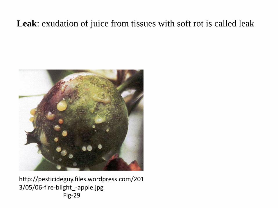
Leak: exudation of juice from tissues with soft rot is called leak
http://pesticideguy.files.wordpress.com/2013/05/06-fire-blight_-apple.jpg
Fig-29
Hyperplastic & Hypertrophic symptoms
Hyperplasia
A plant overgrowth due to increased cell division.
Hypertrophy
A plant overgrowth due to abnormal cell enlargement.
Wound tumors
• Galls
• Witches Broom
• Enations
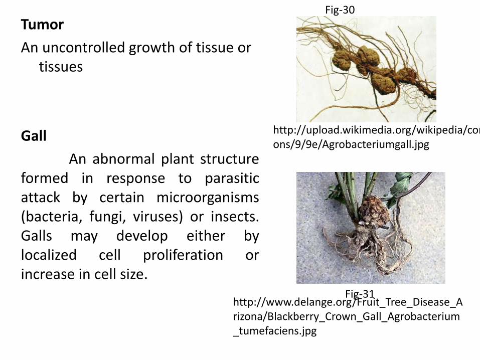
Tumor
An uncontrolled growth of tissue or tissues
Gall
An abnormal plant structureformed in response to parasiticattack by certain microorganisms(bacteria, fungi, viruses) or insects.Galls may develop either bylocalized cell proliferation orincrease in cell size.
http://upload.wikimedia.org/wikipedia/commons/9/9e/Agrobacteriumgall.jpg
http://www.delange.org/Fruit_Tree_Disease_Arizona/Blackberry_Crown_Gall_Agrobacterium_tumefaciens.jpg
Fig-30
Fig-31
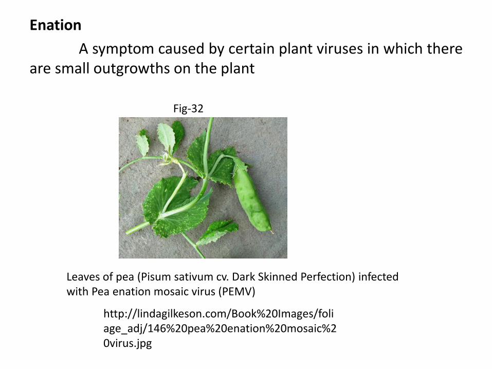
Enation
A symptom caused by certain plant viruses in which thereare small outgrowths on the plant
Leaves of pea (Pisum sativum cv. Dark Skinned Perfection) infectedwith Pea enation mosaic virus (PEMV)
http://lindagilkeson.com/Book%20Images/foliage_adj/146%20pea%20enation%20mosaic%20virus.jpg
Fig-32
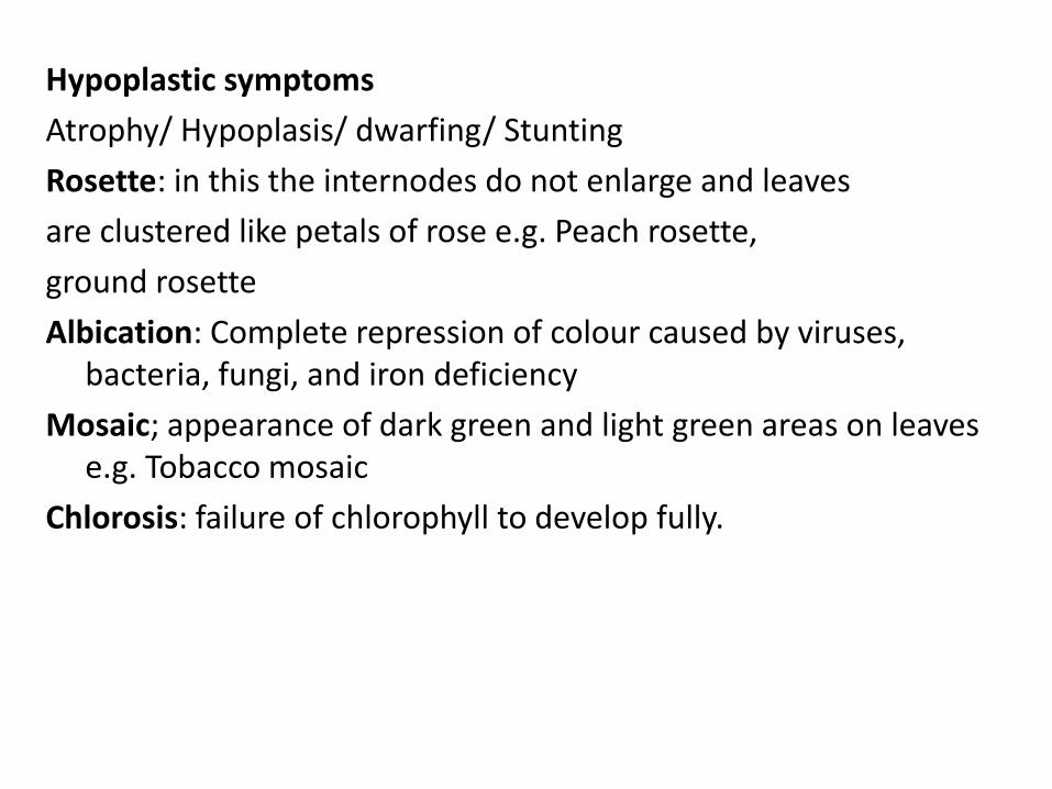
Hypoplastic symptoms
Atrophy/ Hypoplasis/ dwarfing/ Stunting
Rosette: in this the internodes do not enlarge and leaves
are clustered like petals of rose e.g. Peach rosette,
ground rosette
Albication: Complete repression of colour caused by viruses, bacteria, fungi, and iron deficiency
Mosaic; appearance of dark green and light green areas on leaves e.g. Tobacco mosaic
Chlorosis: failure of chlorophyll to develop fully.
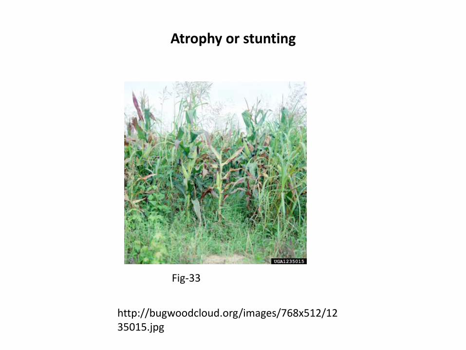
Atrophy or stunting
http://bugwoodcloud.org/images/768x512/1235015.jpg
Fig-33

Chlorosis
The loss of chlorophyll from the tissues of a plant, resulting from
microbial infection,
e.g. viral infection, the action of certain phytotoxins, the lack of
light, to magnesium or iron deficiency, etc.
Chlorotic tissues commonly appear yellowish
MosaicAppearance of dark green, light green pattern or sometimeschlorotic areas on leaves due to virus infection
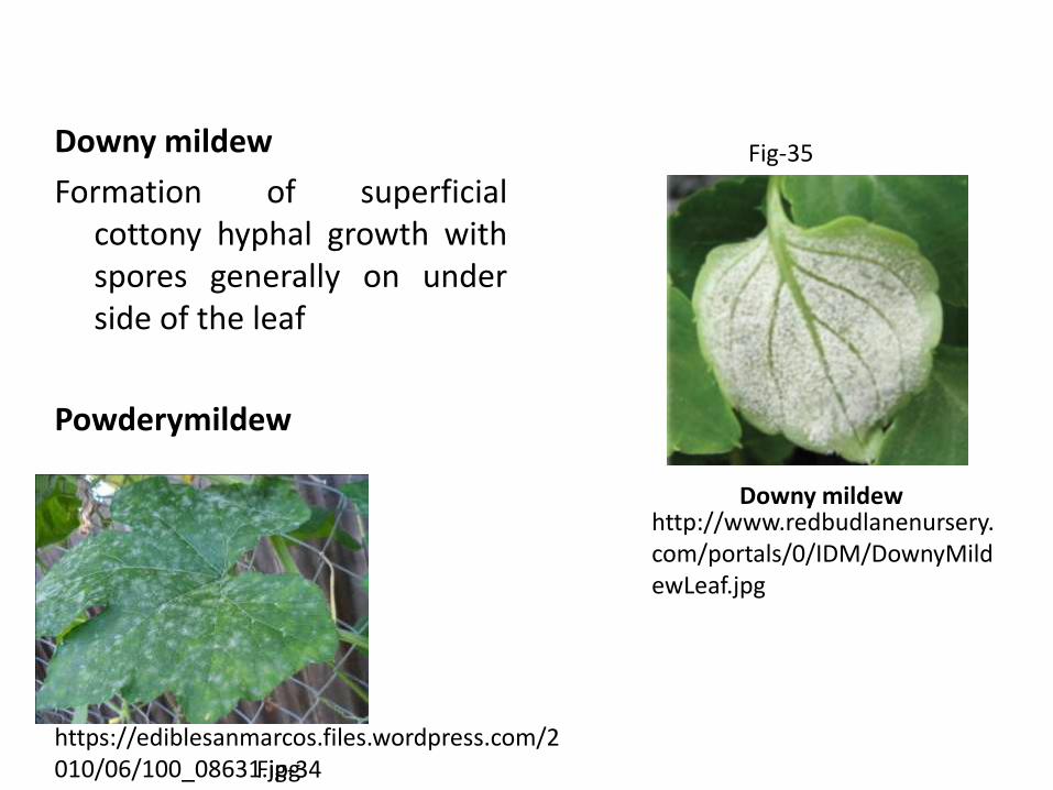
Downy mildew
Formation of superficialcottony hyphal growth withspores generally on underside of the leaf
Powderymildew
http://www.redbudlanenursery.com/portals/0/IDM/DownyMildewLeaf.jpg
Downy mildew
https://ediblesanmarcos.files.wordpress.com/2010/06/100_08631.jpgFig-34
Fig-35
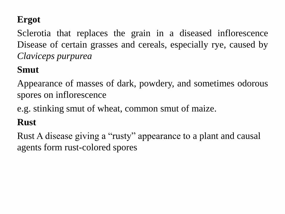
Ergot
Sclerotia that replaces the grain in a diseased inflorescence
Disease of certain grasses and cereals, especially rye, caused by
Claviceps purpurea
Smut
Appearance of masses of dark, powdery, and sometimes odorous
spores on inflorescence
e.g. stinking smut of wheat, common smut of maize.
Rust
Rust A disease giving a “rusty” appearance to a plant and causal
agents form rust-colored spores
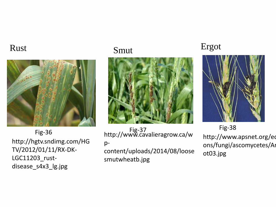
Rust
http://hgtv.sndimg.com/HGTV/2012/01/11/RX-DK-LGC11203_rust-disease_s4x3_lg.jpg
ErgotSmut
http://www.apsnet.org/edcenter/intropp/lessons/fungi/ascomycetes/Article%20Images/ergot03.jpg
Fig-36Fig-38Fig-37
http://www.cavalieragrow.ca/wp-content/uploads/2014/08/loosesmutwheatb.jpg

Refererances
Books
Principles of Plant Pathology by R.S. Singh
Web Resources
www.tnau.com
Image Resources
Fig1- http://archive.agric.wa.gov.au/objtwr/imported_images/f06891e.gif
Fig2- http://uconnladybug.files.wordpress.com/2010/08/dsc01536.jpg
Fig3 http://www.container-gardening-for-you.com//image-files/tomato-wilt-1.jpg
Fig 4- http://www.hawaiiplantdisease.net/glossary/Hydrosis_%28water_soaking%29.htm
Fig 5- http://agropedia.iitk.ac.in/sites/default/files/icrisat/Rhizoctonia_Damping-Off.JPG
Fig 6- http://www.forestryimages.org/browse/detail.cfm?imgnum=5360387
Fig 7- http://www.ipm.iastate.edu/ipm/info/files/images/plantdisease/12/27.jpg
Fig 8-http://cropgenebank.sgrp.cgiar.org/images/management/common_bean_smogs/bsmv.png
Fig 9- http://extension.udel.edu/weeklycropupdate/files/2011/09/SVNV1.jpg
Fig 10- http://www.kiwicare.co.nz/content/images/312/255x255crop/shot-hole-1.jpg
Fig 11- http://www.omafra.gov.on.ca/english/crops/hort/news/hortmatt/2012/19hrt12a6f3.JPG
Fig 12- http://aces.nmsu.edu/pubs/_circulars/CR538/images/Fig07_Purple_blotch.jpg

Fig 13-http://img.scoop.it/dRMheLe_NSXiYKNogG9MUjl72eJkfbmt4t8yenImKBVvK0kTmF0xjctABnaLJIm9
Fig 14-http://cropgenebank.sgrp.cgiar.org/images/management/wheat_smogs/barley_stripe_mosaic_virus_%28bsmv%29.jpg
Fig 15-http://agropedia.iitk.ac.in/sites/default/files/uas%20raichur/Die%20back%20symptoms.jpg
Fig 16- http://www.apsnet.org/publications/apsnetfeatures/Article%20Images/Huanglongbing_Fig01.jpg
Fig 17- http://idtools.org/id/citrus/diseases/images/fs_images/04CitrusCanker.jpg
Fig 18- https://c1.staticflickr.com/5/4062/4712129326_8df483a871.jpg
Fig 19- http://vegetablemdonline.ppath.cornell.edu/Images/Tomato/Tom_BacCanker/Bacterial-cankerNecSpots.jpg
Fig 20- http://cdn.c.photoshelter.com/img-get/I0000i9kw_zx62Wo/s/600/600/510205.jpg
Fig 21- http://plant.daleysfruit.com.au/l/citrus-graft-sap-3420.jpeg
Fig 22- http://th06.deviantart.net/fs70/PRE/i/2012/018/3/c/morning_guttation_by_do7slash-d2qj6xn.jpg
Fig 23- http://themountaineer.villagesoup.com/media/Common/17/E2/1172128/Early%20Blight.jpg
Fig 24- http://www.greaterkashmir.com/news/2008/Nov/4/images/news_4_11_2008_2.jpg
Fig 25- http://agropedia.iitk.ac.in/sites/default/files/13_1.bmp
Fig 26- https://extension.illinois.edu/photolib/lib17//midsize/anthracnose%20-%20silver%20maple.jpg
Fig 27- http://www.invasive.org/collections/viewcollection.cfm?id=81953
Fig 28- http://www7.inra.fr/hyp3/images/6032562.jpgFig 29- http://pesticideguy.files.wordpress.com/2013/05/06-fire-blight_-apple.jpgFig 30- http://upload.wikimedia.org/wikipedia/commons/9/9e/Agrobacteriumgall.jpgFig 31-
http://www.delange.org/Fruit_Tree_Disease_Arizona/Blackberry_Crown_Gall_Agrobacterium_tumefaciens.jpg
Fig 32-http://lindagilkeson.com/Book%20Images/foliage_adj/146%20pea%20enation%20mosaic%20virus.jpg
Fig 33- http://bugwoodcloud.org/images/768x512/1235015.jpgFig 34- https://ediblesanmarcos.files.wordpress.com/2010/06/100_08631.jpgFig 35- http://www.redbudlanenursery.com/portals/0/IDM/DownyMildewLeaf.jpgFig 36-http://hgtv.sndimg.com/HGTV/2012/01/11/RX-DK-LGC11203_rust-
disease_s4x3_lg.jpgFig 37- http://www.cavalieragrow.ca/wp
content/uploads/2014/08/loosesmutwheatb.jpg
Fig 38- http://www.cavalieragrow.ca/wp-content/uploads/2014/08/loosesmutwheatb.jpg


![Plant Pathology & Microbiology...ft.) [27]. Pierce’s disease symptoms were observed two months post inoculation. The symptoms were rated on a scale from 0 to 5 as described previously](https://img.pdfslide.net/doc/110x75/5f9431f772f64a7fe439a5cb/plant-pathology-microbiology-ft-27-pierceas-disease-symptoms-were.jpg)