Embed Size (px)
Citation preview

CONCEPTS OF BIOLOGYChapter 3 CELL STRUCTURE AND FUNCTION
PowerPoint Image Slideshow
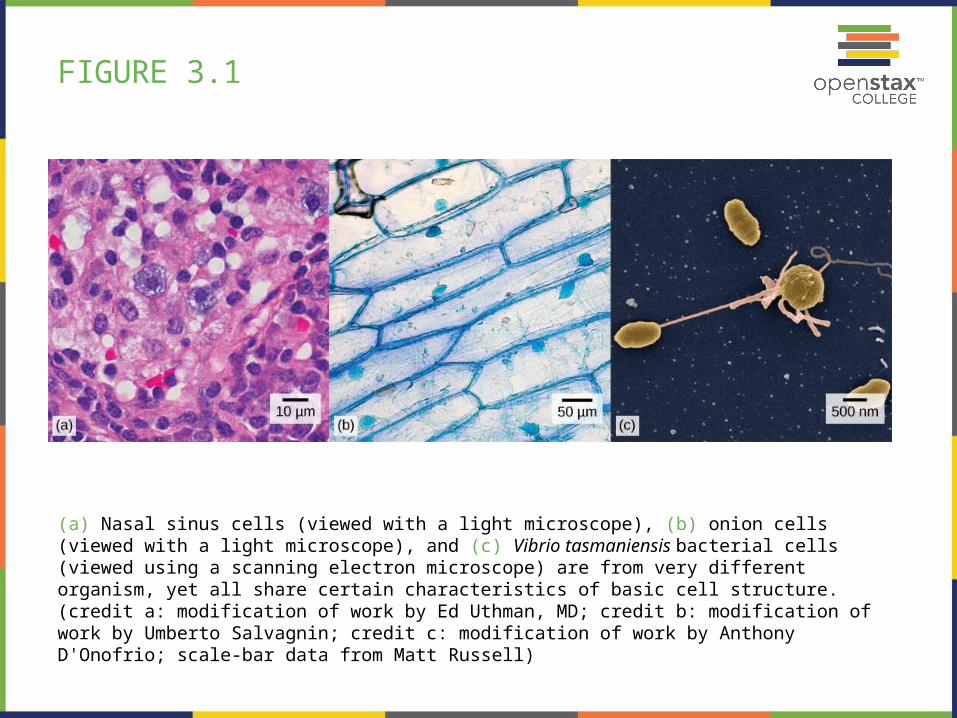
FIGURE 3.1
(a) Nasal sinus cells (viewed with a light microscope), (b) onion cells (viewed with a light microscope), and (c) Vibrio tasmaniensis bacterial cells (viewed using a scanning electron microscope) are from very different organism, yet all share certain characteristics of basic cell structure. (credit a: modification of work by Ed Uthman, MD; credit b: modification of work by Umberto Salvagnin; credit c: modification of work by Anthony D'Onofrio; scale-bar data from Matt Russell)
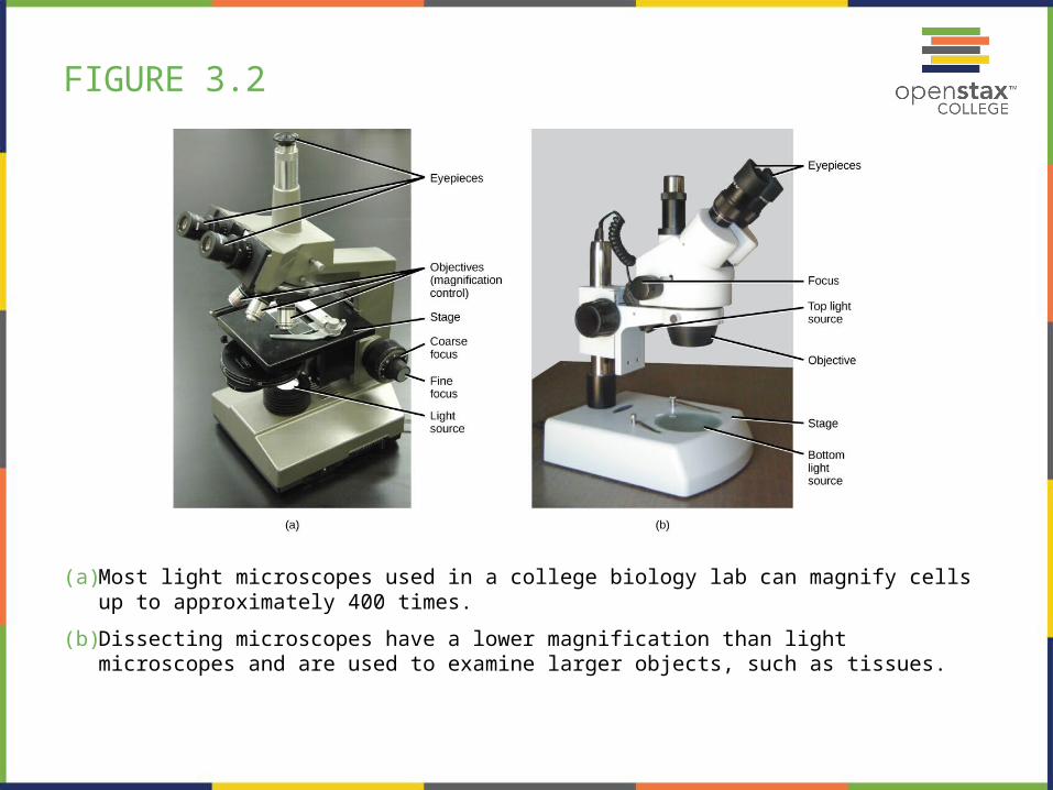
FIGURE 3.2
(a) Most light microscopes used in a college biology lab can magnify cells up to approximately 400 times.
(b) Dissecting microscopes have a lower magnification than light microscopes and are used to examine larger objects, such as tissues.
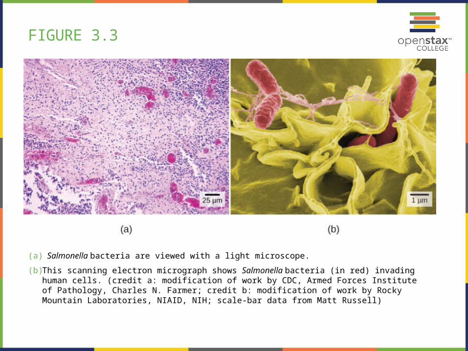
FIGURE 3.3
(a) Salmonella bacteria are viewed with a light microscope.
(b) This scanning electron micrograph shows Salmonella bacteria (in red) invading human cells. (credit a: modification of work by CDC, Armed Forces Institute of Pathology, Charles N. Farmer; credit b: modification of work by Rocky Mountain Laboratories, NIAID, NIH; scale-bar data from Matt Russell)

FIGURE 3.4
These uterine cervix cells, viewed through a light microscope, were obtained from a Pap smear. Normal cells are on the left. The cells on the right are infected with human papillomavirus. (credit: modification of work by Ed Uthman; scale-bar data from Matt Russell)

FIGURE 3.5
This figure shows the generalized structure of a prokaryotic cell.
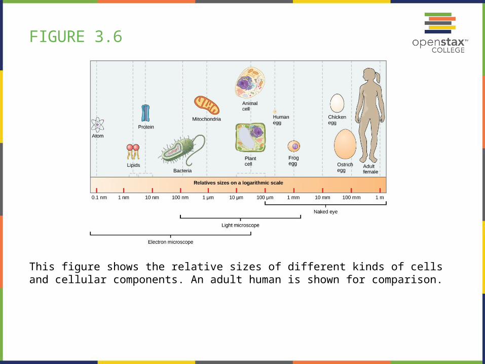
FIGURE 3.6
This figure shows the relative sizes of different kinds of cells and cellular components. An adult human is shown for comparison.

FIGURE 3.7
This figure shows (a) a typical animal cell and (b) a typical plant cell.
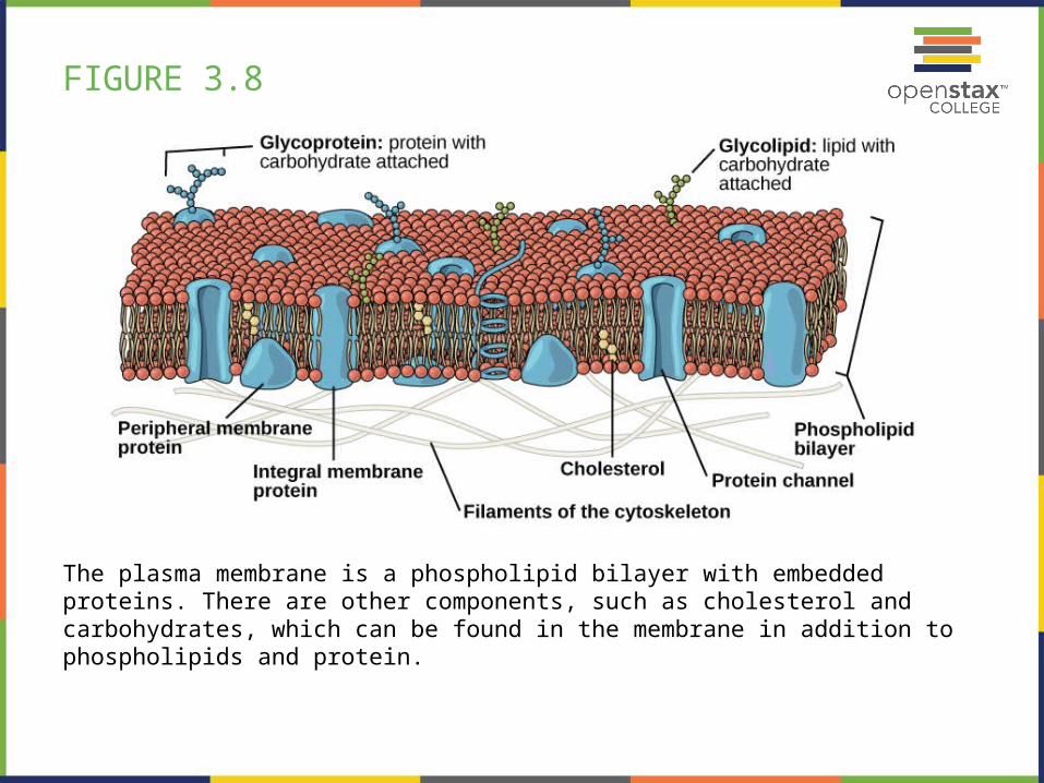
FIGURE 3.8
The plasma membrane is a phospholipid bilayer with embedded proteins. There are other components, such as cholesterol and carbohydrates, which can be found in the membrane in addition to phospholipids and protein.
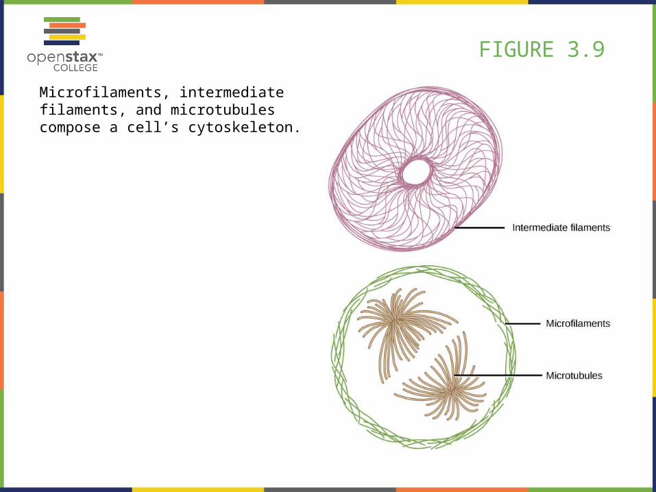
FIGURE 3.9
Microfilaments, intermediate filaments, and microtubules compose a cell’s cytoskeleton.

FIGURE 3.10
The outermost boundary of the nucleus is the nuclear envelope. Notice that the nuclear envelope consists of two phospholipid bilayers (membranes)—an outer membrane and an inner membrane—in contrast to the plasma membrane (Figure 3.8), which consists of only one phospholipid bilayer. (credit: modification of work by NIGMS, NIH)

FIGURE 3.11
The Golgi apparatus in this transmission electron micrograph of a white blood cell is visible as a stack of semicircular flattened rings in the lower portion of this image. Several vesicles can be seen near the Golgi apparatus. (credit: modification of work by Louisa Howard; scale-bar data from Matt Russell)

FIGURE 3.12
A macrophage has phagocytized a potentially pathogenic bacterium into a vesicle, which then fuses with a lysosome within the cell so that the pathogen can be destroyed. Other organelles are present in the cell, but for simplicity, are not shown.

FIGURE 3.13
The endomembrane system works to modify, package, and transport lipids and proteins. (credit: modification of work by Magnus Manske)

FIGURE 3.14
This transmission electron micrograph shows a mitochondrion as viewed with an electron microscope. Notice the inner and outer membranes, the cristae, and the mitochondrial matrix. (credit: modification of work by Matthew Britton; scale-bar data from Matt Russell)

FIGURE 3.15
This simplified diagram of a chloroplast shows the outer membrane, inner membrane, thylakoids, grana, and stroma.
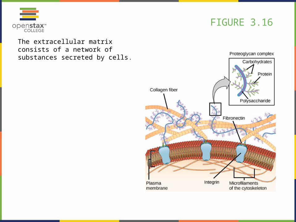
FIGURE 3.16
The extracellular matrix consists of a network of substances secreted by cells.
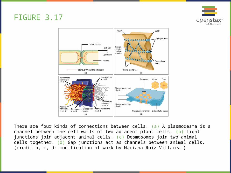
FIGURE 3.17
There are four kinds of connections between cells. (a) A plasmodesma is a channel between the cell walls of two adjacent plant cells. (b) Tight junctions join adjacent animal cells. (c) Desmosomes join two animal cells together. (d) Gap junctions act as channels between animal cells. (credit b, c, d: modification of work by Mariana Ruiz Villareal)

FIGURE 3.18
The fluid mosaic model of the plasma membrane structure describes the plasma membrane as a fluid combination of phospholipids, cholesterol, proteins, and carbohydrates.

FIGURE 3.19
HIV docks at and binds to the CD4 receptor, a glycoprotein on the surface of T cells, before entering, or infecting, the cell. (credit: modification of work by US National Institutes of Health/National Institute of Allergy and Infectious Diseases)

FIGURE 3.20
Diffusion through a permeable membrane follows the concentration gradient of a substance, moving the substance from an area of high concentration to one of low concentration. (credit: modification of work by Mariana Ruiz Villarreal)
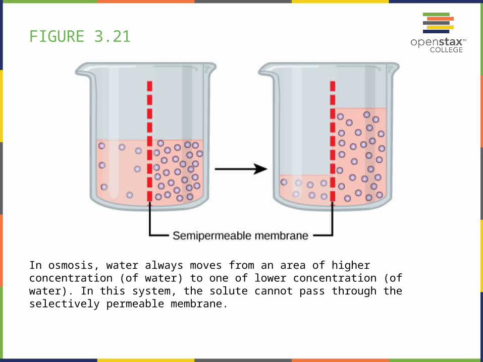
FIGURE 3.21
In osmosis, water always moves from an area of higher concentration (of water) to one of lower concentration (of water). In this system, the solute cannot pass through the selectively permeable membrane.

FIGURE 3.22
Osmotic pressure changes the shape of red blood cells in hypertonic, isotonic, and hypotonic solutions. (credit: modification of work by Mariana Ruiz Villarreal)

FIGURE 3.23
The turgor pressure within a plant cell depends on the tonicity of the solution that it is bathed in. (credit: modification of work by Mariana Ruiz Villarreal)

FIGURE 3.24
Electrochemical gradients arise from the combined effects of concentration gradients and electrical gradients. (credit: modification of work by “Synaptitude”/Wikimedia Commons)

FIGURE 3.25
The sodium-potassium pump move potassium and sodium ions across the plasma membrane. (credit: modification of work by Mariana Ruiz Villarreal)

FIGURE 3.26
Three variations of endocytosis are shown.
(a) In one form of endocytosis, phagocytosis, the cell membrane surrounds the particle and pinches off to form an intracellular vacuole.
(b) In another type of endocytosis, pinocytosis, the cell membrane surrounds a small volume of fluid and pinches off, forming a vesicle.
(c) In receptor-mediated endocytosis, uptake of substances by the cell is targeted to a single type of substance that binds at the receptor on the external cell membrane. (credit: modification of work by Mariana Ruiz Villarreal)

FIGURE 3.27
In exocytosis, a vesicle migrates to the plasma membrane, binds, and releases its contents to the outside of the cell. (credit: modification of work by Mariana Ruiz Villarreal)

This PowerPoint file is copyright 2011-2013, Rice University. All Rights Reserved.