Embed Size (px)
Citation preview

1
2

3
4
• The Digestive System or The Digestive Tract. this is like along tube some nine meters in total, through the middle of the body.
• It starts at the Mouth, where food and drink enter the body, and finishes at the Anus, where food and wastes leave the body.

5
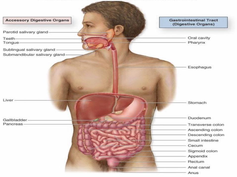
• The main parts of the digestive system :
(Buccal Cavity) (Mouth) - Oesophagus -
Stomach - Small Intestine - (Duodenum + Jejunum + ileum) - Large Intestine ( Caecum + Colon + Rectum ) - Anus.
And the accessory glands : Salivary Glands, Liver and Pancreas.
6

7
• your digestive system is uniquely constructed to perform its specialized function of turning food into the energy you need to survive and packaging the residue for waste disposal.
• To help you understand how the many parts of the digestive system work together, here is an overview of the structure and function of this complex system : -

8
Mouth• The mouth is the beginning of the
digestive tract ; and, in fact, digestion starts here when taking the first bite of food. Chewing breaks the food into pieces that are more easily digested, while saliva mixes with food to begin the process of breaking it down into a form your body can absorb and use.

9

10
What & Where are the Salivary Glands
which help in digestion of the carbohydrates ?

11
• They're glands which found in and around your mouth and throat.
The three major pairs of salivary glands are:
1. parotid glands on the insides of the cheeks
2. submandibular glands at the floor of the mouth
3. sublingual glands under the tongue

12
13
• They all secrete saliva into your mouth, the parotid through tubes that drain saliva, called salivary ducts, near your upper teeth, submandibular under your tongue, and the sublingual through many ducts in the floor of your mouth.

14

15
• Besides these glands, there are many tiny glands called minor salivary glands located in your lips, inner cheek area (buccal mucosa), and extensively in other linings of your mouth and throat.

16

17
What Is the Function of the Salivary Glands?
AnswerAnswer• The main function of the salivary glands
is to manufacture saliva and help the bolus go down the esophagus easily. Salivary glands also secrete amylase, which is an enzyme that breaks down starch into maltose.

18
• Salivary glands produce the saliva used to moisten your mouth, initiate digestion, and help protect your teeth from decay.
• As a good health measure, it is important to drink lots of liquids daily.
• Your salivary glands make as much as a quart of saliva each day. Saliva is important to lubricate your mouth, help with swallowing, protect your teeth against bacteria, and aid in the digestion of food.

19
Esophagus
• Located in your throat near your trachea (windpipe), the esophagus receives food from your mouth when you swallow. By means of a series of muscular contractions called peristalsis, the esophagus delivers food to your stomach.

20

21
Stomach• The stomach is a hollow organ, or
"container," that holds food while it is being mixed with enzymes that continue the process of breaking down food into a usable form. Cells in the lining of the stomach secrete a strong acid and powerful enzymes that are responsible for the breakdown process. When the contents of the stomach are sufficiently processed, they are released into the small intestine.

22

23

24
Digestion in the Stomach
• Digestion in the stomach can be divided into 2 classes: mechanical digestion and chemical digestion.
• Mechanical digestion is the physical division of a mass of food into smaller masses while chemical digestion is the chemical conversion of larger molecules into smaller molecules.

25

26
• The mixing action of the stomach walls allows mechanical digestion to occur in the stomach. The smooth muscles of the stomach produce contractions known as mixing waves that mix the boluses of food with gastric juice. This mixing leads to the production of the thick liquid known as chyme.

27

28

29
• While food is being physically mixed with gastric juice to produce chyme, the enzymes present in the gastric juice chemically digest large molecules into their smaller subunits.
• Gastric lipase splits triglyceride fats into fatty acids and diglycerides. Pepsin breaks proteins into smaller amino acids.
• The chemical digestion begun in the stomach will not be completed until chyme reaches the intestines, but the stomach prepares hard-to-digest proteins and fats for further digestion.

30

31
Small intestine
Made up of three
segments :
• the duodenum
• jejunum
• and ileum

32
• The small intestine is a 22-foot long muscular tube that breaks down food using enzymes released by the pancreas and bile from the liver.
• Peristalsis also is at work in this organ, moving food through and mixing it with digestive secretions from the pancreas and liver.
• The duodenum is largely responsible for the continuous breaking-down process, with the jejunum and ileum mainly responsible for absorption of nutrients into the bloodstream.

33

34
• Contents of the small intestine start out semi-solid, and end in a liquid form after passing through the organ.
• Water, bile, enzymes, and mucous contribute to the change in consistency.
• Once the nutrients have been absorbed and the leftover-food residue liquid has passed through the small intestine, it then moves on to the large intestine, or colon.

35

36

37
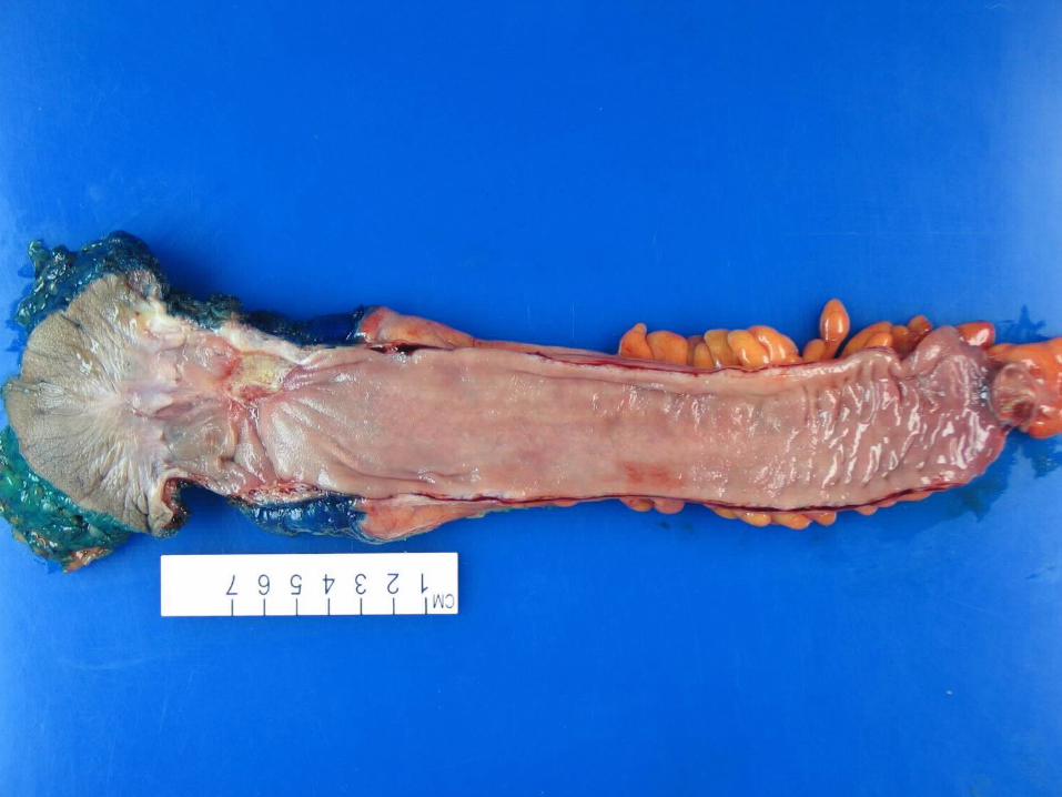
Small Intestine villi :
• Small intestine duodenum with numerous tongue-shaped projections (villi) protruding from the gut wall. The duodenum is the first part of the small intestine, extending after the stomach to the jejunum.

38

39
• As a major secretory region of the gut, the duodenum also receives secretory products from other organs to aid in digestion.
• As in other parts of the small intestine, digestion and absorption are maximized by the large surface area conferred by the presence of villi.

40

41
• The villi are composed mainly of columnar epithelial cells, each of which is covered with many microvilli to increase cell absorption (not seen). Globlet cells are present among the epithelial cells (not seen). They secrete mucus that lubricates the lining of the intestine.

42

43

44
Colon (large intestine)
• The colon is a 6-foot long muscular tube that connects the small intestine to the rectum.
• The large intestine is made up of the cecum, the ascending (right) colon, the transverse (across) colon, the descending (left) colon, and the sigmoid colon, which connects to the rectum.

45

46
• The large intestine is a highly specialized organ that is responsible for processing waste so that emptying the bowels is easy and convenient.
• Stool, or waste left over from the digestive process, is passed through the colon by means of peristalsis, first in a liquid state and ultimately in a solid form.

47

48
• As stool passes through the colon, water is removed. Stool is stored in the sigmoid (S-shaped) colon until a "mass movement" empties it into the rectum once or twice a day. It normally takes about 36 hours for stool to get through the colon.

49

50
• The stool itself is mostly food debris and bacteria. These bacteria perform several useful functions, such as synthesizing various vitamins, processing waste products and food particles, and protecting against harmful bacteria.
• When the descending colon becomes full of stool, or feces, it empties its contents into the rectum to begin the process of elimination.

51
The appendix
• It's a small tube attached to the cecum.
• Originally it was thought that there was no function of the appendix, however doctors now believe that it plays an important role in fetus and adolescent development. The appendix releases hormones and endocrine cells as well as it is involved in the production of good bacteria in the intestines.

52

53
Rectum • The rectum (Latin for "straight")
is an 8-inch chamber that connects the colon to the anus. It is the rectum's job to receive stool from the colon, to let the person know that there is stool to be evacuated, and to hold the stool until evacuation happens.
54

55
• When anything (gas or stool) comes into the rectum, sensors send a message to the brain.
• The brain then decides if the rectal contents can be released or not. If they can, the sphincters relax and the rectum contracts, disposing its contents. If the contents cannot be disposed, the sphincter contracts and the rectum accommodates so that the sensation temporarily goes away.

56

57
Anus• The anus is the last part of the
digestive tract. It is a 2-inch long canal consisting of the pelvic floor muscles and the two anal sphincters (internal and external).
• The lining of the upper anus is specialized to detect rectal contents. It lets you know whether the contents are liquid, gas, or solid.

58

59
• The anus is surrounded by sphincter muscles that are important in allowing control of stool.
• The pelvic floor muscle creates an angle between the rectum and the anus that stops stool from coming out when it is not supposed to.

60
• The internal sphincter is always tight, except when stool enters the rectum. It keeps us continent when we are asleep or otherwise unaware of the presence of stool.
• When we get an urge to go to the bathroom, we rely on our external sphincter to hold the stool until reaching a toilet, where it then relaxes to release the contents.

61
Accessory Glands :-
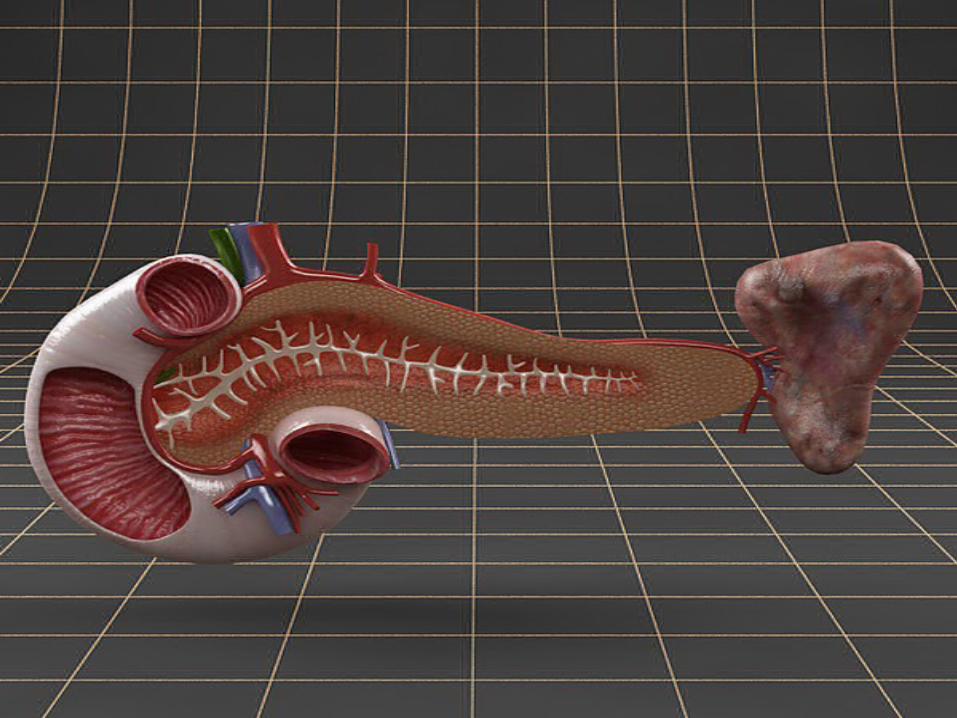
Pancreas :
The pancreas secretes digestive enzymes into the duodenum, the first segment of the small intestine.

62

63
These enzymes break down protein, fats, and carbohydrates. The pancreas also makes insulin, secreting it directly into the bloodstream. Insulin is the chief hormone for metabolizing sugar.
64

65
Liver :The liver is located in the upper right-
hand portion of the abdominal cavity, beneath the diaphragm, and on top of the stomach, right kidney, and intestines. Shaped like a cone, the liver is a dark reddish-brown organ that weighs about 3 pounds.
66
There are two distinct sources that supply blood to the liver, including the following:
a) Oxygenated blood flows in from the hepatic artery
b) Nutrient-rich blood flows in from the hepatic portal vein

67

68
• The liver holds about one pint (13 percent) of the body's blood supply at any given moment.
• The liver consists of two main lobes, both of which are made up of thousands of lobules.
• These lobules are connected to small ducts that connect with larger ducts to ultimately form the hepatic duct.
• The hepatic duct transports the bile produced by the liver cells to the gallbladder and duodenum (the first part of the small intestine).

69
Did you know?
• The liver can lose three-quarters of its cells before it stops functioning.
• In addition, the liver is the only organ in the body that can regenerate itself.

70

71
Functions of the liver :1) Production of bile, which helps carry
away waste and break down fats in the small intestine during digestion
2) Production of certain proteins for blood plasma
3) Production of cholesterol and special proteins to help carry fats through the body

72
4) Conversion of excess glucose into glycogen for storage (glycogen can later be converted back to glucose for energy)
5) Regulation of blood levels of amino acids, which form the building blocks of proteins
6) Processing of hemoglobin for use of its iron content (the liver stores iron)

73

74
7) Conversion of poisonous ammonia to urea (urea is an end product of protein metabolism and is excreted in the urine)
8) Clearing the blood of drugs and other poisonous substances
9) Regulating blood clotting
10) Resisting infections by producing immune factors and removing bacteria from the bloodstream

75
76
Gallbladder :
• The gallbladder stores and concentrates bile, and then releases it into the duodenum to help absorb and digest fats.

77

78
What Are Digestive Enzymes?
• Digestive enzymes are substances produced by our bodies that help us to digest the foods we eat. These enzymes are secreted by the various parts of our digestive system and they help to break down food components such as proteins, carbohydrates, and fats.

79
Types of Digestive Enzymes
• Our bodies produce many different types of digestive enzymes to help our bodies to take advantage of the various nutrients found within the foods we consume. Here are examples of some of the more prevalent types of enzymes:

80
Amylase
• Amylase is a digestive enzyme essential for our digestion of carbohydrates, as amylase breaks down starches into sugars. The measurement of amylase levels in the blood is sometimes used as an aid in diagnosing various pancreas
or other digestive tract diseases.

81
Lactase
• Lactase is a type of enzyme that breaks down the sugar, lactose, found in dairy products. Supplemental lactase may be used to assist people who are lactose intolerant to digest dairy products.

82
Lipase• Lipase is the enzyme responsible
for the breakdown of fats that we consume. Within your body, lipase is produced in small amounts by your mouth and stomach, and in larger amounts by your pancreas.

83
Proteases
Proteases are digestive enzymes that break down proteins. Here are the major types of proteases found within the human digestive tract:
Chymotrypsin Pepsin Trypsin

84

85
The Digestive Process:• The start of the process - the
mouth:
The digestive process begins in the mouth. Food is partly broken down by the process of chewing and by the chemical action of salivary enzymes (these enzymes are produced by the salivary glands and break down starches into smaller molecules).

86
On the way to the stomach: the esophagus-• After being chewed and swallowed, the
food enters the esophagus. The esophagus is a long tube that runs from the mouth to the stomach. It uses rhythmic, wave-like muscle movements (called peristalsis) to force food from the throat into the stomach. This muscle movement gives us the ability to eat or drink even when we're upside-down.

87
In the stomach -
• The stomach is a large, sack-like organ that churns the food and bathes it in a very strong acid (gastric acid). Food in the stomach that is partly digested and mixed with stomach acids is called chyme.
88
In the small intestine -
• After being in the stomach, food enters the duodenum, the first part of the small intestine. It then enters the jejunum and then the ileum (the final part of the small intestine). In the small intestine, bile (produced in the liver and stored in the gall bladder), pancreatic enzymes, and other digestive enzymes produced by the inner wall of the small intestine help in the breakdown of food.

89

90
In the large intestine -
• After passing through the small intestine, food passes into the large intestine. In the large intestine, some of the water and electrolytes (chemicals like sodium) are removed from the food. Many microbes (bacteria like Bacteroides, Lactobacillus acidophilus, Escherichia coli, and Klebsiella) in the large intestine help in the digestion process.

91
• The first part of the large intestine is called the cecum (the appendix is connected to the cecum). Food then travels upward in the ascending colon. The food travels across the abdomen in the transverse colon, goes back down the other side of the body in the descending colon, and then through the sigmoid colon.

92
The end of the process -
• Solid waste is then stored in the rectum until it is excreted via the anus.

93

94
For Your Time ....
Ghofran Alnabilsy / Human Biology Practical / Dentistry batch (18) / semester 1 / UMST / 9-11-2013

95
Group 5
Ghofran Alnabilsy Laden
Al-Atta
Areej Sa'ad
Shahd Abo Bakr
Shahd AlFate
h
Raheeq Omar
Done By :