Embed Size (px)
Citation preview
Radiological Diagnosis of
Pulmonary Hypertension
Fatema Ali Al-Khater
210007782
Radiographic Features
Plain X-Ray
By the time the diagnosis of
pulmonary arterial hypertension is made,
90% of patients have an abnormal chest
radiograph .
-low sensitivity and specificity.
Plain film
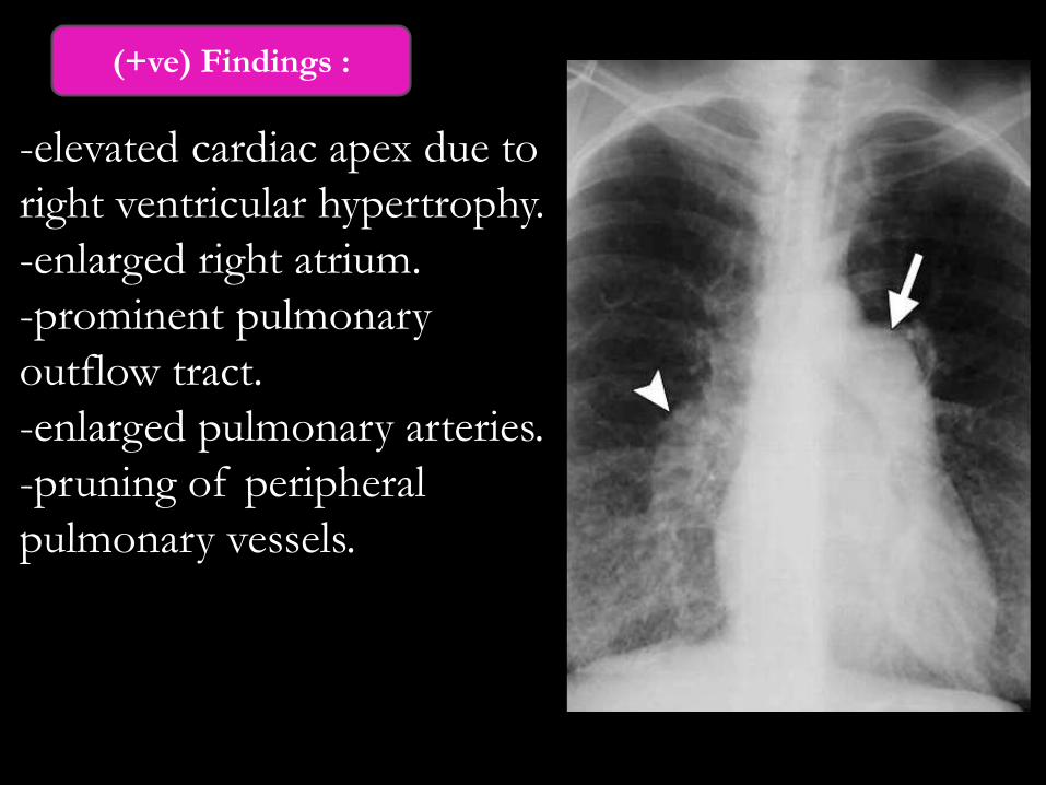
-elevated cardiac apex due to
right ventricular hypertrophy.
-enlarged right atrium.
-prominent pulmonary
outflow tract.
-enlarged pulmonary arteries.
-pruning of peripheral
pulmonary vessels.
(+ve) Findings :
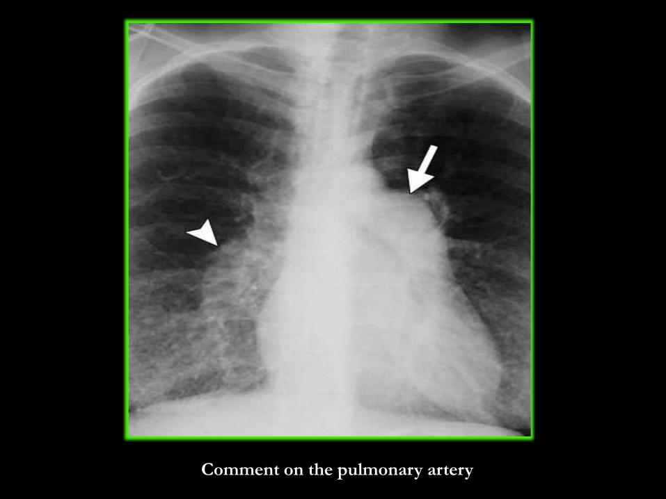
Comment on the pulmonary artery
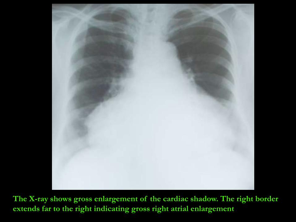
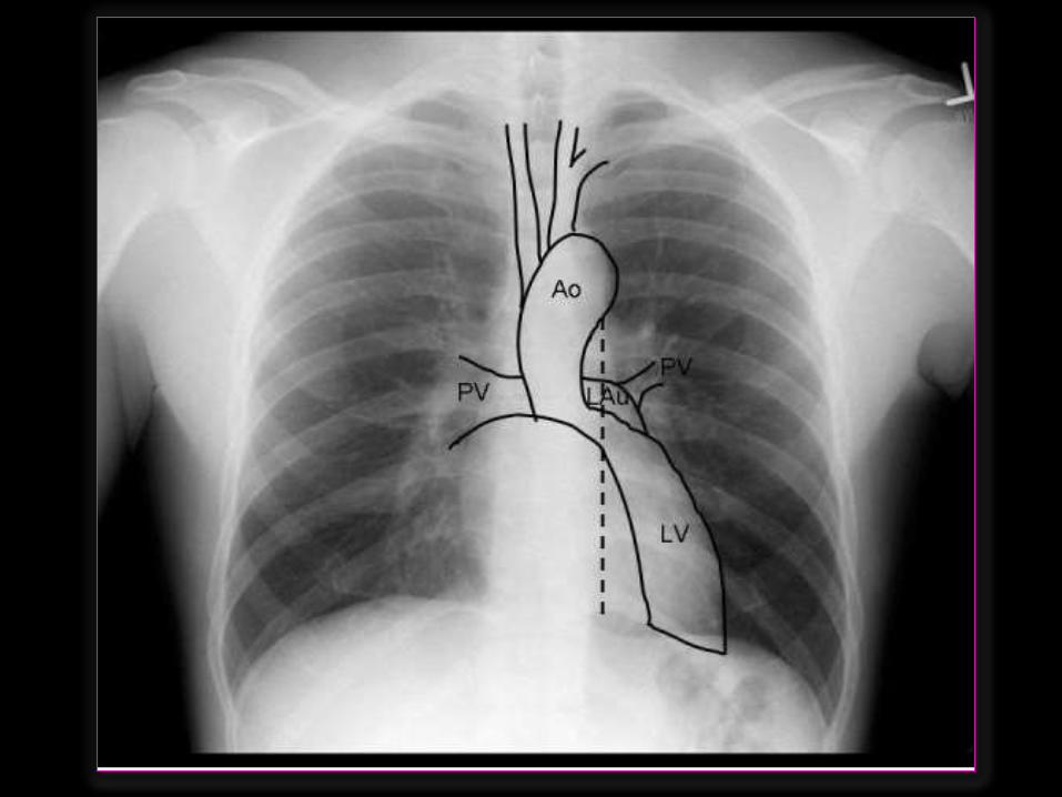
The X-ray shows gross enlargement of the cardiac shadow. The right border
extends far to the right indicating gross right atrial enlargement
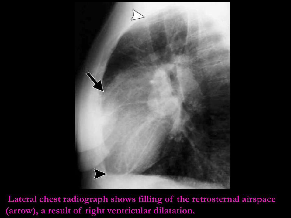
Lateral chest radiograph shows filling of the retrosternal airspace
(arrow), a result of right ventricular dilatation.
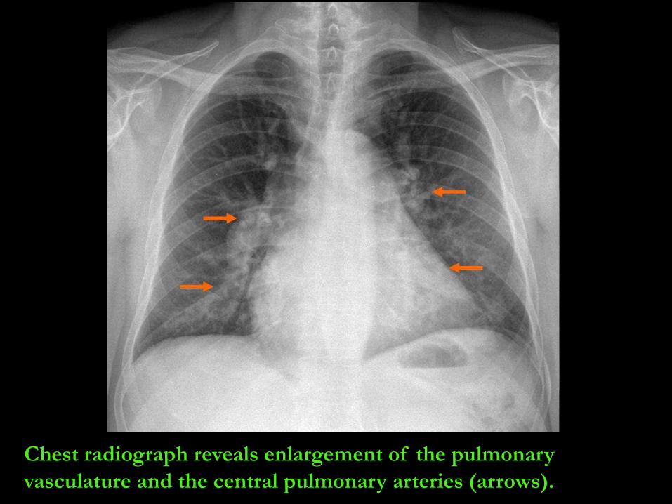
Chest radiograph reveals enlargement of the pulmonary
vasculature and the central pulmonary arteries (arrows).
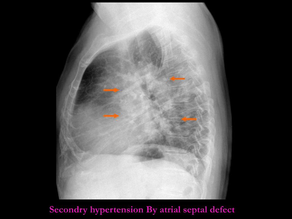
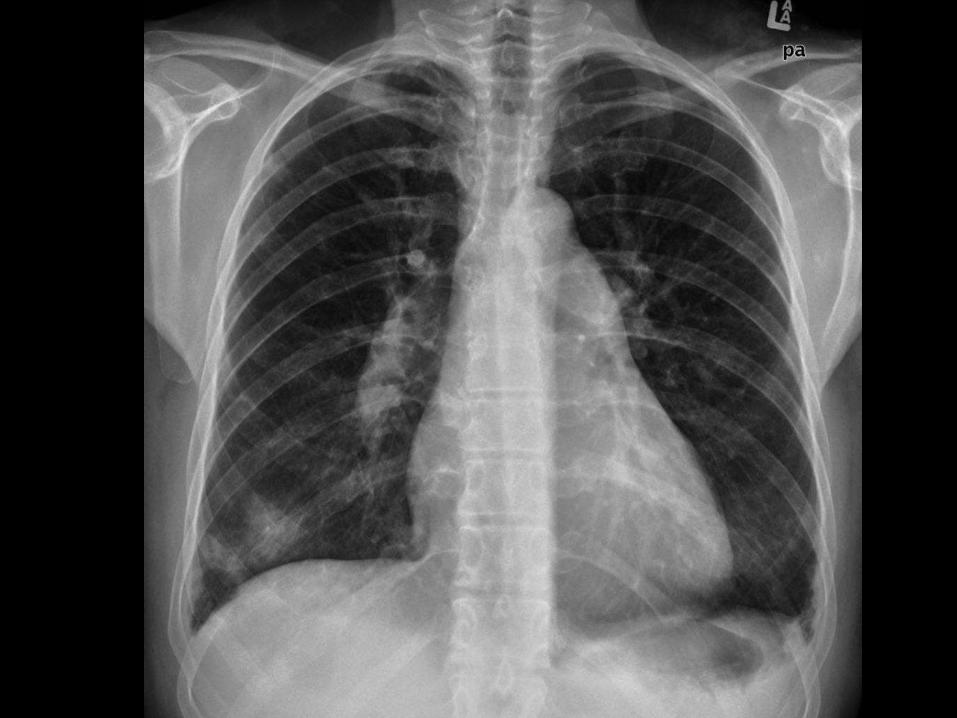
Secondry hypertension By atrial septal defect
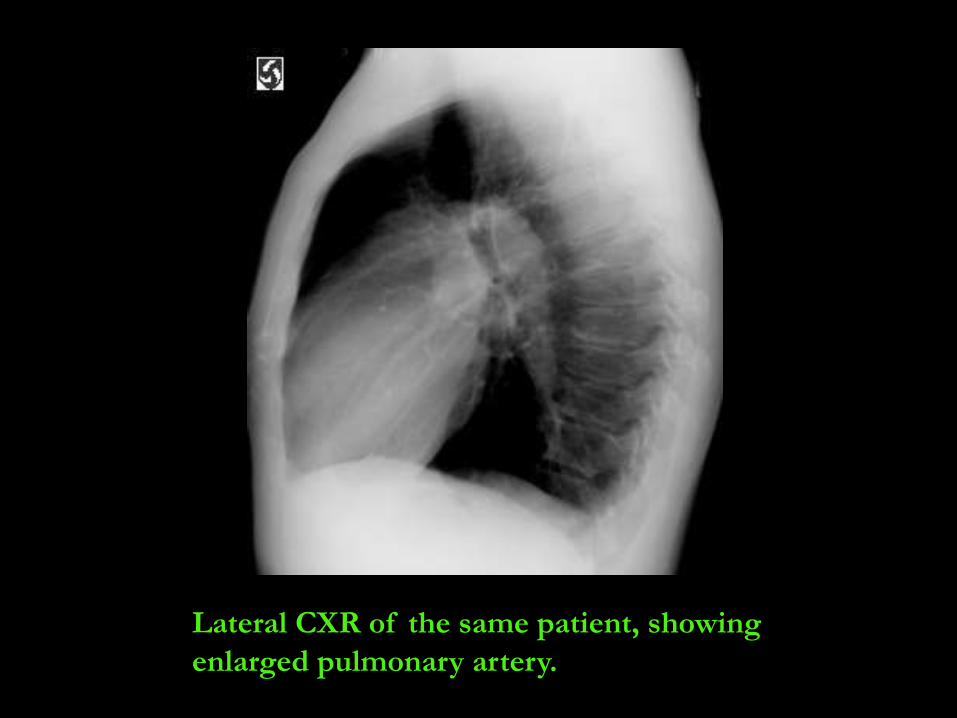
Lateral CXR of the same patient, showing
enlarged pulmonary artery.
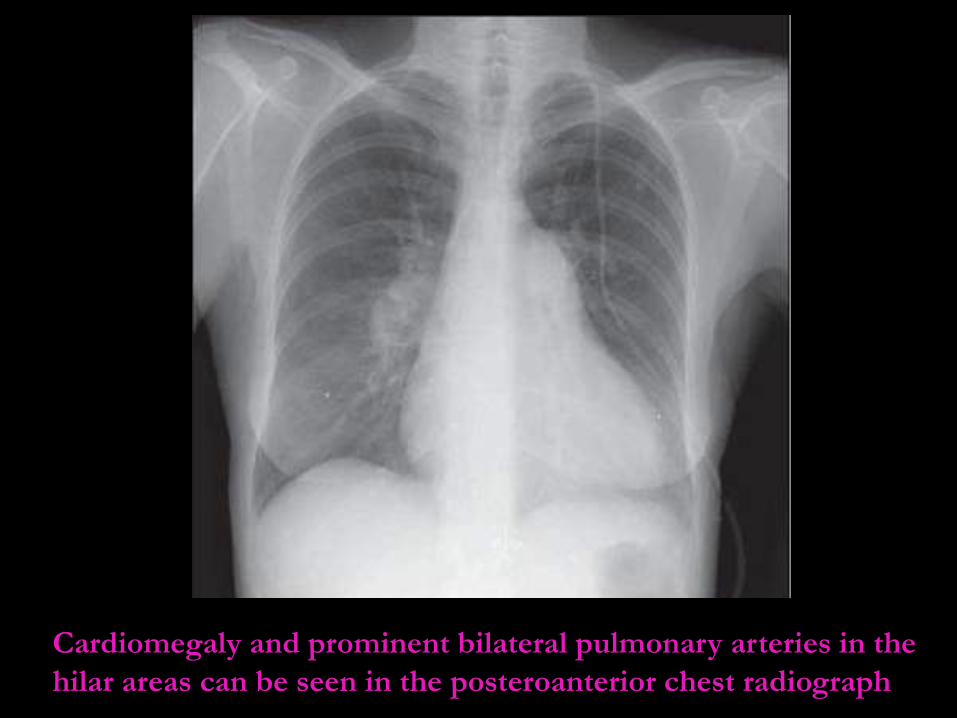
Cardiomegaly and prominent bilateral pulmonary arteries in the
hilar areas can be seen in the posteroanterior chest radiograph
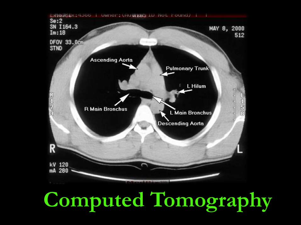
Computed Tomography
1- CT is good , noninvasive , used to
confirm presence of pulmonary
hypertension.
2- It is useful in delineating the anatomic
detail of the pulmonary vasculature.
3-CTPA is the best method for
demonstrating emboli.
4- Contrast-enhanced images may show
intraluminal abnormalities in the arteries and
veins and can detect emboli if it’s large.
Advantages of CT
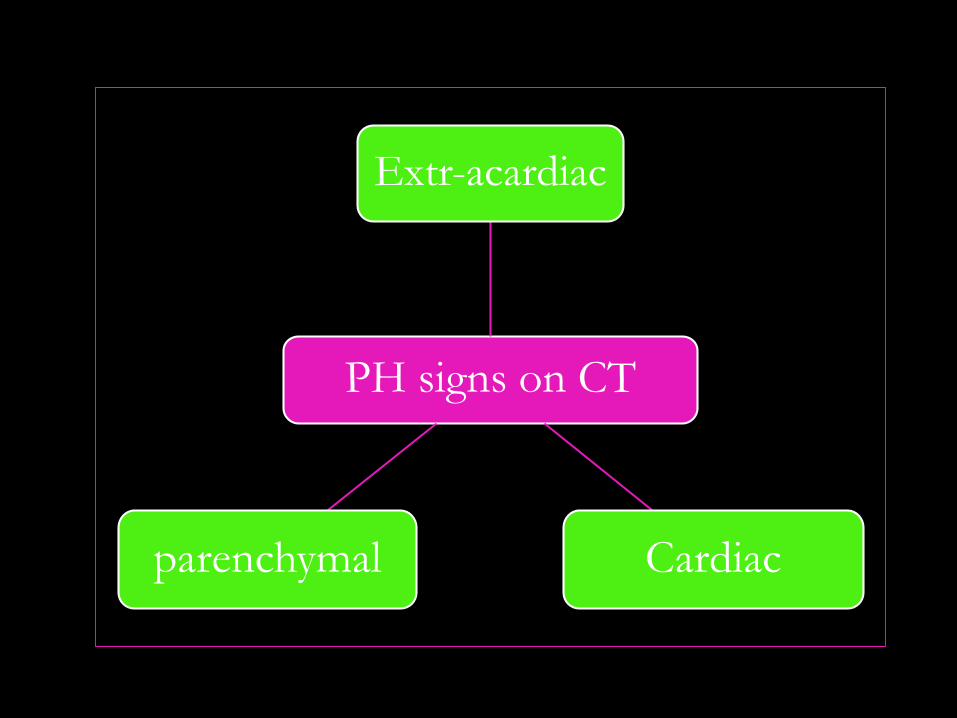
PH signs on CT
Extr-acardiac
Cardiacparenchymal
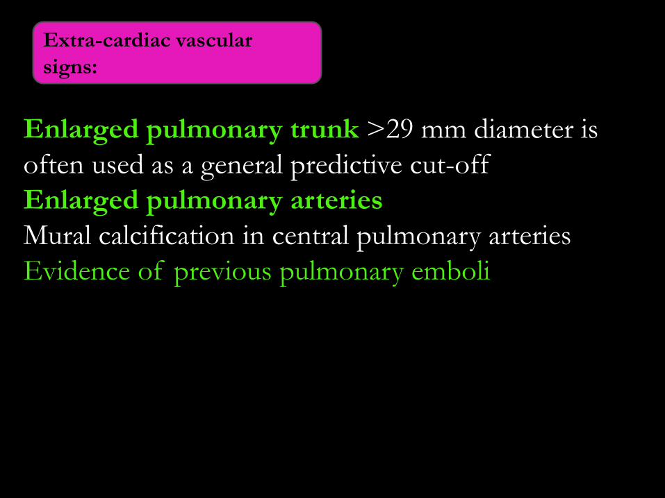
Enlarged pulmonary trunk >29 mm diameter is
often used as a general predictive cut-off
Enlarged pulmonary arteries
Mural calcification in central pulmonary arteries
Evidence of previous pulmonary emboli
Extra-cardiac vascular
signs:
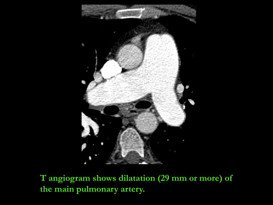
T angiogram shows dilatation (29 mm or more) of
the main pulmonary artery.
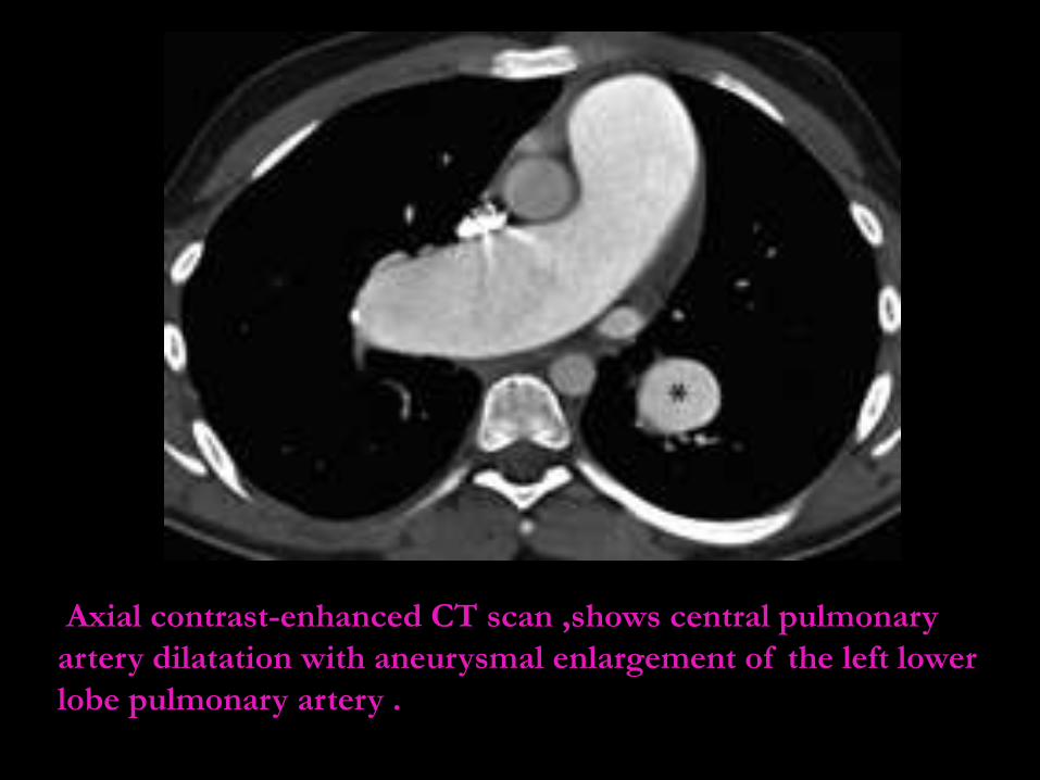
Axial contrast-enhanced CT scan ,shows central pulmonary
artery dilatation with aneurysmal enlargement of the left lower
lobe pulmonary artery .
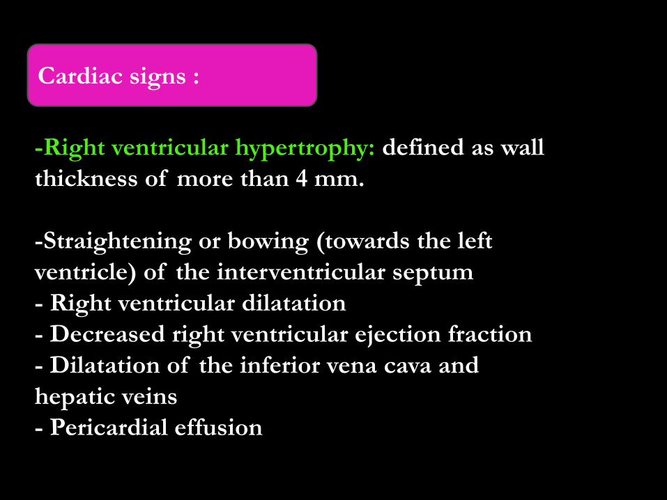
-Right ventricular hypertrophy: defined as wall
thickness of more than 4 mm.
-Straightening or bowing (towards the left
ventricle) of the interventricular septum
- Right ventricular dilatation
- Decreased right ventricular ejection fraction
- Dilatation of the inferior vena cava and
hepatic veins
- Pericardial effusion
Cardiac signs :
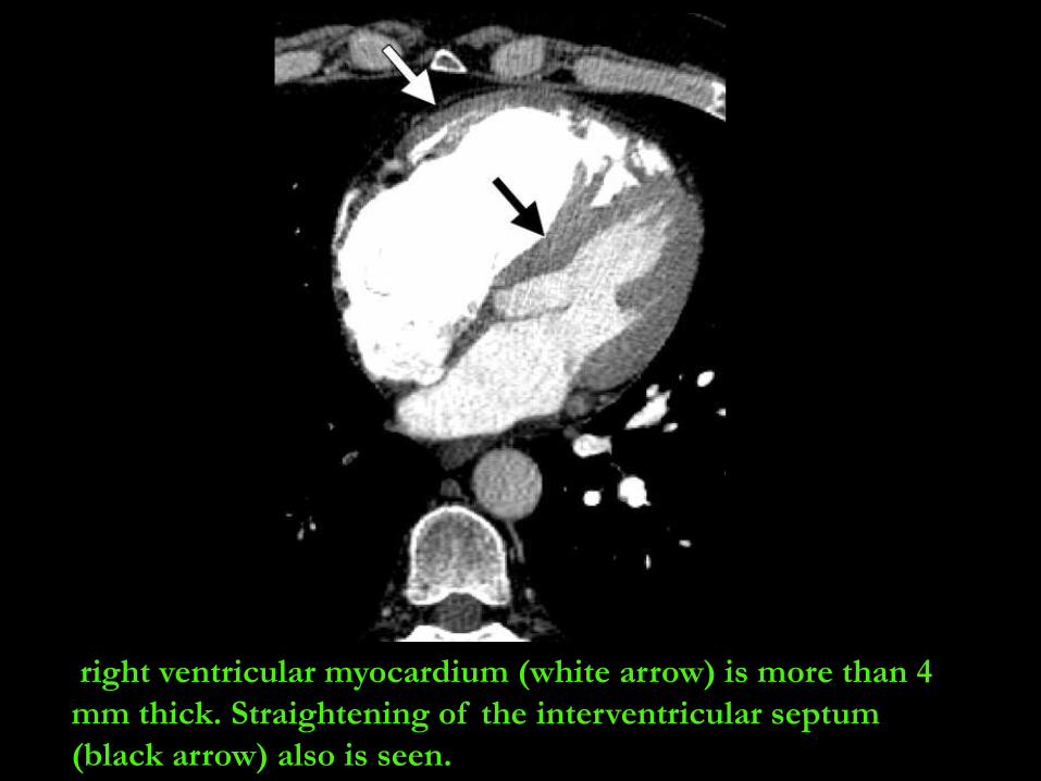
right ventricular myocardium (white arrow) is more than 4
mm thick. Straightening of the interventricular septum
(black arrow) also is seen.
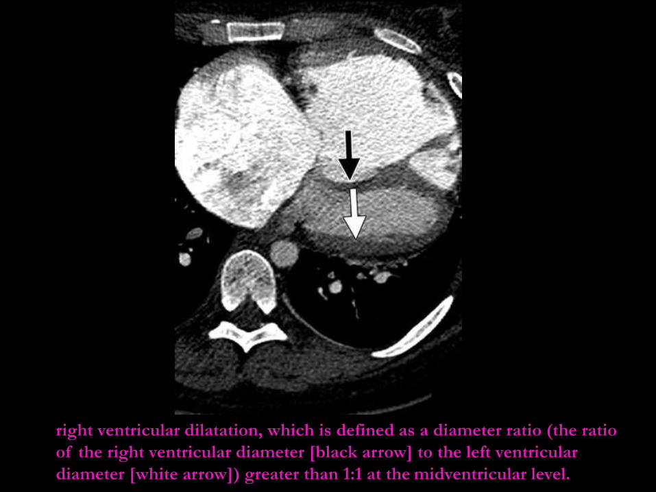
right ventricular dilatation, which is defined as a diameter ratio (the ratio
of the right ventricular diameter [black arrow] to the left ventricular
diameter [white arrow]) greater than 1:1 at the midventricular level.
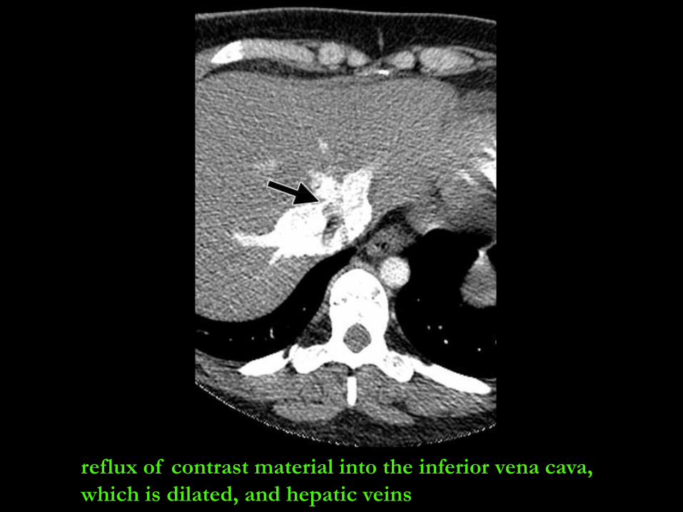
reflux of contrast material into the inferior vena cava,
which is dilated, and hepatic veins

Centrilobular ground-glass nodules (Cholesterol
granuloma).
Neovascularity: tiny serpiginous intrapulmonary
vessels that often emerge from centrilobular
arterioles.
Parenchymal signs:
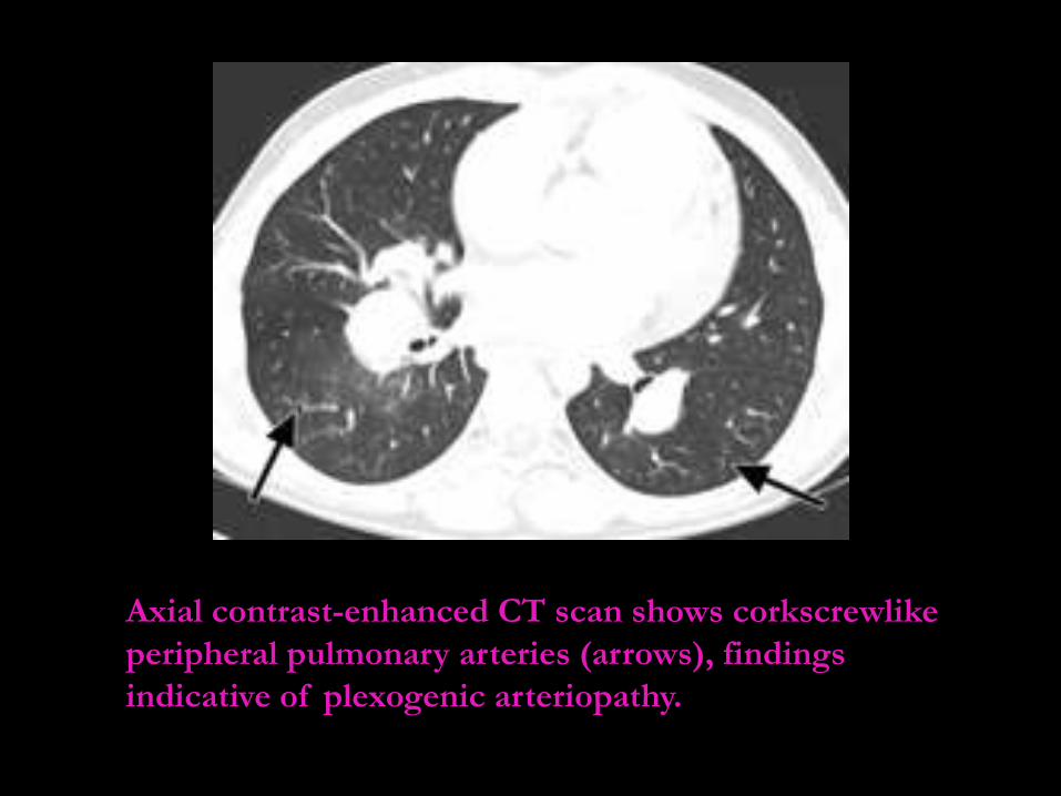
Axial contrast-enhanced CT scan shows corkscrewlike
peripheral pulmonary arteries (arrows), findings
indicative of plexogenic arteriopathy.
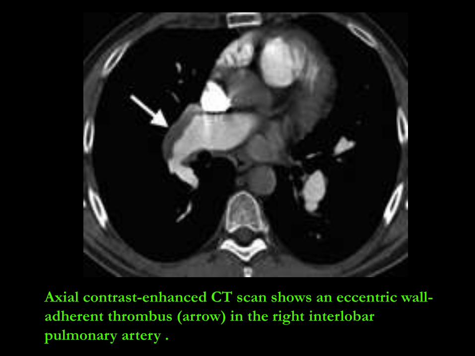
Axial contrast-enhanced CT scan shows an eccentric wall-
adherent thrombus (arrow) in the right interlobar
pulmonary artery .
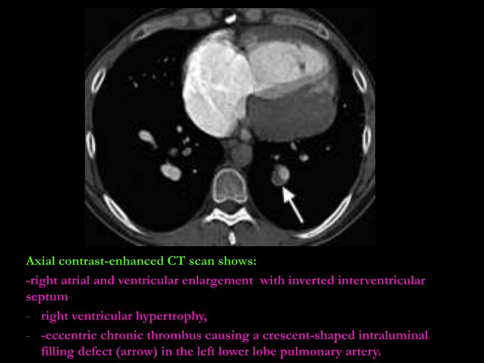
Axial contrast-enhanced CT scan shows:
-right atrial and ventricular enlargement with inverted interventricular
septum
- right ventricular hypertrophy,
- -eccentric chronic thrombus causing a crescent-shaped intraluminal
filling defect (arrow) in the left lower lobe pulmonary artery.
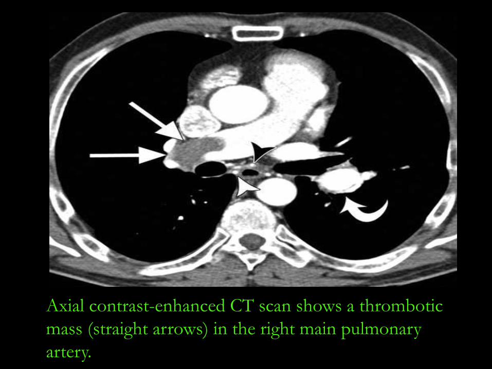
Axial contrast-enhanced CT scan shows a thrombotic
mass (straight arrows) in the right main pulmonary
artery.
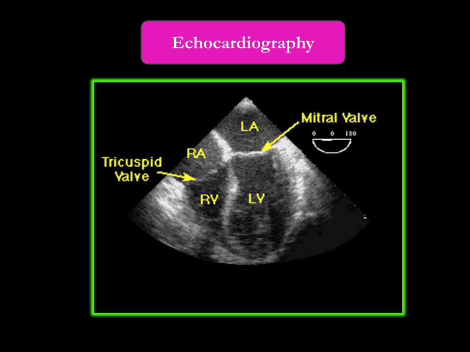
Echocardiography
- It’s performed to estimate the pulmonary artery
systolic pressure and to assess right ventricular size,
thickness, and function.
- evaluate right atrial size, left ventricular systolic
and diastolic function, and valve function.
- detecting pericardial effusions and intracardiac
shunts.
- uses Doppler ultrasound to estimate the
pulmonary artery systolic pressure.
Advantages
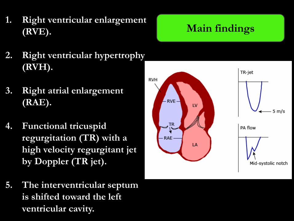
1. Right ventricular enlargement
(RVE).
2. Right ventricular hypertrophy
(RVH).
3. Right atrial enlargement
(RAE).
4. Functional tricuspid
regurgitation (TR) with a
high velocity regurgitant jet
by Doppler (TR jet).
5. The interventricular septum
is shifted toward the left
ventricular cavity.
Main findings
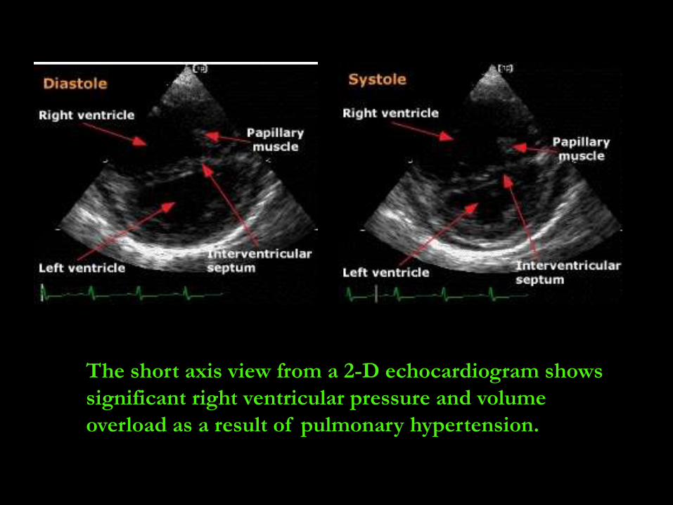
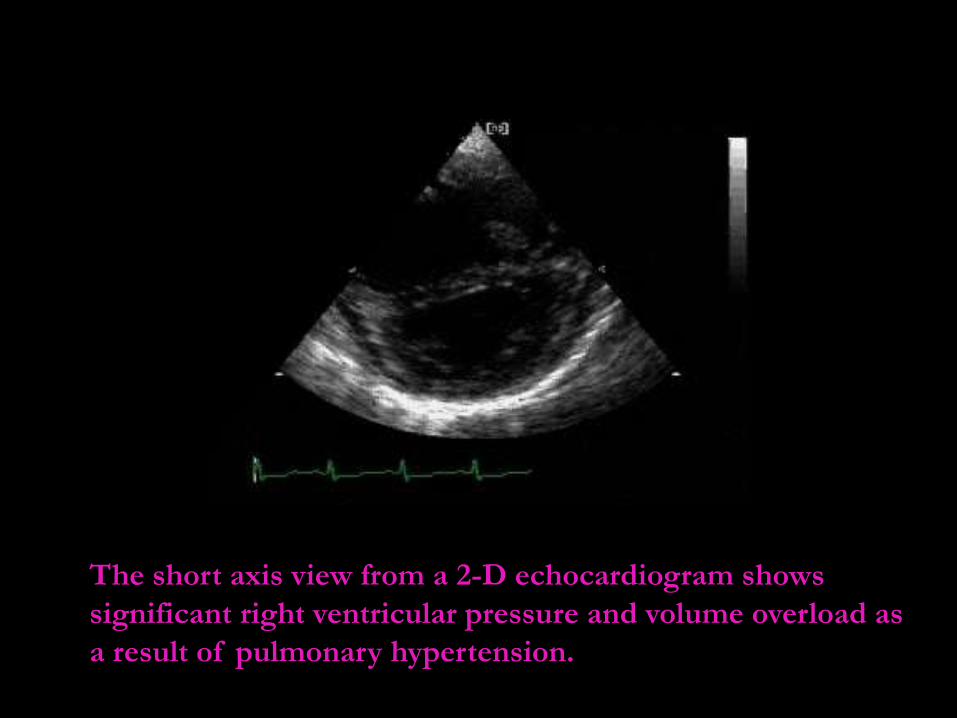
The short axis view from a 2-D echocardiogram shows
significant right ventricular pressure and volume
overload as a result of pulmonary hypertension.
The short axis view from a 2-D echocardiogram shows
significant right ventricular pressure and volume overload as
a result of pulmonary hypertension.

Angiography
Right heart catheterization may be
required.
-Pulmonary angiography is the most
accurate modality for evaluating the
anatomy and pathophysiology of
pulmonary hypertension
-The disadvantage :
it is an invasive procedure as one cannulates
the right side of the heart and thea
pulmonary artery.
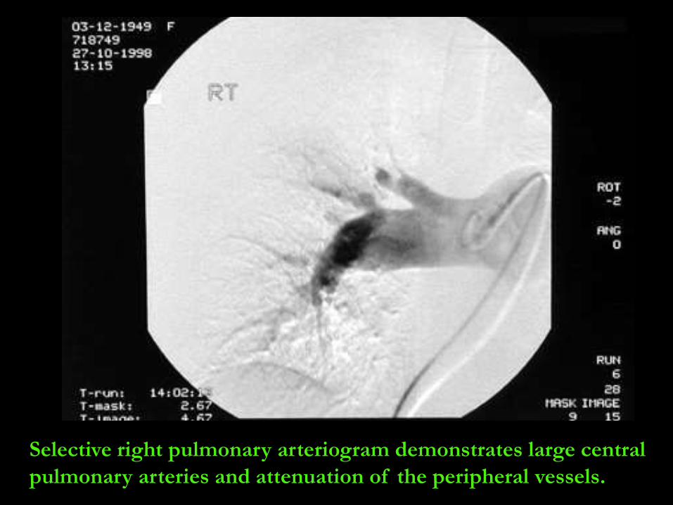
Selective right pulmonary arteriogram demonstrates large central
pulmonary arteries and attenuation of the peripheral vessels.
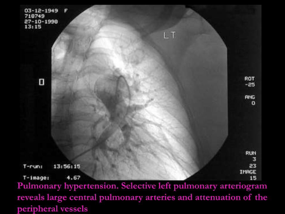
Pulmonary hypertension. Selective left pulmonary arteriogram
reveals large central pulmonary arteries and attenuation of the
peripheral vessels
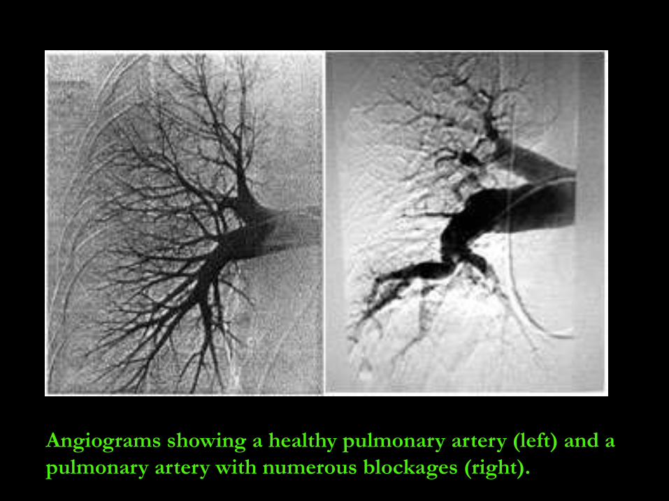
Angiograms showing a healthy pulmonary artery (left) and a
pulmonary artery with numerous blockages (right).

Magnetic resonance
Imaging
The disadvantages with MRI:
-include limitations in individuals with cardiac-
pacemakers and defibrillators.
- its limited availability and cost, and difficulty in
assessing estimate PA pressures with MRI.
MRI with contrast enhancement allows one to
distinguish between the pulmonary vasculature
and mediastinal adenopathy
Advantages :
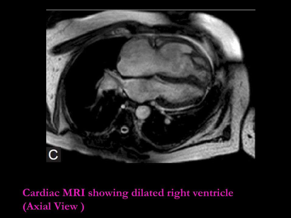
Cardiac MRI showing dilated right ventricle
(Axial View )
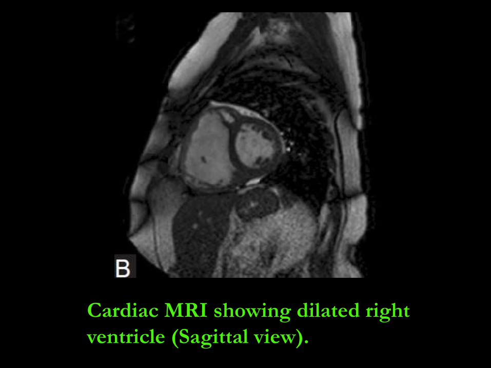
Cardiac MRI showing dilated right
ventricle (Sagittal view).
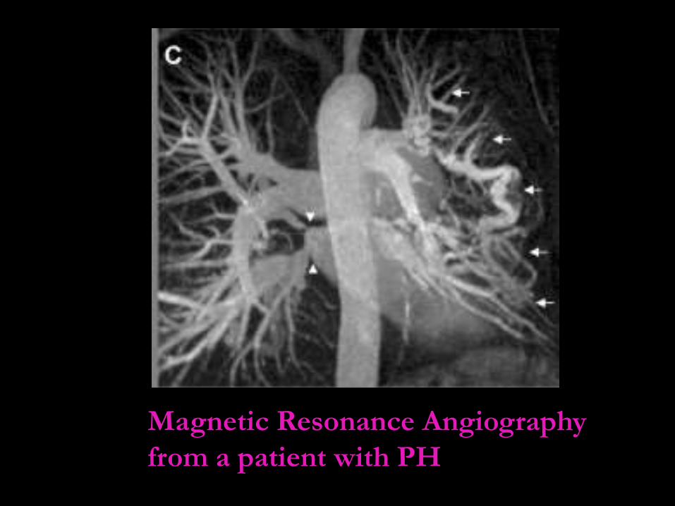
Magnetic Resonance Angiography
from a patient with PH
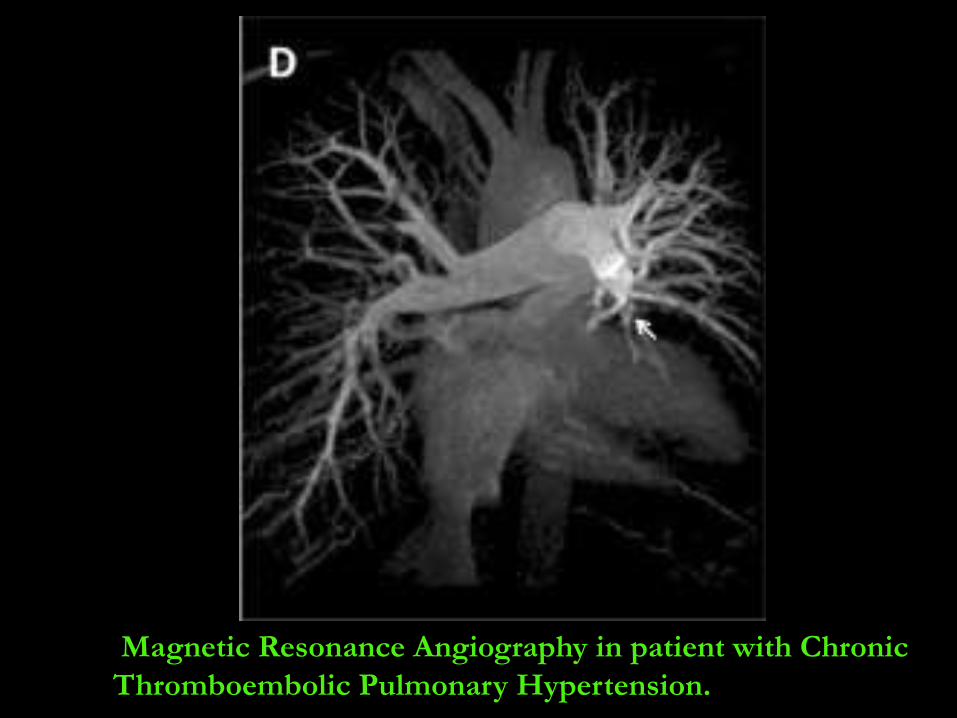
Magnetic Resonance Angiography in patient with Chronic
Thromboembolic Pulmonary Hypertension.
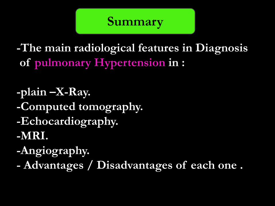
-The main radiological features in Diagnosis
of pulmonary Hypertension in :
-plain –X-Ray.
-Computed tomography.
-Echocardiography.
-MRI.
-Angiography.
- Advantages / Disadvantages of each one .
Summary
References

Thank you
Any Question?