Embed Size (px)
Citation preview
ORIGINAL RESEARCHpublished: 24 May 2016
doi: 10.3389/fphys.2016.00180
Frontiers in Physiology | www.frontiersin.org 1 May 2016 | Volume 7 | Article 180
Edited by:
Mario Diaz,
Universidad de La Laguna, Spain
Reviewed by:
Nick Robert Forsyth,
Keele University, UK
Roman Krawetz,
University of Calgary, Canada
Yolande Ramos,
Leiden University Medical Center,
Netherlands
*Correspondence:
Mohammed Abbas
Ali Mobasheri
Specialty section:
This article was submitted to
Membrane Physiology and Membrane
Biophysics,
a section of the journal
Frontiers in Physiology
Received: 18 January 2016
Accepted: 05 May 2016
Published: 24 May 2016
Citation:
Kalamegam G, Abbas M, Gari M,
Alsehli H, Kadam R, Alkaff M,
Chaudhary A, Al-Qahtani M,
Abuzenadah A, Kafienah W and
Mobasheri A (2016) Pelleted Bone
Marrow Derived Mesenchymal Stem
Cells Are Better Protected from the
Deleterious Effects of Arthroscopic
Heat Shock. Front. Physiol. 7:180.
doi: 10.3389/fphys.2016.00180
Pelleted Bone Marrow DerivedMesenchymal Stem Cells Are BetterProtected from the DeleteriousEffects of Arthroscopic Heat ShockGauthaman Kalamegam 1, 2, Mohammed Abbas 2, 3*, Mamdooh Gari 1, 2, 4, Haneen Alsehli 5,
Roaa Kadam 1, Mohammed Alkaff 2, 3, Adeel Chaudhary 1, Mohammed Al-Qahtani 1,
Adel Abuzenadah 1, 5, Wael Kafienah 6 and Ali Mobasheri 1, 7, 8*
1Center of Excellence in Genomic Medicine Research, King Abdulaziz University, Jeddah, Saudi Arabia, 2 Sheik Salem Bin
Mahfouz Scientific Chair for Treatment of Osteoarthritis by Stem Cells, King Abdulaziz University, Jeddah, Saudi Arabia,3Department of Orthopedic Surgery, Faculty of Medicine, King Abdulaziz University Hospital, Jeddah, Saudi Arabia,4Department of Medical Laboratory Technology, Faculty of Applied Medical Sciences, King Abdulaziz University, Jeddah,
Saudi Arabia, 5 Faculty of Applied Medical Sciences, Center of Innovation in Personalized Medicine, King Abdulaziz University,
Jeddah, Saudi Arabia, 6 School of Cellular and Molecular Medicine, University of Bristol, Bristol, UK, 7 The D-BOARD
European Consortium for Biomarker Discovery, The APPROACH Innovative Medicines Initiative Consortium, Faculty of Health
and Medical Sciences, University of Surrey, Surrey, UK, 8 Arthritis Research UK Centre for Sport, Exercise and Osteoarthritis,
Arthritis Research UK Pain Centre, Medical Research Council and Arthritis Research UK Centre for Musculoskeletal Aging
Research, University of Nottingham, Queen’s Medical Centre, Nottingham, UK
Introduction: The impact of arthroscopic temperature on joint tissues is poorly
understood and it is not known how mesenchymal stem cells (MSCs) respond to
the effects of heat generated by the device during the process of arthroscopy
assisted experimental cell-based therapy. In the present study, we isolated and
phenotypically characterized human bone marrow mesenchymal stem cells (hBMMSCs)
from osteoarthritis (OA) patients, and evaluated the effect of arthroscopic heat on cells
in suspension and pellet cultures.
Methods: Primary cultures of hBMMSCs were isolated from bone marrow aspirates of
OA patients and cultured using DMEM supplemented with 10% FBS and characterized
for their stemness. hBMMSCs (1 6× 10 cells) cultured as single cell suspensions or
cell pellets were exposed to an illuminated arthroscope for 10, 20, or 30min. This was
followed by analysis of cellular proliferation and heat shock related gene expression.
Results: hBMMSCs were viable and exhibited population doubling, short spindle
morphology, MSC related CD surface markers expression and tri-lineage differentiation
into adipocytes, chondrocytes and osteoblasts. Chondrogenic and osteogenic
differentiation increased collagen production and alkaline phosphatase activity. Exposure
of hBMMSCs to an illuminated arthroscope for 10, 20, or 30min for 72 h decreased
metabolic activity of the cells in suspensions (63.27% at 30min) and increased metabolic
activity in cell pellets (62.86% at 10min and 68.57% at 20min). hBMMSCs exposed to
37, 45, and 55◦C for 120 s demonstrated significant upregulation of BAX, P53, Cyclin
A2, Cyclin E1, TNF-α, and HSP70 in cell suspensions compared to cell pellets.
Kalamegam et al. Heat Shock and MSC Viability
Conclusions: hBMMSC cell pellets are better protected from temperature alterations
compared to cell suspensions. Transplantation of hBMMSCs as pellets rather than as
cell suspensions to the cartilage defect site would therefore support their viability and
may aid enhanced cartilage regeneration.
Keywords: mesenchymal stem cells, osteoarthritis (OA), cell viability, population doubling, fluorescence activated
cell sorting (FACS), differentiation, heat shock
INTRODUCTION
Osteoarthritis (OA) is a chronic degenerative disease of load-bearing synovial joints that commonly affects a significant andever increasing proportion of the aging population. OA is themost common joint disorder in the elderly. Its incidence hasbeen rising steadily and is expected to escalate further with theprojected world-wide increase in the aging population, impactingon the existing socio-economic burden (Arden and Nevitt, 2006).Nearly 50 million Americans are already afflicted with OA, with50% of the individuals affected being more than 65 years of age(Center for Disease Control Prevention (CDC), 2010). By 2030,it is estimated that about 20% of the European and Americanpopulations will suffer fromOA (De Bari et al., 2010). In the sameperiod in the Middle East, the indicators of aging, namely thestandardized prospective median age and the average age of thepopulation are projected to increase from 20.9 and 26.0 in 2010 to23.5 and 31.4 (Lutz et al., 2008). Furthermore, the cultural prayerposition and increased obesity in the Middle Eastern populationpose additional risks for the development of OA in the agingpopulation.
OA is characterized by progressive joint degeneration, pain,effusion and limitation of mobility with eventual loss of function(Buckwalter and Lane, 1997). The entire articulating jointtogether with its constituent peri-articular tissues including bone,ligaments, joint capsule and synovium and are involved in theprogression of OA, highlighting the complex nature of thisdisease (Loeser et al., 2012).
Although multiple factors are implicated in theaetiopathogenesis of OA, the underlying molecular mechanismsstill remain unclear. Several hypotheses have been proposedto account for the development of OA including age-related“wear and tear,” chondrocytes’ poor response to growth factors,increased sensitivity to pro-inflammatory stimuli, excessivecross-linking and structural modification of collagens leadingto alterations in the bio-mechanical properties of articularcartilage, mitochondrial dysfunction, oxidative stress and low-level inflammation (Mobasheri et al., 2013). Fundamentally, thenumber of chondrocytes in articular cartilage decreases with ageresulting in impaired extracellular matrix repair and reducedproduction of new matrix proteins. In addition the absence ofblood vessels and stem cells within the cartilage limits the naturalhealing capacity (Henrotin and Reginster, 1999; Mobasheriet al., 2013) although evidence suggests that innervation andvascularization increases in the late stages of disease progression.
Non-steroidal anti-inflammatory drugs, opioids, topicalformulations, intra-articular injections and nutraceuticals are allused in OA treatment, but they are largely symptom modifying
agents and there is little evidence to suggest that any of them
have the capacity to act as disease modifying osteoarthritis drugs
(DMOADs). Pharmacological management with DMOADs may
relieve pain to some degree, but does not offer any structure
modification (Reid et al., 2012). Various surgical methods have
been developed to restore damaged cartilage and improve joint
function. These include microfracture, subchondral drilling,
abrasion arthroplasty and autologus chondrocyte implantation(ACI). The aim of these techniques is to promote intrinsic
healing by promoting vascular invasion, fibrin clot formationand recruitment of stem cells (Brittberg et al., 1994; Vinatier
et al., 2009; Orth et al., 2014). However, the poor biomechanicalproperties of the new scar tissue in microfracture, as well as
donor site morbidity, low cellularity and surrounding cartilage
damage at the transplantation site in ACI limit their clinicalutility and surgical outcomes (Brittberg et al., 1994; Vinatier et al.,
2009; Orth et al., 2014). When pharmacological and surgicalmanagement fail, which is so often the case in the clinicalsetting, the disease progresses to end stage OA, where total jointreplacement becomes the only option. However, the life span ofcurrently available prostheses is limited and there is increasingdemand for safer and more effective surgical treatments andtherapeutic strategies (Kurtz et al., 2007).
Regenerative medicine offers great potential for therapeutic
intervention in OA and could provide an excellent alternative
to total joint replacement. The use of autologus matrix induced
chondrogenesis (AMIC) in OA (Gille et al., 2013) and intra-
articular injection of meniscal stem/progenitors cells (Shen et al.,
2014) are some recent advances in this area. Autologus and
allogeneic stem cells derived from various sources (viz. bone
marrow, synovium, adipose tissue etc.) have been used for
treatment of OA with variable success (Garcia-Alvarez et al.,
2011; Mobasheri et al., 2014). These are either directly injected
into the damaged site or differentiated into cartilage together with
tissue engineered scaffolds or following treatment with growthfactors.
The introduction of biological agents such as stem cells
into the joint normally requires arthroscopic techniques.
Temperature increases of 52.0 and 49.5◦C following monopolar
and bioplar radiofrequency application with irrigation have been
reported during wrist arthroscopy in cadaveric models (Huber
et al., 2013). Animal studies have demonstrated ultrastructural
changes in the size and cross-sectional diameter of the joint
capsular collagen fibrils ranging from 22.5 to 50.4% following
increased temperatures of 45 and 85◦C (Lopez et al., 1998).Bone drilling, a common treatment for operative fracture also
generates heat and temperatures above 47◦C are known to cause
Frontiers in Physiology | www.frontiersin.org 2 May 2016 | Volume 7 | Article 180
Kalamegam et al. Heat Shock and MSC Viability
osteonecrosis (Augustin et al., 2012). The aim of this studywas to investigate the impact of temperature changes associatedwith arthroscopic procedures on cellular activity and/or survivalin suspension and pellet cultures of human bone marrowmesenchymal stem cells (hBMMSCs), which are increasinglystudied as a promising cell source for cartilage regeneration.
MATERIALS AND METHODS
Derivation and Propagation of hBMMSCsBone marrow aspirates were harvested from the iliac crest ofOA patients undergoing surgical treatment in the Departmentof Orthopedics, King Abdulaziz University Hospital, Jeddah,Kingdom of Saudi Arabia. Prior to sample collection informedpatient consent was obtained and the research study was carriedout following Institutional Research Ethics Committee approval[11–557]. hBMMSCs were isolated using earlier publishedprotocols (Brady et al., 2014). Briefly, the bone marrow aspirate(5–6ml) was collected in heparinized tubes (Becton Dickinson,BD) and directly plated into tissue culture flasks (∼2ml ofaspirate/T175 cm2 flask; Greiner) and cultured using Dulbeccos’smodified Eagle’s medium (Sigma), supplemented with 10% fetalbovine serum (Sigma), 2mM Gluta-Max (Life Technologies)and antibiotic solution (penicillin, 100 u/mL; streptomycin100µg/mL - Sigma) under standard culture conditions of 37◦Cand 5% carbon dioxide (CO2) in atmospheric air for 5–7 days.The first media change was carried out on day 5 and subsequentlyevery 2 or 3 days until subculture. Dead/suspended cells andcellular debris were washed away with media changes and thehBMMSCs were retained as a consequence of their plasticadherence. Basic fibroblast growth factor (bFGF; Peprotech) at5 ng/mLwas added to the culture medium to facilitate hBMMSCsexpansion and early passages of the derived hBMMSCs (<P5)were used in the experiment Expanded cells were frozen usingProFreeze (Lonza) in liquid nitrogen and stored for subsequentuse.
Effect of Arthroscope Temperature on CellMorphology and Metabolic ActivityBaseline characterization included assessment of cell morphologyand survival. The hBMMSCs were seeded in 24 well tissueculture plates at 2 × 10⁴ cells/well. Cell morphology and theirmetabolic activity were analyzed using phase contrastmicroscopyand MTT assay respectively. To study the effect of temperaturerelated changes that may result with use of the arthroscope ina clinical setting hBMMSCs (1 × 10⁶ cells) were used either ascell suspensions or cell pellets. The arthroscope was sterilizedand suspended vertically down using a stand with fixed clampand placed within the biosafety cabinet. The suspension heightof the arthroscope from the clamp was kept constant and theilluminated end was placed into the medium (10mL) containingcell suspensions or cell pellets in 50mL graduated Falcon tubes.The samples from both cell suspension and cell pellet groupswere exposed for 10min (Group A), 20min (Group B), or 30min(Group C). The cell suspensions and pellets were then gentlymixed and 2 × 10⁴ cells/well were seeded in a 24 well plateand cultured under standard culture conditions of 37◦C in 5%
atmospheric air for 72 h and both cell morphology and metabolicactivity were assessed.
Cellular activity assays were also performed on cells fromall the groups using the MTT kit (3-(4,5-dimethylthiazolyl-2)-2,5-diphenyltetrazolium bromide; Sigma). Briefly,10µL MTT reagent (0.5 mg/mL) was added to cultures,incubated for 4 h followed by medium removal andaddition of 200µL solubilization reagent. Culture plateswere further incubated for 2 h and absorbance at 570 nm(ref 650 nm) was measured using a spectrophotometer(µQuant; BioTek).
CD Marker AnalysisCultures of hBMMSCs were analyzed for expression ofMSC related cluster of differentiation (CD) markers. Briefly,monolayer cultures of hBMMSCs were dissociated using 0.25%Trypsin-EDTA (Life Technologies) for 3min. Trypsin activitywas inhibited by addition of culture medium containing 10%fetal bovine serum (FBS). The cell suspension was centrifugedat 300 g × 5min and the cell pellet was then resuspendedin phosphate buffered saline without calcium and magnesium(PBS-) containing 3% FBS to obtain single cell suspension.Separate aliquots (2 × 10⁵ cells) were used for MSC isotypecocktail (Miltenyi Biotec), MSC phenotyping cocktail (MiltenyiBiotec) or in combination with other primary monoclonalantibodies (CD44, CD29—BDPharmingen) to avoid interferencewith same fluorochromes. The MSC isotype cocktail comprisedof fluorochrome conjugated monoclonal antibodies, namelymouse IgG1-FITC, mouse IgG1-PE, mouse IgG1-APC, mouseIgG1-PerCp and mouse IG2a-PerCp. The MSC phenotypingcocktail comprised of both positive (CD73-APC, CD90-FITC,CD105-PE) and negative (CD34/CD45/CD14/CD20-PerCp)fluorochrome conjugated monoclonal antibodies. The cellswere incubated with respective antibodies at 1:10 dilutionfor 15min at 4◦C; then washed with 1mL of 3% FBSand centrifuged at 300 g × 5min. The supernatant wasdiscarded and the cells were resuspended in 500µl of 3%FBS before analysis using a FACS Aria III instrument (BDBioSciences), which is equipped with a 488 nM (blue) laserand a 561-nM (yellow-green) laser for uncoupled excitationand detection of FITC and PE fluorochromes. In addition toincreasing the sensitivity of PE detection, this set-up eliminatedthe PE-FITC spill over, thereby eliminating the need forcompensation. As an additional measure, single-stained controltubes for each color was analyzed to rule out the need forcompensation as well as set up the detection range for eachfluorochrome.
Trypan Blue Viability/Population DoublingTimeThe hBMMSCs were seeded at 2 × 10⁴ cells/well in a 24 wellplate and cultured for up to 3 days to determine cell viabilityand population doubling time (PDT). The cells were trypsinizeddaily at the same time and the live/dead cell counts were obtainedfollowing trypan blue vital staining. Three replicates were carriedout for each sample. PDT (http://www.doubling-time.com) was
Frontiers in Physiology | www.frontiersin.org 3 May 2016 | Volume 7 | Article 180
Kalamegam et al. Heat Shock and MSC Viability
calculated using the formula:
Doubling time =
Duration ∗ log(2)
Log(final concentration)− Log(initial concentration)
hBMMSCs Differentiation into AdipocytesThe hBMMSCs (2 × 10⁴ cells/well) were seeded into 24 wellplates and allowed to reach confluence before being stimulatedto differentiate using the StemPro adipocyte differentiationkit (A10070-01, ThermoFisher Scientific). Control cells werecultured using the differentiation basal medium alone while cellsstimulated to undergo adipocyte differentiation were cultured inbasal medium fortified with adipocytic supplement (StemPro R©)for up to 21 days with fresh media change every 3–4 days.Following differentiation, the cells were fixed in 4% formaldehydesolution for 30min, rinsed twice with PBS and stained with oilRed O (Sigma) to visualize lipid vacuoles.
hBMMSCs Differentiation into OsteoblastsThe hBMMSCs (2×10⁴ cells/ well) were seeded into 24 well platesand stimulated to differentiate along the osteoblastic lineageusing (StemPro R©) osteoblast differentiation kit (A10072-01,ThermoFisher Scientific). The control cells were cultured usingthe differentiation basal medium alone while the cells stimulatedto undergo osteoblastic differentiation were cultured in basalmedium fortified with osteogenic supplement (StemPro R©) for upto 21 days with fresh media change every 3–4 days. Followingdifferentiation, the cells were fixed in 4% formaldehyde solutionfor 30min, rinsed twice with PBS and stained with Alizarin red(Sigma) solution, washed and analyzed by light microscopy.
hBMMSCs Differentiation intoChondrocytesThe hBMMSCs (2×10⁴ cells/ well) were seeded into 24 well platesand stimulated to differentiate along the chondrocytic lineageusing (StemPro R©) chondrocyte differentiation kit (A10071-01,ThermoFisher Scientific). The control cells were cultured usingthe differentiation basal medium alone while the cells stimulatedto undergo chondrocyte differentiation were cultured in basalmedium with chondrogenic supplement (StemPro R©) for up to21 days with fresh media change every 3–4 days. Followingdifferentiation, the cells were fixed in 4% formaldehyde solutionfor 30min, rinsed twice with PBS and stained with Alcian bluesolution prepared in 0.1 N HCl, washed and analyzed by lightmicroscopy.
Collagen (Sircol) AssayThe secreted total collagen levels from both chondrocytecontrol and differentiation cultures were evaluated using theSircolTM (chemical collagen assay) kit (Bioclor) according to themanufacturer’s instructions. Briefly, 1ml of the Sircol reagent wasadded to the 100µl of the standards and samples (1:20, dilutedin distilled water) in 1.5ml microcentrifuge tubes (Eppendorf);mixed well and placed on a mechanical shaker for 30min toenable collagen-dye complex precipitation. The contents werethen centrifuged at 12,000 rpm for 10min and the supernatant
was carefully decanted taking care to avoid loss of cell pellets.The unbound dye was removed by layering 750µl of ice-coldacid-salt wash reagent (Kit content) followed by centrifugation at12,000 rpm for 10min. The supernatant was carefully removedand 250µl of alkali reagent (kit content) was added andvortexed to dissolve the bound dye. Absorbance at 555 nmwas spectrophotometrically measured using a microplate ELISAreader (µQuant-BioTek) and the collagen concentration wasdetermined.
Alkaline Phosphatase AssayAlkaline phosphatase (ALP) activity levels from both controland differentiated osteoblast cultures were evaluated bymeasuring the release of p-nitrophenylphosphate (Sigma)as reported earlier (Gauthaman et al., 2011). Absorbance at555 nm was spectrophotometrically measured using a microplateELISA reader (µQuant-BioTek) and ALP concentration wasdetermined.
Quantitative Real-Time Polymerase ChainReaction (QRTPCR)The hBMMSCs (2×10⁶ cells) either as pellets or cell suspensionsin 500µl PBS, were subjected to heat shock for varioustemperatures (37, 45, and 55◦C) for 120 s using the hotplatemethod as reported earlier (Dolan et al., 2012) as the temperaturewas identified to be more accurate. Total RNA was thenextracted from the heat shock treated cells using QiagenTMRNA extraction kit reagent (Invitrogen, Life Technologies).RNA quality and quantity were measured using a NanodropTMspectrophotometer (Nanodrop technologies, Wilmington, DW)and all samples were treated with DNase-I prior to first strandcDNA synthesis with random hexamers using the SuperScriptTM
first strand synthesis system (Invitrogen). Primer sequences weretaken from earlier published studies (Alekseenko et al., 2014;Liang et al., 2015) and the details are given in Table 1. QRT-PCR analysis was performed with the ABI StepOne Plus Real-Time PCR System (Applied Biosystems, Foster City, CA) usingSYBR green and relative quantitation was performed using thecomparative CT (2-11CT) method.
Statistical AnalysisThe differences observed between treated and control cellnumbers, collagen content, alkaline phosphatase and geneexpression assays were analyzed using the Students t-test withthe statistical package for Social Sciences (SPSS 13). The resultswere expressed as mean ± standard error of the mean (SEM)from three different replicates for individual assays and a valueof p < 0.05 was considered to be statistically significant.
RESULTS
Morphology and Growth Characteristics ofhBMMSCsIn primary cultures by day 5–7 the hBMMSCs adhered to theculture surface as multiple colony forming units (CFU) and thecell numbers continued to expand by day 7–9 reaching up to60–70% confluence. The non-adherent cells that were present
Frontiers in Physiology | www.frontiersin.org 4 May 2016 | Volume 7 | Article 180
Kalamegam et al. Heat Shock and MSC Viability
TABLE 1 | The genes and primer sequences used for quantitative real time
PCR.
Genes Primer sequences
GAPDH F: 5′-ACCACAGTCCATGCCATCAC-3′
R: 5′-TCCACCACCCTGTTGCTGTA-3′
BAX F: 5′- TGGAGCTGCAGAGGATGATTG -3′
R: 5′- GCTGCCACTCGGAAAAAGAC -3′
BCL2 F: 5′- GGCTGGGATGCCTTTGTG -3′
R: 5′- CAGCCAGGAGAAATCAAACAGA -3′
P53 F: 5′- GCGCACAGAGGAAGAGAATC -3′
R: 5′- CTCTCGGAACATCTCGAAGC -3′
TNF-α 5′-GGT-GCTTGT-TCC-TCA-GCC-TC-3′
5′-CAG-GCA-GAAGAG-CGT-GGT-G-3′
Cyclin A2 5′-CCT CTC CTC CAT GTC TGT
GTT-AAG-3′
5′-GTG CTC CAT TCT CAG AAC CTG
CTT-3′
Cyclin E1 5′-TGC AGA TCG CAG AGC TTC TA-3′
5′-CTT TCT TTG CTT GGG CTT TG-3′
HSP70 5′-TCTTGGCACCACCTACTCTTG-3′
5′-CATCACCGATCAACCGTTCAG-3′
F, Forward primer; R, Reverse primer.
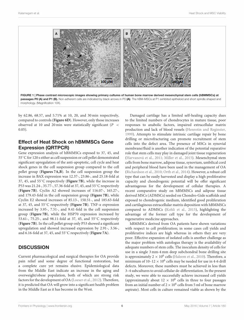
in early cultures were washed away with media changes leavingbehind only adherent hBMMSCs. The hBMMSCs derived fromthe bone marrow aspirate of OA patients showed epitheloid andshort spindle shaped cells in early passages (Figure 1). The initialnumber of cells in primary monolayer cultures varied from 1.4±0.4×10⁶ to 1.9± 0.6×10⁶ cells (from 5mL bonemarrow aspiratecultured in three T175 cm2 flasks). However, with subsequentpassages where uniform monolayer cultures were obtained, thecell numbers could be expanded to 2.1 ± 0.4 × 10⁶ cells perT175 cm2 flask.
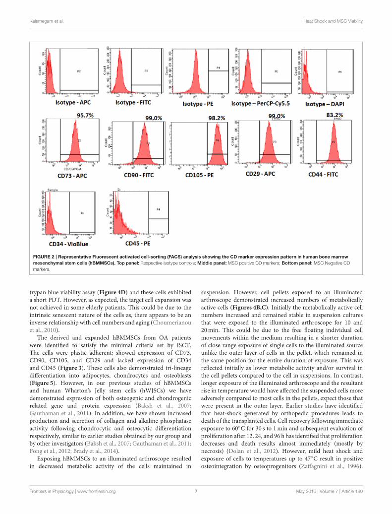
Surface Marker Characterization ofhBMMSCsThe derived cells analyzed for CD markers expressiondemonstrated high percentages of positive MSC related CDmarkers, namely CD73 (95.7%), CD90 (99.0%), CD105 (98.2%),CD44 (99.0%), and CD29 (83.2%) compared with respectiveisotype matched controls (Figure 2). These cells were negativefor CD34 and CD45, the haematopoietic stem cell related CDmarkers (Figure 2).
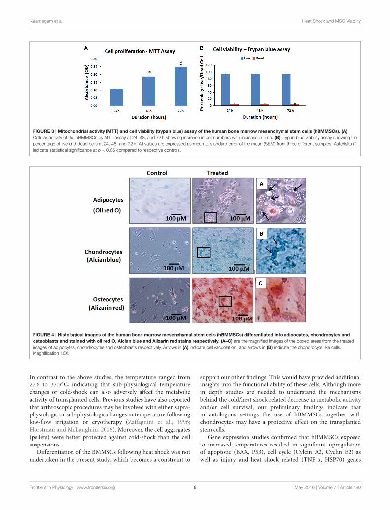
hBMMSCs Population Doubling and CellViabilityThe hBMMSCs demonstrated a mean increase in cell numbersfrom 24 to 72 h. There was a mean increase of 72.73 and 127.27%at 48 and 72 h respectively (Figure 3A). These mean increases incell numbers were statistically significant (P < 0.05).
The hBMMSCs showed an increasing linear growth profileover time with every passage and the PDT was 24.33–29.56 hwith growth rate 0.0285 and 0.0234 (Growth rate = number ofdoublings that occur per unit of time) at P1 and P5 respectively.Cell growth were slower with increase in passage number. Thetrypan blue viability showed that most of the cultured hBMMSCs
remained viable in culture platforms that could be used forin vitro assays. The percentage of viable cells were 94.57, 94.33,and 94.77% at 24, 48, and 72 h respectively (Figure 3B).
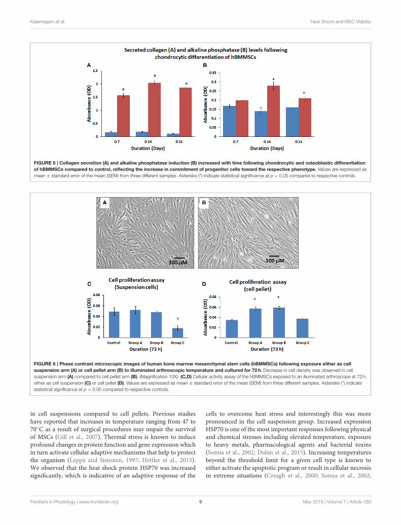
Differentiation Potential of hBMMSCsThe hBMMSCs showed differentiation into adipocytes,chondrocytes and osteoblasts with culture in respectivedifferentiation medium (StemPro R©). The cells differentiatedalong the adipocyte lineage demonstrated lipid vacuolationstarting as early as day 14 and the number of cells with lipidvacuoles increased when cultured until 21 days and these cellsdemonstrated positive staining with oil red O (Figure 4A).hBMMSCs cultured in chondrogenic differentiation mediumdemonstrated aggregation of cells when cultured for up to 21days and they included chondrocyte like cells that demonstratedpositive staining with Alcian blue compared to the control(Figure 4B). Osteogenic differentiation potential of hBMMSCscultured in osteogenic differentiation medium showed positivestaining with Alizarin red indicative of calcium mineralization(Figure 4C).
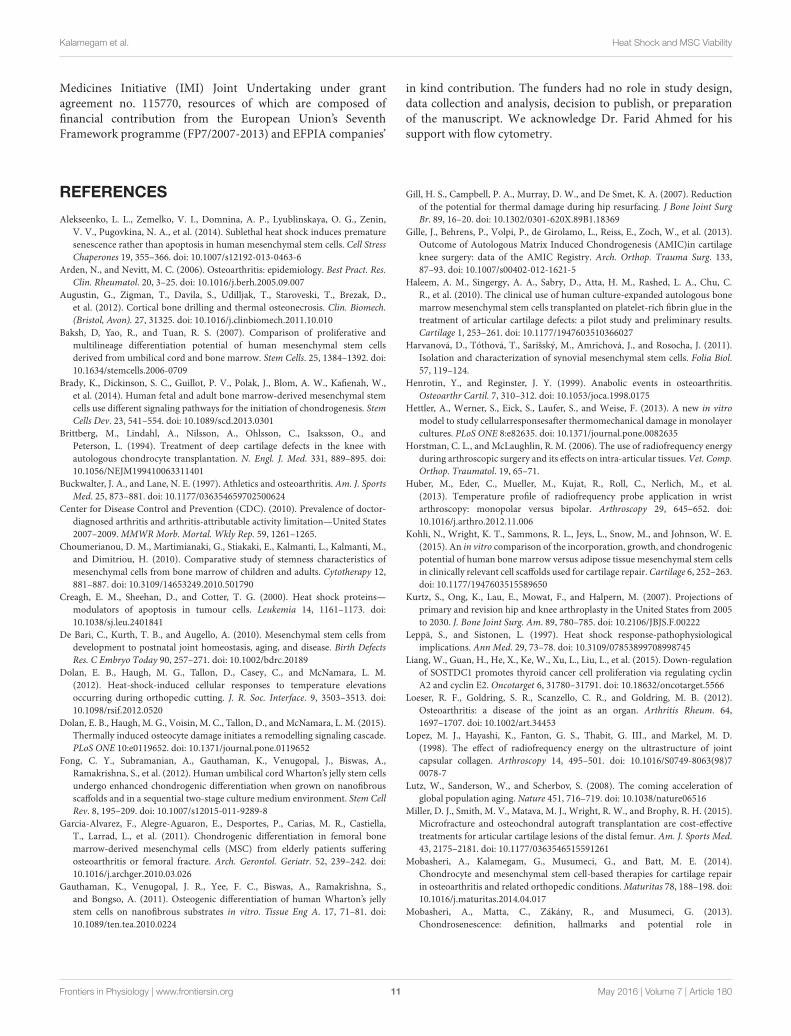
Collagen Secretion and AlkalinePhosphatase Activity of DifferentiatedhBMMSCsThe amount of secreted collagen measured using the Sircol assayconfirmed that chondrocytic differentiating cells secreted largeamounts of collagen at various culture periods compared toundifferentiated control cells (Figure 5A). The mean percentageincreases in collagen levels were 923.53, 1078.96, and 1550.00% at7, 14, and 21 days compared to their respective controls and theseincreases were statistically significant (P < 0.05).
The culture media analyzed for alkaline phosphataseactivity from control and osteocytic differentiated cells showedincrease in alkaline phosphatase levels compared to controlundifferentiated cells (Figure 5B). The mean percentageincreases in alkaline phosphatase levels were 17.65, 200.10, and131.25% at 7, 14, and 21 days compared to their respectivecontrols and only those increases at 14 and 21 days werestatistically significant (P < 0.05).
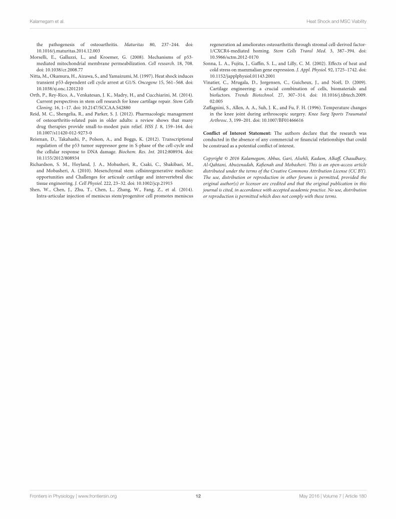
Effect of Arthroscope Temperature on theMetabolic Activity of hBMMSCsThe cells that were exposed to the illuminated arthroscope for10min (Group A), 20min (Group B), and 30min (Group C)demonstrated differences in metabolic activity rates for bothcell suspension group and cell pellet group when subsequentlycultured and assayed for 72 h. There were no changes in cellmorphology between suspensions and cell pellets. However, therewas a decrease in metabolically active cell numbers in the cellsuspensions (Figure 6A) compared to cell pellets (Figure 6B).The cell suspensions displayed a minimal increase of 6.12% anda minimal decrease of 2.04% at 10 and 20min, respectively,compared to controls (Figure 6C). Maximum mean decrease incell numbers were observed at 30min (by 63.27%) comparedto the control (Figure 6C) and this was statistically significant(P < 0.05). The cell pellet showedmean increases in cell numbers
Frontiers in Physiology | www.frontiersin.org 5 May 2016 | Volume 7 | Article 180
Kalamegam et al. Heat Shock and MSC Viability
FIGURE 1 | Phase contrast microscopic images showing primary cultures of human bone marrow derived mesenchymal stem cells (hBMMSCs) at
passages P0 (A) and P1 (B). Non-adherent cells are indicated by black arrows in P0 (A). The hBM-MSCs at P1 exhibited epitheloid and short spindle shaped and
morphology. (Magnification 10X).
by 62.86, 68.57, and 5.71% at 10, 20, and 30min respectively,compared to controls (Figure 6D). However, only those increasesobserved at 10 and 20min were statistically significant (P <
0.05).
Effect of Heat Shock on hBMMSCs GeneExpression (QRTPCR)Gene expression analysis of hBMMSCs exposed to 37, 45, and55◦C for 120 s either as cell suspension or cell pellet demonstratedsignificant upregulation of the anti-apoptotic, cell cycle and heatshock genes in the cell suspension group compared to the cellpellet group (Figures 7A,B). In the cell suspension group theincrease in BAX expression was 12.37-, 23.86-, and 25.18-fold at37, 45, and 55◦C respectively (Figure 7B), while the increase inP53 was 21.24-, 35.77-, 57.36-fold at 37, 45, and 55◦C respectively(Figure 7B). Cyclin A2 showed increases of 116.07-, 165.27-,and 179.43-fold in the cell suspension group (Figure 7B), whileCyclin E2 showed increases of 85.13-, 150.51-, and 185.63-foldat 37, 45, and 55◦C respectively (Figure 7B). TNF-α expressionincreased by 3.95-, 7.51-, and 9.41-fold in the cell suspensiongroup (Figure 7B), while the HSP70 expression increased by53.61-, 75.25-, and 98.11-fold at 37, 45, and 55◦C respectively(Figure 7B). In the cell pellet group only P53 showed a significantupregulation and showed increased expression by 2.91-, 3.56-,and 6.16-fold at 37, 45, and 55◦C respectively (Figure 7A).
DISCUSSION
Current pharmacological and surgical therapies for OA providepain relief and some degree of functional restoration, buta complete cure yet remains elusive. Epidemiological datafrom the Middle East indicate an increase in the aging andoverweight/obese population, both of which are strong riskfactors for the development of OA (Loeser et al., 2012). Therefore,it is predicted that OA will grow into a significant health problemin the Middle East as it has become in the West.
Damaged cartilage has a limited self-healing capacity duesto the limited numbers of chondrocytes in mature tissue, poorresponses to anabolic factors, impaired extracellular matrixproduction and lack of blood vessels (Henrotin and Reginster,1999). Attempts to stimulate intrinsic cartilage repair by bonedrilling or microfracturing can promote recruitment of stemcells into the defect area. The presence of MSCs in synovialmembrane/fluid is another indication of the potential reparativerole that stem cells may play in damaged joint tissue regeneration(Harvanová et al., 2011; Miller et al., 2015). Mesenchymal stemcells from bonemarrow, adipose tissue, synovium, umbilical cordand peripheral blood have been used in the management of OA(Richardson et al., 2010; Orth et al., 2014). However, a robust celltype that can be easily harvested and display a high proliferativecapacity and chondrogenic potential will be offer significantadvantageous for the development of cellular therapies. Arecent comparative study on hBMMSCs and adipose tissuederivedMSCs (ADMSCs) seeded on Chondro-Gide scaffolds andexposed to chondrogenic medium, identified good proliferationand cartilaginous extracellular matrix deposition with hBMMSCscompared to ADMSCs (Kohli et al., 2015), highlighting theadvantage of the former cell type for the development ofregenerative medicine approaches.
hBMMSCs derived from OA patients have shown variationswith respect to cell proliferation; in some cases cell yields andproliferative indices are high whereas in others they are verypoor. Effective expansion of isolated cells is another challenge asthe major problem with autologus therapy is the availability ofadequate numbers of stem cells. The inoculum density of cells foruse in a single 3mm-4mm deep subchondral bone drilling siteis approximately 2× 10⁶ cells (Haleem et al., 2010). Therefore, aminimum of 10–12× 106 cells may be needed for use in 4-6 drilldefects. Moreover, these numbers must be achieved in less than3–4 subcultures to avoid cellular de-differentiation. In the presentstudy, we were able to successfully achieve increased cell yields(approximately about 12 × 106 cells in three to four passagesfrom an initial number of 2×10⁶ cells from 5ml of bone marrowaspirate). Most cells in culture remained viable as shown by the
Frontiers in Physiology | www.frontiersin.org 6 May 2016 | Volume 7 | Article 180
Kalamegam et al. Heat Shock and MSC Viability
FIGURE 2 | Representative Fluorescent activated cell-sorting (FACS) analysis showing the CD marker expression pattern in human bone marrow
mesenchymal stem cells (hBMMSCs). Top panel: Respective isotype controls; Middle panel: MSC positive CD markers; Bottom panel: MSC Negative CD
markers.
trypan blue viability assay (Figure 4D) and these cells exhibiteda short PDT. However, as expected, the target cell expansion wasnot achieved in some elderly patients. This could be due to theintrinsic senescent nature of the cells as, there appears to be aninverse relationship with cell numbers and aging (Choumerianouet al., 2010).
The derived and expanded hBMMSCs from OA patientswere identified to satisfy the minimal criteria set by ISCT.The cells were plastic adherent; showed expression of CD73,CD90, CD105, and CD29 and lacked expression of CD34and CD45 (Figure 3). These cells also demonstrated tri-lineagedifferentiation into adipocytes, chondrocytes and osteoblasts(Figure 5). However, in our previous studies of hBMMSCsand human Wharton’s Jelly stem cells (hWJSCs) we havedemonstrated expression of both osteogenic and chondrogenicrelated gene and protein expression (Baksh et al., 2007;Gauthaman et al., 2011). In addition, we have shown increasedproduction and secretion of collagen and alkaline phosphataseactivity following chondrocytic and osteocytic differentiationrespectively, similar to earlier studies obtained by our group andby other investigators (Baksh et al., 2007; Gauthaman et al., 2011;Fong et al., 2012; Brady et al., 2014).
Exposing hBMMSCs to an illuminated arthroscope resultedin decreased metabolic activity of the cells maintained in
suspension. However, cell pellets exposed to an illuminatedarthroscope demonstrated increased numbers of metabolicallyactive cells (Figures 4B,C). Initially the metabolically active cellnumbers increased and remained stable in suspension culturesthat were exposed to the illuminated arthroscope for 10 and20min. This could be due to the free floating individual cellmovements within the medium resulting in a shorter durationof close range exposure of single cells to the illuminated sourceunlike the outer layer of cells in the pellet, which remained inthe same position for the entire duration of exposure. This wasreflected initially as lower metabolic activity and/or survival inthe cell pellets compared to the cell in suspensions. In contrast,longer exposure of the illuminated arthroscope and the resultantrise in temperature would have affected the suspended cells moreadversely compared to most cells in the pellets, expect those thatwere present in the outer layer. Earlier studies have identifiedthat heat-shock generated by orthopedic procedures leads todeath of the transplanted cells. Cell recovery following immediateexposure to 60◦C for 30 s to 1min and subsequent evaluation ofproliferation after 12, 24, and 96 h has identified that proliferationdecreases and death results almost immediately (mostly bynecrosis) (Dolan et al., 2012). However, mild heat shock andexposure of cells to temperatures up to 47◦C result in positiveosteointegration by osteoprogenitors (Zaffagnini et al., 1996).
Frontiers in Physiology | www.frontiersin.org 7 May 2016 | Volume 7 | Article 180
Kalamegam et al. Heat Shock and MSC Viability
FIGURE 3 | Mitochondrial activity (MTT) and cell viability (trypan blue) assay of the human bone marrow mesenchymal stem cells (hBMMSCs). (A)
Cellular activity of the hBMMSCs by MTT assay at 24, 48, and 72 h showing increase in cell numbers with increase in time. (B) Trypan blue viability assay showing the
percentage of live and dead cells at 24, 48, and 72 h. All values are expressed as mean ± standard error of the mean (SEM) from three different samples. Asterisks (*)
indicate statistical significance at p < 0.05 compared to respective controls.
FIGURE 4 | Histological images of the human bone marrow mesenchymal stem cells (hBMMSCs) differentiated into adipocytes, chondrocytes and
osteoblasts and stained with oil red O, Alcian blue and Alizarin red stains respectively. (A–C) are the magnified images of the boxed areas from the treated
images of adipocytes, chondrocytes and osteoblasts respectively. Arrows in (A) indicate cell vacuolation, and arrows in (B) indicate the chondrocyte like cells.
Magnification 10X.
In contrast to the above studies, the temperature ranged from27.6 to 37.3◦C, indicating that sub-physiological temperaturechanges or cold-shock can also adversely affect the metabolicactivity of transplanted cells. Previous studies have also reportedthat arthroscopic procedures may be involved with either supra-physiologic or sub-physiologic changes in temperature followinglow-flow irrigation or cryotherapy (Zaffagnini et al., 1996;Horstman and McLaughlin, 2006). Moreover, the cell aggregates(pellets) were better protected against cold-shock than the cellsuspensions.
Differentiation of the BMMSCs following heat shock was notundertaken in the present study, which becomes a constraint to
support our other findings. This would have provided additionalinsights into the functional ability of these cells. Although morein depth studies are needed to understand the mechanismsbehind the cold/heat shock related decrease in metabolic activityand/or cell survival, our preliminary findings indicate thatin autologous settings the use of hBMMSCs together withchondrocytes may have a protective effect on the transplantedstem cells.
Gene expression studies confirmed that hBMMSCs exposedto increased temperatures resulted in significant upregulationof apoptotic (BAX, P53), cell cycle (Cylcin A2, Cyclin E2) aswell as injury and heat shock related (TNF-α, HSP70) genes
Frontiers in Physiology | www.frontiersin.org 8 May 2016 | Volume 7 | Article 180
Kalamegam et al. Heat Shock and MSC Viability
FIGURE 5 | Collagen secretion (A) and alkaline phosphatase induction (B) increased with time following chondrocytic and osteoblastic differentiation
of hBMMSCs compared to control, reflecting the increase in commitment of progenitor cells toward the respective phenotype. Values are expressed as
mean ± standard error of the mean (SEM) from three different samples. Asterisks (*) indicate statistical significance at p < 0.05 compared to respective controls.
FIGURE 6 | Phase contrast microscopic images of human bone marrow mesenchymal stem cells (hBMMSCs) following exposure either as cell
suspension arm (A) or cell pellet arm (B) to illuminated arthroscopic temperature and cultured for 72h. Decrease in cell density was observed in cell
suspension arm (A) compared to cell pellet arm (B). (Magnification 10X). (C,D) Cellular activity assay of the hBMMSCs exposed to an illuminated arthroscope at 72 h,
either as cell suspension (C) or cell pellet (D). Values are expressed as mean ± standard error of the mean (SEM) from three different samples. Asterisks (*) indicate
statistical significance at p < 0.05 compared to respective controls.
in cell suspensions compared to cell pellets. Previous studieshave reported that increases in temperature ranging from 47 to70◦C as a result of surgical procedures may impair the survivalof MSCs (Gill et al., 2007). Thermal stress is known to induceprofound changes in protein function and gene expression whichin turn activate cellular adaptive mechanisms that help to protectthe organism (Leppä and Sistonen, 1997; Hettler et al., 2013).We observed that the heat shock protein HSP70 was increasedsignificantly, which is indicative of an adaptive response of the
cells to overcome heat stress and interestingly this was morepronounced in the cell suspension group. Increased expressionHSP70 is one of the most important responses following physicaland chemical stresses including elevated temperature, exposureto heavy metals, pharmacological agents and bacterial toxins(Sonna et al., 2002; Dolan et al., 2015). Increasing temperaturesbeyond the threshold limit for a given cell type is known toeither activate the apoptotic program or result in cellular necrosisin extreme situations (Creagh et al., 2000; Sonna et al., 2002;
Frontiers in Physiology | www.frontiersin.org 9 May 2016 | Volume 7 | Article 180

Kalamegam et al. Heat Shock and MSC Viability
FIGURE 7 | Gene expression profile (QRT-PCR) of BCL2, BAX, P53,
HSP70, TNF-α, CYCLIN A2, and CYCLIN E2 of hBMMSCs exposed to
heat shock either as cell pellet (A) or cell suspension (B). Upregulation of
apoptotic, cell cycle, inflammation and heat shock related genes in hBM-MSCs
cell suspension (B) compared to cell pellet (A) indicate that individual cells (B)
were easily affected by heat shock compared to cell pellet (A). Data analysis
and relative quantitation were performed using the comparative Ct method
(11Ct). The differences in gene expression levels were analyzed using
student’s t-test. Asterisks (*) indicate statistical significance at p < 0.05.
Dolan et al., 2015). We also observed that in cell suspensionsproapoptotic BAX was increased and the antiapoptotic BCL2was decreased, indicating that the hBMMSCs were undergoingcell death. Cyclin E2 is involved in G1/S phase of cell cycleprogression and cyclin A2 is involved in regulation of G2/Mphase of cell cycle. Heat stress following radiation upregulateHSP which in turn augments cell cycle arrest at G1, S, and G2/Mphases (Nitta et al., 1997). The increased expression observed forcyclin E2 and Cyclin A2 in cell suspensions may be indicativeof an attempt to overcome the heat stress and promote cellsurvival. The expression level probably decreased as the heatstressed cells in the suspension group underwent necrotic deathunlike the pellet group. In addition, P53 is known to influenceexpression of numerous target genes that control cell cycle,apoptosis, gene instability, senescence following stress (Morselliet al., 2008). Increased expression of P53 was observed followingheat stress, and P53 is associated with negative regulation of cellcycle progression (Reisman et al., 2012). If the gene expression ofcell cycle inhibitor p21 had been included in the study, it would
have provided additional information on the cell proliferationability following heat shock of hBMMSCs.
The introduction ofMSCs into the joint can be achieved eitherthrough injection as a cell suspension or through loading ontoa scaffold as an implant. Our findings offer caution when theformer approach is used since the cells are not encased by amatrix that can protect them from external factors such as heatfrom the arthroscope. This study suggests that improvizationsin surgical methods that avoid generating large amounts ofheat may result in more favorable outcomes when applyingMSCs in cell suspension to treat an arthritic joint. Alternatively,administration of cells in combination with biodegradablematerials as pellets/encapsulations might enhance the survival oftransplanted cells.
CONCLUSIONS
hBMMSCs from OA patients have the capacity to expand andactively differentiate. However, they are prone to damage ifthe method of delivery to the joint is not optimal and isaccompanied by the generation of excess heat. Less invasive andmore cytoprotective surgical methods that do not generate excessheat are needed to ensure successful therapeutic delivery ofMSCsto the joint.
AUTHOR CONTRIBUTIONS
MAb and MAl are the clinicians and were involved inproviding clinical materials/information and intellectual support.GK and MG were involved in conceptualization, intellectualcontribution, statistical evaluation and manuscript writing. HAand RK was involved in providing technical assistance withexperimental work, manuscript editing and intellectual help. AA,AC, MAQ, and WK were involved in the overall co-ordinationof the work, and also reviewed and edited the manuscript. AMcontributed to the synthesis and editing of the manuscript.
ACKNOWLEDGMENTS
We acknowledge the financial support provided by the“Sheik Salem Bin Mahfouz Scientific Chair for Treatment ofOsteoarthritis by Stem Cells” and the stem cell laboratoryfacility at CEGMR and King Abdulaziz University Hospital’.AM is the co-ordinator of the D-BOARD Consortium fundedby European Commission Framework 7 programme (EUFP7; HEALTH.2012.2.4.5-2, project number 305815, NovelDiagnostics and Biomarkers for Early Identification of ChronicInflammatory Joint Diseases). AM is a member of theApplied Public-Private Research enabling OsteoArthritis ClinicalHeadway (APPROACH) consortium, a 5-year project fundedby the European Commission’s Innovative Medicines Initiative(IMI). APPROACH is a public-private partnership directedtoward osteoarthritis biomarker development through theestablishment of a heavily phenotyped and comprehensivelyanalyzed longitudinal cohort. The research leading to theseresults has received partial support from the Innovative
Frontiers in Physiology | www.frontiersin.org 10 May 2016 | Volume 7 | Article 180
Kalamegam et al. Heat Shock and MSC Viability
Medicines Initiative (IMI) Joint Undertaking under grantagreement no. 115770, resources of which are composed offinancial contribution from the European Union’s SeventhFramework programme (FP7/2007-2013) and EFPIA companies’
in kind contribution. The funders had no role in study design,data collection and analysis, decision to publish, or preparationof the manuscript. We acknowledge Dr. Farid Ahmed for hissupport with flow cytometry.
REFERENCES
Alekseenko, L. L., Zemelko, V. I., Domnina, A. P., Lyublinskaya, O. G., Zenin,
V. V., Pugovkina, N. A., et al. (2014). Sublethal heat shock induces premature
senescence rather than apoptosis in human mesenchymal stem cells. Cell Stress
Chaperones 19, 355–366. doi: 10.1007/s12192-013-0463-6
Arden, N., and Nevitt, M. C. (2006). Osteoarthritis: epidemiology. Best Pract. Res.
Clin. Rheumatol. 20, 3–25. doi: 10.1016/j.berh.2005.09.007
Augustin, G., Zigman, T., Davila, S., Udilljak, T., Staroveski, T., Brezak, D.,
et al. (2012). Cortical bone drilling and thermal osteonecrosis. Clin. Biomech.
(Bristol, Avon). 27, 31325. doi: 10.1016/j.clinbiomech.2011.10.010
Baksh, D, Yao, R., and Tuan, R. S. (2007). Comparison of proliferative and
multilineage differentiation potential of human mesenchymal stem cells
derived from umbilical cord and bone marrow. Stem Cells. 25, 1384–1392. doi:
10.1634/stemcells.2006-0709
Brady, K., Dickinson, S. C., Guillot, P. V., Polak, J., Blom, A. W., Kafienah, W.,
et al. (2014). Human fetal and adult bone marrow-derived mesenchymal stem
cells use different signaling pathways for the initiation of chondrogenesis. Stem
Cells Dev. 23, 541–554. doi: 10.1089/scd.2013.0301
Brittberg, M., Lindahl, A., Nilsson, A., Ohlsson, C., Isaksson, O., and
Peterson, L. (1994). Treatment of deep cartilage defects in the knee with
autologous chondrocyte transplantation. N. Engl. J. Med. 331, 889–895. doi:
10.1056/NEJM199410063311401
Buckwalter, J. A., and Lane, N. E. (1997). Athletics and osteoarthritis. Am. J. Sports
Med. 25, 873–881. doi: 10.1177/036354659702500624
Center for Disease Control and Prevention (CDC). (2010). Prevalence of doctor-
diagnosed arthritis and arthritis-attributable activity limitation—United States
2007–2009.MMWRMorb. Mortal. Wkly Rep. 59, 1261–1265.
Choumerianou, D. M., Martimianaki, G., Stiakaki, E., Kalmanti, L., Kalmanti, M.,
and Dimitriou, H. (2010). Comparative study of stemness characteristics of
mesenchymal cells from bone marrow of children and adults. Cytotherapy 12,
881–887. doi: 10.3109/14653249.2010.501790
Creagh, E. M., Sheehan, D., and Cotter, T. G. (2000). Heat shock proteins—
modulators of apoptosis in tumour cells. Leukemia 14, 1161–1173. doi:
10.1038/sj.leu.2401841
De Bari, C., Kurth, T. B., and Augello, A. (2010). Mesenchymal stem cells from
development to postnatal joint homeostasis, aging, and disease. Birth Defects
Res. C Embryo Today 90, 257–271. doi: 10.1002/bdrc.20189
Dolan, E. B., Haugh, M. G., Tallon, D., Casey, C., and McNamara, L. M.
(2012). Heat-shock-induced cellular responses to temperature elevations
occurring during orthopedic cutting. J. R. Soc. Interface. 9, 3503–3513. doi:
10.1098/rsif.2012.0520
Dolan, E. B., Haugh, M. G., Voisin, M. C., Tallon, D., andMcNamara, L. M. (2015).
Thermally induced osteocyte damage initiates a remodelling signaling cascade.
PLoS ONE 10:e0119652. doi: 10.1371/journal.pone.0119652
Fong, C. Y., Subramanian, A., Gauthaman, K., Venugopal, J., Biswas, A.,
Ramakrishna, S., et al. (2012). Human umbilical cordWharton’s jelly stem cells
undergo enhanced chondrogenic differentiation when grown on nanofibrous
scaffolds and in a sequential two-stage culture medium environment. Stem Cell
Rev. 8, 195–209. doi: 10.1007/s12015-011-9289-8
Garcia-Alvarez, F., Alegre-Aguaron, E., Desportes, P., Carias, M. R., Castiella,
T., Larrad, L., et al. (2011). Chondrogenic differentiation in femoral bone
marrow-derived mesenchymal cells (MSC) from elderly patients suffering
osteoarthritis or femoral fracture. Arch. Gerontol. Geriatr. 52, 239–242. doi:
10.1016/j.archger.2010.03.026
Gauthaman, K., Venugopal, J. R., Yee, F. C., Biswas, A., Ramakrishna, S.,
and Bongso, A. (2011). Osteogenic differentiation of human Wharton’s jelly
stem cells on nanofibrous substrates in vitro. Tissue Eng A. 17, 71–81. doi:
10.1089/ten.tea.2010.0224
Gill, H. S., Campbell, P. A., Murray, D. W., and De Smet, K. A. (2007). Reduction
of the potential for thermal damage during hip resurfacing. J Bone Joint Surg
Br. 89, 16–20. doi: 10.1302/0301-620X.89B1.18369
Gille, J., Behrens, P., Volpi, P., de Girolamo, L., Reiss, E., Zoch, W., et al. (2013).
Outcome of Autologous Matrix Induced Chondrogenesis (AMIC)in cartilage
knee surgery: data of the AMIC Registry. Arch. Orthop. Trauma Surg. 133,
87–93. doi: 10.1007/s00402-012-1621-5
Haleem, A. M., Singergy, A. A., Sabry, D., Atta, H. M., Rashed, L. A., Chu, C.
R., et al. (2010). The clinical use of human culture-expanded autologous bone
marrow mesenchymal stem cells transplanted on platelet-rich fibrin glue in the
treatment of articular cartilage defects: a pilot study and preliminary results.
Cartilage 1, 253–261. doi: 10.1177/1947603510366027
Harvanová, D., Tóthová, T., Sarišský, M., Amrichová, J., and Rosocha, J. (2011).
Isolation and characterization of synovial mesenchymal stem cells. Folia Biol.
57, 119–124.
Henrotin, Y., and Reginster, J. Y. (1999). Anabolic events in osteoarthritis.
Osteoarthr Cartil. 7, 310–312. doi: 10.1053/joca.1998.0175
Hettler, A., Werner, S., Eick, S., Laufer, S., and Weise, F. (2013). A new in vitro
model to study cellularresponsesafter thermomechanical damage in monolayer
cultures. PLoS ONE 8:e82635. doi: 10.1371/journal.pone.0082635
Horstman, C. L., and McLaughlin, R. M. (2006). The use of radiofrequency energy
during arthroscopic surgery and its effects on intra-articular tissues.Vet. Comp.
Orthop. Traumatol. 19, 65–71.
Huber, M., Eder, C., Mueller, M., Kujat, R., Roll, C., Nerlich, M., et al.
(2013). Temperature profile of radiofrequency probe application in wrist
arthroscopy: monopolar versus bipolar. Arthroscopy 29, 645–652. doi:
10.1016/j.arthro.2012.11.006
Kohli, N., Wright, K. T., Sammons, R. L., Jeys, L., Snow, M., and Johnson, W. E.
(2015). An in vitro comparison of the incorporation, growth, and chondrogenic
potential of human bone marrow versus adipose tissue mesenchymal stem cells
in clinically relevant cell scaffolds used for cartilage repair.Cartilage 6, 252–263.
doi: 10.1177/1947603515589650
Kurtz, S., Ong, K., Lau, E., Mowat, F., and Halpern, M. (2007). Projections of
primary and revision hip and knee arthroplasty in the United States from 2005
to 2030. J. Bone Joint Surg. Am. 89, 780–785. doi: 10.2106/JBJS.F.00222
Leppä, S., and Sistonen, L. (1997). Heat shock response-pathophysiological
implications. Ann Med. 29, 73–78. doi: 10.3109/07853899708998745
Liang, W., Guan, H., He, X., Ke, W., Xu, L., Liu, L., et al. (2015). Down-regulation
of SOSTDC1 promotes thyroid cancer cell proliferation via regulating cyclin
A2 and cyclin E2. Oncotarget 6, 31780–31791. doi: 10.18632/oncotarget.5566
Loeser, R. F., Goldring, S. R., Scanzello, C. R., and Goldring, M. B. (2012).
Osteoarthritis: a disease of the joint as an organ. Arthritis Rheum. 64,
1697–1707. doi: 10.1002/art.34453
Lopez, M. J., Hayashi, K., Fanton, G. S., Thabit, G. III., and Markel, M. D.
(1998). The effect of radiofrequency energy on the ultrastructure of joint
capsular collagen. Arthroscopy 14, 495–501. doi: 10.1016/S0749-8063(98)7
0078-7
Lutz, W., Sanderson, W., and Scherbov, S. (2008). The coming acceleration of
global population aging. Nature 451, 716–719. doi: 10.1038/nature06516
Miller, D. J., Smith, M. V., Matava, M. J., Wright, R. W., and Brophy, R. H. (2015).
Microfracture and osteochondral autograft transplantation are cost-effective
treatments for articular cartilage lesions of the distal femur. Am. J. Sports Med.
43, 2175–2181. doi: 10.1177/0363546515591261
Mobasheri, A., Kalamegam, G., Musumeci, G., and Batt, M. E. (2014).
Chondrocyte and mesenchymal stem cell-based therapies for cartilage repair
in osteoarthritis and related orthopedic conditions.Maturitas 78, 188–198. doi:
10.1016/j.maturitas.2014.04.017
Mobasheri, A., Matta, C., Zákány, R., and Musumeci, G. (2013).
Chondrosenescence: definition, hallmarks and potential role in
Frontiers in Physiology | www.frontiersin.org 11 May 2016 | Volume 7 | Article 180
Kalamegam et al. Heat Shock and MSC Viability
the pathogenesis of osteoarthritis. Maturitas 80, 237–244. doi:
10.1016/j.maturitas.2014.12.003
Morselli, E., Galluzzi, L., and Kroemer, G. (2008). Mechanisms of p53-
mediated mitochondrial membrane permeabilization. Cell research. 18, 708.
doi: 10.1038/cr.2008.77
Nitta, M., Okamura, H., Aizawa, S., and Yamaizumi, M. (1997). Heat shock induces
transient p53-dependent cell cycle arrest at G1/S. Oncogene 15, 561–568. doi:
10.1038/sj.onc.1201210
Orth, P., Rey-Rico, A., Venkatesan, J. K., Madry, H., and Cucchiarini, M. (2014).
Current perspectives in stem cell research for knee cartilage repair. Stem Cells
Cloning. 16, 1–17. doi: 10.2147/SCCAA.S42880
Reid, M. C., Shengelia, R., and Parker, S. J. (2012). Pharmacologic management
of osteoarthritis-related pain in older adults: a review shows that many
drug therapies provide small-to-modest pain relief. HSS J. 8, 159–164. doi:
10.1007/s11420-012-9273-0
Reisman, D., Takahashi, P., Polson, A., and Boggs, K. (2012). Transcriptional
regulation of the p53 tumor suppressor gene in S-phase of the cell-cycle and
the cellular response to DNA damage. Biochem. Res. Int. 2012:808934. doi:
10.1155/2012/808934
Richardson, S. M., Hoyland, J. A., Mobasheri, R., Csaki, C., Shakibaei, M.,
and Mobasheri, A. (2010). Mesenchymal stem cellsinregenerative medicne:
opportunities and Challenges for articualr cartilage and intervertebral disc
tissue engineering. J. Cell Physiol. 222, 23–32. doi: 10.1002/jcp.21915
Shen, W., Chen, J., Zhu, T., Chen, L., Zhang, W., Fang, Z., et al. (2014).
Intra-articular injection of meniscus stem/progenitor cell promotes meniscus
regeneration ad ameliorates osteoarthritis through stromal cell-derived factor-
1/CXCR4-mediated homing. Stem Cells Transl Med. 3, 387–394. doi:
10.5966/sctm.2012-0170
Sonna, L. A., Fujita, J., Gaffin, S. L., and Lilly, C. M. (2002). Effects of heat and
cold stress on mammalian gene expression. J. Appl. Physiol. 92, 1725–1742. doi:
10.1152/japplphysiol.01143.2001
Vinatier, C., Mrugala, D., Jorgensen, C., Guicheux, J., and Noël, D. (2009).
Cartilage engineering: a crucial combination of cells, biomaterials and
biofactors. Trends Biotechnol. 27, 307–314. doi: 10.1016/j.tibtech.2009.
02.005
Zaffagnini, S., Allen, A. A., Suh, J. K., and Fu, F. H. (1996). Temperature changes
in the knee joint during arthroscopic surgery. Knee Surg Sports Traumatol
Arthrosc. 3, 199–201. doi: 10.1007/BF01466616
Conflict of Interest Statement: The authors declare that the research was
conducted in the absence of any commercial or financial relationships that could
be construed as a potential conflict of interest.
Copyright © 2016 Kalamegam, Abbas, Gari, Alsehli, Kadam, Alkaff, Chaudhary,
Al-Qahtani, Abuzenadah, Kafienah and Mobasheri. This is an open-access article
distributed under the terms of the Creative Commons Attribution License (CC BY).
The use, distribution or reproduction in other forums is permitted, provided the
original author(s) or licensor are credited and that the original publication in this
journal is cited, in accordance with accepted academic practice. No use, distribution
or reproduction is permitted which does not comply with these terms.
Frontiers in Physiology | www.frontiersin.org 12 May 2016 | Volume 7 | Article 180


![Ocala Banner. (Ocala, Florida) 1901-05-24 [p ].ufdcimages.uflib.ufl.edu/UF/00/04/87/34/00589/00180.pdf · 2009-07-14 · SSS SADDLERYO-CALA EXCHANGE THAGHERS OCALA BINE Liquors SYRUP-For](https://img.pdfslide.net/doc/110x75/5f7b445908715e4efd161734/ocala-banner-ocala-florida-1901-05-24-p-2009-07-14-sss-saddleryo-cala.jpg)


![Ocala Evening Star. (Ocala, Florida) 1900-08-24 [p ].ufdcimages.uflib.ufl.edu/UF/00/07/59/08/02563/00180.pdf · digest sheeps foun-tain Kidney ingredi William ... la-The Paris Store](https://img.pdfslide.net/doc/110x75/5ac797d77f8b9acb7c8c0397/ocala-evening-star-ocala-florida-1900-08-24-p-sheeps-foun-tain-kidney-ingredi.jpg)