Embed Size (px)
Citation preview

KERATOPLASTY- Dr. Narang
Keratoplasty/corneal grafting /corneal transplantation
• Definition: It is an operation in which the patient's diseased cornea is replaced by the donor's healthy clear cornea.
Types:• 1. Penetrating keratoplasty (full-thickness grafting)
• 2. Lamellar keratoplasty (partial-thickness grafting) -anterior or posterior lamellar
• Indications:
• Optical ,i.e., to improve vision - corneal opacity, bullous keratopathy, corneal dystrophies, advanced keratoconus.
• Therapeutic, i.e., to replace inflamed cornea not responding to conventional therapy
• Tectonic graft, i.e., to restore integrity of eyeball in eyes with severe structural changes such as severe thinning with descemetocele.
• Cosmetic, i.e., to improve the appearance of the eye.
Donor tissue :
• removed as early as possible (12–24 hours of death).
• Corneas from infants (3 years and under) are rarely used -surgical, refractive and rejection problems.
• It should be stored under sterile conditions.
• Evaluation –medical history review and donor blood screening to exclude contraindications, and microscopic examination of the cornea including endothelial cell count determination
Methods of corneal preservation
• Short-term storage (up to 2 days) -The whole globe is preserved at 40C in a moist chamber.
• Intermediate storage (up to 2 weeks) -McCarey-Kaufman (MK) medium and various chondroitin sulfate enriched media such as optisol medium used.
• Long-term storage (up to 35 days) -It is done by organ culture method.
Contraindications to ocular tissue donation• Death of unknown cause.
• Certain systemic infections such as HIV, viral hepatitis, syphilis, congenital rubella, tuberculosis, septicaemia and active malaria.
• Prior high-risk behavior for HIV and hepatitis.
• infectious diseases of the CNS.
• Receipt of a transplanted organ.
• Most hematological malignancies.
• Ocular disease such as inflammation and malignancies (e.g. retinoblastoma) and corneal refractive surgery.
Host factors may adversely affect the prognosis:
• Severe stromal vascularization, extreme thinning at the proposed host-graft junction and active corneal inflammation.
• Abnormalities of the eyelids (blepharitis, ectropion, entropionand trichiasis).
• Recurrent or progressive forms of conjunctival inflammation.
• Tear film dysfunction.
• Anterior synechiae.
• Uncontrolled glaucoma.
• Uveitis.
Penetrating keratoplasty
• most commonly performed corneal transplantation procedure.
INDICATIONS INCLUDE:
• Disease involving all layers of the cornea.
• Specific common indications: keratoconus, pseudophakicbullous keratopathy, Fuchs endothelial and other dystrophies.
Technique:
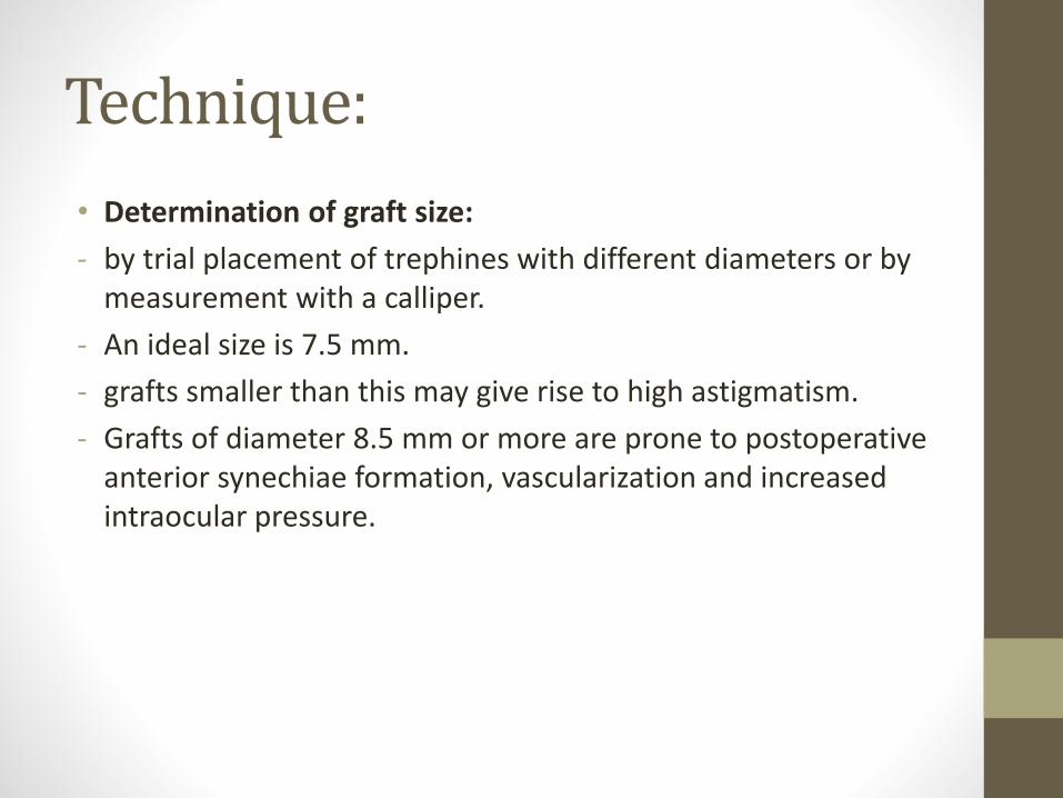
• Determination of graft size:
- by trial placement of trephines with different diameters or by measurement with a calliper.
- An ideal size is 7.5 mm.
- grafts smaller than this may give rise to high astigmatism.
- Grafts of diameter 8.5 mm or more are prone to postoperative anterior synechiae formation, vascularization and increased intraocular pressure.

• Excision of donor corneal button -The donor corneal button should be trephined 0.25 mm larger than the recipient, taking care not to damage the endothelium.
- to facilitate watertight closure, minimize postoperative flattening and reduce the possibility of postoperative glaucoma.
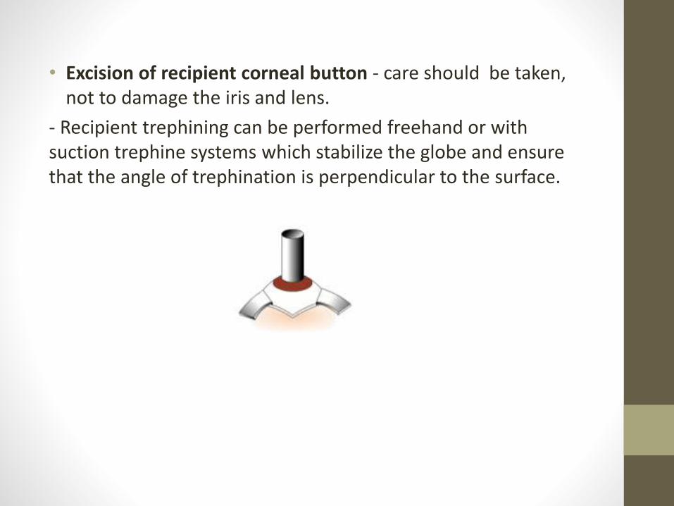
• Excision of recipient corneal button - care should be taken, not to damage the iris and lens.
- Recipient trephining can be performed freehand or with suction trephine systems which stabilize the globe and ensure that the angle of trephination is perpendicular to the surface.
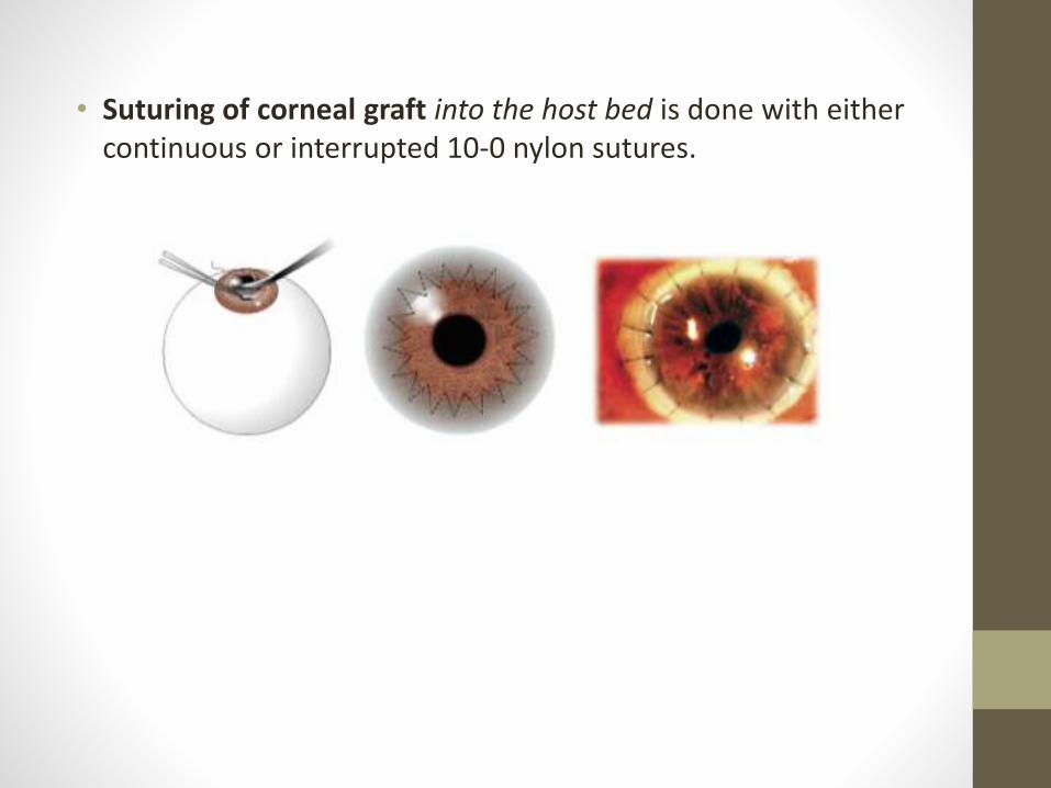
• Suturing of corneal graft into the host bed is done with either continuous or interrupted 10-0 nylon sutures.
Postoperative management:
• Topical steroids are used to decrease the risk of immunological graft rejection.
• Other immunosuppressants –azathioprine, ciclosporin may be rarely used in high-risk for prevention of rejection.
• Mydriatics - if uveitis persists.
• Monitoring of IOP is performed during the early postoperative period.
• Removal of sutures when the graft-host junction has healed. This is usually after 12–18 months.
• Rigid contact lenses -to optimize visual acuity in eyes with astigmatism.
Postoperative complications:
• Early complications: persistent epithelial defects, irritation by protruding sutures, wound leak, flat anterior chamber, iris prolapse, uveitis, elevation of intraocular pressure, microbial keratitis and endophthalmitis .
• Late: astigmatism, recurrence of initial disease process, late wound separation, retro-corneal membrane formation, glaucoma and cystoid macular oedema.
Superficial lamellar keratoplasty
• This involves partial thickness excision of the corneal epithelium and stroma.
• endothelium and part of the deep stroma are left behind.
Indications:
• Opacification of the superficial one-third of the corneal stroma.
• Marginal corneal thinning or infiltration as in recurrent pterygium, marginal degeneration.
• Localized thinning or descemetocele formation.
Deep anterior lamellar keratoplasty
• Opaque corneal tissue is removed almost to the level of Descemet membrane.
• decreased risk of rejection because the endothelium, a major target for rejection, is not transplanted.
Indications:• Disease involving the anterior 95% of corneal thickness with a
normal endothelium and absence of breaks or scars in Descemet membrane .
• Chronic inflammatory disease such as atopic keratoconjunctivitis which carries an increased risk of graft rejection.
Descemet stripping endothelial keratoplasty
• It involves removal only of diseased endothelium along with Descemet membrane, through a corneoscleral or corneal incision.
• Folded donor tissue is introduced through the same small (about 5 mm) incision.
Indications:• include endothelial disease such as pseudophakic bullous
keratopathy.